Embed Size (px)
Citation preview
IMMUNOCHEMICAL METHODS IN BIOCHEMISTRY
Kateřina DunovskáDepartment of Medical Chemistry and Clinical Biochemistry
2nd Faculty of Medicine and University Hospital Motol

TERMINOLOGY
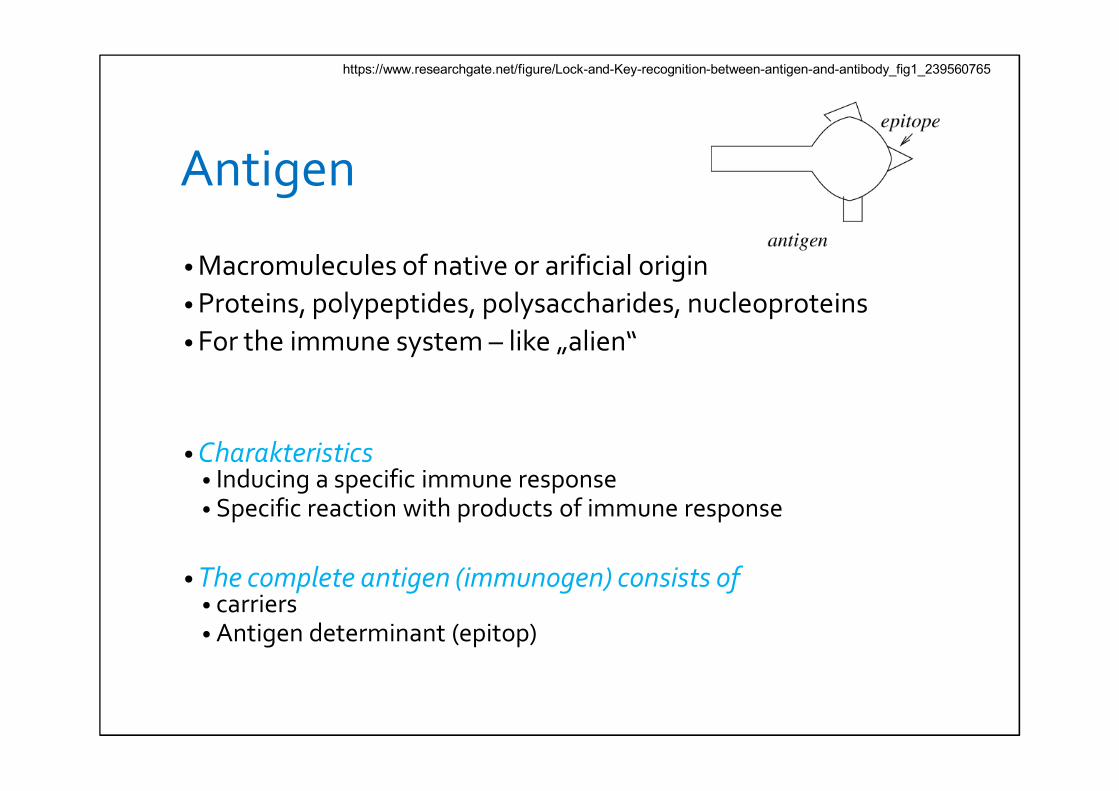
Antigen
• Macromulecules of native or arificial origin• Proteins, polypeptides, polysaccharides, nucleoproteins• For the immune system – like „alien“
• Charakteristics• Inducing a specific immune response• Specific reaction with products of immune response
• The complete antigen (immunogen) consists of• carriers• Antigen determinant (epitop)
https://www.researchgate.net/figure/Lock-and-Key-recognition-between-antigen-and-antibody_fig1_239560765
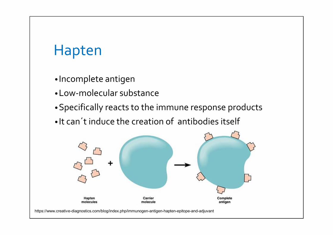
Hapten
• Incomplete antigen
• Low-molecular substance
• Specifically reacts to the immune response products
• It can´t induce the creation of antibodies itself
https://www.creative-diagnostics.com/blog/index.php/immunogen-antigen-hapten-epitope-and-adjuvant
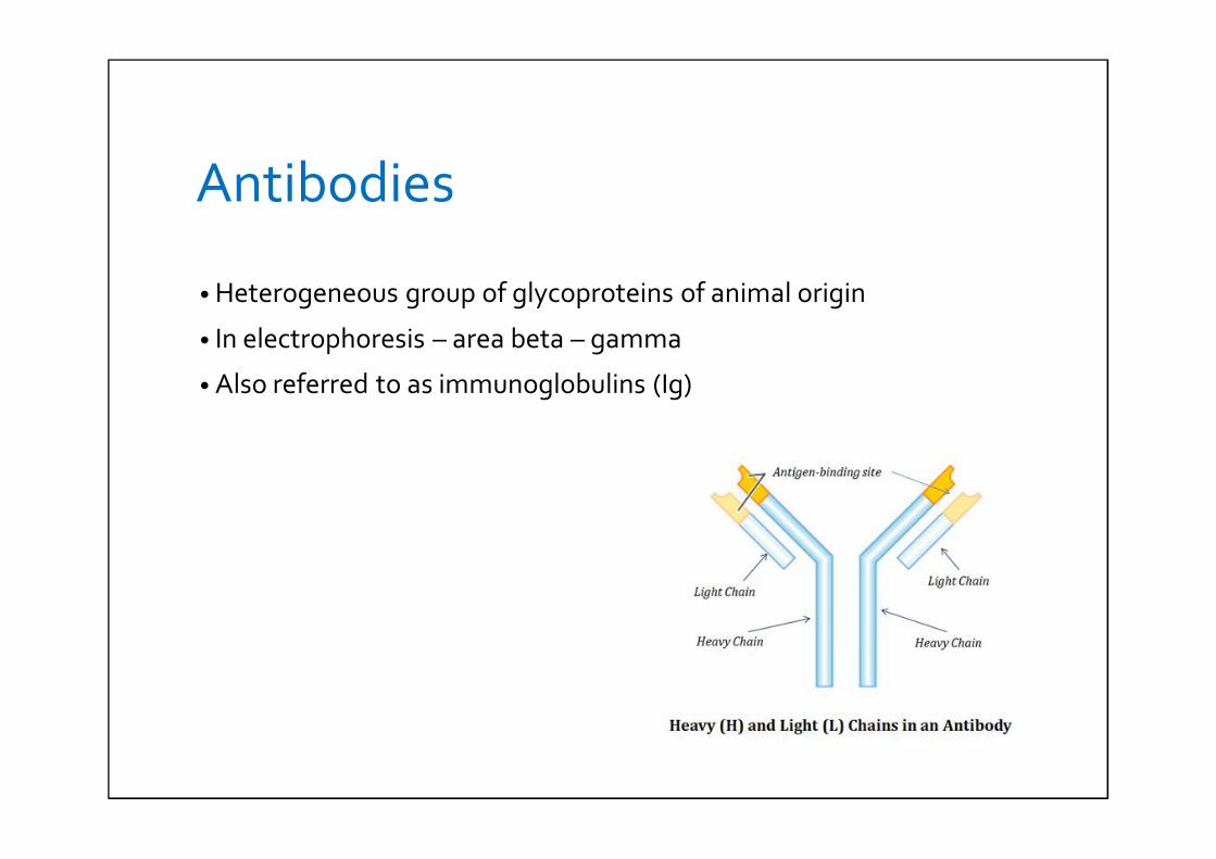
Antibodies
• Heterogeneous group of glycoproteins of animal origin
• In electrophoresis – area beta – gamma
• Also referred to as immunoglobulins (Ig)
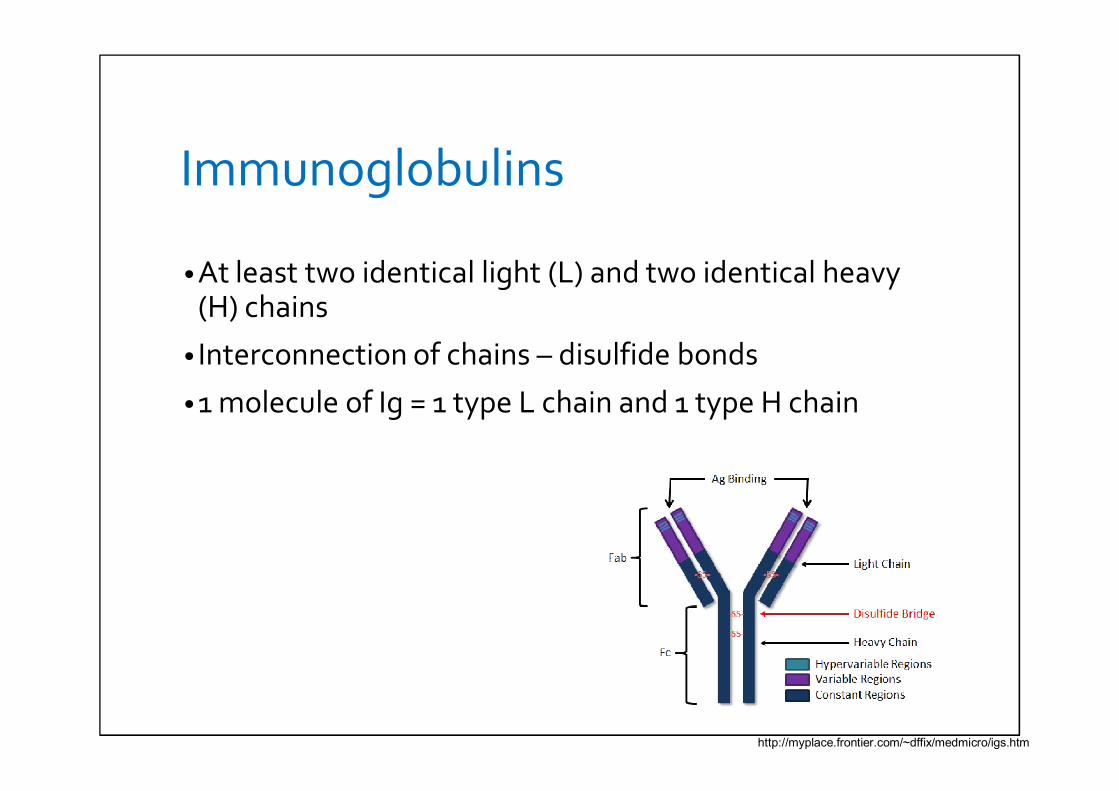
Immunoglobulins
• At least two identical light (L) and two identical heavy(H) chains
• Interconnection of chains – disulfide bonds
• 1 molecule of Ig = 1 type L chain and 1 type H chain
http://myplace.frontier.com/~dffix/medmicro/igs.htm
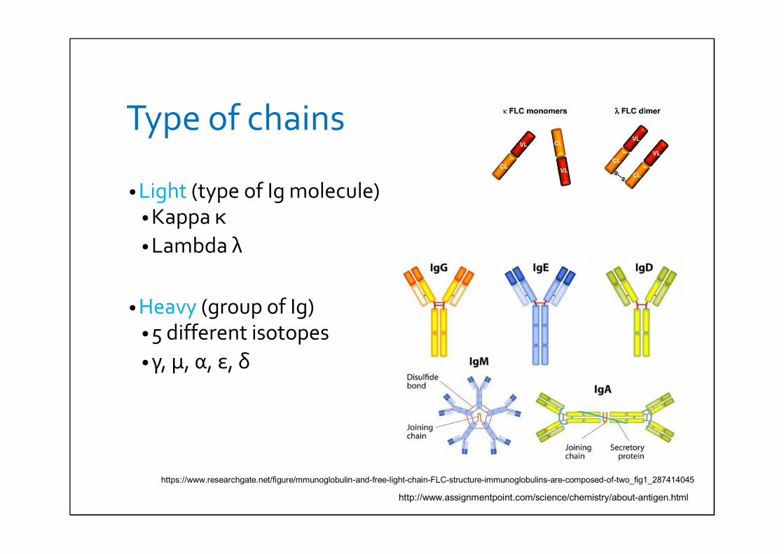
Type of chains
• Light (type of Ig molecule)• Kappa κ• Lambda λ
• Heavy (group of Ig)• 5 different isotopes• γ, μ, α, ε, δ
http://www.assignmentpoint.com/science/chemistry/about-antigen.html
https://www.researchgate.net/figure/mmunoglobulin-and-free-light-chain-FLC-structure-immunoglobulins-are-composed-of-two_fig1_287414045
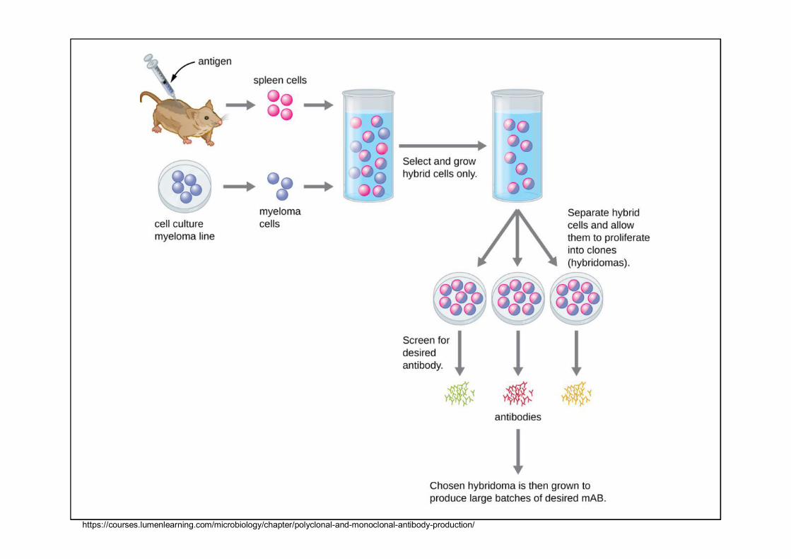
Monoclonal antibodies• Products of one clone of plasma cells derived from B-
lymphocytes
• Preparation: hybrid technology
• Principle: • Cell fusion of tumor cells with splenic lymphocytes
from immunized with mice
• Against 1 epitope of a particular antigen – identicalcopy of immunoglobin• The same primary structure• The same specificity of binding sites
• Disadvantage: a bad precipitation
https://courses.lumenlearning.com/microbiology/chapter/polyclonal-and-monoclonal-antibody-production/
Polyclonal antibodies
• Conventional antibodies
• Preparation: • Immunizing animals with an antigen• Blood serum of the immunized animal = antiserum
(contains antibodies against the antigen used forimmunization)
• Use 1 antigen for immunization – monospecific antibodies• Use of antigens mixture for immunization– polyspecific
antibodies
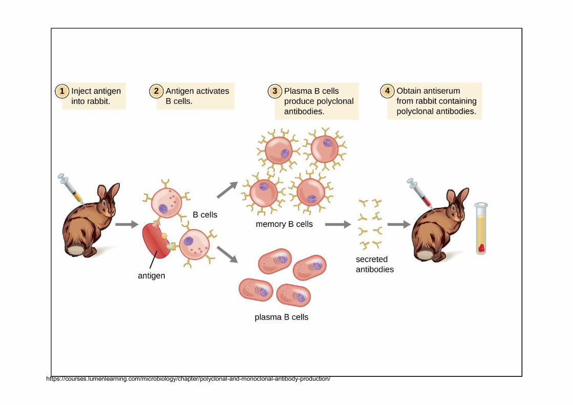
https://courses.lumenlearning.com/microbiology/chapter/polyclonal-and-monoclonal-antibody-production/
Affinity
• It expresses the energy of binding between one bindingsite on the antibody and the correspondint epitope ofantigen
• Sum of all repulsive and attractive forces between twocomplemantry structures
Avidity
• It expresses the total energy of the bonds between antibodies and antigens
• Sum of the bindins affinities of all individual antibodies binding sites with all corresponding epitopes on antigens
Immunochemical methods
• Are based on the reakce between antibody and antigen
• We can demonstrate the presence of antibodies orantigens in samples
• Basic division• Without a marker • With markers
IMMUNOCHEMICALMETHODS
I. Without a marker
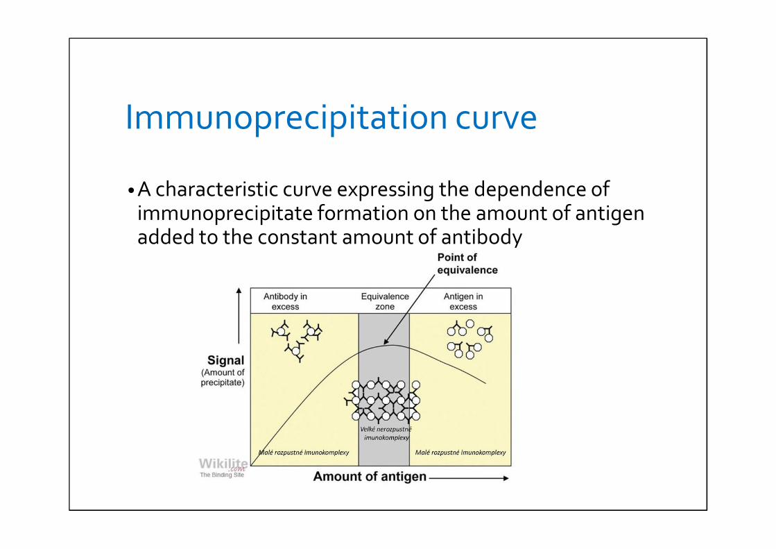
Immunoprecipitation curve
• A characteristic curve expressing the dependence of immunoprecipitate formation on the amount of antigen added to the constant amount of antibody
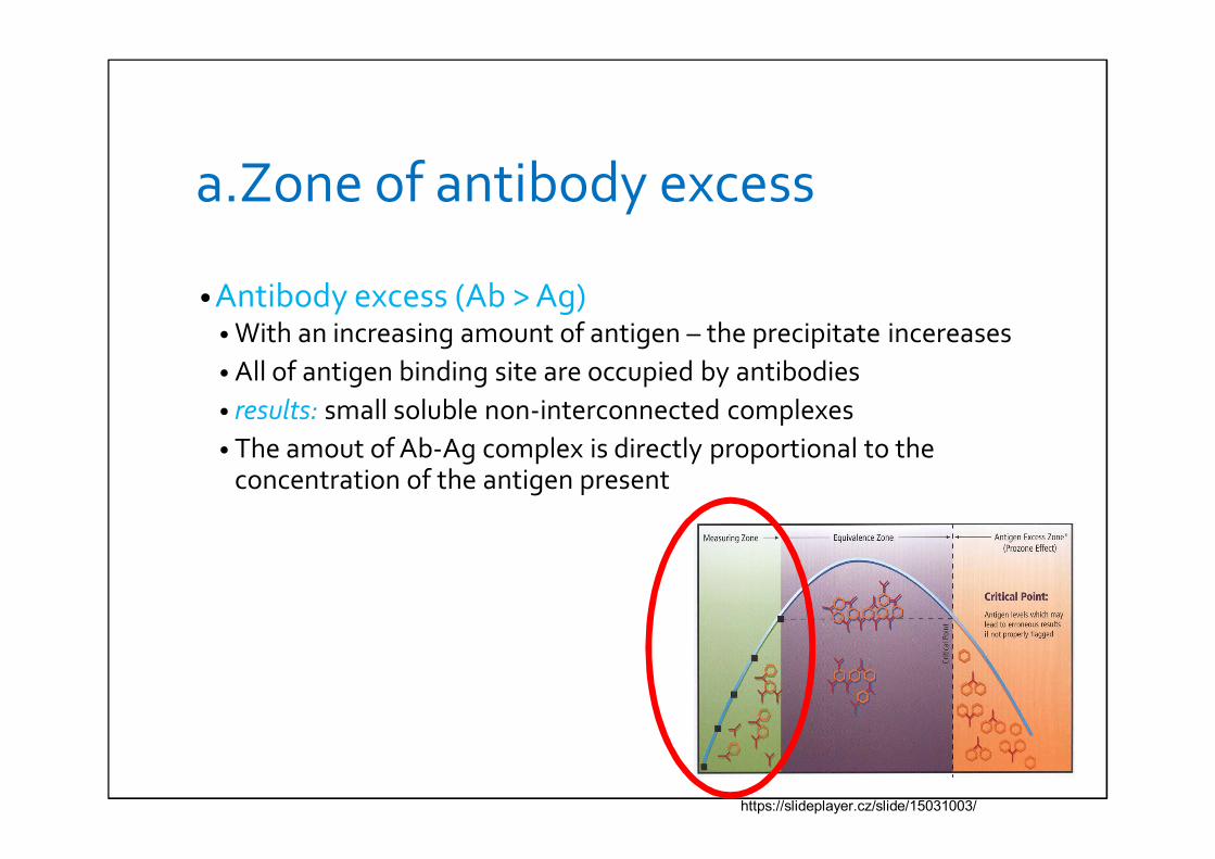
a.Zone of antibody excess
• Antibody excess (Ab > Ag)• With an increasing amount of antigen – the precipitate incereases• All of antigen binding site are occupied by antibodies• results: small soluble non-interconnected complexes• The amout of Ab-Ag complex is directly proportional to the
concentration of the antigen present
https://slideplayer.cz/slide/15031003/
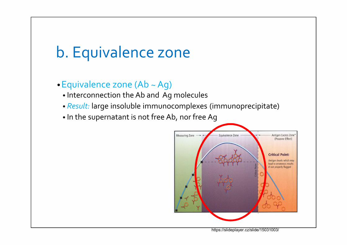
b. Equivalence zone
• Equivalence zone (Ab ~ Ag)• Interconnection the Ab and Ag molecules• Result: large insoluble immunocomplexes (immunoprecipitate)• In the supernatant is not free Ab, nor free Ag
https://slideplayer.cz/slide/15031003/
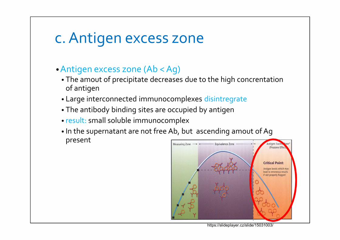
c. Antigen excess zone
• Antigen excess zone (Ab < Ag)• The amout of precipitate decreases due to the high concrentation
of antigen• Large interconnected immunocomplexes disintregrate• The antibody binding sites are occupied by antigen• result: small soluble immunocomplex• In the supernatant are not free Ab, but ascending amout of Ag
present
https://slideplayer.cz/slide/15031003/
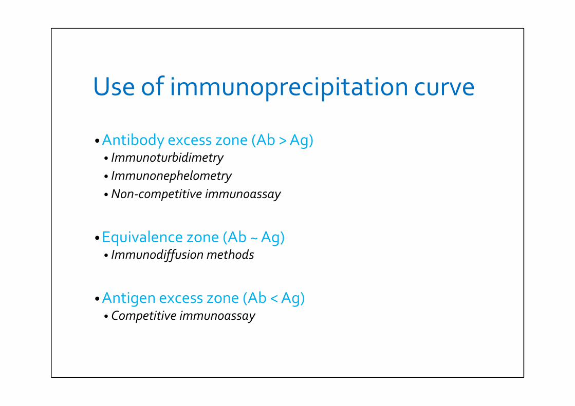
Use of immunoprecipitation curve
• Antibody excess zone (Ab > Ag)• Immunoturbidimetry• Immunonephelometry• Non-competitive immunoassay
• Equivalence zone (Ab ~ Ag)• Immunodiffusion methods
• Antigen excess zone (Ab < Ag)• Competitive immunoassay
Factors influencing precipitation
• type of Ab (e.g. IgG)
• temperature – with inceasing temperature precipitation is accelarated (e.g. 38°C)
• The relative concentration of Ag and Ab
• pH
• Ion charge
• Shape and size of the part
Immunoprecipitation reactions in gels• Gel enviroment: agar, agarose
Single immunodiffusion
• 1 component is moving (second component is evenly dispersed in the gel)
Double immunodiffusion
• Ab and Ag are moving freely
Simple radial immunodiffusion
• quantitative method• Wells in the gel contain monospecific Ab• Ag is applied to the wells• Ag diffuses radially from the wells into the gel in which
the Ab concentration is constant – until an equivalentratio between Ag and Ab
• Result: precipitate Ag-Ab complex (sharp white ring)• Ring area = the second power of the ting diameter
(directly proportional to the concentration of Ag)• These days replaced by immunonephelometry a
immunoturbidimetry
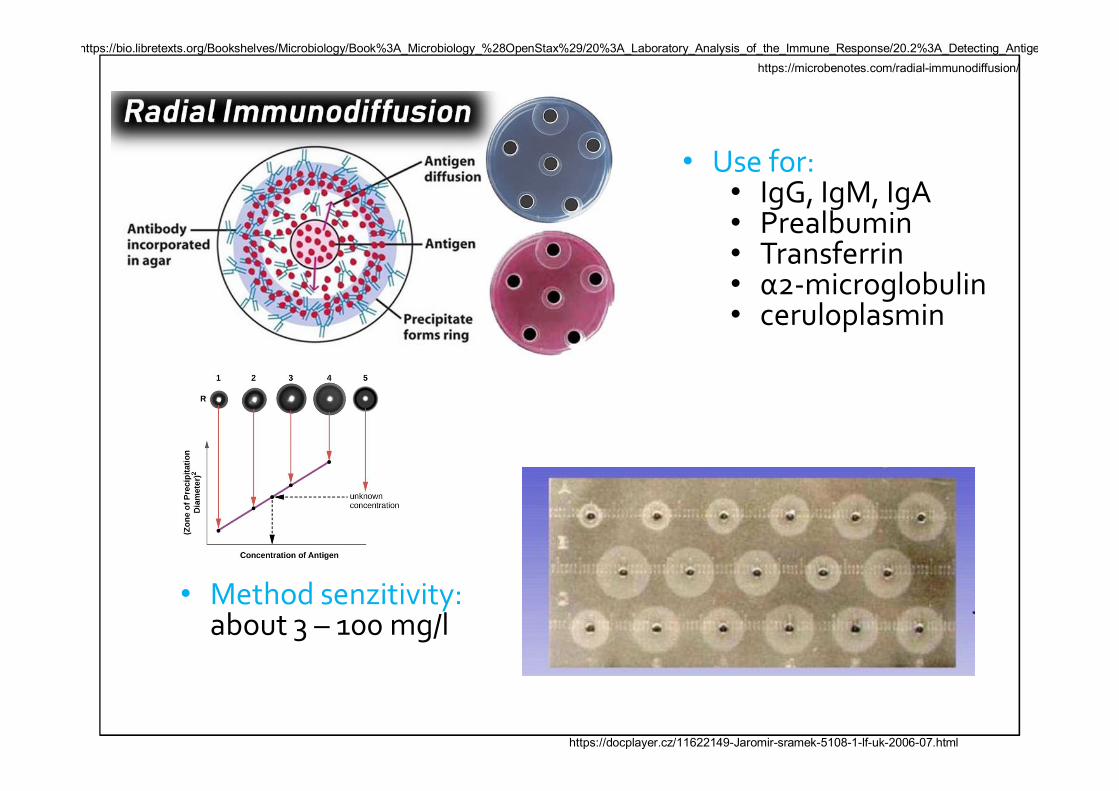
• Method senzitivity: about 3 – 100 mg/l
• Use for:• IgG, IgM, IgA• Prealbumin• Transferrin• α2-microglobulin• ceruloplasmin
https://docplayer.cz/11622149-Jaromir-sramek-5108-1-lf-uk-2006-07.html
https://bio.libretexts.org/Bookshelves/Microbiology/Book%3A_Microbiology_%28OpenStax%29/20%3A_Laboratory_Analysis_of_the_Immune_Response/20.2%3A_Detecting_Antigenhttps://microbenotes.com/radial-immunodiffusion/

Double immunodiffusion
• qualitative method• Ab and Ag diffuse against each other• Ouchterlony technique is often use• In the gel there are the wells containing antigens or
antibodies (form of a rosette)• Ag or Ab diffuse radial into surroundings• result: precipitate is formed at the Ab and Ag sites• In the presence of multiple Ag – identification of several
lines
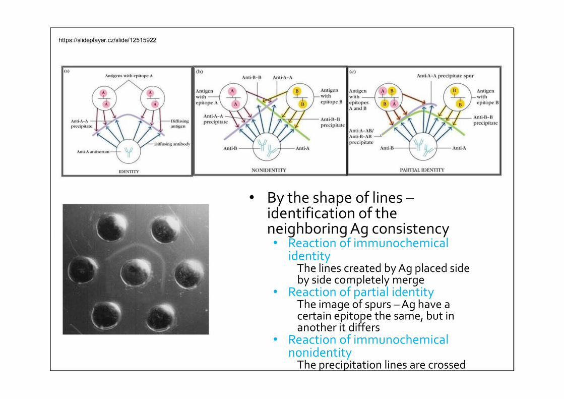
• By the shape of lines –identification of the neighboring Ag consistency • Reaction of immunochemical
identity The lines created by Ag placed side by side completely merge
• Reaction of partial identityThe image of spurs – Ag have a certain epitope the same, but in another it differs
• Reaction of immunochemical nonidentity
The precipitation lines are crossed
https://slideplayer.cz/slide/12515922
Immunoelectrophoresis
• Combination of immunodiffusion a electrophoresis
• Faster than immunodiffusion methods
• Low sensitivity and more laborious than immunoassays
• Types:• Clasical immunoelectrophoresis (qualitative)• rocket immunoelectrophoresis (quantitative)• inverse immunoelectrophoresis (qualitative)• 2D immunoelectrophoresis (quantitative)
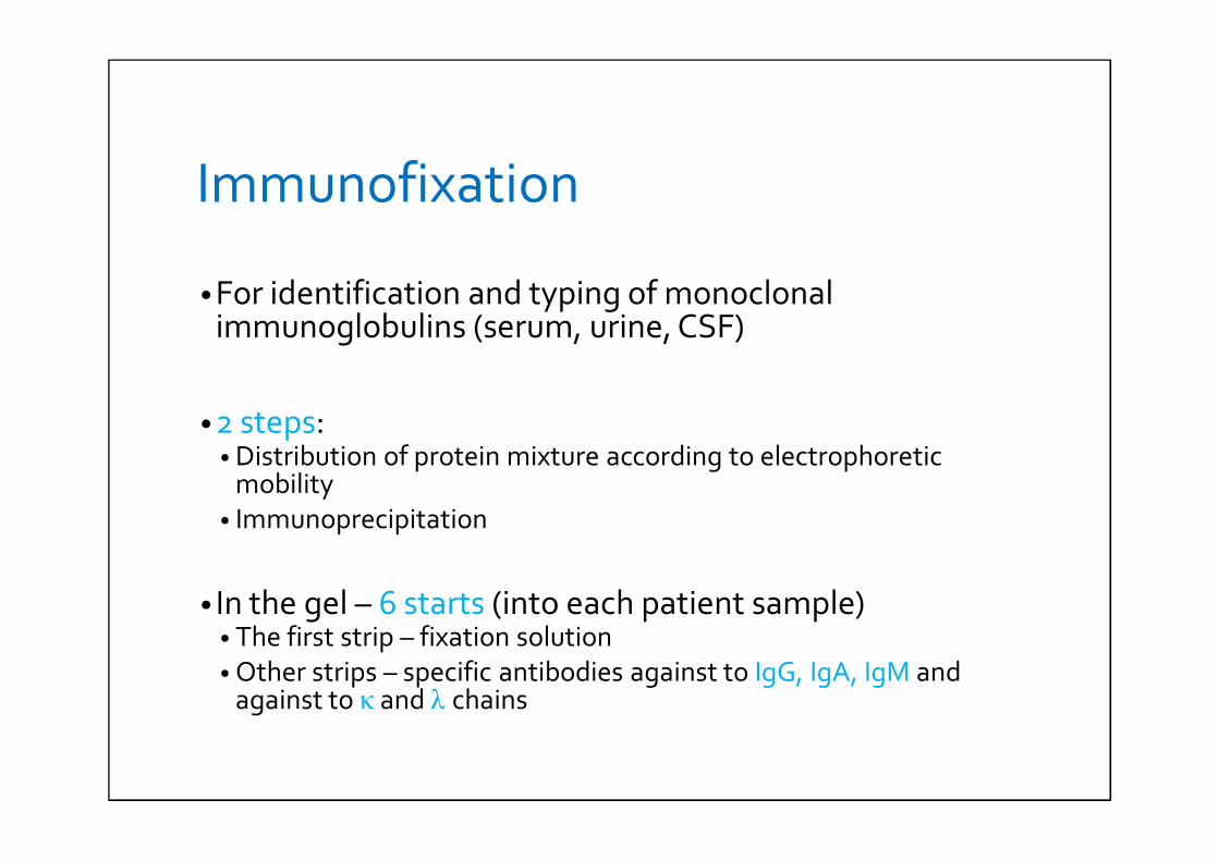
Immunofixation
• For identification and typing of monoclonalimmunoglobulins (serum, urine, CSF)
• 2 steps:• Distribution of protein mixture according to electrophoretic
mobility• Immunoprecipitation
• In the gel – 6 starts (into each patient sample)• The first strip – fixation solution• Other strips – specific antibodies against to IgG, IgA, IgM and
against to κ and λ chains
https://slideplayer.com/slide/7467983/
Use of immunofixation
• Demonstration of monoclonal immunoglobulins
• 10x higher senzitivity than immunoelectrophoresis
• IF – after immunoelectrophoresis with unclear findings
M-komponent (papaprotein)
•Ab-producing cells are insensitive to regulatory signals
•Continuous reproduction and production of antibodies
•Monoclonal gammapathy• One clone of plasma cells is uncontrollably mutiplied• May be benign • Malignant (multiple myeloma)
• Production of complete monoclonal Ig• Sometimes along with Bence-Jones protein (light chain
monomer/dimer)
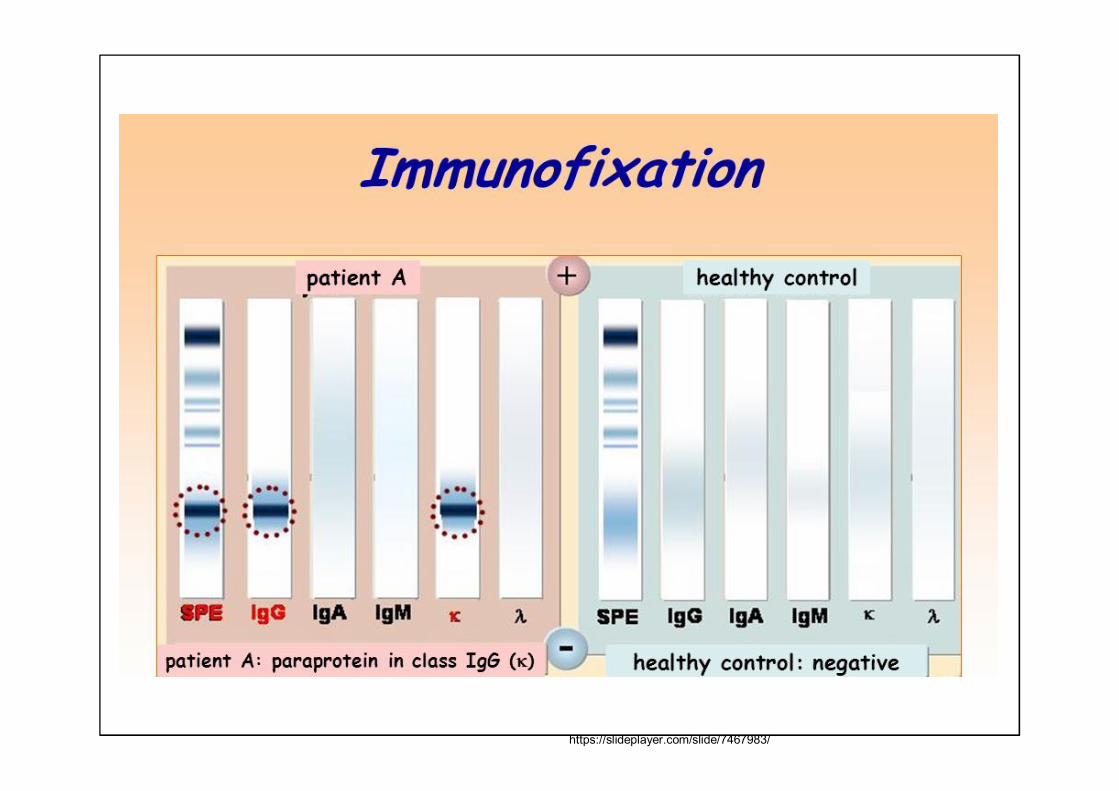
Results of immunofixation
• Polyclonal Ig• Formation of diffuse-colored precipitates
• Monoclonal Ig• Narrow, strongly stained strips in diffusion precipitate• Usually a reaction with one heavy and one light chain
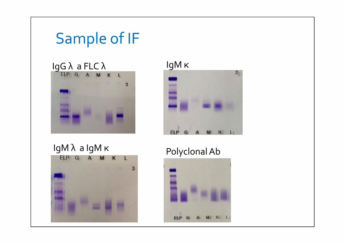
Sample of IF
IgG λ a FLC λ IgM κ
IgM λ a IgM κ Polyclonal Ab
Immunoprecipitation reactions in solution• Measurement of turbidity in solution
• The light passes through a cloudy solution and diffusses againts to the solution where is no immunochemical reaction
• Necessary antibody excess
• In antigen excess zone – falsely low value
• Modern analyzers – measures the rate of turbidity
Immunoturbidimetry, immunonephelometry• Turbidimetry
• Measures the intensity of the light transmitted in the straight line (as a conventional spectrophotometer)
• Nephelometry• Measures the intensity of scattered light that comes out of
solution in all directions• It is measured at an angle which is different from the direction of
the incident radiation• Usually 45 ° or 90 °
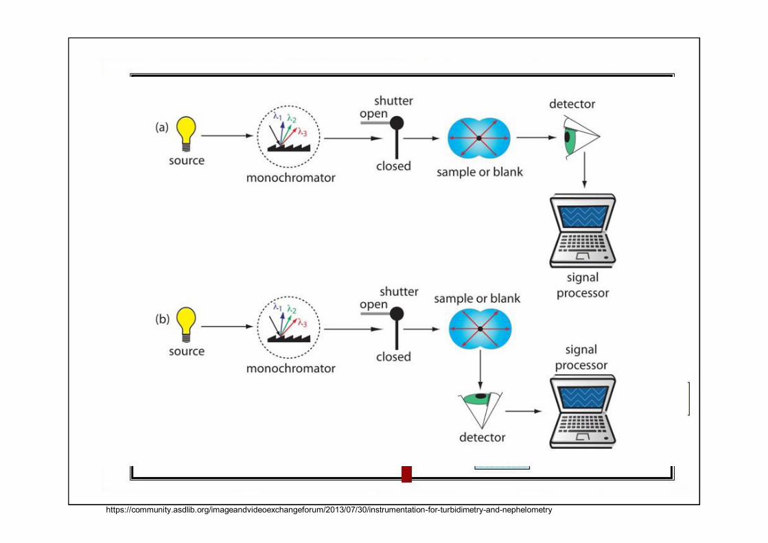
https://community.asdlib.org/imageandvideoexchangeforum/2013/07/30/instrumentation-for-turbidimetry-and-nephelometry
IMMUNOCHEMICALMETHODS
I. With markers
Immunomethods using markers
• No immunoprecipitation
• Reaction Ab + Ag – immunocomplex binding marker
• Marker:• Radioisotope• Enzyme• Fluorescent substance• Chemiluminiscent substance
Use of radioisotope
• RIA radioimmunoassay• Radioisotope-labeled Ag/hapten
• IRMA immunoradiometric assay • Radioizotopically labeled Ab
• EPRIA Enzyme Potentiated RIA• Ab with a bound enzyme that releases the radioisotope from the
added substrate
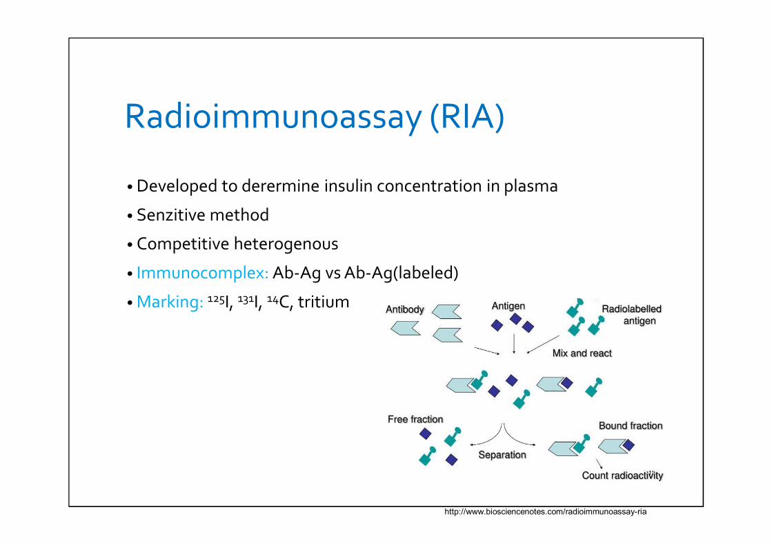
Radioimmunoassay (RIA)
• Developed to derermine insulin concentration in plasma
• Senzitive method
• Competitive heterogenous
• Immunocomplex: Ab-Ag vs Ab-Ag(labeled)
• Marking: 125I, 131I, 14C, tritium
http://www.biosciencenotes.com/radioimmunoassay-ria

Use of RIA
• Endocrinology
• Medicaments - Digitoxin, digoxin
• Toxicology – presence of drugs
• Hepatitida B in blood donors
• Immunology: systemic lupus erythematosus
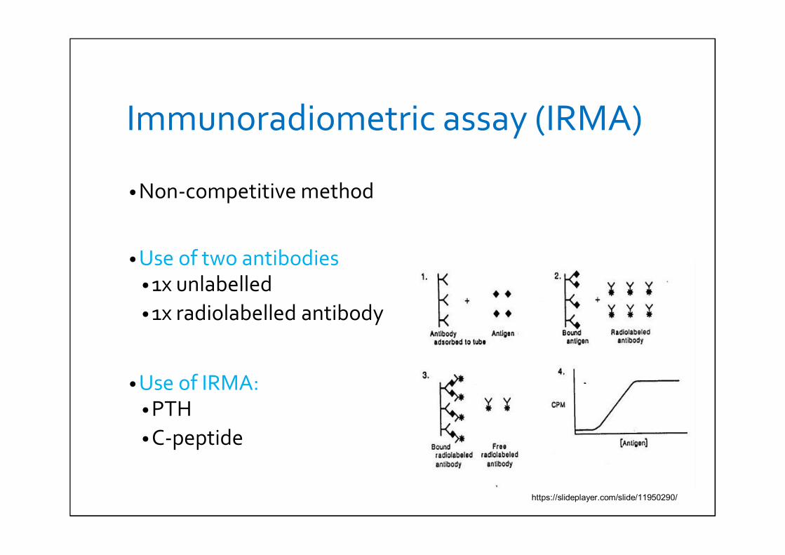
Immunoradiometric assay (IRMA)
• Non-competitive method
• Use of two antibodies• 1x unlabelled• 1x radiolabelled antibody
• Use of IRMA:• PTH• C-peptide
https://slideplayer.com/slide/11950290/

Enzyme Potentiated RIA (EPRIA)
• 100x higher senzitivy than RIA
• Enzyme labelled Ab
• Disadvantage: expensive operation
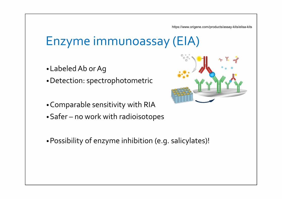
Enzyme immunoassay (EIA)
• Labeled Ab or Ag
• Detection: spectrophotometric
• Comparable sensitivity with RIA
• Safer – no work with radioisotopes
• Possibility of enzyme inhibition (e.g. salicylates)!
https://www.origene.com/products/assay-kits/elisa-kits
Enzyme immunoassay
• Enzyme labeling (peroxidase, alkaline phospatase)• Heterogenous
• Ag or Ab nonspecificifically bound to the surface of the solid phase (tubes, magnetic particles)
• types: competitive and non-competitive
• Homogeneous• Separation the bondend and labeled Ag (or Ab) is not necessary• The known amout of Ag with bound enzyme + Ab + patient
sample with unknown amout of Ag → Ag in patient sample and labeled Ag „competes“ for binding to Ab → in Ag-Ab binding -labeled Ag loses its enzyme activity
• Higher concentration of Ag in patient sample (unlabelled) – more Ab-unlabelled Ag binding → in the sample more labeled Agremains (teraining greater enzyme aktivity)
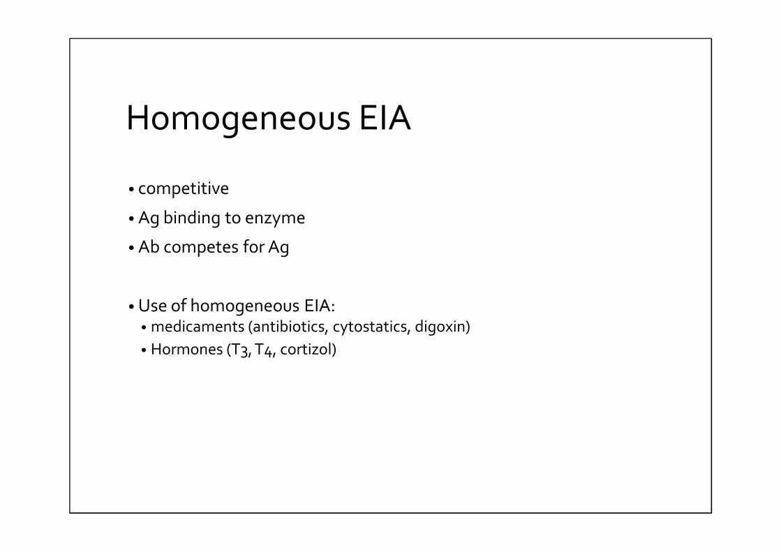
Homogeneous EIA
• competitive
• Ag binding to enzyme
• Ab competes for Ag
• Use of homogeneous EIA:• medicaments (antibiotics, cytostatics, digoxin)• Hormones (T3, T4, cortizol)
Heterogeneous enzyme immunoassays (ELISA)• The most common IA in biochemistry
• Determination of concentrations of Ag or Ab
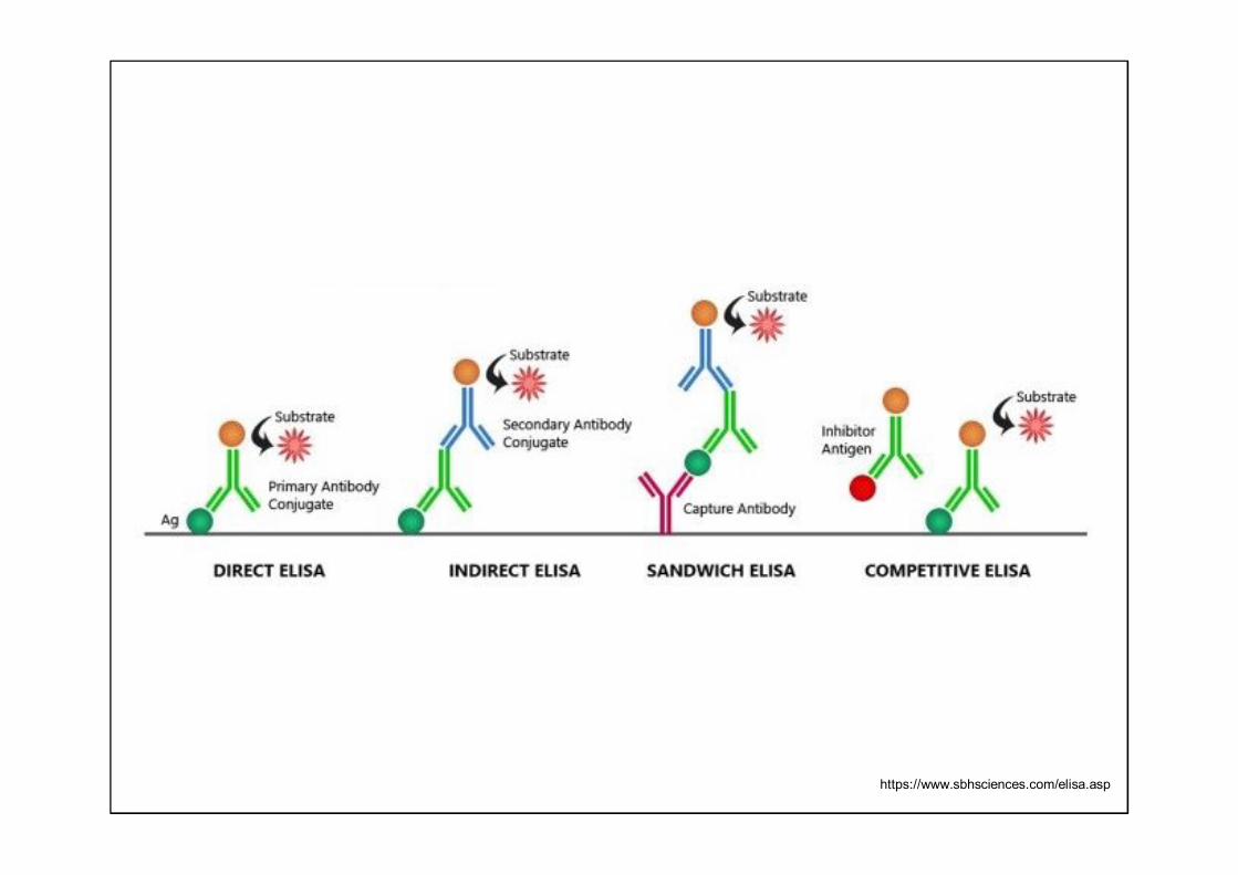
• Classification:• Direct• Indirect (non-competitive)• Competitive• Sandwich
https://www.sbhsciences.com/elisa.asp
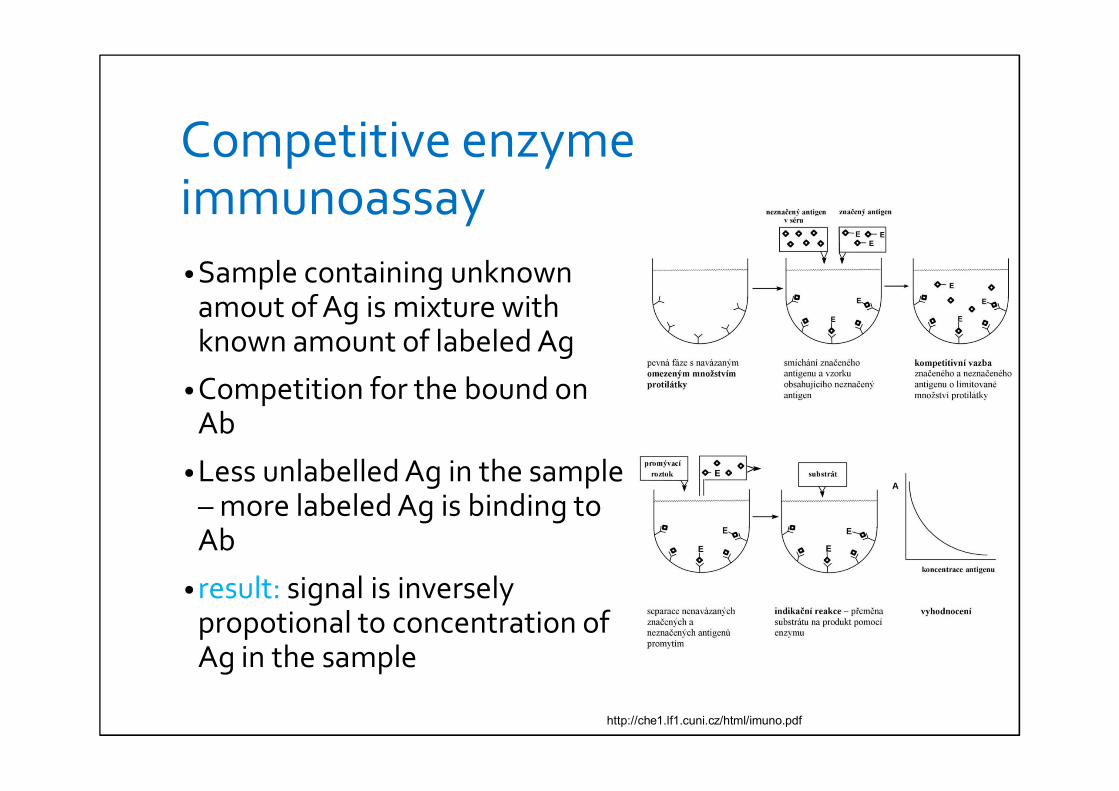
Competitive enzyme immunoassay• Sample containing unknown
amout of Ag is mixture with known amount of labeled Ag
• Competition for the bound on Ab
• Less unlabelled Ag in the sample – more labeled Ag is binding to Ab
• result: signal is inversely propotional to concentration of Ag in the sample
http://che1.lf1.cuni.cz/html/imuno.pdf
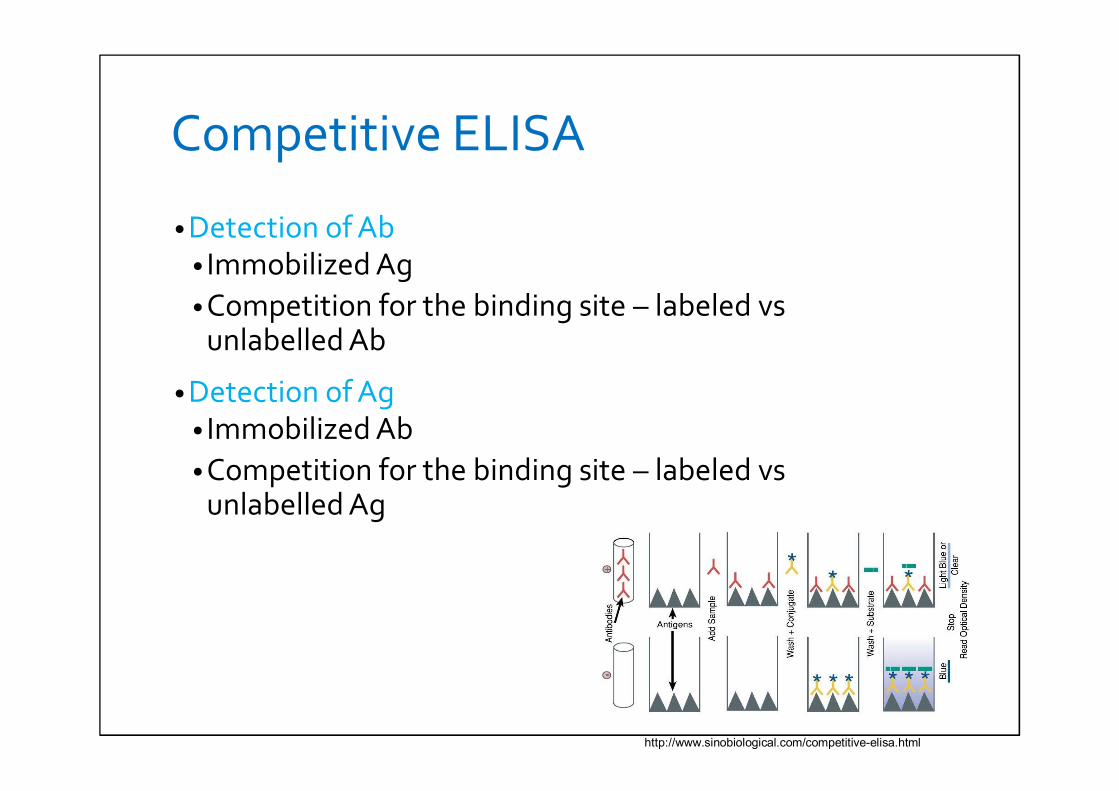
Competitive ELISA
• Detection of Ab• Immobilized Ag• Competition for the binding site – labeled vs
unlabelled Ab
• Detection of Ag• Immobilized Ab• Competition for the binding site – labeled vs
unlabelled Ag
http://www.sinobiological.com/competitive-elisa.html
Use of competitive enzyme immunoassays• Determination of small molecule concentrations with
one antigen determinant• Steroids• Thyroid hormones
• Determination of medicament levels in biological fluids
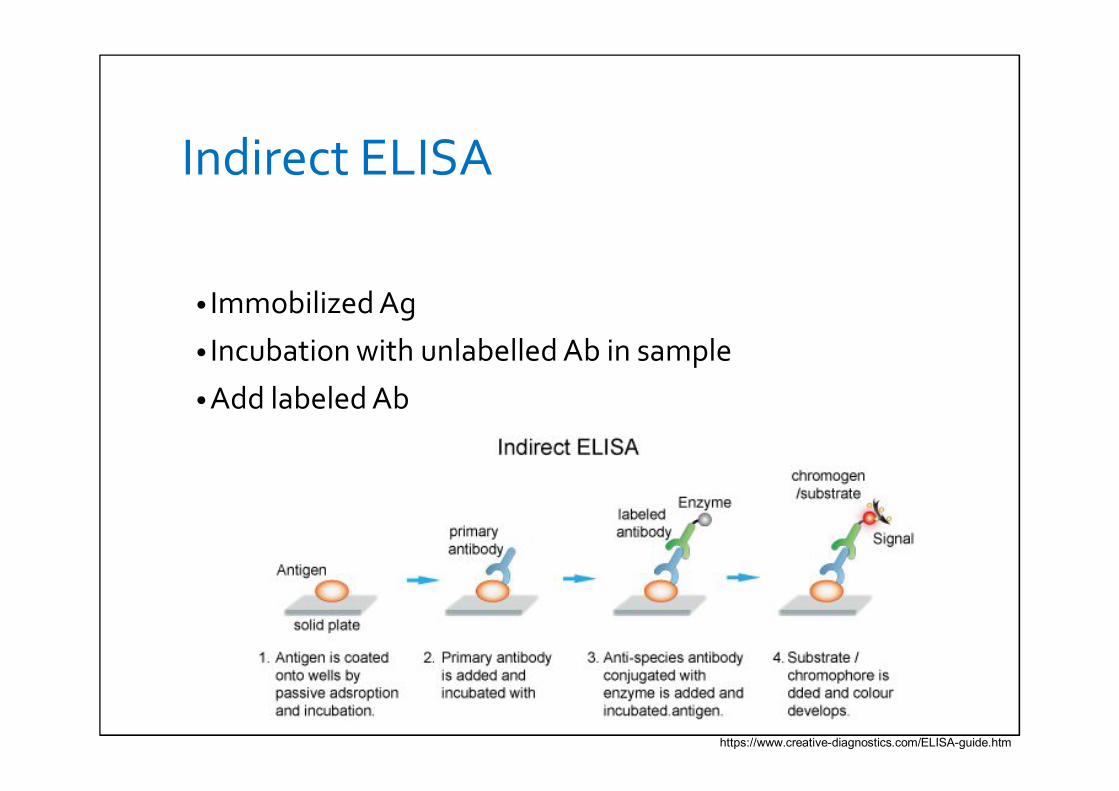
Indirect ELISA
• Immobilized Ag
• Incubation with unlabelled Ab in sample
• Add labeled Ab
https://www.creative-diagnostics.com/ELISA-guide.htm
Non-competitive enzyme immunoassays („sandwich method“)
• To determine the antigen or antibody
• Solid phase with the bound Ab or Ag which are in excess against the analyte
Non-competitive enzyme immunoassays(„sandwich method“)for determination of antigen
• Necessary 2 different Ab against different antigen determinant
• The first Ab excess is bound to solid phase (all of Ag can bebound)
• Add patient sample – the first reaction• wash• Add the second labeled Ab excess againts different antigen
determinant - the second reaction• Result: complex Ab on the solid phase – Ag – labeledAb• Interpretaion: absorbance is directly proportional to amout of
Ag
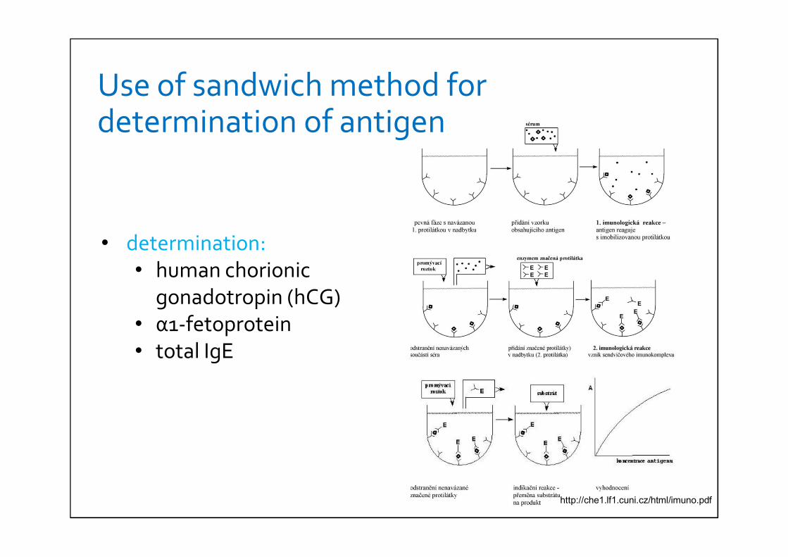
• determination:• human chorionic
gonadotropin (hCG)• α1-fetoprotein• total IgE
Use of sandwich method for determination of antigen
http://che1.lf1.cuni.cz/html/imuno.pdf
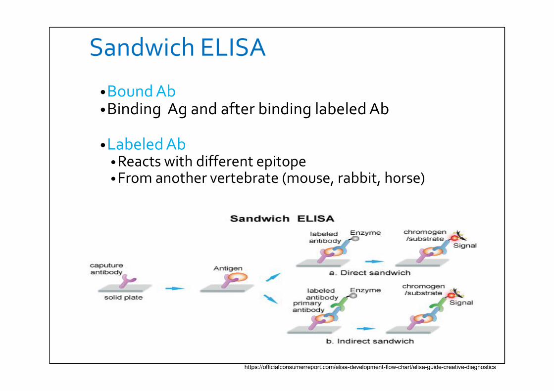
Sandwich ELISA
•Bound Ab•Binding Ag and after binding labeled Ab
•Labeled Ab • Reacts with different epitope• From another vertebrate (mouse, rabbit, horse)
https://officialconsumerreport.com/elisa-development-flow-chart/elisa-guide-creative-diagnostics
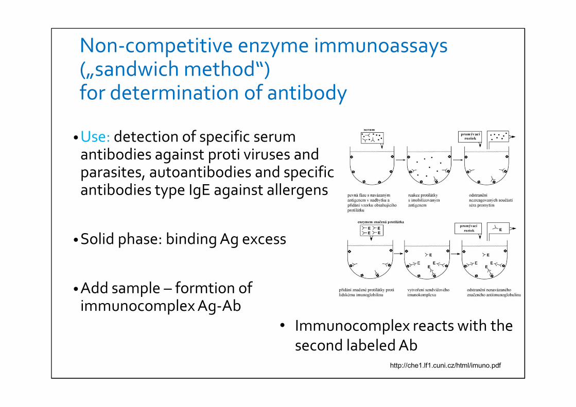
Non-competitive enzyme immunoassays(„sandwich method“)for determination of antibody
• Use: detection of specific serumantibodies against proti viruses and parasites, autoantibodies and specificantibodies type IgE against allergens
• Solid phase: binding Ag excess
• Add sample – formtion ofimmunocomplex Ag-Ab
• Immunocomplex reacts with the second labeled Ab
http://che1.lf1.cuni.cz/html/imuno.pdf
Use of ELISA I.
• Screening of blood donor• presence anti-HIV antibody• Presence hep. C antibody• hep. B antibody/antigen
• Hormones• hCG, LH• Thyroid function: TSH, T3, T4• Hormones in athletes (anabolics)
Use of ELISA II.
• Detection of allergens
• Determination of RF
• Determination of autoantibody (SLE)
• Measurement of toxins
• Detection of drugs• cocaine, opiates, THC
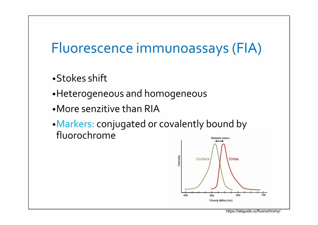
Fluorescence immunoassays (FIA)
•Stokes shift•Heterogeneous and homogeneous•More senzitive than RIA•Markers: conjugated or covalently bound by fluorochrome
https://labguide.cz/fluorochromy/
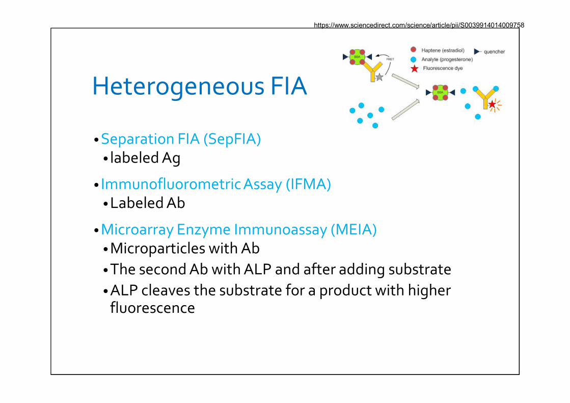
Heterogeneous FIA
• Separation FIA (SepFIA)• labeled Ag
• Immunofluorometric Assay (IFMA)• Labeled Ab
• Microarray Enzyme Immunoassay (MEIA)• Microparticles with Ab• The second Ab with ALP and after adding substrate• ALP cleaves the substrate for a product with higher
fluorescence
https://www.sciencedirect.com/science/article/pii/S0039914014009758
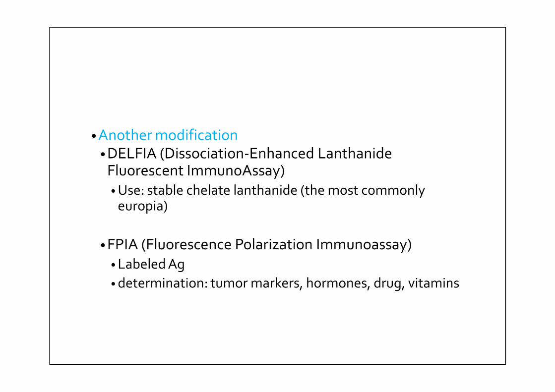
• Another modification• DELFIA (Dissociation-Enhanced Lanthanide
Fluorescent ImmunoAssay)• Use: stable chelate lanthanide (the most commonly
europia)
• FPIA (Fluorescence Polarization Immunoassay)• Labeled Ag• determination: tumor markers, hormones, drug, vitamins
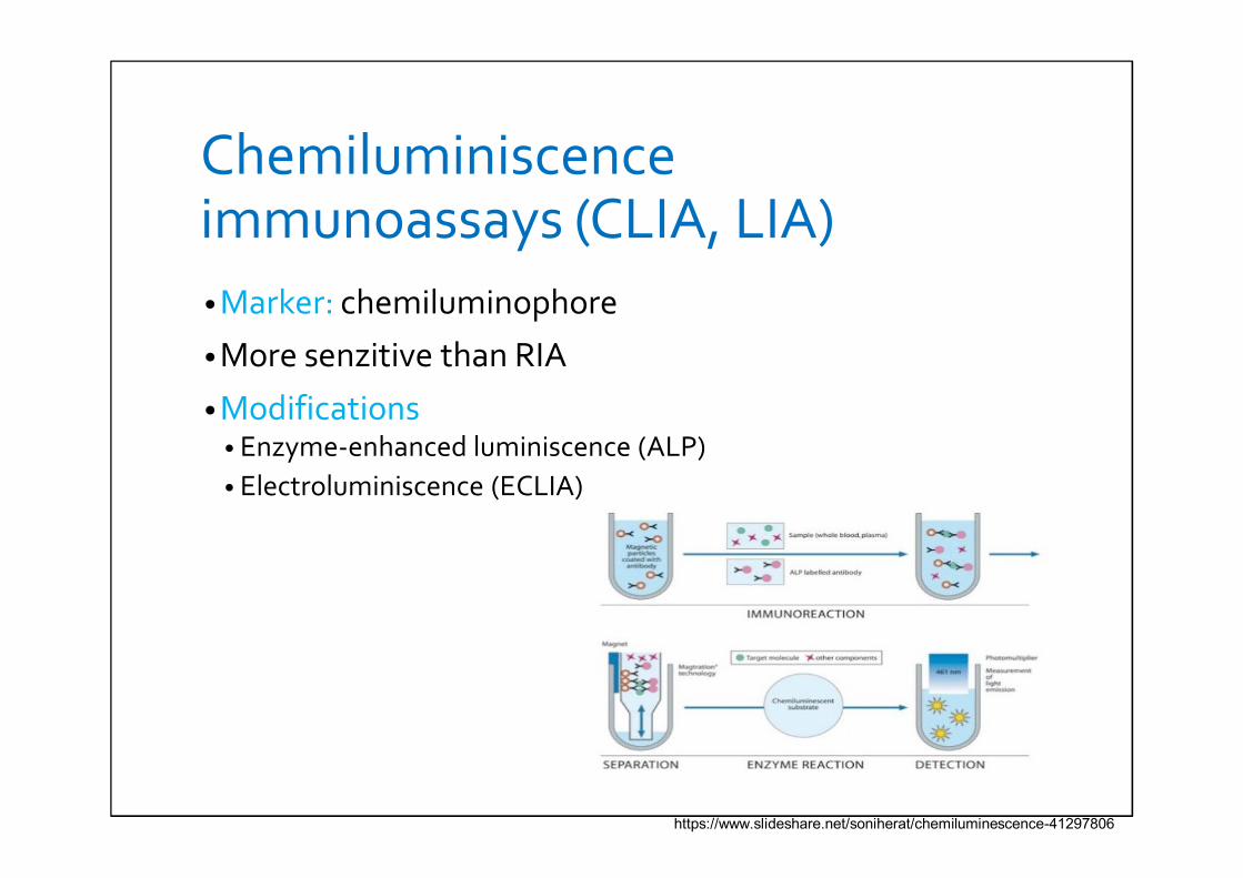
Chemiluminiscence immunoassays (CLIA, LIA)• Marker: chemiluminophore
• More senzitive than RIA
• Modifications• Enzyme-enhanced luminiscence (ALP)• Electroluminiscence (ECLIA)
https://www.slideshare.net/soniherat/chemiluminescence-41297806
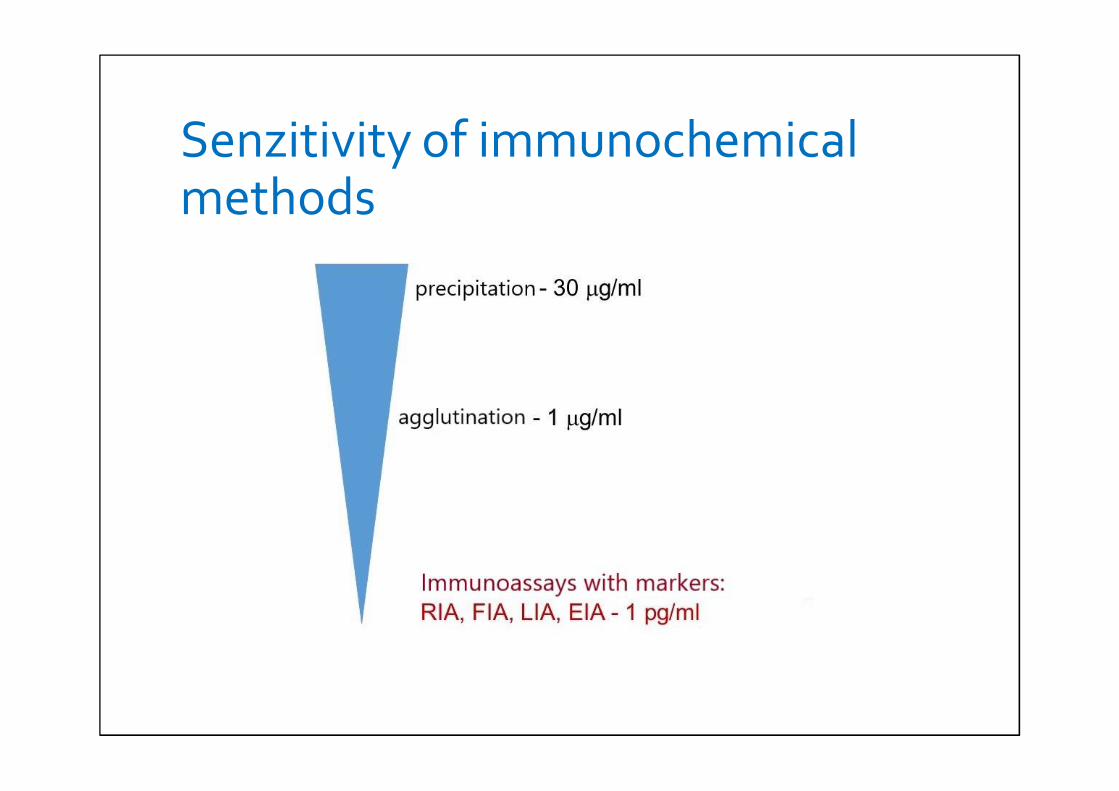
Senzitivity of immunochemical methods
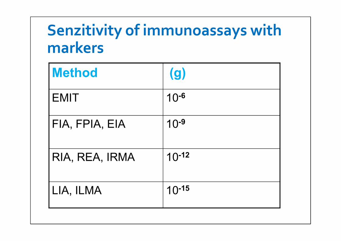
Senzitivity of immunoassays with markersMethod (g)
EMIT 10-6
FIA, FPIA, EIA 10-9
RIA, REA, IRMA 10-12
LIA, ILMA 10-15
PRACTICAL EXERCISE
1) Blood groups
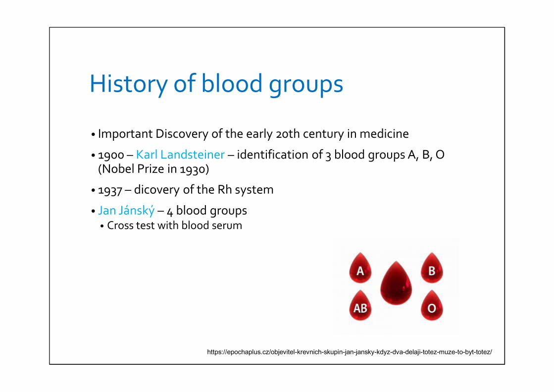
History of blood groups
• Important Discovery of the early 20th century in medicine
• 1900 – Karl Landsteiner – identification of 3 blood groups A, B, O (Nobel Prize in 1930)
• 1937 – dicovery of the Rh system
• Jan Jánský – 4 blood groups• Cross test with blood serum
https://epochaplus.cz/objevitel-krevnich-skupin-jan-jansky-kdyz-dva-delaji-totez-muze-to-byt-totez/
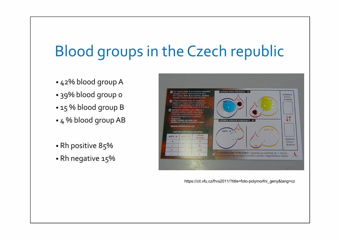
Blood groups in the Czech republic
• 42% blood group A
• 39% blood group 0
• 15 % blood group B
• 4 % blood group AB
• Rh positive 85%
• Rh negative 15%
https://cit.vfu.cz/frvs2011/?title=foto-polymorfni_geny&lang=cz
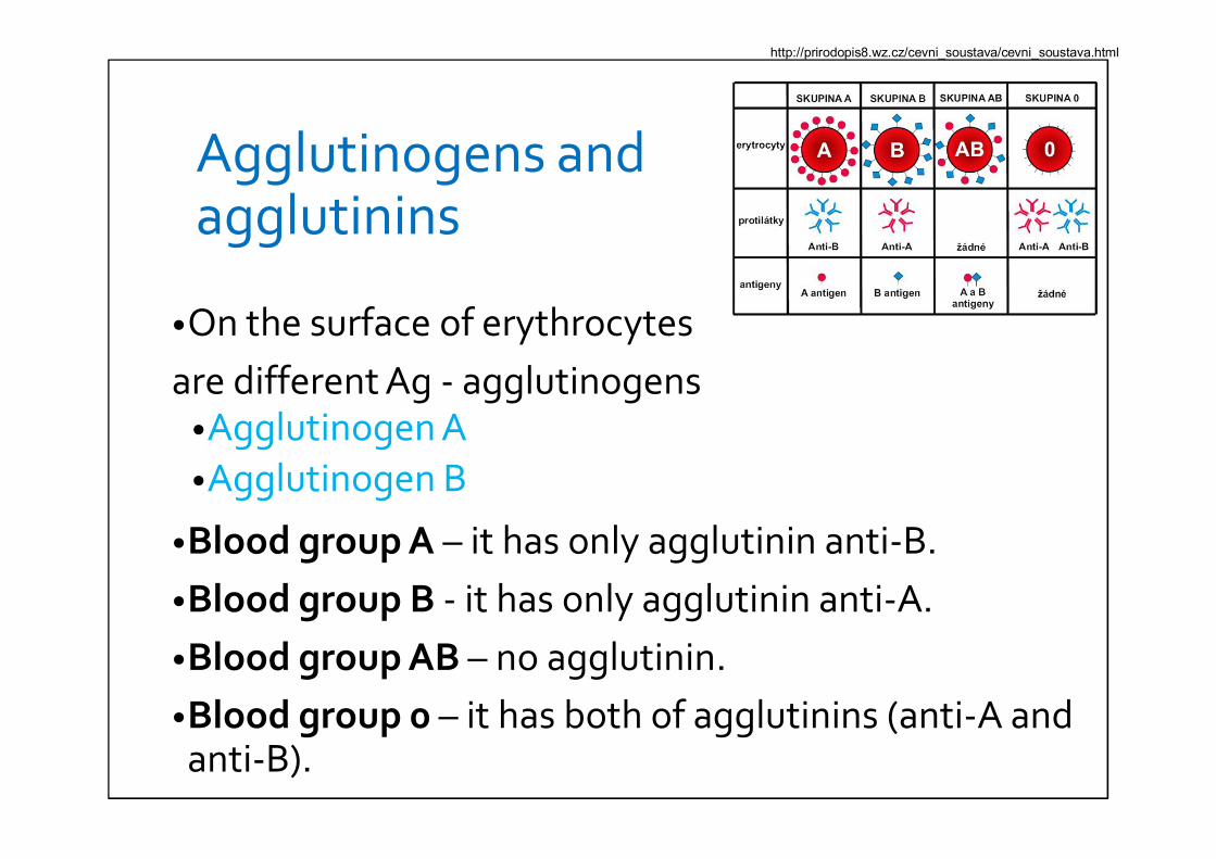
Agglutinogens and agglutinins
•On the surface of erythrocytesare different Ag - agglutinogens
•Agglutinogen A•Agglutinogen B
•Blood group A – it has only agglutinin anti-B.•Blood group B - it has only agglutinin anti-A.•Blood group AB – no agglutinin.•Blood group 0 – it has both of agglutinins (anti-A and anti-B).
http://prirodopis8.wz.cz/cevni_soustava/cevni_soustava.html
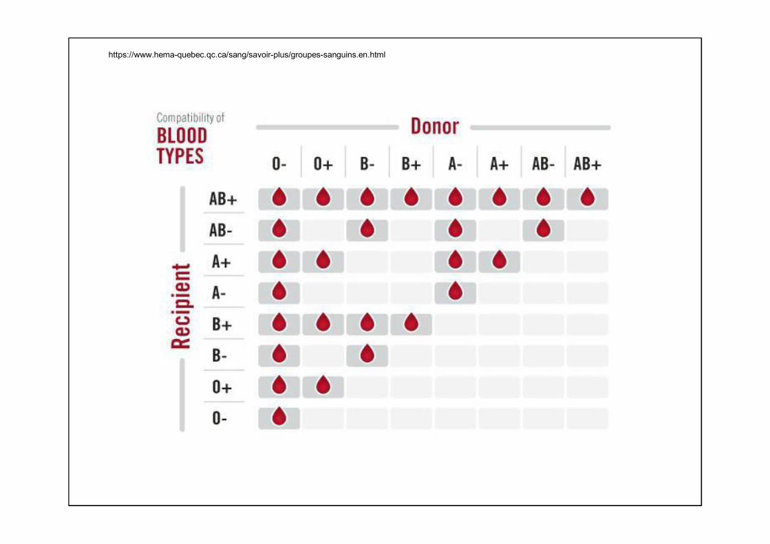
https://www.hema-quebec.qc.ca/sang/savoir-plus/groupes-sanguins.en.html
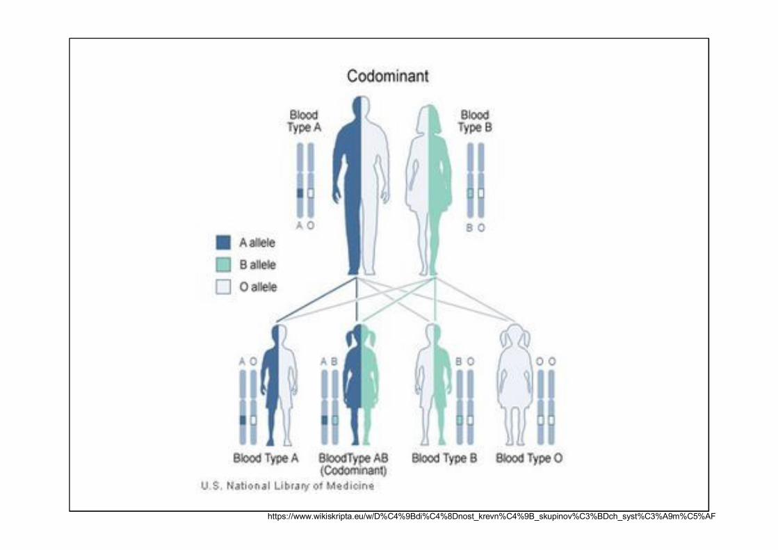
https://www.wikiskripta.eu/w/D%C4%9Bdi%C4%8Dnost_krevn%C4%9B_skupinov%C3%BDch_syst%C3%A9m%C5%AF
PRACTICAL EXERCISE
2) CRP
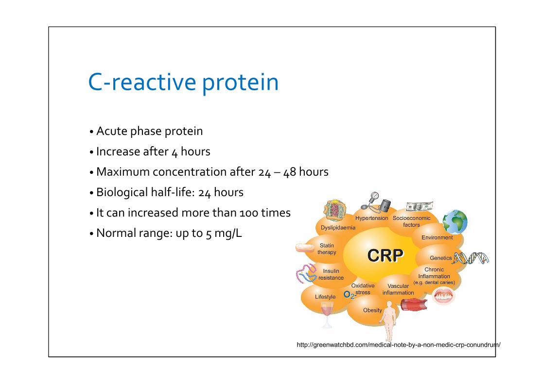
C-reactive protein
• Acute phase protein
• Increase after 4 hours
• Maximum concentration after 24 – 48 hours
• Biological half-life: 24 hours
• It can increased more than 100 times
• Normal range: up to 5 mg/L
http://greenwatchbd.com/medical-note-by-a-non-medic-crp-conundrum/
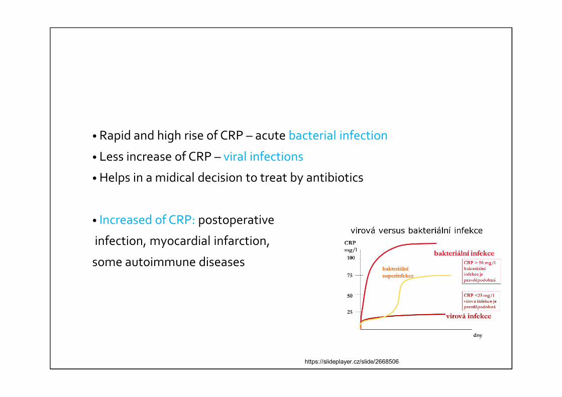
• Rapid and high rise of CRP – acute bacterial infection
• Less increase of CRP – viral infections
• Helps in a midical decision to treat by antibiotics
• Increased of CRP: postoperative
infection, myocardial infarction,
some autoimmune diseases
https://slideplayer.cz/slide/2668506

PRACTICE GUIDELINES
Determination of blood groups
• Chapter 5.3.
• Blood groups:• A, B, AB, 0
• On your desk – one microscope glass slide• 1x drop of anti-A (blue solution)• 1x drop of anti-B (yellow solution)
• 2 drops of your blood next to both diagnostic sera
• Mixture each drop of your blood with a drop of anti-A or anti-B
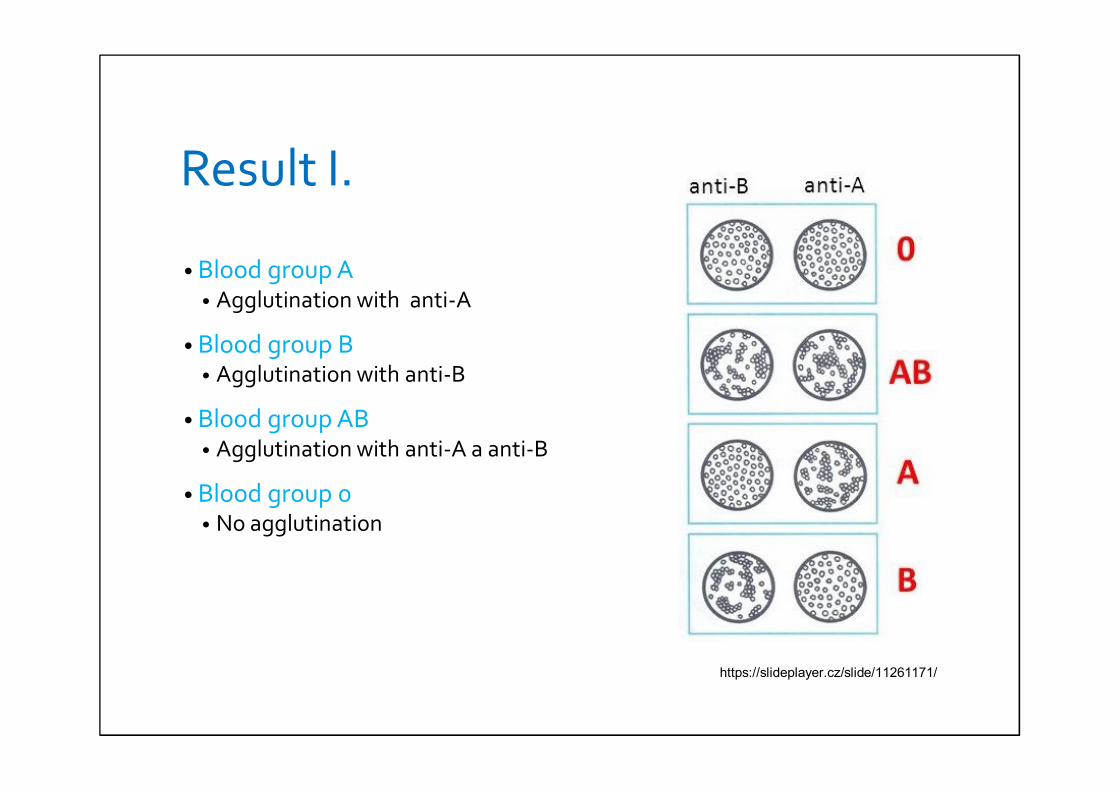
Result I.
• Blood group A• Agglutination with anti-A
• Blood group B• Agglutination with anti-B
• Blood group AB• Agglutination with anti-A a anti-B
• Blood group 0• No agglutination
https://slideplayer.cz/slide/11261171/
Determination of CRP
• Chapter 5.4
• On your desk – test card with 3 black circle
• Into each circle – 1 drop of reagent A (white bottle)
• Into the first black circle – 50 μL of patient sample• Into the second black circle - 1 drop of positive control (red bottle)• Into the third black circle – 1 drop of negative control (blue bottle)
• In each black circle – mixture reagents by a disposable stiring rod (or use tips for pipettes)
• Use a new stirring rod for each circle!!!

Determination of your CRP
• You can determinate your own CRP.
• You need at least 100 μL of your blood (from your fingertip).
Result II.
• After two minutes of mixing by waving the card check the presence of visible agglutination.
• Agglutination – concentration of CRP is higher than 6 mg/L
• No agglutination - concentration of CRP is lower than 6 mg/l
Conclusion of PT
• Blood groups• What did you see after mixture of blood drop with anti-A or anti-B?• Determinate your blood group.• You can work in couples (one determination of blood group in couple is
enough).
• CRP determination• What did you see after mixture of patient serum (or your serum) with
reagent A?• Did a positive/negative control agglutinate?• Was patient sample (or your sample) positive or negative?• You can work in couples (one determination of CRP in couple is enough).