Embed Size (px)
Citation preview
Immuno-Golgi as a Tool for Analyzing Neuronal 3D-Dendritic Structure in Phenotypically CharacterizedNeuronsLuısa Pinto1,2*, Antonio Mateus-Pinheiro1,2, Monica Morais1,2, Joao Miguel Bessa1,2, Nuno Sousa1,2*
1 School of Health Sciences, Life and Health Sciences Research Institute, University of Minho, Braga, Portugal, 2 PT Government Associate Laboratory, Life and Health
Sciences Research Institute/3B’s, Braga/Guimaraes, Portugal
Abstract
Characterization of neuronal dendritic structure in combination with the determination of specific neuronal phenotype ortemporal generation is a challenging task. Here we present a novel method that combines bromodioxyuridine (BrdU)immunohistochemistry with Golgi-impregnation technique; with this simple non-invasive method, we are able to determinethe tridimensional structure of dendritic arborization and spine shape of neurons born at a specific time in the hippocampusof adult animals. This analysis is relevant in physiological and pathological conditions in which altered neurogenesis isimplicated, such as aging or emotional disorders.
Citation: Pinto L, Mateus-Pinheiro A, Morais M, Bessa JM, Sousa N (2012) Immuno-Golgi as a Tool for Analyzing Neuronal 3D-Dendritic Structure in PhenotypicallyCharacterized Neurons. PLoS ONE 7(3): e33114. doi:10.1371/journal.pone.0033114
Editor: Anna Dunaevsky, University of Nebraska Medical Center, United States of America
Received December 21, 2011; Accepted February 4, 2012; Published March 12, 2012
Copyright: � 2012 Pinto et al. This is an open-access article distributed under the terms of the Creative Commons Attribution License, which permitsunrestricted use, distribution, and reproduction in any medium, provided the original author and source are credited.
Funding: This work was supported by the Portuguese Foundation for Science and Technology (FCT) (PTDC/SAU-NEU/105180/2008). Luisa Pinto is funded by afellowship from the FCT (SFRH/BPD/47174/2008). The funders had no role in study design, data collection and analysis, decision to publish, or preparation of themanuscript.
Competing Interests: The authors have declared that no competing interests exist.
* E-mail: [email protected] (LP); [email protected] (NS)
Introduction
The unique phenomenon of adult neurogenesis adds a new
dimension to neuroplasticity in the adult brain [1,2]. By definition,
adult neurogenesis comprises proliferation, migration and differ-
entiation phases, before the newly-born neurons are incorporated
in pre-existent neuronal networks. Using distinct tools to
monitorize adult neurogenesis many researchers try to unveil the
functional implications of adult neurogenesis. Strikingly, this
neuroplastic phenomenon is disrupted in many disorders [3,4]
and, thus, its analysis, particularly when combined with comple-
mentary methods became of great relevance to dissect the
underlying mechanisms of those disorders.
Neuronal proliferation and survival can be regulated by several
factors [5,6]. Previous studies have proposed that imbalances in
hippocampal adult neurogenesis could be involved in the
pathophysiology of depression and in the actions of antidepressant
drugs, thus giving rise to a ‘‘neurogenic hypothesis’’ of depression
[7]. Decreased proliferation and neuronal structural changes,
within the hippocampus and other brain regions, are also
increasingly recognized as key to the pathophysiology of
depression [8].
To study stem cell activity in vivo, it is necessary to identify
proliferating cells, but also to stably label their progeny so that
other components of neurogenesis can be appreciated. Herein we
describe a novel, non-invasive, method that combines BrdU
immunohistochemistry with Golgi-impregnation; with this new
method we can trace newborn neurons (BrdU labeling) and study
their dendritic and spine structure, (3D morphometric analysis of
Golgi-impregnated neurons) in distinct experimental conditions.
Methods
Ethics StatementAll procedures were carried out in accordance with National
guidelines (Portaria nu 1005/92), with the European Union
Directive 2010/63/EU and NIH guidelines on animal care and
experimentation. This study was approved by the Ethical
committee board of the Portuguese Veterinary Direction (DGV)
as stated in the document with the reference 023248 in 11th of
October of 2005.
Chronic mild stress protocolAdult male Wistar rats (Charles-River Laboratories) were either
handled (control) or submitted to a chronic mild stress (CMS)
protocol [9]. To assure interference with neuronal proliferation,
we administered methyazoxymethanol (MAM), an alkylating
agent that arrests cellular proliferation (see Methods S1).
Golgi-Cox staining4 weeks after BrdU injections (100 mg/kg), rats were anaesthe-
tized with sodium pentobarbital (Eutasil, 60 mg/Kg i.p.; Ceva
Saude Animal, Portugal) and perfused with 0.9% saline. Brains
were removed, dropped into Golgi-Cox solution and kept in the
dark for 15 days. Next, they were transferred to a 30% sucrose
solution and kept in the refrigerator for 2 to 5 days in the dark
until they sink. Sections (200 mm) were obtained in a vibratome
(MicromHM-650V) and transferred to 24-well multiwell plate
(Nunc) filled with distilled water for 15 min and then dipped in
ammonium hydroxide (Sigma Aldrich) for 5 min in the dark.
Sections were washed with distilled water twice, 10 min each, and
PLoS ONE | www.plosone.org 1 March 2012 | Volume 7 | Issue 3 | e33114

dipped in Kodak Fix solution (Rapid fixer; Sigma Aldrich) for
20 min. After washes in distilled water, 10 min each, sections were
dipped in PBS 16, and kept cool in the refrigerator.
BrdU StainingAfter Golgi-Cox staining, sections were transferred to 6-well
multiwell plates with citrate buffer (10 mM; pH = 6). For antigen
retrieval sections were heated for 5 min in the microwave to near
100u to expose the BrdU epitope in the tissue, resulting in brighter
fluorescent labeling of the cells that incorporated BrdU [10,11]
filter your current search. Sections were then rinsed in TBS 3
times, for 10 min and incubated with primary BrdU antibody
(1:50 in 0.5% TritonH-X 100 and 10% normal goat serum (NGS);
rat anti-mouse, Novocastra) overnight at 4uC. The next day,
sections were rinsed with TBS and incubated with secondary
antibody (1:1000 in 0.5% TritonH-X 100 and 10% NGS; anti-
mouse Alexa Fluor 488, Invitrogen) for 2 h at RT. Finally, sections
were incubated in DAPI (1 mg/ml) for 10 min at RT and then
rinsed in TBS. Sections were mounted in superfrost slides (Menzel-
Glaser) using Vectashield mounting medium (Vector Labs).
Confocal microscopy and stereological analysesImaging of neurons was performed using an Olympus FV1000
laser scanning confocal microscope (emission wavelength 488 and
brightfield) at high magnification (640). Sections were optically
sectioned using 1–2 mm intervals and cells rotated in orthogonal
planes to verify double labeling. For each selected neuron that
showed co-localization of Golgi-Cox with BrdU, all branches of
the dendritic tree were reconstructed at 606 (oil) magnification
using a motorized microscope (Axioplan2; Carl Zeiss) and
Neurolucida software with the AutoNeuron extension module. A
three-dimensional analysis of the reconstructed neurons was
performed using NeuroExplorer software (Microbrightfield). For
control rats, with and without MAM administration, 25 and 28
newborn neurons were reconstructed, respectively. For rats
exposed to CMS, with and without MAM administration, 26
and 23 newborn neurons were reconstructed, respectively. For
each neuron we examined the total dendritic length and the
percentage of dendritic spine types. Three-dimensional Sholl
analysis was used to evaluate the arrangement of the dendritic
material. The analysis of spines was performed in segments visible
for at least 30 mm in both proximal and distal branches of dentate
granule cells. To assess changes in spine morphology, spines in the
selected segments were classified into mushroom, thin, wide and
ramified spines [8,12] and the proportion of spines in each
category was calculated for each neuron. In total, 30 branches
were examined per experimental group. For control rats, with and
without MAM administration, 420 and 215 dendritic spines were
analyzed, respectively. For rats exposed to CMS, with and without
MAM administration, 316 and 239 dendritic spines were
analyzed, respectively.
Statistical analysesTwo-way ANOVA was used to evaluate the dendritic
arborisation and spine shape of newborn BrdU+ neuronal cells.
Differences between groups were subsequently determined by
Tukey’s honestly significant difference test (Tukey HSD) post hoc
analysis. Statistical significance was accepted for P,0.05. Results
are expressed as mean6s.e.m.
Results and Discussion
We studied neurons that showed co-labeling of BrdU and Golgi-
Cox staining using confocal microscopy (Figure 1, 2) and
performed three-dimensional morphometric analysis of the
Golgi-impregnated neurons using computer-assisted reconstruc-
tions (Figure 1). To validate this method, we studied the structure
Figure 1. Three-dimensional analysis of a neuron double-labeled with BrdU and Golgi-Cox. Three-dimensional morphometric analysis ofa Golgi-impregnated neuron co-labeled with BrdU (depicted with green dot) and Dapi (staining of nuclei depicted with blue dots) (A) usingcomputer-assisted reconstructions. Neuronal reconstruction was performed using a motorized microscope and Neurolucida software with theautomatic AutoNeuron extension module directly from the confocal image (red colour in B and C) and using manual reconstruction (white colour inD). Different colours on C depict distinct dendritic branches and black arrows in B and D depict the differences detected between the automatic andmanual reconstructions. Image E depicts a neuronal segment showing all different spine types (mushroom, thin, wide and ramified). Scale bars:50 mm.doi:10.1371/journal.pone.0033114.g001
Immuno-Golgi to Analyze Neuronal 3D Structures
PLoS ONE | www.plosone.org 2 March 2012 | Volume 7 | Issue 3 | e33114
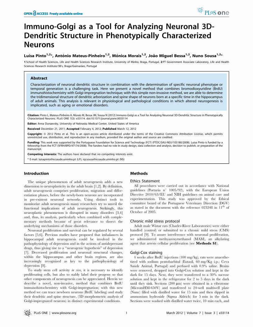
of newborn neurons in the dentate gyrus (DG) of control rats
(Figure 1, 2) and compared to those of rats displaying depressive-
like symptoms after exposure to CMS and/or to the administra-
tion of MAM, an alkylating agent that arrests cellular proliferation.
All branches of the dendritic tree of the selected neurons were
reconstructed using a motorized microscope and Neurolucida
software with the AutoNeuron extension module (Figure 1B, C)
and using manual reconstruction without the AutoNeuron
extension (Figure 1D). No significant differences in total dendritic
length per neuron were found between the automatic AutoNeuron
and manual reconstructions, thus showing that distinct recon-
struction strategies can be used to study the structure of these
neurons. A three-dimensional analysis of the reconstructed
neurons was performed using NeuroExplorer software (FigureS1). This method also allows the analysis of spine types
(Figure 1E) which provides information on neuronal connectivity
and synaptic plasticity [13].
The potential contribution of synaptic plasticity and neuronal
connectivity to the development of, and recovery from, depressive-
like behavior, has been scrutinized [8]. In these studies, the
assessment of neurons dendritic arborization and spine shape was
performed by three-dimensional morphometric analysis of Golgi-
impregnated hippocampal neurons without making possible to
distinguish between old and newly-born neurons. With this new
method we could label neurons that were born during the CMS
period using staining for BrdU and simultaneously study their cell
shape, including dendritic processes. Thus, we could compare not
only the morphology of newborn neurons in rats subjected to
CMS, with and without MAM administration (Figure 2B, C, E,F), with those of control rats (Figure 2A, D), but also to compare
them with adjacent pre-existent granule cells in the same animal.
Here, we show that newborn neurons from the DG of rats
subjected to CMS without blockage of neurogenesis (Figure 2B,E, G) are morphologically comparable to those of control
untreated rats (Figure 2A, D, G). These findings also show for
the first time that the newborn neurons from the DG of rats
subjected to CMS and injected with MAM are unable to reach full
dendritic development (Figure 2C, F, G). In contrast, control rats
treated with MAM present normal dendritic architecture
(Figure 2G).
Comparing the spine morphology of DG newborn neurons of
control rats with those of rats exposed to CMS, with and without
MAM administration, we found a significant increase in the
percentage of thin spines in all animals treated with MAM (control
and CMS) and in untreated CMS animals (Figure 2H).
Moreover, CMS exposed rats present a significant reduction in
the percentage of mushroom spines in newborn neurons when
comparing to those of control rats. Newborn neurons of control
and CMS exposed rats treated with MAM show a significant
decrease in the percentage of thick and ramified spines in
comparison to those of control untreated rats (Figure 2H). These
alterations in the spine morphology of newborn neurons also
suggest severe in the dendritic maturation of neurons in rats
exposed to CMS and/or treated with MAM.
Figure 2. Confocal images and three-dimensional morphometric analysis of neurons double-labeled with BrdU and Golgi-Cox. (A,B, C) Confocal images of three dentate gyrus neurons double-labeled with BrdU (depicted with green dots) and Golgi-Cox (black staining). BrdU wasadministered for five consecutive days to 4 months old Wistar rats non-stressed and injected with saline for two weeks (Control) (A) or exposed tounpredictable chronic mild stress (uCMS) and injected with saline (B) or methyazoxymethanol (uCMS+MAM) (C) in the last two weeks of the stressprotocol. Immunohistochemical analyses were performed 4 weeks after the injections. Nuclei (depicted with blue dots) were stained with Dapi. (D, E,F) Three-dimensional morphometric reconstruction analysis of the Golgi-impregnated dentate granule neurons double-labeled with BrdU shown in A(D) in B (E) and in C (F). (G) Graph showing the total dendritic length of newborn dentate granule neurons in the subgranular zone of differentexperimental groups (control and uCMS exposed rats untreated (2MAM) and treated with MAM (+MAM)). (H) Graph showing the percentage ofdifferent types of spines (thin, mushroom, wide and ramified) present in newborn dentate granule neurons in the subgranular zone of differentexperimental groups (control and uCMS exposed rats untreated (2MAM) and treated with MAM (+MAM)). Data represented as mean6se.m. Asteriskrepresents the comparison between control and all other experimental groups; *P,0.05 and **P,0.01. Scale bars: 50 mm.doi:10.1371/journal.pone.0033114.g002
Immuno-Golgi to Analyze Neuronal 3D Structures
PLoS ONE | www.plosone.org 3 March 2012 | Volume 7 | Issue 3 | e33114
BrdU labeling is currently one of the prevailing methods to
study proliferation and neurogenesis in vivo. Once injected
systemically, BrdU is incorporated as a thymidine analog into
the DNA of all cells undergoing DNA synthesis allowing its
detection in postmitotic cells for the remainder of their life [14].
This technique was essential to the identification of the origin of
newly-generated neuronal cells in the adult hippocampal SGZ
[2,15,16]. So far, however, the analysis of the dendritic and
synaptic structure of newly-born neurons could only be achieved
by retroviral labeling [17,18]. Retroviral labeling provides several
advantages compared to BrdU labeling; for example, it allows
distinguishing between cell division and DNA repair, as the stable
integration of the retroviral genome into the chromosomal DNA
can only happen after nuclear membrane breakdown. However,
retroviral labeling has also several disadvantages that make it less
suitable for in vivo studies comparing to BrdU labeling. Since the
blood-brain barrier is an obstacle, retroviruses have to be applied
directly into the region of interest through stereotaxic surgeries,
causing brain lesions from the procedure and possible local
inflammatory reactions. These lesions and inflammation may
induce alterations on neurogenesis, thus raising some cautions
when using this method to study stem cell activity in the brain.
Moreover, this approach only permits the study of the region
where the virus was injected and does not allow for precise
temporal resolution of proliferation nor for the comparison of the
neuronal morphology between old and newly-dividing cells.
Analysis of dendritic branching and spines by three-dimensional
morphometric assessment of Golgi-impregnated neurons using
computer-assisted reconstructions enables to generate a unique
picture of the effect of different diseases and treatments on the fine
neuronal structure. In fact, dendritic and synaptic pathology is a
hallmark in several neuropsychiatric conditions. Standard histo-
pathological techniques used to label neurons do not stain
dendrites and spines and, thus, may miss aberrant dendritic
branching and synaptic loss in neurodegenerative processes. In
contrast, the Golgi-Cox staining is a simple and valuable method
that provides detailed information on neuronal morphology
allowing the detection of subtle damage. As shown herein, if
combined with BrdU staining, this technique will further allow
distinguishing whether these changes target specific neuronal
populations (e.g. in the present study, whether they affect
differently old versus newly-born neurons). Obviously, the
applicability of this method is much broader, as the combination
of other markers with Golgi staining will allow the analysis of the
neuronal dendritic structure with its phenotypic characterization
in a wide spectrum of experimental conditions. Indeed, a recent
study shows the potential of combining Golgi-Cox staining with
immunocytochemical staining, which permits the analysis of the
morphological patterns of biochemically characterized neurons
[19].
In summary, this novel simple and non-invasive method is an
useful tool to study the fine neuronal structure in phenotypically
characterized neurons in both physiological and pathological
conditions. Its applicability is likely to be broad, if one considers all
those conditions in which neurogenesis and dendritic/synaptic
plasticity might be affected, such as in depression [8], Alzheimer’s
disease and schizophrenia [20].
Supporting Information
Figure S1 Three-dimensional analysis of a reconstruct-ed neuron. (A–E) Three-dimensional analyses of a reconstructed
neuron using NeuroExplorer software. Panels B–E show the
neuron from distinct 3D perspectives (coordinates).
(TIF)
Methods S1 Chronic mild stress protocol.(PDF)
Acknowledgments
We thank L. Martins and D. Teixeira for excellent technical assistance.
Author Contributions
Conceived and designed the experiments: LP JB NS. Performed the
experiments: LP AP MM JB. Analyzed the data: LP JB. Contributed
reagents/materials/analysis tools: LP JB NS. Wrote the paper: LP NS.
References
1. Alvarez-Buylla A, Garcia-Verdugo JM, Tramontin AD (2001) A unifiedhypothesis on the lineage of neural stem cells. Nat Rev Neurosci 2: 287–293.
2. Seri B, Garcia-Verdugo JM, McEwen BS, Alvarez-Buylla A (2001) Astrocytesgive rise to new neurons in the adult mammalian hippocampus. J Neurosci 21:
7153–7160.
3. Pittenger C, Duman RS (2008) Stress, depression, and neuroplasticity: aconvergence of mechanisms. Neuropsychopharmacology 33: 88–109.
4. Dranovsky A, Hen R (2006) Hippocampal neurogenesis: regulation by stress andantidepressants. Biol Psychiatry 59: 1136–1143.
5. Airan RD, Meltzer LA, Roy M, Gong Y, Chen H, et al. (2007) High-speedimaging reveals neurophysiological links to behavior in an animal model of
depression. Science 317: 819–823.
6. van Praag H, Kempermann G, Gage FH (1999) Running increases cellproliferation and neurogenesis in the adult mouse dentate gyrus. Nat Neurosci 2:
266–270.7. Malberg JE, Eisch AJ, Nestler EJ, Duman RS (2000) Chronic antidepressant
treatment increases neurogenesis in adult rat hippocampus. J Neurosci 20:
9104–9110.8. Bessa JM, Ferreira D, Melo I, Marques F, Cerqueira JJ, et al. (2009) The mood-
improving actions of antidepressants do not depend on neurogenesis but areassociated with neuronal remodeling. Mol Psychiatry 14: 764–773, 739.
9. Bessa JM, Mesquita AR, Oliveira M, Pego JM, Cerqueira JJ, et al. (2009) A
trans-dimensional approach to the behavioral aspects of depression. Front BehavNeurosci 3: 1.
10. Evers P, Uylings HB (1997) An optimal antigen retrieval method suitable fordifferent antibodies on human brain tissue stored for several years in
formaldehyde fixative. J Neurosci Methods 72: 197–207.11. Tang X, Falls DL, Li X, Lane T, Luskin MB (2007) Antigen-retrieval procedure
for bromodeoxyuridine immunolabeling with concurrent labeling of nuclear
DNA and antigens damaged by HCl pretreatment. J Neurosci 27: 5837–5844.
12. Harris KM, Jensen FE, Tsao B (1992) Three-dimensional structure of dendritic
spines and synapses in rat hippocampus (CA1) at postnatal day 15 and adult
ages: implications for the maturation of synaptic physiology and long-term
potentiation. J Neurosci 12: 2685–2705.
13. Cerqueira JJ, Taipa R, Uylings HB, Almeida OF, Sousa N (2007) Specific
configuration of dendritic degeneration in pyramidal neurons of the medial
prefrontal cortex induced by differing corticosteroid regimens. Cereb Cortex 17:
1998–2006.
14. Landgren H, Curtis MA (2011) Locating and labeling neural stem cells in the
brain. J Cell Physiol 226: 1–7.
15. Doetsch F, Garcia-Verdugo JM, Alvarez-Buylla A (1999) Regeneration of a
germinal layer in the adult mammalian brain. Proc Natl Acad Sci U S A 96:
11619–11624.
16. Johansson CB, Momma S, Clarke DL, Risling M, Lendahl U, et al. (1999)
Identification of a neural stem cell in the adult mammalian central nervous
system. Cell 96: 25–34.
17. Price J, Grove E, Williams B, Hajihosseini M, Iavachev L, et al. (1994) Labelling
neural precursor cells with retroviruses. Gene Ther 1 Suppl 1: S4–5.
18. Jessberger S, Zhao C, Toni N, Clemenson GD, Jr., Li Y, et al. (2007) Seizure-
associated, aberrant neurogenesis in adult rats characterized with retrovirus-
mediated cell labeling. J Neurosci 27: 9400–9407.
19. Spiga S, Acquas E, Puddu MC, Mulas G, Lintas A, et al. (2011) Simultaneous
Golgi-Cox and immunofluorescence using confocal microscopy. Brain Struct
Funct 216: 171–182.
20. DeCarolis NA, Eisch AJ (2010) Hippocampal neurogenesis as a target for the
treatment of mental illness: a critical evaluation. Neuropharmacology 58:
884–893.
Immuno-Golgi to Analyze Neuronal 3D Structures
PLoS ONE | www.plosone.org 4 March 2012 | Volume 7 | Issue 3 | e33114