Embed Size (px)
Citation preview

Immunity
Innate & Adaptive

Immunity• Innate: response to attack is always
the same– Mechanical mechanisms– Chemical mediators– Cellular response– Inflammatory response
• Adaptive: response to attack improves with each exposure– Specific– Has memory

Innate immunity
• Mechanical mechanisms– Skin & mucus membranes form physical
barriers to prevent entry– Tears, saliva, urine wash away pathogens
from surface or body or dilute invading army of pathogens

Innate immunity• Chemical mediators: Some prevent
entry to cells, kill bacteria, or produce inflammation– Complement proteins
• ~ 20 in plasma• normally inactive; activated by combining with
parts of bacterial cells or antibodies• Leads to chain rxn activation of neighboring
compliments & inflammation, phagocytosis, or lysis

Innate immunity• Chemical mediators
– Interferons protect against viral infection• Virus-infected cells place interferons on their
surface (SOS signal)• These bind to neighboring cells & stimulate
neighbors to produce antiviral proteins• Intiviral proteins inhibit production of new viral
RNA• Some interferons activate macrophages and
NKC

Innate immunity• Cellular mechanisms
– WBC & derivatives are most important cellular components• Attracted to invading bacteria and
microorganisms through chemotaxis
– Phagocytic cells (who are these?)– Inflammatory cells (…and these?)– Natural Killer Cells (NKC)

White Blood Cells

Innate immunity• Phagocytic cells
– Neutrophils• Small; first to enter infected tissue from blood;
ingest, then die --> pus accumulation
– Macrophages• Monocytes leave blood & enlarge; arrive after
neutrophils; do most eating & cleanup• Also hang out at “entry points” (gate-keepers)
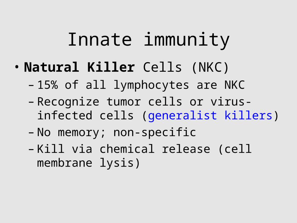
Innate immunity• Natural Killer Cells (NKC)
– 15% of all lymphocytes are NKC– Recognize tumor cells or virus-infected
cells (generalist killers)– No memory; non-specific– Kill via chemical release (cell membrane
lysis)
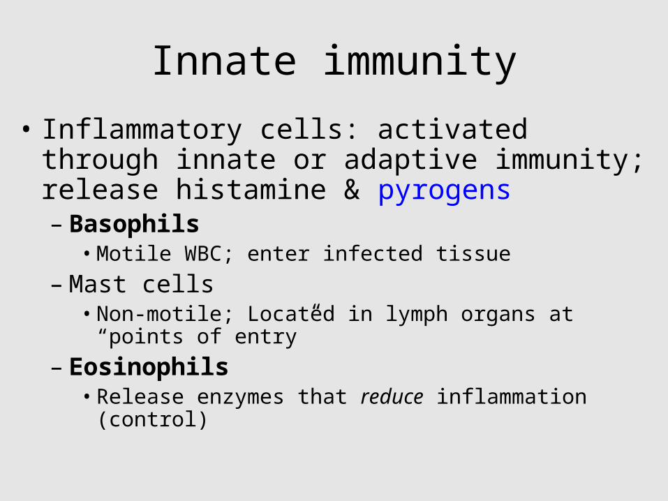
Innate immunity
• Inflammatory cells: activated through innate or adaptive immunity; release histamine & pyrogens– Basophils
• Motile WBC; enter infected tissue
– Mast cells• Non-motile; Located in lymph organs at “points
of entry”
– Eosinophils• Release enzymes that reduce inflammation
(control)

Inflammation is useful!
• Local inflammation: Redness, heat, swelling– caused by increased blood flow & vascular
permeability– Chemicals and swelling activate pain receptors
• Systemic inflammation– Red marrow increases neutrophil production– Pyrogens stimulate fever by increasing heat
production & conservation– Vascular permeability increases

Adaptive immunity
• Lymphocytes– Origin & development– Activation & multiplication
• Antibody-mediated• Cell-mediated

Adaptive immunity• Antigens stimulate adaptive immune
response– Self– Foreign
• MHC molecules display antigens• Lymphocytes
– Origin & development– Activation & multiplication
• Types of Adaptive Immunity– Antibody-mediated
• B cells; produce cells that make antibodies
– Cell-mediated• T cells; cytotoxic & helper T cells

Antigens
• Foreign– Components of bacteria, viruses, and
their chemical byproducts– Pollen, animal hair, foods produce allergic
response (overreaction of immune system)
• Self– Produced by our bodies
• Recognize tumor antigens

B & T-cells (Lymphocytes)
• Contain antigen receptors in their cell membranes
• We each have thousands of different populations of B & T-cells, each with unique antigen receptors
• Cells are stimulated by binding of antigens to their unique receptors

Cell Developmen
t• Red marrow produces:– Pre B-cells:
released into bloodstream
– migrate to lymph organs
– Pre T-cells: migrate to thymus & mature there
• Mature T-cells migrate to lymph organs
Lymph nodes,spleen, and
other lymphaticorgans
Final maturationof B and T cells inlymphatic organ
Viablood
T cellB cell
Viablood
Antigenreceptor
Thymus
Antigenreceptor
Immaturelymphocytes
Stem cell
Bonemarrow

Activation & multiplication• Macrophages present
antigens– Phagocytize invaders, process
& display antigens (with help of MHC molecules)
• MHC/Antigen complex binds to receptors on B or T-cells
• T-cells auto-stimulate– Produce cytokines (peptides;
e.g. interleukin) that up-regulate growth & division

B cell activation• Antibody-mediated• B-cells can also
phagocytize & process antigens– same antigen that
stimulated a Th
• Th binds to B-cell
• Interleukins are produced– stimulate B-cell division &
proliferation– Daughter (plasma) cells
produce antibodies

Effects of Antibodies
• Direct– Antibodies bind antigens = inactivation
• Indirect– Activate Complement cascade
• Inflammation (mast cells and basophils release histamine)
• Chemotaxis (attracts white blood cells)• Phagocytosis or lysis (macrophage eats
antibody & antigen

Binding of antibodies to antigensinactivates antigens by
NeutralizationAgglutinationof microbes
Precipitation ofdissolved antigens
Phagocytosis Cell lysis
Activation of complement system
Leads to
Foreign cell Hole
Complement moleculeBacteria
Antigenmolecules
Enhances
Bacterium
Virus
Macrophage

Antibody production
• Differs following first and second exposure to antigen– First exposure = primary response
• B-cells bind antigen; produce plasma cells (produce antibodies) and memory B-cells
• Response time = 3-14 days; disease symptoms develop; SLOW

Fig. 24-7aa-1
Primary immuneresponse
B cells withdifferentantigenreceptors
Antigen receptor(antibody on cellsurface)
1

Fig. 24-7aa-2
Primary immuneresponse
B cells withdifferentantigenreceptors
Antigen receptor(antibody on cellsurface)
1 Antigenmolecules
2

Fig. 24-7aa-3
Primary immuneresponse
B cells withdifferentantigenreceptors
Antigen receptor(antibody on cellsurface)
1 Antigenmolecules
2
First exposureto antigen
Cell activation:growth,division, anddifferentiation
3

Fig. 24-7aa-4
Primary immuneresponse
B cells withdifferentantigenreceptors
Antigen receptor(antibody on cellsurface)
1 Antigenmolecules
2
First exposureto antigen
Cell activation:growth,division, anddifferentiation
3
Antibodymolecules
EndoplasmicreticulumFirst clone
Plasma (effector) cells secreting antibodies
4

Fig. 24-7aa-5
Primary immuneresponse
B cells withdifferentantigenreceptors
Antigen receptor(antibody on cellsurface)
1 Antigenmolecules
2
First exposureto antigen
Cell activation:growth,division, anddifferentiation
3
Antibodymolecules
EndoplasmicreticulumFirst clone
Plasma (effector) cells secreting antibodies
4
Memory cells
5

Antibody production• Differs following first and second
exposure to antigen– Second exposure = secondary response
• Memory cells quickly induce plasma cells to produce antibodies
• Time to antibody production is reduced• More plasma cells & antibodies produced• RAPID response, no disease symptoms =
immunity
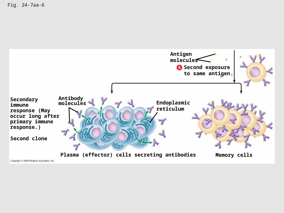
Fig. 24-7aa-6
Second clone
Plasma (effector) cells secreting antibodies Memory cells
6
Antigenmolecules
Second exposureto same antigen
Endoplasmicreticulum
Antibodymolecules
Secondaryimmuneresponse (Mayoccur long afterprimary immuneresponse.)

Fig. 24-7b
Time (days)
Second exposureto antigen X,
first exposureto antigen Y
First exposureto antigen X
Secondary immuneresponse to
antigen X
Primary immuneresponse to
antigen Y
Primary immuneresponse to
antigen X
Antibodiesto X
Antibodiesto Y
An
tib
od
y c
on
cen
tra
tio
n
5649423528211470

Cell-mediated immunity
• Cytotoxic T cells (Tc)
• Effective against viruses, bacteria
• Infected cells display antigens, and Tc binds to MHC/antigen combo– Stimulates production of more Tc
– Costimulation by Th which were stimulated by macrophage display of antigens

Fig. 24-11b
Self-nonselfcomplex
T cellreceptor
Interleukin-2stimulatescell division
Interleukin-1stimulateshelper T cell
Bindingsite forantigen
Bindingsite forself protein
HelperT cell
Antigen-presentingcell
23
4
5 6
7
B cell
CytotoxicT cell
Cell-mediatedimmuneresponse(attack oninfected cells)
Interleukin-2activates B cellsand other T cells
Humoralimmuneresponse(secretion ofantibodies byplasma cells)

Fig. 24-12-1
1 Cytotoxic T cell bindsto infected cell
Self-nonselfcomplex
CytotoxicT cell
Foreignantigen
Perforinmolecule
Infected cell

Fig. 24-12-2
1 Cytotoxic T cell bindsto infected cell
Self-nonselfcomplex
CytotoxicT cell
Foreignantigen
Perforinmolecule
Infected cell
Perforin makes holes ininfected cell’s membraneand enzyme enters
Enzyme thatcan promoteapoptosis
Holeforming
2

Fig. 24-12-3
1 Cytotoxic T cell bindsto infected cell
Self-nonselfcomplex
CytotoxicT cell
Foreignantigen
Perforinmolecule
Infected cell
Perforin makes holes ininfected cell’s membraneand enzyme enters
Enzyme thatcan promoteapoptosis
Holeforming
2 Infected cellis destroyed
3

Show “Immune Response”Mcgraw Hill
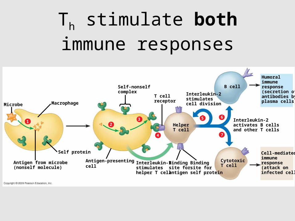
Microbe Macrophage
Self protein
Self-nonselfcomplex
T cellreceptor
Interleukin-2stimulatescell division
Interleukin-1stimulateshelper T cell
Bindingsite forantigen
Bindingsite forself protein
HelperT cell
Antigen-presentingcell
Antigen from microbe(nonself molecule)
12
3
4
5 6
7
B cell
CytotoxicT cell
Cell-mediatedimmuneresponse(attack oninfected cells)
Interleukin-2activates B cellsand other T cells
Humoralimmuneresponse(secretion ofantibodies byplasma cells)
Th stimulate both immune responses

Acquiring Adaptive immunity

Antigens, Antibodies & Vaccines
• Inject host with inactive or attenuated virus (usually bits & pieces aka antigens)
• Host immune cells grab antigens & stimulate other cells (B cells) to engineer antibodies to bind to antigens

Antigens, Antibodies & Vaccines
• B cells “remember” how to make this antibody forever
• On subsequent infection, live virus is mobbed by antibodies, targeted for termination and eaten by macrophages