Embed Size (px)
Citation preview

Immunity

Body Defenses• First line - barriers
• Skin and mucous membranes
• Flushing action
– Antimicrobial substances
• Lysozyme, acids, salts, normal microbiota

Second line defenses• Cells – neutrophils, macrophages, natural
killer cells– Toll-like receptors on phagocytes recognize
carbohydrates on the surface of bacteria
• Inflammation
• Proteins– Complement– Interferon
• Fever
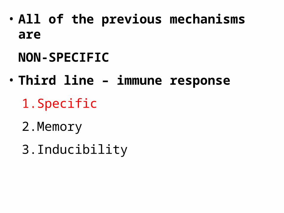
• All of the previous mechanisms are
NON-SPECIFIC
• Third line – immune response
1.Specific
2.Memory
3.Inducibility

•Antigens - substances recognized as “non-self” These can be:
•Infectious agents - bacteria, viruses, fungi or parasites
•Noninfectious substances – •Environmental - pollen, foods, bee
venoms•Drugs, vaccines, transfusions and transplanted tissues

AntigenAntibody GeneratorThe best antigens are:
1. large
2. recognized as foreign
3. complex

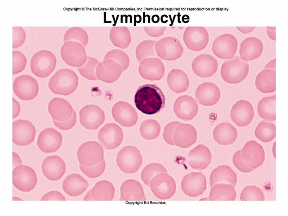
Two cell types give us the immune response;both are lymphocytes, which are a type of leukocyte, or white blood cell.
B lymphocytes or B cellsT lymphocytes or T cells

The cells of the immune response differ from the cells of the inflammatory response in three ways:
1. They are SPECIFIC and each cell recognizes only one specific antigen.
B cells produce antibodies
Tc cells attack antigen directly

2. Both produce groups of cells called “memory cells” that act quickly the second time the antigen is encountered.
3. An antigen induces an immune response. Only small amounts of antibodies or T Cells are present before encountering an antigen.

Long lasting protection against a specific antigen is immunity.
Natural immunity:
Not produced by the immune response
Species specific
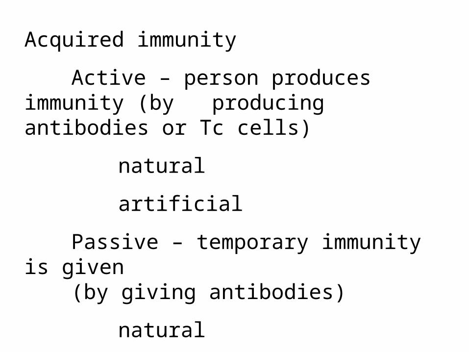
Acquired immunity
Active – person produces immunity (by producing antibodies or Tc cells)
natural
artificial
Passive – temporary immunity is given(by giving antibodies)
natural
artificial

LymphocytesOriginate :
in liver, spleen and bone marrow of fetus
in bone marrow after birth
From stem cells – hemocytoblasts – that produce all blood cells.
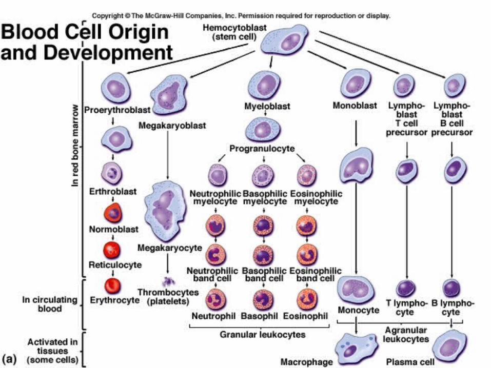

To become mature, immunocompetent cells, they must pass through lymphoid tissues in other parts of the body.
As they do so, they become committed to becoming either T cells or B cells
Cells that migrate through the bone marrow (bursal equivalent) become B cells, and will produce antigens and participate in humoral immunity.


Cells that migrate through the thymus glands become T cells and participate in Cell-mediated immunity.
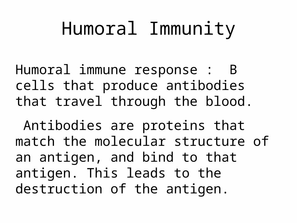
Humoral Immunity
Humoral immune response : B cells that produce antibodies that travel through the blood.
Antibodies are proteins that match the molecular structure of an antigen, and bind to that antigen. This leads to the destruction of the antigen.

Antibody

When antigen binds to antibody receptors on the surface of the B cell, the B cell divides and differentiates into antibody producing plasma cells and also memory cells.


Immunoglobulins
• IgG - monomer (crosses placenta)
• IgA – dimer – 2 units - in secretions
• IgM – pentamer – 5 units
• IgD – monomer – on surface of B cells
• IgE – monomer – involved in hypersensitivities


Cell-mediated immunity
• Produced through Tcytoxic or Tc Cells (T8 cell)
• DO NOT produce antibodies
• Attack invaders directly
• May produce toxic chemicals – such as perforins, cytolytic enzymes, etc.
• May stimulate cell’s self-destruct mechanism

Primary and Secondary Immune Responses
• Primary response – Latent period– IgM produced– IgG produced later

•Secondary response
•Anamnestic response –much more rapid due to memory cells
•Primarily IgG


Antigen-Presenting cells (macrophages) place antigen on their cell surface in combination with the MHC II complex
Antigen is presented to a specific helper T cell that has receptors that match the antigen – MHC II complex


Hypersensitivities
“The Immune System Gone Bad”

Hypersensitivities
1. Allergies – Exaggerated immune response against environmental antigens
2. Autoimmunity – immune response against host’s own cells
3. Alloimmunity – immune response against beneficial foreign tissues, such as transfusions or transplants

These immune processes initiate inflammation and destroy healthy tissue. Four types:
Type I – IgE-mediated allergic reactions immediate type hypersensitivity
Type II – tissue-specific reactions antibody-dependent cytotoxicity
Type III – immune-complex-mediated reactions
Type IV - cell-mediated reactionsdelayed-type hypersensitivity
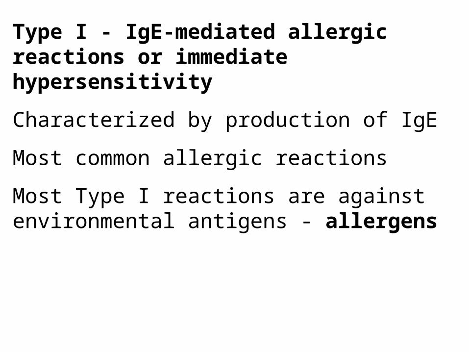
Type I - IgE-mediated allergic reactions or immediate hypersensitivity
Characterized by production of IgE
Most common allergic reactions
Most Type I reactions are against environmental antigens - allergens

Selected B cells produce IgE
Need repeated exposure to large quantities of allergen to become sensitized
IgE binds by Fc end to mast cells after first exposure

Second exposure (and subsequent exposures) – antigen binds with Fab portion of antibody on mast cells, and cross-links adjacent antibodies, causing mast cell to release granules.
Response is immediate ( 5- 30 minutes)


Sometimes beneficial to host – IgE-mediated destruction of parasites, especially parasitic worms.

Histamine release:• Increases vascular permeability, causing
edema
• Causes vasodilation
• Constricts bronchial smooth muscle
• Stimulates secretion from nasal, bronchial and gastric glands
• Also hives (skin), conjunctivitis (eyes) and rhinitis (mucous membranes of nose).

Late phase reaction• 2 – 8 hours; lasts for 2 - 3 days
• Other mediators that take longer to be released or act:– Chemotactic factors for eosinophils and
neutrophils– Leukotrienes– Prostaglandins– Platelet-activating factor – Protein-digesting enzymes


Genetic predisposition
• Allergy prone or atopic
• Can be life threatening, so individuals should be aware
• Skin tests – injection – see wheal and flare
• Lab tests for circulating IgE

Treatment• First wave – antihistamines or epinephrine
(blocks mast cell degranulation)
• Second wave – corticosteroids and nonsteroidal anti-inflammatory agents that block synthesis of leukotrienes and prostaglandins
• Desensitization by repeated injections of allergen – formation of IgG

Anaphylaxis – Type I allergic reaction
may be localized or general
immediate – within a few minutes of exposure
Systemic anaphylaxis:pruritus(intense itching)urticaria (hives)Wheezing; dyspnea; swelling of the larynx
Give epinephrine

Anaphylactic shock
• Hypotension, edema (esp. of larynx), rash, tachycardia, pale cool skin, convulsions and cyanosis
• Treatment:– Maintain airway– Epinephrine, antihistamines, corticosteroids– Fluids– Oxygen
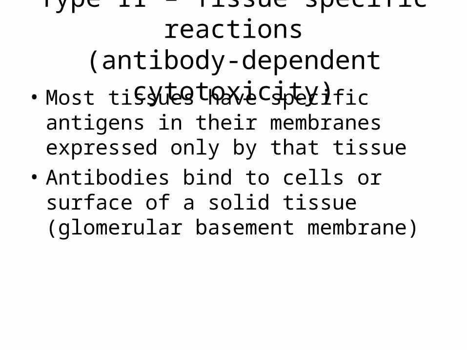
Type II – Tissue specific reactions(antibody-dependent cytotoxicity)
• Most tissues have specific antigens in their membranes expressed only by that tissue
• Antibodies bind to cells or surface of a solid tissue (glomerular basement membrane)

Destruction of tissue occurs:– Destruction by Tc Cells which are not
antigen specific- K cells
– Complement-mediated lysis
– Phagocytosis by macrophages(“frustrated phagocytosis”)
– Binding of antibody causes cell to malfunction


Type III – Immune-complex-mediated reactions
• Caused by antigen-antibody complexes formed in circulation and deposited in vessel walls or other tissues
• Not organ specific
• Effects caused by activation of complement – chemotaxis of neutrophils
• Neutrophils release lysosomal enzymes into tissues (“frustrated phagocytosis”)

Type IV- Cell-mediated reactions• Sensitized T lymphocytes – either Tc Cells
or lymphokine producing Td cells• Takes 24 – 72 hours to develop• Damage by Tc Cell or inflammatory
response by Td Cells (lymphokines)• Graft rejection, tumor rejection, TB reaction,
poison ivy and metal reactions• Immune diseases• Tissue rejection


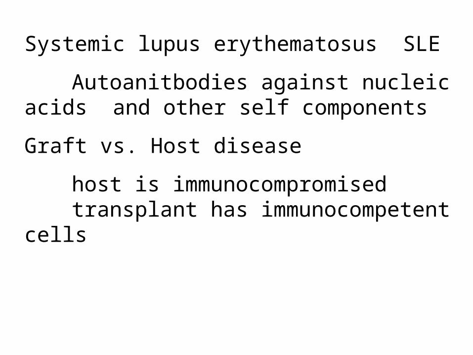
Systemic lupus erythematosus SLE
Autoanitbodies against nucleic acids and other self components
Graft vs. Host disease
host is immunocompromisedtransplant has immunocompetent cells