Embed Size (px)
Citation preview
1
IMMUNE EVASION BY THE EBOLA VIRUS
LI T ER A TU R E R ES EA R C H BY D A NI D E LOU W
Bachelor thesis by Dani de Louw (951105530050) Chair group: cell biology and health
Date: 7 – 10 – 2017 Tutor: Inge Palm
Zoonotic transmission of the Ebola virus from a non-human species to humans causes severe haemorrhagic fever in infected individuals with mortality rates varying from 50 to 90%. The 2014-2016 Ebola virus outbreak in West-Africa is the largest and most complex one in history. Without the availability of vaccines or therapeutic treatments the virus constitutes an important global public health threat. Growing understanding about the pathogenic mechanisms of Ebola virus enabled Public Health Canada to develop the first vaccine which showed protective efficacy, immunogenicity and safety in a ring vaccination clinical trial. Many studies have been conducted to examine the pathogenic effects of the Ebola virus but not all the processes are understood yet. The virulence of Ebola virus may be partially attributed by the primary infection and replication in macrophages and dendritic cells. The Ebola virus glycoprotein likely interacts with TLR-4 found on the surface of macrophages and dendritic cells and thereby influences viral entry and infection. This has a huge impact on the entire immune system. On the one hand, the innate immune response is aberrant, characterized by the hypersecretion of cytokines (i.e. cytokine storm), the lack of an interferon response and impaired maturation of dendritic cells. The infected macrophages and dendritic cells are still able to induce an inflammatory response and blood coagulation, but cannot prevent the systemic spread of the virus. The inflammatory response results in the attraction of additional immune cells to the site of infection which are potential new host cells for the virus. If these cells also become infected, then even more inflammatory cytokines are released. Therefore, the hypersecretion of cytokines (i.e. cytokine storm) paradoxically enhances the viral systemic spread throughout the body. Furthermore, it is presumed that VP24 and VP35 play a crucial role in the disruption of the interferon response. VP24 blocks the cellular responses to exogenous type I and II interferons while VP35 inhibits the production of type I interferons and the maturation of dendritic cells. On the other hand, the adaptive immune response is suppressed due to the decreased production of interferons, the impaired maturation of dendritic cells and the apoptosis of lymphocytes. The apoptosis of lymphocytes further impairs the adaptive immune response due to decreased levels of circulating cytokines produced by T lymphocytes and a decreased T cell dependent activation of B cells. To conclude, an infection by the Ebola virus is characterized by the dysregulation of the host’s normal immune responses. The purpose of this thesis is to review recent research about the mechanisms of immune evasion by the Ebola virus. If you understand the mechanisms of immune evasion, then you are better able to look for possibilities or treatments to prevent or inhibit the viral infection. Nowadays, only one vaccine exists and there are no therapeutic treatments available. By giving an overview of the current knowledge this thesis helps to create a better understanding of the pathogenic mechanisms of the Ebola virus and stresses the importance of developing more appropriate vaccines and therapeutic treatments.
2
TABLE OF CONTENTS
1. PREFACE ............................................................................................................................................. 3
2. THE EBOLA VIRUS .............................................................................................................................. 4
3. THE HOST IMMUNE RESPONSE AGAINST VIRUSES ........................................................................... 6
3.1 THE INNATE IMMUNE RESPONSE ............................................................................................ 7
3.2 THE ADAPTIVE IMMUNE RESPONSE ........................................................................................ 8
4. PATHOGENIC EFFECTS OF THE EBOLA VIRUS .................................................................................. 11
4.1 INFECTION OF MACROPHAGES AND DENDRITIC CELLS......................................................... 11
4.2 ABERRANT INNATE IMMUNE RESPONSE ............................................................................... 13
4.3 SUPPRESSION OF THE ADAPTIVE IMMUNE RESPONSE ......................................................... 20
5. VACCINE DEVELOPMENT ................................................................................................................. 24
6. DISCUSSION ................................................................................................................................. 25
7. CONCLUSION ................................................................................................................................... 28
8. REFERENCES ..................................................................................................................................... 29
3
1. PREFACE
Viruses have always had a major impact on mankind [1]. For centuries they were a mystery but since the discovery of molecular biology and the availability of recombinant DNA technology more knowledge about viruses was gained [2]. Viruses are described as the smallest genetic organisms with three characteristic properties: infection of host cells, replication inside the host cell and survival in an inert state outside the host cell. Nowadays, one of the most deadly viruses is the Ebola virus with mortality rates varying from 50% to 90% [3, 4]. Ebola virus infections are characterised by the disruption of an effective host immune response. The Ebola virus is not only a local problem in African countries but also worldwide due to the risk of imported infections and the fear of misuse as a biological weapon for terrorists [3]. Therefore, there is an urgent need to address the pathogenic mechanisms of the Ebola virus so an appropriate vaccine or therapeutic treatment can be developed. Until this year, neither vaccines nor therapeutic treatments were available making the Ebola virus a growing public health concern [3, 5]. But this year, Public Health Canada developed the first vaccine which showed protective efficacy, immunogenicity and safety in a ring vaccination clinical trial [6]. However, the 2014-2016 Ebola virus outbreak in West-Africa is the largest and most complex one in history suggesting that Ebola virus outbreaks are not under control yet. The purpose of this thesis is to review recent research about the mechanisms of immune evasion by the Ebola virus. If you understand the mechanisms of immune evasion, then you are better able to look for possibilities or treatments to prevent or inhibit the viral infection. Therefore, this thesis helps to create a better understanding of the pathogenic mechanisms of the Ebola virus and stresses the importance of developing more appropriate vaccines and therapeutic treatments. The first section of this thesis will give an overview of the Ebola virus and viral infections in general. The second section will go into more detail on how the Ebola virus invades and interferes with the human immune response by explaining some pathogenic processes. The last part will address the recently developed vaccine.
4
2. THE EBOLA VIRUS
Since the discovery of the Ebola virus in 1976, several outbreaks have occurred sporadically in Africa. The first two outbreaks took place simultaneously in Nzara (South Sudan) and Yambuku (Democratic Republic of Congo) [7, 8]. The outbreak in Yambuku was located nearby the Ebola river and therefore it was referred to as the Ebola virus. These outbreaks were found to be caused by different species of the Ebola virus. Nowadays, five species of the Ebola virus have been identified: Zaire ebolavirus (ZEBOV), Sudan ebolavirus (SEBOV), Côte d'Ivoire ebolavirus (CIEBOV), Bundibugyo ebolavirus (BEBOV) and Reston ebolavirus (REBOV) [3, 5, 7, 9]. The different species seem to cause different clinical syndromes. For instance, the Reston ebolavirus is not infectious in humans. The first outbreaks were located in remote villages, but the latest outbreak in 2014 took place in multiple countries in West Africa involving major urban areas. This led to the largest and most complex Ebola virus outbreak in history [10].
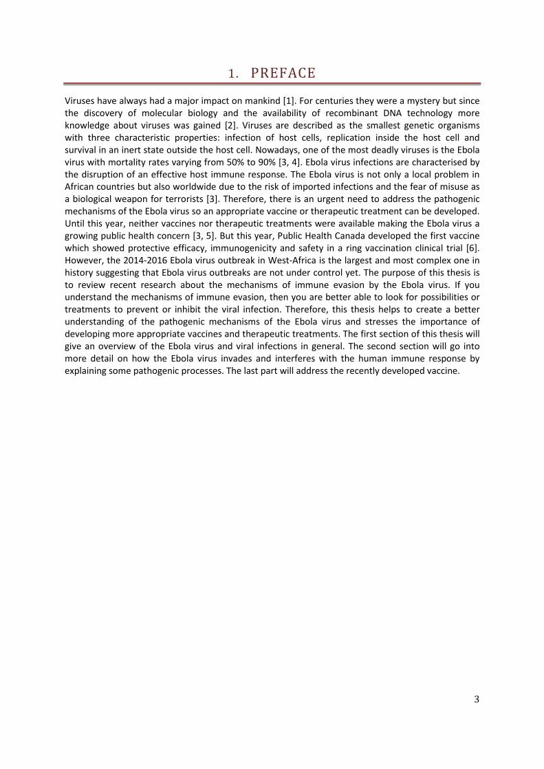
The structure of a virus determines its specific properties. Every virus consists of genetic material which is protected by a protein coat and/or a lipid envelope [2]. But among viruses, big differences exist in the genetic material and the surrounding coat. The Ebola virus is a member of the Filoviridae family which are characterised by long filamentous particles (see FIGURE 1) [3, 4]. It is an enveloped, non-segmented, single-stranded negative sense RNA virus (ssRNA-) [3, 4, 7]. In other words, the Ebola virus is surrounded by a lipid envelope and needs to encapsulate viral RNA polymerase within the viral particle since the negative-stranded RNA is not directly translatable. The Ebola virus is called the elephant among the viruses since it is a very big virus with a length up to 14.000 nm [11]. However, the genome is 19 kb long and consists of only seven open reading frames encoding for structural proteins: envelope glycoprotein (GP), nucleoprotein (NP), viral polymerase (L) and four viral proteins (VP24, VP40, VP30, VP35) [3-5, 7]. The Ebola virus proteins carry out different functions in the replication cycle. Some of the viral proteins (VP24 and VP35) play a crucial role in the interaction with the human immune system whereas the GP seems to be involved in viral entry in the target cell. This will be further elaborated in chapter 3 [7].
FIGURE 1 [7]. THE STRUCTURE OF THE EBOLA VIRUS. THE EBOLA VIRUS CONSISTS OF A LIPID ENVELOPE WHICH ENCAPSULATES THE SSRNA- STRAND ENCODING SEVEN OPEN READING FRAMES FOR STRUCTURAL PROTEINS. THE GENOME HAS THE FOLLOWING STRUCTURE: ‘3-LEADER-NP-VP35-VP40-GP-VP30-VP24-L-TRAILER-5.
5
But where does the Ebola virus hide and how is it transmitted to humans? Normally a virus causes no or little disease in their natural host. This is logical because by killing their host the virus indirectly kills itself. But when a virus is transmitted from one species to another one, it could cause serious pathogenic effects in the new host [12]. Presumably, humans are only ‘dead-end’ hosts for the Ebola virus while other species are the reservoir species [3, 7, 13]. These reservoir species are still unknown but there are indications that fruits bats are most likely involved [5, 7, 14, 15]. Ebola virus epidemics were temporarily associated with bat infections and migrations. It also appeared that fruits bats do not develop filovirus disease which made them the prime reservoir suspects [7]. Nevertheless, attempts to implicate fruit bats as reservoir species appeared to be problematic since it was not possible, with some exceptions, to detect viral RNA sequences or isolate the Ebola virus during and after epidemics [7].
An infection or disease which is transmitted from a non-human species to a human is called a zoonosis [3, 16]. How this primary transmission from the unknown reservoir species to humans occurs remains unknown. However, the route of secondary transmission is better understood. The Ebola virus can spread from person to person via exposure of mucous membranes, abrasions and injuries in the skin to infectious body fluids or tissues [17]. Therefore, the main route of infection is through direct contact with infected persons or cadavers since their body fluids contain large amounts of the virus particles [3]. Caregiving or unhygienic practices during funerals enhance the risk of transmission enormously [7, 14]. For example, the risk of transmission ranged from 8% for minimal contact to 83% for touching a cadaver [18]. This risk was calculated using histories of household members of Ebola virus disease survivors in Sierra Leone [18]. Transmission also occurs when the virus is directly injected into the bloodstream. This was the case in the first Ebola virus outbreak in 1976, where the reuse of contaminated needles lead to the spread of infection. Moreover, one outbreak of Zaire Ebola virus in DRC was associated with the consumption of contaminated foods (e.g. infected bats). Normally, proper cooking of foods should inactivate the virus. However, ingestion of contaminated food cannot fully be ruled out as a possible mode of transmission [3].
After transmission to a new host, the Ebola virus causes Ebola haemorrhagic fever (EHF) [3, 5]. In general, the onset of EHF is abrupt with an incubation period varying from 2 to 21 days [3, 14]. In the beginning of the infection the Ebola virus causes flu-like symptoms such as fever, chills, headache, malaise and myalgia [3, 4, 14]. These early symptoms are similar to those of other diseases, like malaria and influenza, so laboratory confirmation is needed to confirm Ebola infection [14]. When the infection progresses it leads to more severe symptoms and multi-organ failure, including gastrointestinal, respiratory, neurological, vascular and systemic manifestations [3]. In most of the cases the infection finally results in disseminated intravascular coagulation (DIC) [3]. DIC is a condition which is characterised by the widespread activation of clotting cascades [19]. This leads to the intravascular deposition of fibrin and the occlusion of small and midsize vessels by blood clots. A decreased blood flow could contribute to organ damage [20]. Furthermore, the normal coagulation is disrupted since many platelets and clotting factors are used in the formation of blood clots. Only a small amount is left and circulating in the blood. This could lead to severe internal and external bleedings which eventually lead to hypotensive shock [21]. Besides DIC, the Ebola virus infection frequently causes death of the host due to a disturbed immune response [22]. The Ebola virus will mostly target the macrophages and dendritic cells which are important cells in the immune response (DCs) [14, 22]. More details on these cells will be given in the next section. Replication inside of the macrophages and DCs impairs their function and leads to immune suppression and systemic inflammatory responses which probably contribute to the disease progression [3]. The treatment of infected patients mainly consisted of supportive therapy such as the maintenance of hydration and sufficient oxygen levels [14]. According to the Dr. Margaret Chan, who is the Director-General of the World Health Organization, the world is far better prepared for another Ebola outbreak due the recently developed vaccine [23].
6
3. THE HOST IMMUNE RESPONSE AGAINST VIRUSES
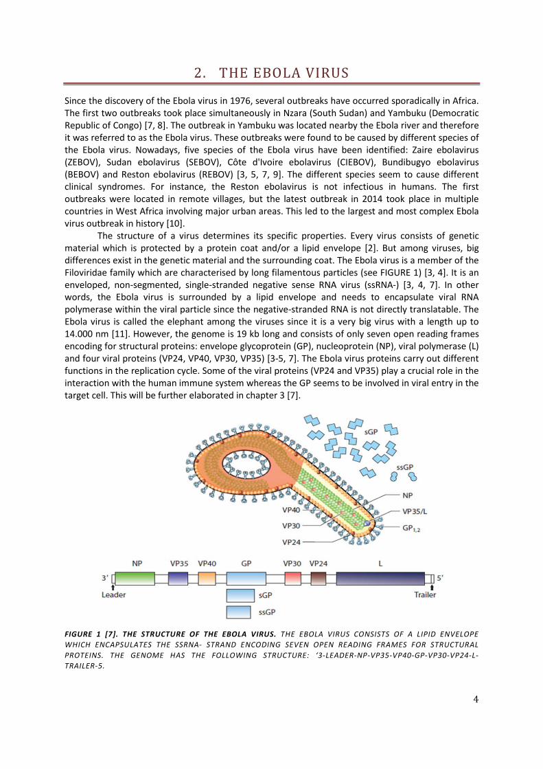
The human body constantly faces pathogens which are capable of causing an infection or disease. Examples of pathogens are bacteria, fungi and viruses. Since the Ebola virus is a virus, this chapter will focus on the host immune response against viruses. The body has several external and internal defence mechanisms to prevent the virus from entering and causing harm. The first line of defence is a physical and chemical barrier which consists of the skin, stomach acid and mucous membranes of the digestive system, respiratory and reproductive tracts [24]. If a virus successfully penetrates the body a second line of defence is activated: the immune system. The immune system of humans consists of two different responses: the innate response and the adaptive response [25]. The innate response is not specific to one particular virus but recognizes proteins found on and produced by pathogens. It is very important for a quick immune response and acts within minutes after the virus has entered the body (see FIGURE 2). Thereby, the innate immune response slows down the viral spread in the beginning of the infection. Due to the fast innate response, the adaptive response has enough time to develop a more specific and potent response. In general, it takes days or even weeks before an adaptive response is established (see FIGURE 2). The two immune responses need each other to function properly and together they ensure that viruses are eliminated from the body and homeostasis is maintained. The following sections will explain in more detail these two responses of the immune system during viral infections.
FIGURE 2 [26]. TIME LINE ACTIVATION INNATE AND ADAPTIVE IMMUNE RESPONSE. THE INNATE IMMUNE RESPONSE ACTS WITHIN MINUTES AFTER A PATHOGEN HAS ENTERED THE BODY WHILE THE ADAPTIVE IMMUNE RESPONSE ACTS WITHIN DAYS OR EVEN WEEKS.
7
3.1 THE INNATE IMMUNE RESPONSE
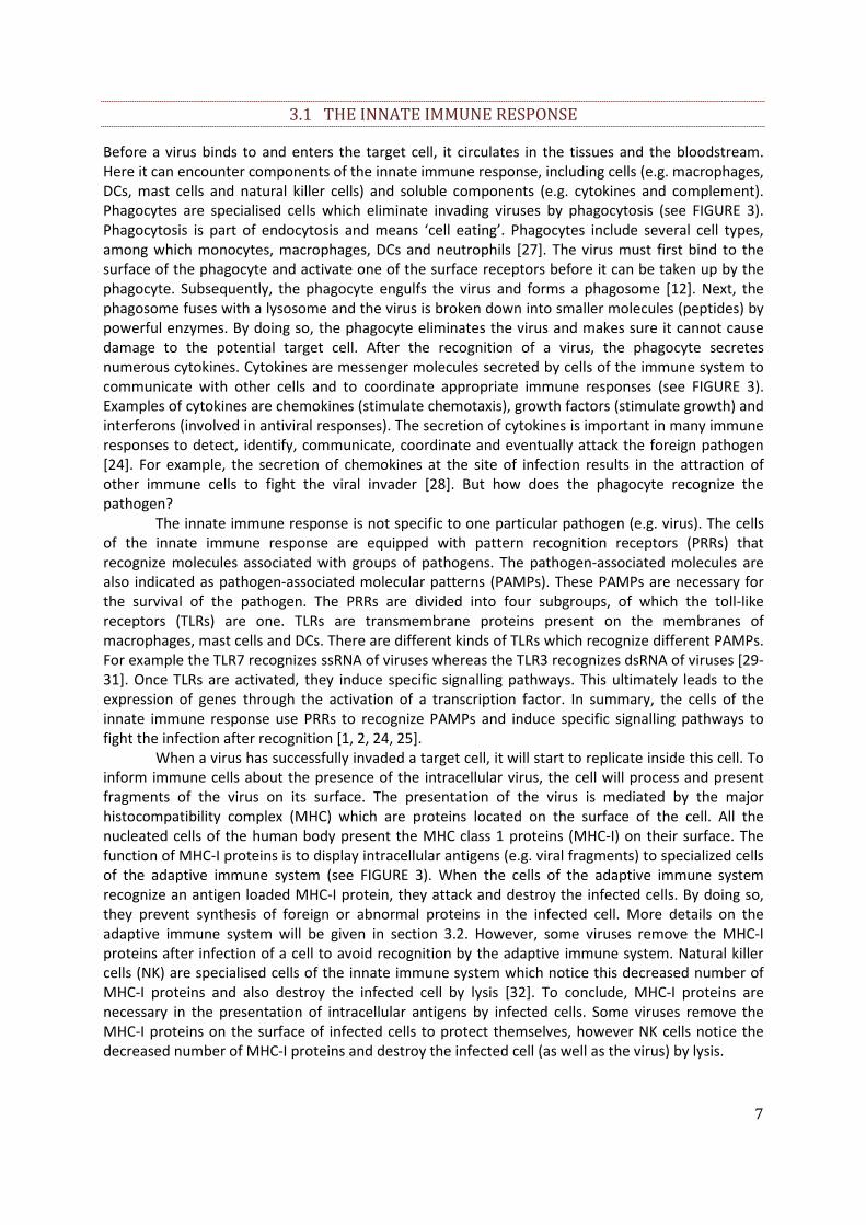
Before a virus binds to and enters the target cell, it circulates in the tissues and the bloodstream. Here it can encounter components of the innate immune response, including cells (e.g. macrophages, DCs, mast cells and natural killer cells) and soluble components (e.g. cytokines and complement). Phagocytes are specialised cells which eliminate invading viruses by phagocytosis (see FIGURE 3). Phagocytosis is part of endocytosis and means ‘cell eating’. Phagocytes include several cell types, among which monocytes, macrophages, DCs and neutrophils [27]. The virus must first bind to the surface of the phagocyte and activate one of the surface receptors before it can be taken up by the phagocyte. Subsequently, the phagocyte engulfs the virus and forms a phagosome [12]. Next, the phagosome fuses with a lysosome and the virus is broken down into smaller molecules (peptides) by powerful enzymes. By doing so, the phagocyte eliminates the virus and makes sure it cannot cause damage to the potential target cell. After the recognition of a virus, the phagocyte secretes numerous cytokines. Cytokines are messenger molecules secreted by cells of the immune system to communicate with other cells and to coordinate appropriate immune responses (see FIGURE 3). Examples of cytokines are chemokines (stimulate chemotaxis), growth factors (stimulate growth) and interferons (involved in antiviral responses). The secretion of cytokines is important in many immune responses to detect, identify, communicate, coordinate and eventually attack the foreign pathogen [24]. For example, the secretion of chemokines at the site of infection results in the attraction of other immune cells to fight the viral invader [28]. But how does the phagocyte recognize the pathogen?
The innate immune response is not specific to one particular pathogen (e.g. virus). The cells of the innate immune response are equipped with pattern recognition receptors (PRRs) that recognize molecules associated with groups of pathogens. The pathogen-associated molecules are also indicated as pathogen-associated molecular patterns (PAMPs). These PAMPs are necessary for the survival of the pathogen. The PRRs are divided into four subgroups, of which the toll-like receptors (TLRs) are one. TLRs are transmembrane proteins present on the membranes of macrophages, mast cells and DCs. There are different kinds of TLRs which recognize different PAMPs. For example the TLR7 recognizes ssRNA of viruses whereas the TLR3 recognizes dsRNA of viruses [29-31]. Once TLRs are activated, they induce specific signalling pathways. This ultimately leads to the expression of genes through the activation of a transcription factor. In summary, the cells of the innate immune response use PRRs to recognize PAMPs and induce specific signalling pathways to fight the infection after recognition [1, 2, 24, 25].
When a virus has successfully invaded a target cell, it will start to replicate inside this cell. To inform immune cells about the presence of the intracellular virus, the cell will process and present fragments of the virus on its surface. The presentation of the virus is mediated by the major histocompatibility complex (MHC) which are proteins located on the surface of the cell. All the nucleated cells of the human body present the MHC class 1 proteins (MHC-I) on their surface. The function of MHC-I proteins is to display intracellular antigens (e.g. viral fragments) to specialized cells of the adaptive immune system (see FIGURE 3). When the cells of the adaptive immune system recognize an antigen loaded MHC-I protein, they attack and destroy the infected cells. By doing so, they prevent synthesis of foreign or abnormal proteins in the infected cell. More details on the adaptive immune system will be given in section 3.2. However, some viruses remove the MHC-I proteins after infection of a cell to avoid recognition by the adaptive immune system. Natural killer cells (NK) are specialised cells of the innate immune system which notice this decreased number of MHC-I proteins and also destroy the infected cell by lysis [32]. To conclude, MHC-I proteins are necessary in the presentation of intracellular antigens by infected cells. Some viruses remove the MHC-I proteins on the surface of infected cells to protect themselves, however NK cells notice the decreased number of MHC-I proteins and destroy the infected cell (as well as the virus) by lysis.
8
Furthermore, interferons (IFNs) are released when the PRRs of the IFN response recognize (components of) a virus [33]. IFNs are a family of anti-viral cytokines which block the replication of the virus at many levels without killing the host cell. IFNs can be divided into three classes according to their cognate receptors. Type I IFNs (e.g. IFNα and IFNβ) are secreted by virus-infected cells to stimulate the synthesis of antiviral proteins [24]. The antiviral proteins prevent the replication of the virus and hereby induce an antiviral state in the cell (autocrine effect). Furthermore, type I IFNs alarm neighbouring cells (paracrine effect) about the coming threat so they can take measures to protect themselves, for example by the upregulation of MHC proteins. This also amplifies antigen presentation. These IFN-induced measures give the immune system more time to develop an appropriate response. Type II IFNs (IFNУ) are secreted by activated macrophages, NK cells and T lymphocytes (cells of the adaptive immune response) and are important for the quality of the adaptive immune response [5, 34]. Finally, type III IFNs (IFNλ1, IFNλ2 and IFNλ3) are secreted by nearly all cell types that are able to produce type I IFNs after virus infection. In conclusion, the IFN system is a crucial part of the innate immune response against viral infections.
Another important function of the innate immune response is the stimulation of the adaptive immune response by the presentation of pathogens on the surface of monocytes, macrophages and DCs. Therefore, they are called antigen-presenting cells (APCs). After phagocytosis, these cells present small peptides of the virus on their surface by MHC-II proteins. DCs migrate to the lymph nodes to present their message while macrophages remain in the tissues. The MHC-II protein, loaded with viral fragments, can be recognized by specific cells of the adaptive immune system which initiate further immune responses (see FIGURE 3). By doing so, the innate response is of great importance for the stimulation of an adaptive response [24, 35].
3.2 THE ADAPTIVE IMMUNE RESPONSE
Unlike the innate response, the adaptive response is known to be specific and acts within days or weeks after the pathogen has entered the body. The adaptive immune response includes B cells (humoral immunity) and T cells (cell-mediated immunity) which have B or T cell receptors respectively. These receptors are specific for only one small region (an epitope) of a virus particle. First, the B and T cells are naïve, meaning that they never encountered an antigen before. They are inactive and nondividing until they are activated by the binding of their receptor to its specific epitope. Then they will undergo clonal expansion (also referred to as proliferation) to create identical clones of themselves with the same epitope specific receptor. Some of these clones become effector cells which are ready to generate immune responses. Others will act as long-term memory cells which are useful during reinfection with the same antigen. Then they can quickly provide effector cells that are able to control the infection before it can cause disease. This whole process takes some time in which the virus is able to multiply and cause adverse health effects. Therefore, it is important to have a fast and good functioning innate response so the adaptive response has enough time to develop an adequate response [1, 2, 24]. APCs are part of the innate response and trigger the activation of the adaptive response. One very important antigen presenting cell is the DC. As mentioned in section 3.1, the DC travels to the lymph nodes to present the message of the invader. There, the extracellular viral peptide together with the MHC-II protein are recognized by T helper (Th) cells (see FIGURE 3). Th cells express the CD4 protein on their membrane and therefore they are called CD4+ cells. Activated Th cells indirectly coordinate immune responses by providing help to antigen-stimulated B cells and cytotoxic T cells (CTL) (see FIGURE 3). This means that the Th cell releases growth and differentiation factors (cytokines) and transmits intracellular maturation signals by cell-cell contact. When the T helper cell binds to an APC, it differentiates into a specific type of Th cell to induce an appropriate response. This
9
can either be a T helper 1 (Th-1) cell or a T helper 2 (Th-2) cell. The differentiation step is controlled by the viral antigen and the balance of cytokines produced by the innate immune response and CTLs. Th-1 cells initiate a cytotoxic response and produce primarily interferon ү, interleukin (IL) 2 and tumor necrosis factor alpha (TNFα) which stimulates the differentiation of CTLs. The secretion of cytokines also attracts macrophages and granulocytes to the site of antigenic stimulation. Th-2 cells initiate an inflammatory response and produce different kinds of interleukins (IL-4, IL-5, IL-9, IL-10 and IL-13) which stimulate nearby B cells to produce antibodies [1, 2, 24]. In summary, stimulation of Th cells by APCs is an important step in the direct and indirect coordination of an immune response.
As mentioned before, infected cells can present peptides of the intracellular virus on their surface by MHC-I proteins. The MHC-I protein loaded with viral fragments is recognized by cytotoxic T lymphocytes (CTL). CTLs express the CD8 protein on their membrane and are therefore called CD8+ cells. After recognition, the CTLs directly attack and destroy the infected cells (and thus the virus) by lysis of the cell (see FIGURE 3). This will prevent synthesis of foreign or abnormal proteins and the spread of the virus [1, 2, 24].
Just like T lymphocytes, B lymphocytes need a trigger to be activated (see FIGURE 3). This trigger can either be the release of cytokines by the Th-2 cells and direct contact with Th cells (T cell dependent activation) or the recognition of extracellular antigens by the B cell receptor (T cell independent activation). An activated B cell differentiates into a plasma cell or a memory cell. A plasma cell will increase in size to be able to produce huge amounts of antigen-specific antibodies (see FIGURE 3). An antibody, also called an immunoglobulin (Ig), is defined as a protein which binds to invading antigens very tightly and specifically, thereby marking it for destruction or inactivating it [12]. There are five different types of antibodies (IgA, IgD, IgE, IgG and IgM) with different structures and functions. When a naïve B cell is activated it will first produce IgM and IgD antibodies. As the B cell matures isotope switching occurs. This is the process in which the production of antibodies switches from one type to another. The antibody remains specific for the same epitope, but can interact with other effector molecules. An antibody can fight viral infections in several ways. Some antibodies work indirectly by opsonisation of the virus. Phagocytes recognize the antibodies which cover the virus and phagocytosis will occur. Another way to indirectly fight a viral infection is by the activation of the complement system which is part of the innate immune response. Other antibodies directly neutralize the virus by covering the epitope with antibodies and therefore disabling the virus to attach to a target cell. Therefore, the virus loses its infectivity [1, 2, 24]. To conclude, B cells fight viral infections through the production of antigen-specific antibodies which directly or indirectly inactivate the virus.
10
FIGURE 3 [24] HUMAN IMMUNE RESPONSE TO VIRUSES. THE VIRUS INVADES A HOST. (1) PREEXISTING ANTIBODIES DIRECTLY NEUTRALIZE THE VIRUS BY COVERING IT. THEREFORE, THE VIRUS CANNOT INFECT CELLS ANYMORE. (2) MACROPHAGES RECOGNIZE ANTIBODIES WHICH COVER THE VIRUS AND INGEST THE VIRUS. AS A RESULT, THE MACROPHAGE SECRETES CYTOKINES AND PRESENTS VIRAL FRAGMENTS ON ITS SURFACE BY MHC-II PROTEINS TO T HELPER CELLS (3). T HELPER CELLS ACTIVATE B CELLS (4) OR CYTOTOXIC T CELLS (5). CYTOTOXIC T CELLS RECOGNIZE VIRAL FRAGMENTS PRESENTED BY MHC-I PROTEINS ON INFECTED CELLS AND INDUCE APOPTOSIS IN THE INFECTED CELL.
11
4. PATHOGENIC EFFECTS OF THE EBOLA VIRUS
So far this thesis has discussed background information of the Ebola virus and the interaction between a virus and the human immune system. The Ebola virus often causes death in humans with mortality rates running up to 90% which indicates that the human immune response is not able to control Ebola virus replication [3, 4]. Recent evidence demonstrated that macrophages and DCs are the two main target cells for early viral replication of the Ebola virus [7, 13, 35-41]. As a consequence, their functions are suppressed and/or modified [13]. This has a huge impact on the entire immune system. The innate immune response is aberrant, characterized by the hypersecretion of cytokines (i.e. cytokine storm), the lack of an interferon response and the impaired maturation of DCs. The adaptive immune response is suppressed due to the decreased production of antiviral interferons, the decreased antigen presentation and the apoptosis of lymphocytes. In summary, the Ebola virus disrupts the development and activation of an immune response in several ways, both innate and adaptive. The following sections will elaborate on the pathogenic effects of the Ebola virus in more detail explaining some of these specific properties during viral infection.
4.1 INFECTION OF MACROPHAGES AND DENDRITIC CELLS
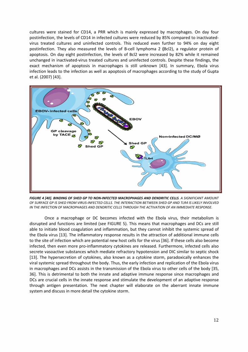
Very little was found in the literature on the question how the Ebola virus enters and infects cells. However, some research suggests that glycoprotein (GP) is involved in this process. GP is one of the seven structural proteins of the Ebola virus and is located on the surface of the Ebola virus [40, 41]. Several studies have been conducted to investigate the role of GP in the entry and infection of the Ebola virus [40, 41]. First, the study of Okumura et al. (2009) showed that surface GP interacts with TLR-4 [41]. TLR-4 is a member of the TLR family and is abundantly expressed on the surface of macrophages and DCs. The TLR4 recognizes the highly glycosylated molecules containing O-linked mannosyl residues which are well-known from the lipopolysaccharides found on gram-negative bacteria. However, these are also found in viral GP. Furthermore, Dolnik et al. (2004) demonstrated that a significant amount of surface GP is shed from virus-infected cells [42]. Thus, Ebola infected cells release both shed GP and new virions (see FIGURE 4). Whether this soluble shed GP influences virus pathogenicity and replication has not been exactly defined. Therefore, the study of Escudero-Pérez et al. (2014) investigated the role of shed GP in Ebola virus infection [40]. The results of this study suggest that the binding of shed GP to the surface of macrophages and DCs also involves an interaction with TLR4 (see FIGURE 4). Since the shed GP has a soluble nature, it could be possible that it infects remote and additional target cells by binding to their TLR4 [40]. The binding of shed GP to TLR4 activates the release of pro- and anti-inflammatory cytokines by macrophages and DCs.
One interesting finding is that viral replication might not be required for the activation of macrophages. The binding of GP to macrophages and DCs already activates an immediate cellular response [38]. The study of Wahl-Jensen et al. (2011) demonstrated that the incubation of macrophages with virus-like particles (VLPs) containing VP40 and GP led to similar levels of cytokine expression as an infection with live Ebola virus. These results are in agreement with those obtained by Escudero-Pérez et al. (2014) which showed that the binding of shed GP to macrophages and DCs led to the transcriptional activation of several genes in a dose-dependent manner, among which TNFα, IL-6, IL-10 and IL-12 [40]. This suggests that binding of GP to macrophages is important in the activation of an immediate cellular response, just like the entrance of viral virions into the cell [38].
In contrast to earlier findings, the study of Gupta et al. (2007) demonstrated that a majority of macrophages is killed by the Ebola virus [43]. They infected peripheral blood mononuclear cell (PBMCs), among which macrophages, with live virus, inactivated virus or without virus. The three
12
cultures were stained for CD14, a PRR which is mainly expressed by macrophages. On day four postinfection, the levels of CD14 in infected cultures were reduced by 85% compared to inactivated-virus treated cultures and uninfected controls. This reduced even further to 94% on day eight postinfection. They also measured the levels of B-cell lymphoma 2 (Bcl2), a regulator protein of apoptosis. On day eight postinfection, the levels of Bcl2 were increased by 82% while it remained unchanged in inactivated-virus treated cultures and uninfected controls. Despite these findings, the exact mechanism of apoptosis in macrophages is still unknown [43]. In summary, Ebola virus infection leads to the infection as well as apoptosis of macrophages according to the study of Gupta et al. (2007) [43].
FIGURE 4 [40]. BINDING OF SHED GP TO NON-INFECTED MACROPHAGES AND DENDRITIC CELLS. A SIGNIFICANT AMOUNT OF SURFACE GP IS SHED FROM VIRUS-INFECTED CELLS. THE INTERACTION BETWEEN SHED GP AND TLR4 IS LIKELY INVOLVED IN THE INFECTION OF MACROPHAGES AND DENDRITIC CELLS THROUGH THE ACTIVATION OF AN IMMEDIATE RESPONSE.
Once a macrophage or DC becomes infected with the Ebola virus, their metabolism is disrupted and functions are limited (see FIGURE 5). This means that macrophages and DCs are still able to initiate blood coagulation and inflammation, but they cannot inhibit the systemic spread of the Ebola virus [13]. The inflammatory response results in the attraction of additional immune cells to the site of infection which are potential new host cells for the virus [36]. If these cells also become infected, then even more pro-inflammatory cytokines are released. Furthermore, infected cells also secrete vasoactive substances which mediate refractory hypotension and DIC similar to septic shock [13]. The hypersecretion of cytokines, also known as a cytokine storm, paradoxically enhances the viral systemic spread throughout the body. Thus, the early infection and replication of the Ebola virus in macrophages and DCs assists in the transmission of the Ebola virus to other cells of the body [35, 36]. This is detrimental to both the innate and adaptive immune response since macrophages and DCs are crucial cells in the innate response and stimulate the development of an adaptive response through antigen presentation. The next chapter will elaborate on the aberrant innate immune system and discuss in more detail the cytokine storm.
13
4.2 ABERRANT INNATE IMMUNE RESPONSE
4.2.1. CYTOKINE STORM
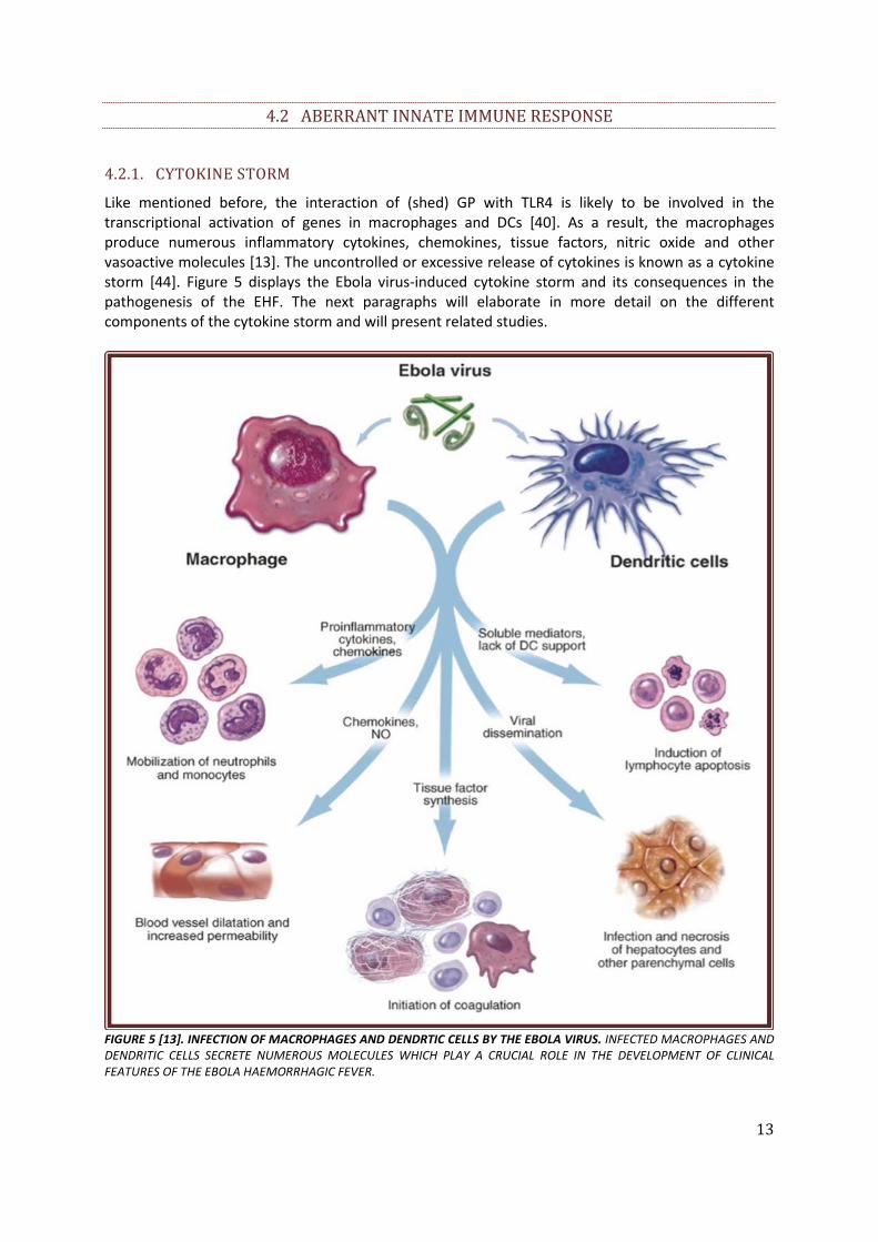
Like mentioned before, the interaction of (shed) GP with TLR4 is likely to be involved in the transcriptional activation of genes in macrophages and DCs [40]. As a result, the macrophages produce numerous inflammatory cytokines, chemokines, tissue factors, nitric oxide and other vasoactive molecules [13]. The uncontrolled or excessive release of cytokines is known as a cytokine storm [44]. Figure 5 displays the Ebola virus-induced cytokine storm and its consequences in the pathogenesis of the EHF. The next paragraphs will elaborate in more detail on the different components of the cytokine storm and will present related studies.
FIGURE 5 [13]. INFECTION OF MACROPHAGES AND DENDRTIC CELLS BY THE EBOLA VIRUS. INFECTED MACROPHAGES AND DENDRITIC CELLS SECRETE NUMEROUS MOLECULES WHICH PLAY A CRUCIAL ROLE IN THE DEVELOPMENT OF CLINICAL FEATURES OF THE EBOLA HAEMORRHAGIC FEVER.
14
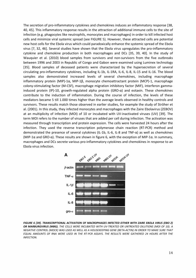
The secretion of pro-inflammatory cytokines and chemokines induces an inflammatory response [38, 40, 45]. This inflammatory response results in the attraction of additional immune cells to the site of infection (e.g. phagocytes like neutrophils, monocytes and macrophages) in order to kill infected host cells and minimize viral systemic spread (see FIGURE 5). However, these attracted cells are potential new host cells for the Ebola virus which could paradoxically enhance the systemic spread of the Ebola virus [7, 32, 46]. Several studies have shown that the Ebola virus upregulates the pro-inflammatory cytokine and chemokine production in both macrophages and DCs [35, 39, 40]. In the study of Wauquier et al. (2010) blood samples from survivors and non-survivors from the five outbreaks between 1996 and 2003 in Republic of Congo and Gabon were examined using Luminex technology [35]. Blood samples of deceased patients were characterized by the hypersecretion of several circulating pro-inflammatory cytokines, including IL-1b, IL-1RA, IL-6, IL-8, IL-15 and IL-16. The blood samples also demonstrated increased levels of several chemokines, including macrophage inflammatory protein (MIP)-1α, MIP-1β, monocyte chemoattractant protein (MCP)-1, macrophage colony-stimulating factor (M-CSF), macrophage migration inhibitory factor (MIF), interferon gamma-induced protein (IP)-10, growth-regulated alpha protein (GRO-α) and eotaxin. These chemokines contribute to the induction of inflammation. During the course of infection, the levels of these mediators became 5 till 1.000 times higher than the average levels observed in healthy controls and survivors. These results match those observed in earlier studies, for example the study of Strôher et al. (2001). In this study, they infected monocytes and macrophages with the Zaire Ebolavirus (ZEBOV) at an multiplicity of infection (MOI) of 10 or incubated with UV-inactivated viruses (UV) [39]. The term MOI refers to the number of viruses that are added per cell during infection. The activation was measured through transcription and protein expression. The cells were harvested 24 hours after the infection. They used the reverse transcription polymerase chain reaction (RT-PCR) method and demonstrated the presence of several cytokines (IL-1b, IL-6, IL-8 and TNF-α) as well as chemokines (MIP-1α and GRO-α). These results are shown in figure 6, with the exception of MIP-1α. In summary, macrophages and DCs secrete various pro-inflammatory cytokines and chemokines in response to an Ebola virus infection.
FIGURE 6 [39]. TRANSCRIPTIONAL ACTIVATION OF MACROPHAGES INFECTED EITHER WITH ZAIRE EBOLA VIRUS (EBO Z) OR MARBURGVIRUS (MBG). THE CELLS WERE INCUBATED WITH UV-TREATED OR UNTREATED DILUTIONS (MOI OF 10). A NEGATIVE CONTROL (MOCK) WAS USED AS WELL AS A HOUSEKEEPING GENE (BETA-ACTIN) IN ORDER TO MAKE SURE THAT EQUAL AMOUNTS OF RNA WERE USED IN THE RT-PCR ASSAYS. THE RESULTS WERE GATHERED 24 HOURS AFTER THE INFECTION.
15
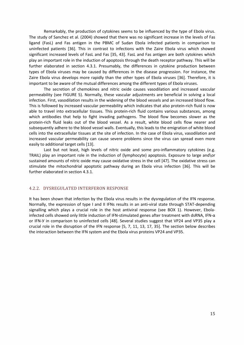
Remarkably, the production of cytokines seems to be influenced by the type of Ebola virus. The study of Sanchez et al. (2004) showed that there was no significant increase in the levels of Fas ligand (FasL) and Fas antigen in the PBMC of Sudan Ebola infected patients in comparison to uninfected patients [36]. This in contrast to infections with the Zaire Ebola virus which showed significant increased levels of FasL and Fas [35, 43]. FasL and Fas antigen are both cytokines which play an important role in the induction of apoptosis through the death receptor pathway. This will be further elaborated in section 4.3.1. Presumably, the differences in cytokine production between types of Ebola viruses may be caused by differences in the disease progression. For instance, the Zaire Ebola virus develops more rapidly than the other types of Ebola viruses [36]. Therefore, it is important to be aware of the mutual differences among the different types of Ebola viruses.
The secretion of chemokines and nitric oxide causes vasodilation and increased vascular permeability (see FIGURE 5). Normally, these vascular adjustments are beneficial in solving a local infection. First, vasodilation results in the widening of the blood vessels and an increased blood flow. This is followed by increased vascular permeability which indicates that also protein-rich fluid is now able to travel into extracellular tissues. This protein-rich fluid contains various substances, among which antibodies that help to fight invading pathogens. The blood flow becomes slower as the protein-rich fluid leaks out of the blood vessel. As a result, white blood cells flow nearer and subsequently adhere to the blood vessel walls. Eventually, this leads to the emigration of white blood cells into the extracellular tissues at the site of infection. In the case of Ebola virus, vasodilation and increased vascular permeability can cause severe problems since the virus can spread even more easily to additional target cells [13].
Last but not least, high levels of nitric oxide and some pro-inflammatory cytokines (e.g. TRAIL) play an important role in the induction of (lymphocyte) apoptosis. Exposure to large and\or sustained amounts of nitric oxide may cause oxidative stress in the cell [47]. The oxidative stress can stimulate the mitochondrial apoptotic pathway during an Ebola virus infection [36]. This will be further elaborated in section 4.3.1.
4.2.2. DYSREGULATED INTERFERON RESPONSE
It has been shown that infection by the Ebola virus results in the dysregulation of the IFN response. Normally, the expression of type I and II IFNs results in an anti-viral state through STAT-depending signalling which plays a crucial role in the host antiviral response (see BOX 1). However, Ebola-infected cells showed only little induction of IFN-stimulated genes after treatment with dsRNA, IFN-α or IFN-У in comparison to uninfected cells [48]. Several studies suggest that VP24 and VP35 play a crucial role in the disruption of the IFN response [5, 7, 11, 13, 17, 35]. The section below describes the interaction between the IFN system and the Ebola virus proteins VP24 and VP35.
16
BOX 1: SIGNALLING PATHWAY OF THE IFN SYSTEM WITHOUT AN EBOLA INFECTION
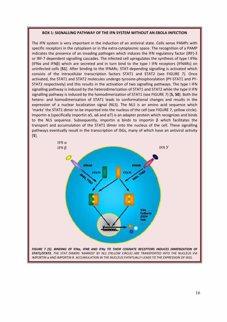
The IFN system is very important in the induction of an antiviral state. Cells sense PAMPs with specific receptors in the cytoplasm or in the extra-cytoplasmic space. The recognition of a PAMP indicates the presence of an invading pathogen which induces the IFN regulatory factor (IRF)-3 or IRF-7 dependent signalling cascades. The infected cell upregulates the synthesis of type I IFNs (IFNα and IFNβ) which are secreted and in turn bind to the type I IFN receptors (IFNARs) on uninfected cells [51]. After binding to the IFNARs, STAT-depending signalling is activated which consists of the intracellular transcription factors STAT1 and STAT2 (see FIGURE 7). Once activated, the STAT1 and STAT2 molecules undergo tyrosine-phosphorylation (PY-STAT1 and PY-STAT2 respectively) and this results in the activation of two signalling pathways. The type I IFN signalling pathway is induced by the heterodimerization of STAT1 and STAT2 while the type II IFN signalling pathway is induced by the homodimerization of STAT1 (see FIGURE 7) [5, 50]. Both the hetero- and homodimerization of STAT1 leads to conformational changes and results in the expression of a nuclear localization signal (NLS). The NLS is an amino acid sequence which ‘marks’ the STAT1 dimer to be imported into the nucleus of the cell (see FIGURE 7, yellow circle). Importin α (specifically importin α5, α6 and α7) is an adapter protein which recognizes and binds to the NLS sequence. Subsequently, importin α binds to importin β which facilitates the transport and accumulation of the STAT1 dimer into the nucleus of the cell. These signalling pathways eventually result in the transcription of ISGs, many of which have an antiviral activity [5].
FIGURE 7 [5]. BINDING OF IFNα, IFNβ AND IFNy TO THEIR COGNATE RECEPTORS INDUCES DIMERIZATION OF STAT1/STAT2. THE STAT DIMERS ‘MARKED’ BY NLS (YELLOW CIRCLE) ARE TRANSPORTED INTO THE NUCLEUS VIA IMPORTIN α AND IMPORTIN β. ACCUMULATION IN THE NUCLEUS EVENTUALLY LEADS TO THE EXPRESSION OF ISGS.
IFN У IFN α IFN β
17
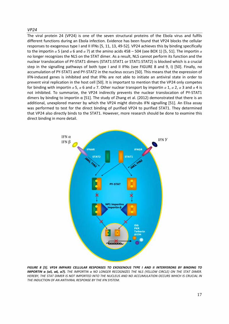
VP24 The viral protein 24 (VP24) is one of the seven structural proteins of the Ebola virus and fulfils different functions during an Ebola infection. Evidence has been found that VP24 blocks the cellular responses to exogenous type I and II IFNs [5, 11, 13, 49-52]. VP24 achieves this by binding specifically to the importin α 5 (and α 6 and α 7) at the amino acids 458 – 504 (see BOX 1) [5, 51]. The importin α no longer recognizes the NLS on the STAT dimer. As a result, NLS cannot perform its function and the nuclear translocation of PY-STAT1 dimers (STAT1:STAT1 or STAT1:STAT2) is blocked which is a crucial step in the signalling pathways of both type I and II IFNs (see FIGURE 8 and 9, I) [50]. Finally, no accumulation of PY-STAT1 and PY-STAT2 in the nucleus occurs [50]. This means that the expression of IFN-induced genes is inhibited and that IFNs are not able to initiate an antiviral state in order to prevent viral replication in the host cell [50]. It is important to mention that the VP24 only competes for binding with importin α 5, α 6 and α 7. Other nuclear transport by importin α 1, α 2, α 3 and α 4 is not inhibited. To summarize, the VP24 indirectly prevents the nuclear translocation of PY-STAT1 dimers by binding to importin α [51]. The study of Zhang et al. (2012) demonstrated that there is an additional, unexplored manner by which the VP24 might distrubs IFN signalling [51]. An Elisa assay was performed to test for the direct binding of purified VP24 to purified STAT1. They determined that VP24 also directly binds to the STAT1. However, more research should be done to examine this direct binding in more detail.
FIGURE 8 [5]. VP24 IMPAIRS CELLULAR RESPONSES TO EXOGENOUS TYPE I AND II INTERFERONS BY BINDING TO IMPORTIN α (α5, α6, α7). THE IMPORTIN α NO LONGER RECOGNIZES THE NLS (YELLOW CIRCLE) ON THE STAT DIMER. HEREBY, THE STAT DIMER IS NOT IMPORTED INTO THE NUCLEUS AND NO ACCUMULATION OCCURS WHICH IS CRUCIAL IN THE INDUCTION OF AN ANTIVIRAL RESPONSE BY THE IFN SYSTEM.
IFN α IFN β IFN У
18
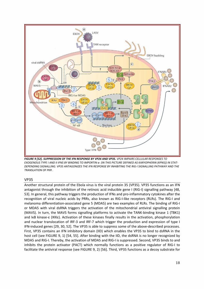
FIGURE 9 [52]. SUPPRESSION OF THE IFN RESPONSE BY VP24 AND VP35. VP24 IMPAIRS CELLULAR RESPONSES TO EXOGENOUS TYPE I AND II IFNS BY BINDING TO IMPORTIN α (IN THIS PICTURE DEFINED AS KARYOPHERIN (KPN5)) IN STAT-DEPENDING SIGNALLING. VP35 ANTAGONIZES THE IFN RESPONSE BY INHIBITING THE RIG-I SIGNALLING PATHWAY AND THE TRANSLATION OF PKR . VP35 Another structural protein of the Ebola virus is the viral protein 35 (VP35). VP35 functions as an IFN antagonist through the inhibition of the retinoic acid inducible gene I (RIG-I) signalling pathway [48, 53]. In general, this pathway triggers the production of IFNs and pro-inflammatory cytokines after the recognition of viral nucleic acids by PRRs, also known as RIG-I-like receptors (RLRs). The RIG-I and melanoma differentiation-associated gene 5 (MDA5) are two examples of RLRs. The binding of RIG-I or MDA5 with viral dsRNA triggers the activation of the mitochondrial antiviral signalling protein (MAVS). In turn, the MAVS forms signalling platforms to activate the TANK-binding kinase 1 (TBK1) and IκB kinase-ε (IKKε). Activation of these kinases finally results in the activation, phosphorylation and nuclear translocation of IRF-3 and IRF-7 which trigger the production and expression of type I IFN-induced genes [29, 30, 52]. The VP35 is able to suppress some of the above-described processes. First, VP35 contains an IFN inhibitory domain (IID) which enables the VP35 to bind to dsRNA in the host cell (see FIGURE 9, 1) [54, 55]. After binding with the IID, the dsRNA is no longer recognized by MDA5 and RIG-I. Thereby, the activation of MDA5 and RIG-I is suppressed. Second, VP35 binds to and inhibits the protein activator (PACT) which normally functions as a positive regulator of RIG-I to facilitate the antiviral response (see FIGURE 9, 2) [56]. Third, VP35 functions as a decoy substrate for
1 2
3 4
5
I
19
TBK1 and IKKε. This means that VP35 binds to the kinases instead of the real substrate, thus blocking their kinase activity (see FIGURE 9, 3). Therefore, VP35 blocks the phosphorylation and nuclear accumulation of IRF-3. At last, VP35 promotes the sumoylation of IRF-3 and IRF-7 (see FIGURE 9, 4). This is a post-translational modification which further impairs the transcriptional activity of IRF-3 and IRF-7 [30, 52]. As a result, the production of type I IFNs and pro-inflammatory cytokines as well as the expression of type I IFN-induced genes is disturbed [5].
VP35 also functions as an IFN antagonist by inhibiting the translation of some individual ISGs, for example the protein kinase R (PKR) [5, 7, 11, 13, 52]. Normally, the PKR is activated once it recognizes dsRNA during viral infection. The activated PKR phosphorylates the eukaryotic translation initiation factor 2 a (eIF2a). As a result, the synthesis of cellular and viral mRNAs is stopped. However, VP35 inhibits the activation of PKR causing the synthesis of cellular and viral mRNAs to continue (see FIGURE 9, 5). Hereby, the VP35 again antagonizes the IFN response. 4.2.3. IMPAIRED DENDRITIC CELL MATURATION
Another function of VP35 is the inhibition of DC maturation by interfering with the RIG-I signalling pathway [5, 7, 36, 40, 52, 53, 57]. As a result, the production of RLR-mediated pro-inflammatory cytokines is impaired. VP35 also blocks the upregulation of MHC I and MHC II as well as the costimulatory molecules CD40, CD80 and CD86. This leads to impaired antigen presentation by DCs to CD4+ and CD8+ cells and limited T cell activation. A study in human myeloid DCs demonstrated the difference in response to non-infectious virus like particles (VLP) or to live virus [13, 58]. When the immature DCs were exposed to non-infectious VLPs a strong inflammatory response was triggered. The DCs started to secrete several mediators, such as TNF-a, IL-6, IL-8 and MIP-1a. Also the MHC-I and II proteins as well as costimulatory molecules such as CD40, CD80 and CD86 were upregulated to transform in an antigen presenting phenotype [40]. All these adjustments enabled the DC to induce proliferation of naïve lymphocytes. However, when the immature DC was exposed to live virus it could not perform these adjustments properly. Only a few chemokines were secreted, the MHC and costimulatory molecules were not upregulated and no differentiation of allogenic lymphocytes was induced. To conclude, VP35 disrupts the crucial bridge between the innate and adaptive immune response. Besides this, the impaired functioning of DCs also contributes the lymphocyte apoptosis. This will be further discussed in the next chapter.
20
4.3 SUPPRESSION OF THE ADAPTIVE IMMUNE RESPONSE
As discussed above, the Ebola virus disrupts the innate immune response due to the early infection of macrophages and DCs. This leads to a cytokine storm and the inhibition of an IFN response. Furthermore, the macrophages and DCs are limited in their function to present antigens and to initiate antigen-specific responses. Altogether, this has a huge impact on the development of an adaptive immune response. The following part of this thesis will describe in more detail the suppression of an adaptive immune response by the Ebola virus.
Many viruses enhance their survival by the regulation of the host cell apoptotic machinery. Some viruses block the apoptosis, so they are able to maximize viral replication inside the host cell [46]. The Ebola virus also interferes with the host cell apoptotic machinery, but in a different manner. Without replicating inside the lymphocytes, the Ebola virus induces lymphocyte apoptosis. The loss of immune cells is beneficial for the virus since it will encounter less resistance. The excessive and uncontrolled release of several mediators (such as FasL, TNF-a and nitric oxygen) by Ebola-infected macrophages and DCs also contributes to the apoptotic processes [13, 59]. Moreover, VP24 and VP35 block the stimulation of latent lymphocytes by interfering with the IFN response. These pathogenic events are all very disadvantageous since the development of a potent cell-mediated immune response is a key factor in surviving Ebola infections [13].
4.3.1 LYMPHOCYTE APOPTOSIS
The apoptosis of lymphocytes is a pathological feature of the Ebola virus infection which occurs early in infection [13, 43, 60]. Even though the virus does not replicate inside the lymphocytes, they induce massive bystander apoptosis which further impairs immunity. Several studies have demonstrated the Ebola-mediated apoptosis of lymphocytes. The study of Wauquier et al. (2010) examined blood samples obtained during the five outbreaks between 1996 and 2003 in Republic of Congo and Gabon [35]. Lymphocyte apoptosis was demonstrated by comparing the percentages of CD4+ and CD8+ T cells in blood samples of survivors, nonsurvivors and controls. Normal percentages of CD4+ and CD8+ T cells were found in the blood samples of healthy controls (respectively 43.6% and 22.4%). Similar percentages of CD4+ and CD8+ T cells were found in the blood samples of survivors during acute phase (respectively 46.2% and 24.1%) and after recovery (respectively 36.6% and 17.4%). However, significantly lower percentages of CD4+ and CD8+ T cells were found in the blood samples of fatally infected patients (respectively 9.4% and 6%). The study of Sanchez et al. (2004) also examined blood samples from infected patients. In this study the blood samples were obtained during a Sudan Ebola virus outbreak in Uganda in 2000 [36]. Again, a reduced number of CD4+ and CD8+ T cells was detected in the blood samples of nonsurvivors. As the severity of the disease progressed, the number of CD4+ T cells and CD8+ T cells decreased below normal levels. This in contrast to the blood samples of survivors, which showed an increase in T lymphocytes as the disease progressed. These results are consistent with those of Gupta et al. (2007) who infected human PBMCs with Ebola virus in vitro [43]. They investigated a variety of cell types (e.g. macrophages, CD4+ T cells, CD8+ T cells, B cells and NK cells) for activation and apoptotic signals several days postinfection. They demonstrated that 20 to 30% of the CD4+ and CD8+ T cells underwent apoptosis during the course of infection. Besides lymphocyte apoptosis, they demonstrated that the majority (70 to 80%) of the macrophages underwent apoptosis by day eight after infection. But how does the Ebola virus induce apoptosis?

21
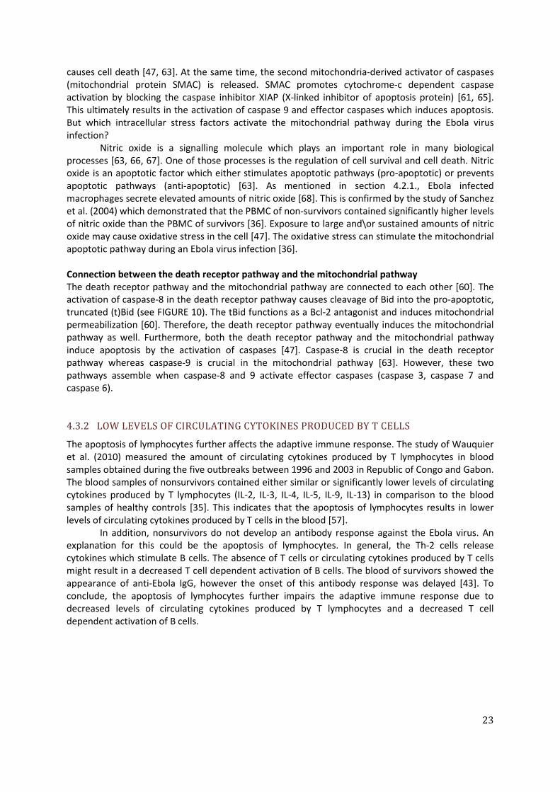
PATHWAYS OF LYMPHOCYTE APOPTOSIS There are still many questions concerning the process of lymphocyte apoptosis in Ebola virus infection [13]. Neither the systemic implications nor the cellular apoptotic pathway(s) of lymphocyte apoptosis are completely understood [60]. However, data suggests that several pathways are involved, including the death receptor pathway (extrinsic) and the mitochondrial pathway (intrinsic). Both pathways induce apoptosis by the activation of caspases (see FIGURE 10) [47]. Caspases are cysteine proteases which are synthesized as inactive proenzymes. Upon activation, they act as common death effector molecules and cleave various substrates in the cytoplasm or nucleus. Besides the extrinsic and intrinsic pathway, it is presumed that infected macrophages and DCs also contribute to lymphocyte apoptosis through the secretion of specific mediators and their impaired functioning [60]. The following paragraphs will elaborate on the activation of the death receptor and mitochondrial pathway during an Ebola virus infection.
FIGURE 10 [61]. THE DEATH RECEPTOR PATHWAY AND THE MITOCHONDRIAL PATHWAY. EVEN THOUGH LYMPHOCYTES ARE NOT INFECTED BY THE EBOLA VIRUS, THEY INDUCE MASSIVE BYSTANDER APOPTOSIS. THE DEATH RECEPTOR PATHWAY AS WELL AS THE MITOCHONDRIAL PATHWAY ARE LIKELY TO BE INVOLVED IN THE APOPTOSIS OF LYMPHOCYTES. PRESUMABLY, INFECTED MACRPHAGES AND DENDRITIC CELLS SECRETE MOLECULES WHICH COULD STIMULATE THESE APOPTOTIC PATHWAYS.
22
The death receptor pathway The death receptor pathway exists of ‘death receptors’ located on the surface of the cell. Examples of death receptors are TNF-R1, Fas (also known as CD95) and TRAIL receptors [62]. In general, all the different death receptors induce apoptosis in approximately the same way. First, the death receptor becomes activated by binding with its cognate ligand (see FIGURE 10). This leads to the activation of caspase-8 via the FAS-associated death domain protein (FADD) [63]. Some death receptors (such as TNF-R1) also need the TNFR-associated death domain protein (TRADD) to induce apoptosis. Finally, the caspase-8 activates effector caspases (such as caspase 3, caspase 6 and caspase 7) which eventually induce apoptosis [47, 61]. Apoptosis occurs within minutes after activation of the death receptors [46].
Several studies have been conducted to examine the role of the death receptor pathway during Ebola virus infection. The study of Bradfute et al. (2017) concluded that all the death receptors use the adaptor protein FADD to initiate apoptosis [60]. Therefore, transgenic mice carrying a dominant negative mutant of FADD (FADD-dn) in T cells were used to examine the role of the death receptor pathway. According to H&E and TUNEL staining of the thymus and spleens, the degree of lymphocyte apoptosis was less in the FADD-dn mice compared to the control mice on day seven postinfection. This confirms the hypothesis that the death receptor pathway contributes to the lymphocyte apoptosis during Ebola virus infection.
Now the question remains which death receptor(s) contribute to this pathway. Again, Bradfute et al. (2017) infected transgenic mice either lacking TRAIL, Fas or both FasL and TRAIL [60]. Since there was no inhibition of lymphocyte apoptosis, it was suggested that multiple death receptors are involved. In addition, Wauquier et al. (2010) conducted a study in which they examined human blood samples for the presence of CD95. High percentages of Fas could confirm the presence of apoptosis. The blood samples of healthy controls showed 5.6% of CD4+CD95+ T cells and 6.8% of CD8+CD95+ T cells in comparison to a significant increase of 54.1% and 75.8% respectively in the blood of fatally infected patients [35]. Also the study of Gupta et al. (2007) demonstrated induced apoptotic signals in infected CD4+CD95+ T cells and CD4+CD95+ T cells [43]. This further suggests that the apoptosis of lymphocytes during the Ebola virus infection among others is caused by the Fas/FasL interactions [35]. Moreover, Gupta et al. (2007) showed that the TRAIL mRNA was upregulated in CD4+ and CD8+ T cells [43]. These studies suggest that Fas/FasL and TRAIL play a role in the lymphocyte apoptosis.
Even though lymphocytes are not infected by the Ebola virus, they induce massive bystander apoptosis which further impairs immunity. One hypothesis is that infected macrophages and DCs secrete or express molecules (such as TRAIL, TNF-a, and Fas ligand) which cause the apoptosis of lymphocytes. The study of Hensley et al. (2004) supported this hypothesis since data shows that TRAIL is expressed in higher amounts by Zaire Ebola infected monocytes [64]. Furthermore, infected monkeys show higher amounts of soluble Fas in their sera and an increased expression of TRAIL and Fas mRNA in their PBMC [47, 59, 60, 63]. To conclude, infection of macrophages and DCs is likely involved in the increased lymphocyte apoptosis during an Ebola virus infection.
The mitochondrial pathway Another pathway which induces apoptosis is the mitochondrial pathway. Multiple steps are involved to eventually induce apoptosis. First, the pathway is stimulated by intracellular stress factors, such as DNA damage, oxidative stress and radiation (see FIGURE 10) [47]. These stimuli activate the BH3-only proteins which are initiators of apoptosis. The activated BH3-only proteins can either directly or indirectly activate the pro-apoptotic effectors Bax and Bak [47]. This results in the release of cytochrome c into the cytosol which in turn binds to apoptosis protease activating factor 1 (Apaf-1). Together they form an apoptosome that recruits and activates the caspase cascade beginning with caspase-9 [60]. Finally, the caspase-9 activates effector caspases (such as caspase 3) which eventually
23
causes cell death [47, 63]. At the same time, the second mitochondria-derived activator of caspases (mitochondrial protein SMAC) is released. SMAC promotes cytochrome-c dependent caspase activation by blocking the caspase inhibitor XIAP (X-linked inhibitor of apoptosis protein) [61, 65]. This ultimately results in the activation of caspase 9 and effector caspases which induces apoptosis. But which intracellular stress factors activate the mitochondrial pathway during the Ebola virus infection?
Nitric oxide is a signalling molecule which plays an important role in many biological processes [63, 66, 67]. One of those processes is the regulation of cell survival and cell death. Nitric oxide is an apoptotic factor which either stimulates apoptotic pathways (pro-apoptotic) or prevents apoptotic pathways (anti-apoptotic) [63]. As mentioned in section 4.2.1., Ebola infected macrophages secrete elevated amounts of nitric oxide [68]. This is confirmed by the study of Sanchez et al. (2004) which demonstrated that the PBMC of non-survivors contained significantly higher levels of nitric oxide than the PBMC of survivors [36]. Exposure to large and\or sustained amounts of nitric oxide may cause oxidative stress in the cell [47]. The oxidative stress can stimulate the mitochondrial apoptotic pathway during an Ebola virus infection [36]. Connection between the death receptor pathway and the mitochondrial pathway The death receptor pathway and the mitochondrial pathway are connected to each other [60]. The activation of caspase-8 in the death receptor pathway causes cleavage of Bid into the pro-apoptotic, truncated (t)Bid (see FIGURE 10). The tBid functions as a Bcl-2 antagonist and induces mitochondrial permeabilization [60]. Therefore, the death receptor pathway eventually induces the mitochondrial pathway as well. Furthermore, both the death receptor pathway and the mitochondrial pathway induce apoptosis by the activation of caspases [47]. Caspase-8 is crucial in the death receptor pathway whereas caspase-9 is crucial in the mitochondrial pathway [63]. However, these two pathways assemble when caspase-8 and 9 activate effector caspases (caspase 3, caspase 7 and caspase 6). 4.3.2 LOW LEVELS OF CIRCULATING CYTOKINES PRODUCED BY T CELLS
The apoptosis of lymphocytes further affects the adaptive immune response. The study of Wauquier et al. (2010) measured the amount of circulating cytokines produced by T lymphocytes in blood samples obtained during the five outbreaks between 1996 and 2003 in Republic of Congo and Gabon. The blood samples of nonsurvivors contained either similar or significantly lower levels of circulating cytokines produced by T lymphocytes (IL-2, IL-3, IL-4, IL-5, IL-9, IL-13) in comparison to the blood samples of healthy controls [35]. This indicates that the apoptosis of lymphocytes results in lower levels of circulating cytokines produced by T cells in the blood [57].
In addition, nonsurvivors do not develop an antibody response against the Ebola virus. An explanation for this could be the apoptosis of lymphocytes. In general, the Th-2 cells release cytokines which stimulate B cells. The absence of T cells or circulating cytokines produced by T cells might result in a decreased T cell dependent activation of B cells. The blood of survivors showed the appearance of anti-Ebola IgG, however the onset of this antibody response was delayed [43]. To conclude, the apoptosis of lymphocytes further impairs the adaptive immune response due to decreased levels of circulating cytokines produced by T lymphocytes and a decreased T cell dependent activation of B cells.
24
5. VACCINE DEVELOPMENT
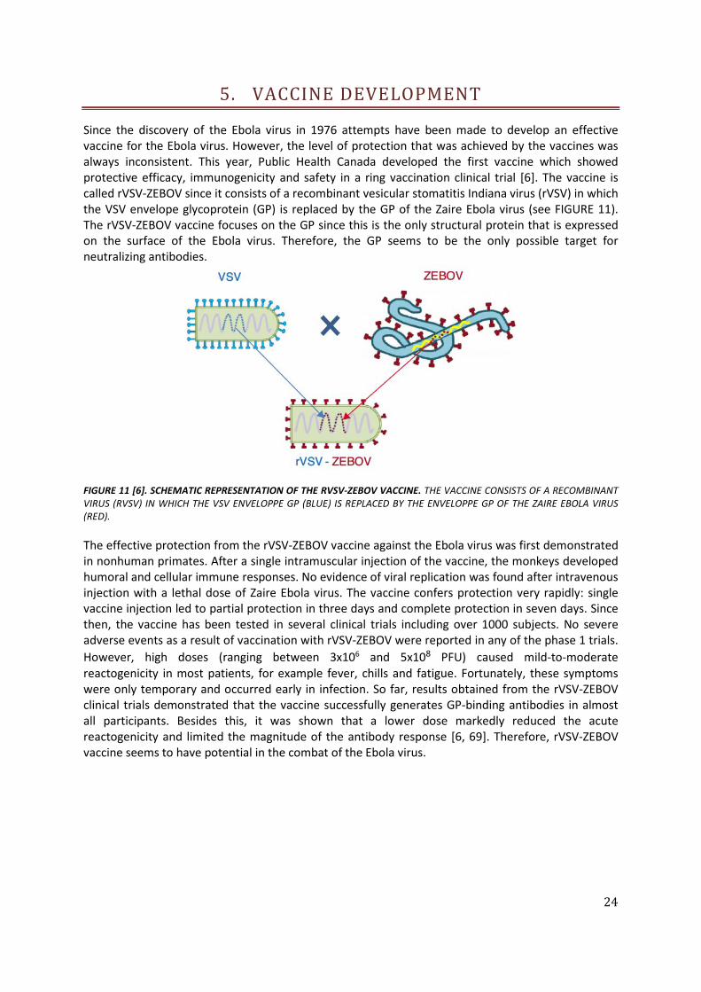
Since the discovery of the Ebola virus in 1976 attempts have been made to develop an effective vaccine for the Ebola virus. However, the level of protection that was achieved by the vaccines was always inconsistent. This year, Public Health Canada developed the first vaccine which showed protective efficacy, immunogenicity and safety in a ring vaccination clinical trial [6]. The vaccine is called rVSV-ZEBOV since it consists of a recombinant vesicular stomatitis Indiana virus (rVSV) in which the VSV envelope glycoprotein (GP) is replaced by the GP of the Zaire Ebola virus (see FIGURE 11). The rVSV-ZEBOV vaccine focuses on the GP since this is the only structural protein that is expressed on the surface of the Ebola virus. Therefore, the GP seems to be the only possible target for neutralizing antibodies.
FIGURE 11 [6]. SCHEMATIC REPRESENTATION OF THE RVSV-ZEBOV VACCINE. THE VACCINE CONSISTS OF A RECOMBINANT VIRUS (RVSV) IN WHICH THE VSV ENVELOPPE GP (BLUE) IS REPLACED BY THE ENVELOPPE GP OF THE ZAIRE EBOLA VIRUS (RED). The effective protection from the rVSV-ZEBOV vaccine against the Ebola virus was first demonstrated in nonhuman primates. After a single intramuscular injection of the vaccine, the monkeys developed humoral and cellular immune responses. No evidence of viral replication was found after intravenous injection with a lethal dose of Zaire Ebola virus. The vaccine confers protection very rapidly: single vaccine injection led to partial protection in three days and complete protection in seven days. Since then, the vaccine has been tested in several clinical trials including over 1000 subjects. No severe adverse events as a result of vaccination with rVSV-ZEBOV were reported in any of the phase 1 trials. However, high doses (ranging between 3x10⁶ and 5x10⁸ PFU) caused mild-to-moderate reactogenicity in most patients, for example fever, chills and fatigue. Fortunately, these symptoms were only temporary and occurred early in infection. So far, results obtained from the rVSV-ZEBOV clinical trials demonstrated that the vaccine successfully generates GP-binding antibodies in almost all participants. Besides this, it was shown that a lower dose markedly reduced the acute reactogenicity and limited the magnitude of the antibody response [6, 69]. Therefore, rVSV-ZEBOV vaccine seems to have potential in the combat of the Ebola virus.
25
6. DISCUSSION
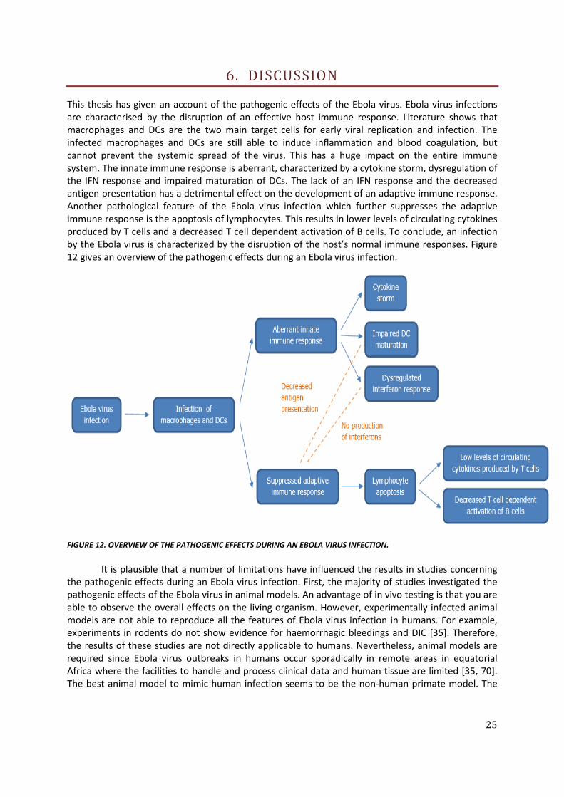
This thesis has given an account of the pathogenic effects of the Ebola virus. Ebola virus infections are characterised by the disruption of an effective host immune response. Literature shows that macrophages and DCs are the two main target cells for early viral replication and infection. The infected macrophages and DCs are still able to induce inflammation and blood coagulation, but cannot prevent the systemic spread of the virus. This has a huge impact on the entire immune system. The innate immune response is aberrant, characterized by a cytokine storm, dysregulation of the IFN response and impaired maturation of DCs. The lack of an IFN response and the decreased antigen presentation has a detrimental effect on the development of an adaptive immune response. Another pathological feature of the Ebola virus infection which further suppresses the adaptive immune response is the apoptosis of lymphocytes. This results in lower levels of circulating cytokines produced by T cells and a decreased T cell dependent activation of B cells. To conclude, an infection by the Ebola virus is characterized by the disruption of the host’s normal immune responses. Figure 12 gives an overview of the pathogenic effects during an Ebola virus infection.
FIGURE 12. OVERVIEW OF THE PATHOGENIC EFFECTS DURING AN EBOLA VIRUS INFECTION.
It is plausible that a number of limitations have influenced the results in studies concerning
the pathogenic effects during an Ebola virus infection. First, the majority of studies investigated the pathogenic effects of the Ebola virus in animal models. An advantage of in vivo testing is that you are able to observe the overall effects on the living organism. However, experimentally infected animal models are not able to reproduce all the features of Ebola virus infection in humans. For example, experiments in rodents do not show evidence for haemorrhagic bleedings and DIC [35]. Therefore, the results of these studies are not directly applicable to humans. Nevertheless, animal models are required since Ebola virus outbreaks in humans occur sporadically in remote areas in equatorial Africa where the facilities to handle and process clinical data and human tissue are limited [35, 70]. The best animal model to mimic human infection seems to be the non-human primate model. The
26
Ebola virus causes similar symptoms in non-human primates as in humans, including hypotensive shock and haemorrhagic bleedings. However, this model is also limited since even small doses of the Ebola virus cause uniformly lethal infections in non-human primates whereas a small percentage of humans (10 to 20%) survives infection by the Ebola virus [13, 35]. Second, most information on human Ebola virus infections comes from in vitro studies. Results from in vitro studies may not correspond to the circumstances occurring a living organism. However, in vivo studies in humans are too risky due to the high mortality rates observed during Ebola virus outbreaks. These two limitations underline the difficulty of collecting data on the human immune response to Ebola virus infections. Further in vivo research and data collection is required to determine exactly the pathogenic effects of the Ebola virus in humans. This knowledge is necessary in the development of therapeutic treatments and vaccines.
More research is required to determine the reservoir species of the Ebola virus. Presumably, the Ebola virus infection does not cause disease in the reservoir species which makes the immune system of the reservoir species of great interest. Revealing the protective immune mechanisms of the reservoir species could provide knowledge for the development of therapeutic treatments and vaccines. Nowadays, the fruits bats are the prime reservoir suspects since they do not develop filovirus disease after infection [5]. Moreover, discovery of the reservoir species could improve the prevention strategies against Ebola virus infection. Therefore, it is of great concern to find out where the Ebola virus is hiding.
This thesis mainly focussed on the pathogenic effects during an Ebola virus infection and less on possible therapeutic treatments. Survival from an Ebola virus infection seems to correlate with the development of an antigen-specific immune response [13]. However, the decreased production of IFNs, the impaired maturation of DCs and the apoptosis of lymphocytes decreases the development of an antigen-specific adaptive immune response during an Ebola virus infection. Further research should be conducted to examine the importance of an antigen-specific immune response during an Ebola virus infection and to develop therapeutic treatments and vaccines which counteract the mechanisms of immune evasion by the Ebola virus. The Public Health Canada recently developed the rVSV-ZEBOV vaccine which generates GP-binding antibodies in a dose-dependent manner. The vaccine already showed protective efficacy, immunogenicity and safety in a ring vaccination clinical trial [6]. The vaccine contributes to the development of humoral and cellular immunity and seems to have potential in the prevention of Ebola virus infections. Further work needs to be done to establish policies for the use of the vaccine in (pre-)epidemic situations [7]. However, it is also important to develop a treatment which helps infected individuals to overcome the Ebola virus infection in future epidemics. This could include the establishment of early and effective epidemic control and the improvement of treatment and patient care in remote, resource-poor areas where the Ebola virus typically re-emerges [7]. To conclude, there is a growing understanding about the pathogenic effects of the Ebola virus. Now, it is necessary to combine all the knowledge in the development of efficient vaccines and treatments.
While developing therapeutic treatments and vaccines, it is important to take into consideration that the human target population in African regions is the most affected by Human Immunodeficiency Virus (HIV) infections. In 2016, approximately 25 million people with HIV were living in African regions [71]. Individuals who suffer from HIV have a weakened immune system and show increased susceptibility to many infections. The Public Health Canada already evaluated rVSV-vaccine safety in immunocompromised hosts by using a few rhesus macaques infected with simian-human immunodeficiency virus (SHIV) [6]. Four out of the six SHIV macaques were protected against Ebola virus infection after immunization and none showed evidence of illness. These results are very promising in macaques infected with SHIV and further research must show the efficacy in humans infected with HIV.

27
Another aspect that should be taken into account during the development of therapeutic treatments and vaccines are the differences between the Ebola virus species. All known Ebola virus species are related, but differ genetically from each other [7]. Therefore, the different species cause (slightly) different clinical syndromes. The Zaire Ebola virus is the most dangerous of the five Ebola virus species with mortality rates varying between 80 to 90% [13]. The Sudan Ebola virus is less dangerous with mortality rates varying between 50 to 60% [13]. Almost all human cases in regions of Gabon, Republic of Congo and Democratic Republic of the Congo are due to the (re)emergence of Zaire Ebola virus, while epidemics in Sudan and Uganda are caused by the Sudan Ebola virus [3]. The role of the Côte d'Ivoire Ebola virus and Bundibugyo Ebola virus in the occurrence of EHF in equatorial Africa is not clear since both of them never re-emerged after their discovery [3]. At last, the Reston Ebola virus is not infectious in humans. The differences between the Ebola virus species should be taken into account while developing and administering therapeutic treatments and vaccines.
28
7. CONCLUSION
Since the discovery in 1976, much knowledge was gained about the Ebola virus. Ebola virus infections are characterised by the disruption of an effective host immune response, both innate and adaptive. Many of these processes have been described, for example the infection of macrophages and DCs, the induction of a cytokine storm, the dysregulation of an IFN response or the apoptosis of lymphocytes. However, not all the mechanisms are completely understood yet. Therefore, the Ebola virus remains a mysterious enemy to human survival. This thesis has reviewed the existing knowledge about the disruption of the human immune response during an Ebola virus infection. By understanding the immune evasion you are better able to look for possibilities or solutions to prevent or inhibit it. Last year, a major step was made through the development of the first vaccine which showed protective efficacy, immunogenicity and safety in a ring vaccination clinical trial. The development of a vaccine is very beneficial in the prevention of new epidemics but therapeutic treatments are also needed to treat infected individuals. Furthermore, there are still questions concerning the reservoir species of the Ebola virus. To conclude, despite the growing understanding about the Ebola virus still much research needs to be done.
29
8. REFERENCES
1. Wagner, E.K., Basic virology. 2004, Malden, MA: Blackwell. 2. Wageningen University and Research Centre, Cell Biology and Immunology, Virology and
Toxicology. 2014: Wageningen University. 3. Feldmann, H. and Geisbert, T.W., Ebola haemorrhagic fever. The Lancet, 2011.
377(9768): p. 849-862. 4. Sullivan, N., Yang, Z.-Y., and Nabel, G.J., Ebola virus pathogenesis: implications for vaccines
and therapies. Journal of virology, 2003. 77(18): p. 9733-9737. 5. Kühl, A. and Pöhlmann, S., How Ebola virus counters the interferon system. Zoonoses and
public health, 2012. 59(s2): p. 116-131. 6. Medaglini, D. and Siegrist, C.-A., Immunomonitoring of human responses to the rVSV-
ZEBOV Ebola vaccine. Current Opinion in Virology, 2017. 23: p. 88-94. 7. Baseler, L., The pathogenesis of Ebola virus disease. Annual Review of Pathology:
Mechanisms of Disease, 2017. 12: p. 387-418. 8. World Health Organization. Ebola virus disease. 2017; Available from:
http://www.who.int/mediacentre/factsheets/fs103/en/. 9. Centers for Disease Control and Prevention. Ebola (Ebola Virus Disease). 2016; Available
from: https://www.cdc.gov/vhf/ebola/about.html. 10. Centers for Disease Control and Prevention. 2014-2016 Ebola Outbreak in West Africa.
2016; Available from: https://www.cdc.gov/vhf/ebola/outbreaks/2014-west-africa/index.html.
11. Wilson, J.A., Vaccine potential of Ebola virus VP24, VP30, VP35, and VP40 proteins. Virology, 2001. 286(2): p. 384-390.
12. Alberts, B., Essential cell biology. 2013: Garland Science. 13. Bray, M. and Geisbert, T.W., Ebola virus: the role of macrophages and dendritic cells in the
pathogenesis of Ebola hemorrhagic fever. The international journal of biochemistry & cell biology, 2005. 37(8): p. 1560-1566.
14. Casillas, A.M., A current review of Ebola virus: pathogenesis, clinical presentation, and diagnostic assessment. Biological research for nursing, 2003. 4(4): p. 268-275.
15. Centers for Disease Control and Prevention. Virus Ecology Graphic. 2016; Available from: https://www.cdc.gov/vhf/ebola/resources/virus-ecology.html.
16. World Health Organization, The Control of Neglected Zoonotic Diseases. 2005: Geneva. 17. Falasca, L., Molecular mechanisms of Ebola virus pathogenesis: focus on cell death. Cell
death and differentiation, 2015. 22(8): p. 1250. 18. Bower, H., Exposure-specific and age-specific attack rates for Ebola virus disease in Ebola-
affected households, Sierra Leone. Emerging infectious diseases, 2016. 22(8): p. 1403. 19. Geisbert, T.W., Mechanisms underlying coagulation abnormalities in ebola hemorrhagic
fever: overexpression of tissue factor in primate monocytes/macrophages is a key event. The Journal of infectious diseases, 2003. 188(11): p. 1618-1629.
20. Levi, M. and Ten Cate, H., Disseminated intravascular coagulation. New England Journal of Medicine, 1999. 341(8): p. 586-592.
21. National Heart, L., and Blood Institute,. What Is Disseminated Intravascular Coagulation? 2011; Available from: https://www.nhlbi.nih.gov/health/health-topics/topics/dic.
22. Leung, D.W., Structural basis for dsRNA recognition and interferon antagonism by Ebola VP35. Nature structural & molecular biology, 2010. 17(2): p. 165-172.
23. World Health Organization. Leaders gather in Guinea to celebrate Ebola vaccine successes. 2017; Available from: http://www.who.int/features/2017/ebola-vaccine-guinea/en/.
30
24. Silverthorn, D.U., Human physiology: An integrated approach. 2007, San Francisco: Pearson/Benjamin Cummings.
25. Janeway, C. and Travers, P., Immunobiology: The Immune System in Health and Disease. 2001, New York.
26. Marie Curie Initial Training Network in Infection Biology. Innate immunity, our sentinel. 2016; Available from: http://www.inbionet.eu/outreach-news/innate-immunity--our-sentinel/n.
27. Savina, A. and Amigorena, S., Phagocytosis and antigen presentation in dendritic cells. Immunological reviews, 2007. 219(1): p. 143-156.
28. Alberts, B., Johnson, A., and Lewis, J. Molecular Biology of the Cell. 4th edition. 2002; Available from: https://www.ncbi.nlm.nih.gov/books/NBK26846/
29. Baum, A. and García-Sastre, A., Induction of type I interferon by RNA viruses: cellular receptors and their substrates. Amino acids, 2010. 38(5): p. 1283-1299.
30. Kawai, T. and Akira, S., Toll‐like Receptor and RIG‐1‐like Receptor Signaling. Annals of the New York Academy of Sciences, 2008. 1143(1): p. 1-20.
31. Xagorari, A. and Chlichlia, K., Toll-like receptors and viruses: induction of innate antiviral immune responses. The open microbiology journal, 2008. 2: p. 49.
32. Topham, N.J. and Hewitt, E.W., Natural killer cell cytotoxicity: how do they pull the trigger? Immunology, 2009. 128(1): p. 7-15.
33. Haller, O., Kochs, G., and Weber, F., The interferon response circuit: induction and suppression by pathogenic viruses. Virology, 2006. 344(1): p. 119-130.
34. Le Page, C., Interferon activation and innate immunity. 2000; Available from: https://www.ncbi.nlm.nih.gov/pubmed/11256746.
35. Wauquier, N., Human fatal zaire ebola virus infection is associated with an aberrant innate immunity and with massive lymphocyte apoptosis. PLoS Negl Trop Dis, 2010. 4(10): p. e837.
36. Sanchez, A., Analysis of human peripheral blood samples from fatal and nonfatal cases of Ebola (Sudan) hemorrhagic fever: cellular responses, virus load, and nitric oxide levels. Journal of virology, 2004. 78(19): p. 10370-10377.
37. Iversen, M.B. and Paludan, S.R., Mechanisms of type III interferon expression. Journal of Interferon & Cytokine Research, 2010. 30(8): p. 573-578.
38. Wahl-Jensen, V., Ebola virion attachment and entry into human macrophages profoundly effects early cellular gene expression. PLoS neglected tropical diseases, 2011. 5(10): p. e1359.
39. Ströher, U., Infection and activation of monocytes by Marburg and Ebola viruses. Journal of virology, 2001. 75(22): p. 11025-11033.
40. Escudero-Pérez, B., Shed GP of Ebola virus triggers immune activation and increased vascular permeability. PLoS pathogens, 2014. 10(11): p. e1004509.
41. Okumura, A., Interaction between Ebola virus glycoprotein and host toll-like receptor 4 leads to induction of proinflammatory cytokines and SOCS1. Journal of virology, 2010. 84(1): p. 27-33.
42. Dolnik, O., Ectodomain shedding of the glycoprotein GP of Ebola virus. The EMBO journal, 2004. 23(10): p. 2175-2184.
43. Gupta, M., Spiropoulou, C., and Rollin, P.E., Ebola virus infection of human PBMCs causes massive death of macrophages, CD4 and CD8 T cell sub-populations in vitro. Virology, 2007. 364(1): p. 45-54.
44. Tisoncik, J.R., Into the eye of the cytokine storm. Microbiology and Molecular Biology Reviews, 2012. 76(1): p. 16-32.
45. Cameron, M.J. and Kelvin, D.J. Cytokines, Chemokines and Their Receptors. 2013; Available from: https://www.ncbi.nlm.nih.gov/books/NBK6294/.
31
46. Edwards, K.M., Anti-viral strategies of cytotoxic T lymphocytes are manifested through a variety of granule-bound pathways of apoptosis induction. Immunology & Cell Biology, 1999. 77(1).
47. Fulda, S., Cellular stress responses: cell survival and cell death. International journal of cell biology, 2010. 2010.
48. Basler, C.F., The Ebola virus VP35 protein functions as a type I IFN antagonist. Proceedings of the National Academy of Sciences, 2000. 97(22): p. 12289-12294.
49. Mateo, M., Ebolavirus VP24 binding to karyopherins is required for inhibition of interferon signaling. Journal of virology, 2010. 84(2): p. 1169-1175.
50. Leung, L.W., Ebola virus VP24 binds karyopherin α1 and blocks STAT1 nuclear accumulation. Journal of virology, 2006. 80(11): p. 5156-5167.
51. Zhang, A.P., The ebola virus interferon antagonist VP24 directly binds STAT1 and has a novel, pyramidal fold. PLoS pathogens, 2012. 8(2): p. e1002550.
52. Prescott, J.B., Immunobiology of Ebola and Lassa virus infections. Nature Reviews Immunology, 2017. 17(3): p. 195-207.
53. Yen, B., Molecular basis for ebolavirus VP35 suppression of human dendritic cell maturation. Journal of virology, 2014. 88(21): p. 12500-12510.
54. Leung, D.W., Ebolavirus VP35 is a multifunctional virulence factor. Virulence, 2010. 1(6): p. 526-531.
55. Leung, D.W., Structure of the Ebola VP35 interferon inhibitory domain. Proceedings of the National Academy of Sciences, 2009. 106(2): p. 411-416.
56. Kok, K.-H., The double-stranded RNA-binding protein PACT functions as a cellular activator of RIG-I to facilitate innate antiviral response. Cell host & microbe, 2011. 9(4): p. 299-309.
57. Lubaki, N.M., The Ebola Interferon Inhibiting Domains Attenuate and Dysregulate Cell-Mediated Immune Responses. PLoS Pathogens, 2016. 12(12): p. e1006031.
58. Bosio, C.M., Ebola and Marburg virus-like particles activate human myeloid dendritic cells. Virology, 2004. 326(2): p. 280-287.
59. Baize, S., Apoptosis in fatal Ebola infection. Does the virus toll the bell for immune system? Apoptosis, 2000. 5(1): p. 5-7.
60. Bradfute, S.B., Mechanisms and consequences of ebolavirus-induced lymphocyte apoptosis. The journal of immunology, 2010. 184(1): p. 327-335.
61. Czabotar, P.E., Control of apoptosis by the BCL-2 protein family: implications for physiology and therapy. Nature reviews. Molecular cell biology, 2014. 15(1): p. 49.
62. Guicciardi, M.E. and Gores, G.J., Life and death by death receptors. The FASEB Journal, 2009. 23(6): p. 1625-1637.
63. Brüne, B., Nitric oxide: NO apoptosis or turning it ON? Cell death and differentiation, 2003. 10(8): p. 864.
64. Hensley, L.E., Proinflammatory response during Ebola virus infection of primate models: possible involvement of the tumor necrosis factor receptor superfamily. Immunology letters, 2002. 80(3): p. 169-179.
65. Du, C., Smac, a mitochondrial protein that promotes cytochrome c–dependent caspase activation by eliminating IAP inhibition. Cell, 2000. 102(1): p. 33-42.
66. Wei, T., Nitric oxide induces oxidative stress and apoptosis in neuronal cells. Biochimica et Biophysica Acta (BBA)-Molecular Cell Research, 2000. 1498(1): p. 72-79.
67. Boscá, L. and Hortelano, S., Mechanisms of nitric oxide-dependent apoptosis: involvement of mitochondrial mediators. Cellular signalling, 1999. 11(4): p. 239-244.
68. Niedbala, W., Cai, B., and Liew, F., Role of nitric oxide in the regulation of T cell functions. Annals of the rheumatic diseases, 2006. 65(suppl 3): p. iii37-iii40.
69. Rechtien, A., Systems Vaccinology Identifies an Early Innate Immune Signature as a Correlate of Antibody Responses to the Ebola Vaccine rVSV-ZEBOV. Cell Reports, 2017. 20(9): p. 2251-2261.

32
70. Nakayama, E. and Saijo, M., Animal models for Ebola and Marburg virus infections. Frontiers in microbiology, 2013. 4.
71. World Health Organization. HIV/AIDS 2017; Available from: http://www.who.int/mediacentre/factsheets/fs360/en/.