Embed Size (px)
Citation preview
ww.sciencedirect.com
wat e r r e s e a r c h 4 6 ( 2 0 1 2 ) 3 9 8 9e3 9 9 8
Available online at w
journal homepage: www.elsevier .com/locate/watres
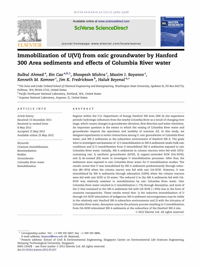
Immobilization of U(VI) from oxic groundwater by Hanford300 Area sediments and effects of Columbia River water
Bulbul Ahmed a, Bin Cao a,b,1, Bhoopesh Mishra c, Maxim I. Boyanov c,Kenneth M. Kemner c, Jim K. Fredrickson b, Haluk Beyenal a,*aThe Gene and Linda Voiland School of Chemical Engineering and Bioengineering, Washington State University, Spokane St, PO Box 642710,
Pullman, WA 99164-2710, United Statesb Pacific Northwest National Laboratory, Richland, WA, United StatescArgonne National Laboratory, Argonne, IL, United States
a r t i c l e i n f o
Article history:
Received 15 December 2011
Received in revised form
8 May 2012
Accepted 15 May 2012
Available online 23 May 2012
Keywords:
Uranium immobilization
Bioremediation
Biofilm
Groundwater
Columbia River water
Remobilization
* Corresponding author. Tel.: þ1 509 335 660E-mail address: [email protected] (H. Bey
1 Present address: School of Civil & EnvNanyang Technological University, Singapor0043-1354/$ e see front matter ª 2012 Elsevdoi:10.1016/j.watres.2012.05.027
a b s t r a c t
Regions within the U.S. Department of Energy Hanford 300 Area (300 A) site experience
periodic hydrologic influences from the nearby Columbia River as a result of changing river
stage, which causes changes in groundwater elevation, flow direction and water chemistry.
An important question is the extent to which the mixing of Columbia River water and
groundwater impacts the speciation and mobility of uranium (U). In this study, we
designed experiments to mimic interactions among U, oxic groundwater or Columbia River
water, and 300 A sediments in the subsurface environment of Hanford 300 A. The goals
were to investigate mechanisms of: 1) U immobilization in 300 A sediments under bulk oxic
conditions and 2) U remobilization from U-immobilized 300 A sediments exposed to oxic
Columbia River water. Initially, 300 A sediments in column reactors were fed with U(VI)-
containing oxic 1) synthetic groundwater (SGW), 2) organic-amended SGW (OA-SGW),
and 3) de-ionized (DI) water to investigate U immobilization processes. After that, the
sediments were exposed to oxic Columbia River water for U remobilization studies. The
results reveal that U was immobilized by 300 A sediments predominantly through reduc-
tion (80e85%) when the column reactor was fed with oxic OA-SGW. However, U was
immobilized by 300 A sediments through adsorption (100%) when the column reactors
were fed with oxic SGW or DI water. The reduced U in the 300 A sediments fed with OA-
SGW was relatively resistant to remobilization by oxic Columbia River water. Oxic
Columbia River water resulted in U remobilization (w7%) through desorption, and most of
the U that remained in the 300 A sediments fed with OA-SGW (w93%) was in the form of
uraninite nanoparticles. These results reveal that: 1) the reductive immobilization of U
through OA-SGW stimulation of indigenous 300 A sediment microorganisms may be viable
in the relatively oxic Hanford 300 A subsurface environments and 2) with the intrusion of
Columbia River water, desorption may be the primary process resulting in U remobilization
from OA-SGW-stimulated 300 A sediments at the subsurface of the Hanford 300 A site.
ª 2012 Elsevier Ltd. All rights reserved.
7; fax: þ1 509 335 4806.enal).ironmental Engineering,e.ier Ltd. All rights reserved
Singapore Centre on Environmental Life Sciences Engineering,
.
wat e r r e s e a r c h 4 6 ( 2 0 1 2 ) 3 9 8 9e3 9 9 83990
1. Introduction
Because of historical nuclear materials processing, storage and
waste disposal facilities, soils, sediments, and groundwater at
DOE sites such as Hanford have been contaminated with
uranium (U) (Palmisano and Hazen, 2003; Riley and Zachara,
1992). Although several thousand tons of sediment have been
excavated and removed from the Hanford 300 Area (300 A),
significant U contamination persists in the groundwater
beneath the 300 A site and there is a risk of it migrating to the
Columbia River (Brown et al., 2008; McKinley et al., 2007;
Peterson, 2001). Furthermore, the Hanford 300 A subsurface is
subject tohydrologicfluctuationsassociatedwithchanges inthe
stage of the ColumbiaRiver. These fluctuations include changes
in groundwater elevation and flow direction (Brown et al., 2008;
Peterson, 2001). Thus, the area is highly subject to U remobili-
zation due to changes in water chemistry attributed to periodic
saturation of the base region of the vadose zone by changes in
the water levels of the Columbia River (Brown et al., 2008;
Peterson et al., 2008; Yabusaki et al., 2008; Zachara et al., 2005).
These mixing events can potentially transport U contaminants
and pose a serious threat to the natural aquatic environment of
the Columbia River and to human health (Peterson, 2001).
Natural attenuation, the strategy of harnessing indigenous
microbial processes for cleanup, has been extensively studied
in relation to U remediation (Abdelouas et al., 1998; Anderson
et al., 2003; Istok et al., 2004; North et al., 2004; Senko et al.,
2002). The approach is to promote the reduction of soluble
U(VI) to sparingly soluble U(IV) species through indigenous
microbial activities, thereby preventing U migration in the
subsurface environment (Anderson et al., 2003). Dissimilatory
metal-reducingmicroorganisms typically reduce U(VI) to form
nanocrystalline uraninite (UO2) in the periplasm and extra-
cellularmaterial (Burgos et al., 2008; Marshall et al., 2006, 2009;
Marsili et al., 2005; Suzuki et al., 2002). However, microbially
mediated non-uraninite reduction products have also been
reported (Bernier-Latmani et al., 2010; Boyanov et al., 2011;
Dalla Vecchia et al., 2010; Fletcher et al., 2010; Sivaswamy
et al., 2011). U(VI) reduction through the stimulation of
dissimilatory metal-reducing microbial activities has been
proposed for the in situ immobilization of U in the subsurface
(Lovley, 1993; Lovley et al., 1991; N’Guessan et al., 2008). The
presence of naturally occurring electron acceptors such as O2,
Fe(III) and Mn(IV) may impede U immobilization by buffering
against redox changes and/or reoxidizing U(IV) through
abiotic or indirect biotic reactions (Fredrickson et al., 2002;
Moon et al., 2009; Sani et al., 2005). To understand the effects
of alternative electron acceptors on the stability of bioreduced
U, column studies using sediments (Moon et al., 2009, 2007;
Tokunaga et al., 2008; Wan et al., 2005) and groundwater
(Moon et al., 2009; Tokunaga et al., 2008) from DOE contami-
nated sites have been carried out. In these studies, U was
reduced through biostimulation under anaerobic conditions
and then various oxidants were introduced to determine their
effects on U remobilization (Moon et al., 2009, 2007; Tokunaga
et al., 2008). Moon et al. (2009) showed that biogenic iron
sulfide precipitates are more effective at protecting biogenic
U(IV) in the presence of O2 and nitrate. In another study, Moon
et al. (2007) showed that acetate stimulation of sediment from
Old Rifle, CO resulted in the immobilization of 62e92% of the
influent U under anaerobic conditions. However, once the
supply of acetate stopped, the presence of dissolved oxygen
(DO) (0.27 mM) or nitrate (1.6 mM) in the influent medium
caused the reoxidation of 88% or 97% of the immobilized U,
respectively. Takunaga et al. (2008) reported changes in the
oxidation states of U, Fe and Mn in U-contaminated sediment
from Oak Ridge National Laboratory, TN and concluded that
Fe(III) was responsible for U(IV) reoxidation. These studies
demonstrated that U immobilized in biostimulated sediment
can be remobilized from the sediment when exposed to
electron acceptors such as O2, nitrate and Fe(III). However,
data generated in different sediment experimentsmay lead to
different conclusions about the stability of bioreduced U
exposed to O2, depending on sediment microbial, chemical
and physical properties.
A high DO concentration usually leads to U remobilization
through reoxidation (Moon et al., 2007). However, microorgan-
isms in natural environments (e.g., wateresediment interfaces
in the subsurface) are commonly found in the form of biofilms
(Decho, 2000; Jones et al., 2010; Weidler et al., 2007), and the
heterogeneity of geochemical gradients in biofilms in subsur-
face sediments (also known as sediment biofilms) may play an
important role in controlling U mobility (Nguyen et al., 2012).
Based on previous studies on microenvironments in model
biofilms (Lewandowski and Beyenal, 2007; McLean et al., 2008;
Nguyen et al., 2012), we hypothesize that, under bulk aerobic
conditions,withO2 limitedorevendepleted inside thesediment
biofilms because of microbial O2 consumption and diffusion
limitation, U(VI) can be immobilized within the sediment bio-
films as U(IV) and remain resistant to the O2 present in the bulk
aqueous phase as long as the active biofilms are maintained.
Since the groundwater in the Hanford 300 A site has been
reported to be at or near saturation with O2 throughout the
year (DO up to 8.22 � 0.26 mg/L) (Zachara, 2009), it is crucial to
understand U mobility in the Hanford 300 A site in the pres-
ence of oxic groundwater. In addition, because of the periodic
hydrologic mixing of Columbia River water with groundwater,
knowledge of the effects of Columbia River water on U
mobility is also crucial to a comprehensive understanding of
the fate of U in the subsurface at this site. Therefore, the goals
of this study were to investigate mechanisms of (1) U immo-
bilization in 300 A sediments in synthetic groundwater (SGW)
column reactors and (2) U remobilization from U-immobilized
300A sediments under bulk oxic conditions by oxic Columbia
Riverwater. X-ray absorption near edge structure (XANES) and
extended X-ray absorption fine structure (EXAFS) spectros-
copies were used to characterize U oxidation states, elucidate
the local coordination environment around solid-phase-
associated U atoms and to determine the average particle
sizes of reduced uraninite species in the 300A sediments.
2. Materials and methods
2.1. Sediment and groundwater characteristics
A core sample (well ID# C-6190, 2-13/C/330-340, Hanford
formation) was collected using resonant sonic drilling from
the DOE Hanford 300 A, Richland, WA (http://ifchanford.pnl.
wat e r r e s e a r c h 4 6 ( 2 0 1 2 ) 3 9 8 9e3 9 9 8 3991
gov). A site map showing the Hanford 300A and the Columbia
River and the conceptual model for the interaction between
300A groundwater and Columbia River water are shown in
Fig. S1. The lithology of the Hanford formation within 300A is
a gravel-dominated, poorly sorted mixture with lesser
amounts of sand and silt (Bjornstad and Horner, 2008;
Bjornstad et al., 2009). The groundwater in this well has a DO
content that varies with season from 5.9 mg/L to 8.3 mg/L
because of Columbia River water infiltration (Zachara, 2009).
2.2. Sediment column reactors
The 300 A sediments were prepared in column reactors. Three
column reactors were prepared by packing the sediments
from the core sample into polycarbonate tubes (internal
diameter of 2.54 cm and length of 40 cm) and fitting themwith
flow distributors. These were made of 0.3-cm glass beads
entrapped between two plastic sieves for uniform flow
distribution and placed in the inlet and outlet of each column
reactor (Marsili et al., 2007). A schematic illustration and
a picture of the 300 A sediments in column reactors are shown
in Supplemental Information (SI) Fig. S2. Each column reactor
contained 350 � 15 g of sediment, and the porosity of the
packed sediment was 38.7 � 5.0%. The column reactors were
fed with oxic SGW, organics-amended SGW (OA-SGW e 2 mM
lactate, 2 mM malate, 2 mM succinate and 2 mM fumarate in
SGW) or de-ionized (DI) water. Table S1 summarizes the
medium pH and DO used in the column reactors. The SGW
consisted of 7.0 mg/L KHCO3, 25.2 mg/L MgSO4, 42.4 mg/L
Ca(NO3)2$4H2O, 61.7 mg/L CaCl2$2H2O, 19.9 mg/L Na2SO4,
92.4mg/L NaHCO3 (Nguyen et al., 2012; Stoliker et al., 2011; Yin
et al., 2011), which was based on the groundwater composi-
tion at the Hanford 300 A site. Themediumflow for all column
reactors was in an upward direction and maintained at a flow
rate of 115 mL/day. The experimental steps during the period
of the study are given in Fig. S3. After a five-month operation
of these column reactors, the 300A sediments from each
column reactor were imaged and then the column reactors
were used for U immobilization for seven more months.
During the five-month operation, samples were collected
periodically from the effluent to measure soluble total iron
and manganese concentrations.
2.3. Scanning electron microscopy
After fivemonths of operation of the column reactors fed with
oxic OA-SGW, SGW, and DI water (Fig. S3), sediments from
each reactorwere imaged using scanning electronmicroscopy
(SEM e FEI 200F, FEI Company, Hillsboro, OR). Low-vacuum
SEM was used, and the images were taken directly using wet
samples with no dehydration or staining at an acceleration
voltage of 10 kV, a vacuum pressure of 120 Pa, and a spot size
of 3.5 nm (Cao et al., 2011a, 2011b).
2.4. U immobilization in 300 A sediments under bulkoxic conditions
The column reactors were used for U immobilization with the
same flow rate that was used during 300 A sediment prepa-
ration. The composition of the medium supplied to each
column did not change, except that uranyl chloride (UO2Cl2)
was amended to a final concentration of 126 mM. Samples
were taken periodically from the effluent, and concentrations
of soluble total iron, manganese, lactate and U were
determined.
2.5. U remobilization from 300 A sediments by oxicColumbia River water
U remobilization was studied using syringe columns (3 mL)
that were packed with 300 A sediments taken from the
column reactors, which had been exposed to U(VI) for seven
months (Fig. S3). A schematic illustration and a picture of the
syringe columns are shown in Fig. S4. Columbia River water
was collected from the river bank close to the Hanford 300 A
and kept in a bucket in darkness at room temperature
(22 � 2 �C) with less than 5% v/v air as headspace. The back-
ground U(VI) concentration in the river water was 0.004 mM
(�5%). After particles were removed through filtration (5 mm),
Columbia River water with a DO concentration of 7.6 �0.05 mg/L was continuously fed into the syringe columns at
a flow rate of 0.1 mL/h Table S1 lists the pH and DO of the
Columbia River water used in syringe columns for the U
remobilization experiments. The chemical composition of the
Columbia River water has previously been reported (Poston
et al., 2009; Yin et al., 2011). Effluent samples were collected
periodically to determine total U concentration. At the end of
the remobilization experiment, the DO concentrations at the
outlets of the syringe columns were measured using micro-
electrodes (Lewandowski and Beyenal, 2007) to assess the O2
consumption by the 300 A sediments, and U LIII-edge X-ray
absorption fine structure (XAFS) spectroscopy was used to
examine U speciation in the solids.
2.6. XAFS measurement and data analysis
U-immobilized 300 A sedimentswere collected from the inlets
and the outlets of the column reactors and kept in serum
bottles with an O2-free N2 headspace to maintain redox
integrity for XAFS analyses. Data collection and analysis were
performed as described previously (Boyanov et al., 2007;
Kemner and Kelly, 2007). The samples were mounted under
anoxic conditions inside a void cut in a plexiglass slide and
sealed with Kapton film windows. U LIII edge (17166-eV) XAFS
measurements were performed at sector 10-ID of the
Advanced Photon Source (Segre et al., 2000). The beamline
undulator was tapered, and the energy of the incident X rays
was scanned using a Si(111) cryogenically cooled double-
crystal monochromator. Harmonic content was removed by
reflection from a Rh-coated harmonic rejection mirror. XAFS
scans were collected in quick scanning mode (3 min each).
Energy calibration was maintained by the simultaneous
collection of data from a hydrogen uranyl phosphate stan-
dard. The final spectrum was produced by averaging 30 quick
XAFS scans. Processing of the raw data was done using
ATHENA (Ravel and Newville, 2005). The resulting c(k) k3
XAFS data are included in the Supplementary Information
section to illustrate the quality of the data (Fig. S5). The EXAFS
data were refined using theoretical models. The crystal
structure of uraninite, UO2, was used to generate theoretical
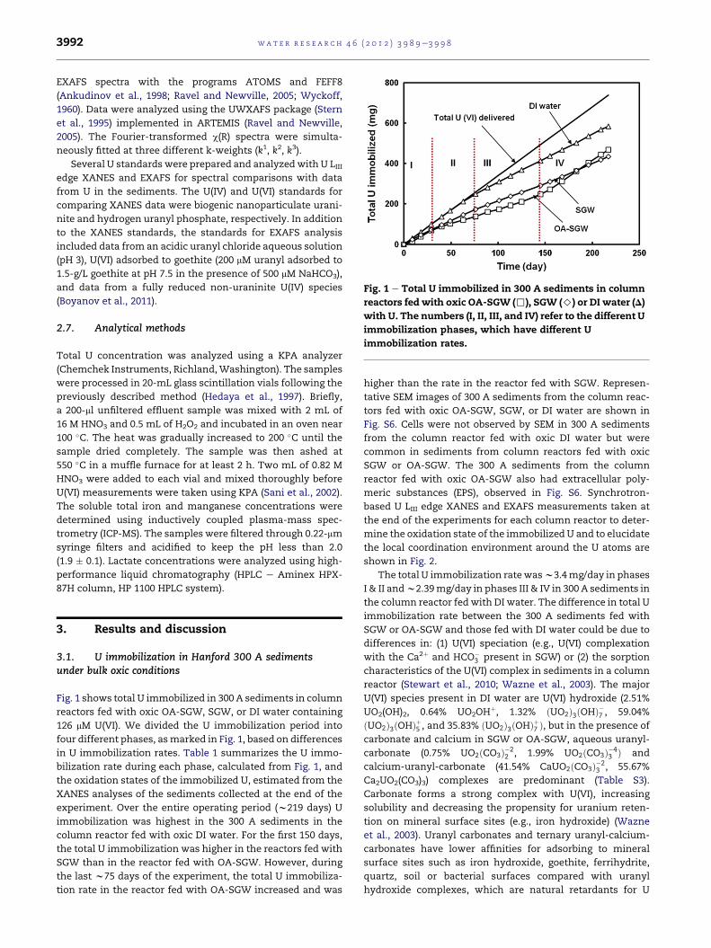
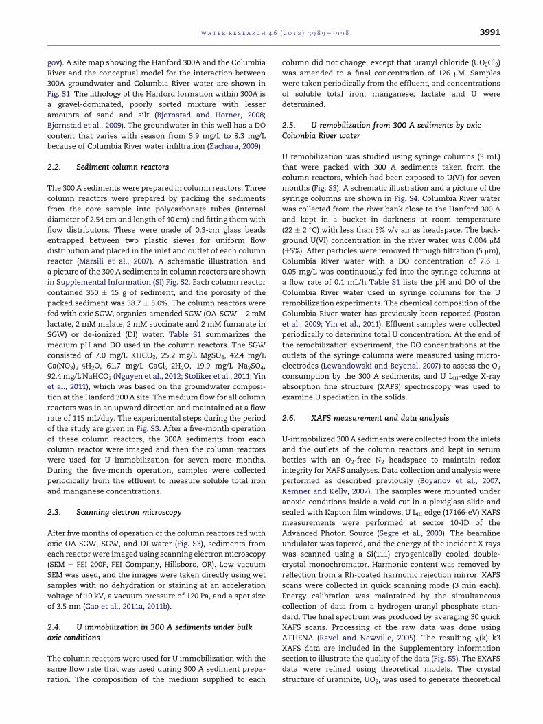
Fig. 1 e Total U immobilized in 300 A sediments in column
reactors fed with oxic OA-SGW (,), SGW (>) or DI water (D)
with U. The numbers (I, II, III, and IV) refer to the different U
immobilization phases, which have different U
immobilization rates.
wat e r r e s e a r c h 4 6 ( 2 0 1 2 ) 3 9 8 9e3 9 9 83992
EXAFS spectra with the programs ATOMS and FEFF8
(Ankudinov et al., 1998; Ravel and Newville, 2005; Wyckoff,
1960). Data were analyzed using the UWXAFS package (Stern
et al., 1995) implemented in ARTEMIS (Ravel and Newville,
2005). The Fourier-transformed c(R) spectra were simulta-
neously fitted at three different k-weights (k1, k2, k3).
Several U standards were prepared and analyzed with U LIIIedge XANES and EXAFS for spectral comparisons with data
from U in the sediments. The U(IV) and U(VI) standards for
comparing XANES data were biogenic nanoparticulate urani-
nite and hydrogen uranyl phosphate, respectively. In addition
to the XANES standards, the standards for EXAFS analysis
included data from an acidic uranyl chloride aqueous solution
(pH 3), U(VI) adsorbed to goethite (200 mM uranyl adsorbed to
1.5-g/L goethite at pH 7.5 in the presence of 500 mM NaHCO3),
and data from a fully reduced non-uraninite U(IV) species
(Boyanov et al., 2011).
2.7. Analytical methods
Total U concentration was analyzed using a KPA analyzer
(Chemchek Instruments, Richland,Washington). The samples
were processed in 20-mL glass scintillation vials following the
previously described method (Hedaya et al., 1997). Briefly,
a 200-ml unfiltered effluent sample was mixed with 2 mL of
16 M HNO3 and 0.5 mL of H2O2 and incubated in an oven near
100 �C. The heat was gradually increased to 200 �C until the
sample dried completely. The sample was then ashed at
550 �C in a muffle furnace for at least 2 h. Two mL of 0.82 M
HNO3 were added to each vial and mixed thoroughly before
U(VI) measurements were taken using KPA (Sani et al., 2002).
The soluble total iron and manganese concentrations were
determined using inductively coupled plasma-mass spec-
trometry (ICP-MS). The samples were filtered through 0.22-mm
syringe filters and acidified to keep the pH less than 2.0
(1.9 � 0.1). Lactate concentrations were analyzed using high-
performance liquid chromatography (HPLC e Aminex HPX-
87H column, HP 1100 HPLC system).
3. Results and discussion
3.1. U immobilization in Hanford 300 A sedimentsunder bulk oxic conditions
Fig. 1 shows total U immobilized in 300 A sediments in column
reactors fed with oxic OA-SGW, SGW, or DI water containing
126 mM U(VI). We divided the U immobilization period into
four different phases, asmarked in Fig. 1, based on differences
in U immobilization rates. Table 1 summarizes the U immo-
bilization rate during each phase, calculated from Fig. 1, and
the oxidation states of the immobilized U, estimated from the
XANES analyses of the sediments collected at the end of the
experiment. Over the entire operating period (w219 days) U
immobilization was highest in the 300 A sediments in the
column reactor fed with oxic DI water. For the first 150 days,
the total U immobilization was higher in the reactors fed with
SGW than in the reactor fed with OA-SGW. However, during
the last w75 days of the experiment, the total U immobiliza-
tion rate in the reactor fed with OA-SGW increased and was
higher than the rate in the reactor fed with SGW. Represen-
tative SEM images of 300 A sediments from the column reac-
tors fed with oxic OA-SGW, SGW, or DI water are shown in
Fig. S6. Cells were not observed by SEM in 300 A sediments
from the column reactor fed with oxic DI water but were
common in sediments from column reactors fed with oxic
SGW or OA-SGW. The 300 A sediments from the column
reactor fed with oxic OA-SGW also had extracellular poly-
meric substances (EPS), observed in Fig. S6. Synchrotron-
based U LIII edge XANES and EXAFS measurements taken at
the end of the experiments for each column reactor to deter-
mine the oxidation state of the immobilized U and to elucidate
the local coordination environment around the U atoms are
shown in Fig. 2.
The total U immobilization ratewasw3.4mg/day in phases
I & II andw2.39mg/day in phases III & IV in 300 A sediments in
the column reactor fed with DI water. The difference in total U
immobilization rate between the 300 A sediments fed with
SGW or OA-SGW and those fed with DI water could be due to
differences in: (1) U(VI) speciation (e.g., U(VI) complexation
with the Ca2þ and HCO�3 present in SGW) or (2) the sorption
characteristics of the U(VI) complex in sediments in a column
reactor (Stewart et al., 2010; Wazne et al., 2003). The major
U(VI) species present in DI water are U(VI) hydroxide (2.51%
UO2(OH)2, 0.64% UO2OHþ, 1.32% ðUO2Þ3ðOHÞ�7 , 59.04%
ðUO2Þ3ðOHÞþ5 , and 35.83% ðUO2Þ3ðOHÞþ7 Þ, but in the presence of
carbonate and calcium in SGW or OA-SGW, aqueous uranyl-
carbonate (0.75% UO2ðCO3Þ�22 , 1.99% UO2ðCO3Þ�4
3 Þ and
calcium-uranyl-carbonate (41.54% CaUO2ðCO3Þ�23 , 55.67%
Ca2UO2(CO3)3) complexes are predominant (Table S3).
Carbonate forms a strong complex with U(VI), increasing
solubility and decreasing the propensity for uranium reten-
tion on mineral surface sites (e.g., iron hydroxide) (Wazne
et al., 2003). Uranyl carbonates and ternary uranyl-calcium-
carbonates have lower affinities for adsorbing to mineral
surface sites such as iron hydroxide, goethite, ferrihydrite,
quartz, soil or bacterial surfaces compared with uranyl
hydroxide complexes, which are natural retardants for U
Table 1 e U immobilization rates during each phase of the U immobilization experiment and oxidation states of theimmobilized U at the end of the U immobilization experiment for column reactors (% deviation in U immobilization ratewas w3%).
300 A sedimentsfed with
Phases
U immobilization rate (mg/day) U oxidation state
I II III IV
OA-SGW 2.23 1.45 1.55 3.20 80e85 % U(IV)
SGW 2.51 2.16 1.70 2.03 100% U(VI)
DI water 3.40 3.34 2.39 2.39 100% U(VI)
wat e r r e s e a r c h 4 6 ( 2 0 1 2 ) 3 9 8 9e3 9 9 8 3993
migration (Brooks et al., 2003; Fox et al., 2006; Gorman-Lewis
et al., 2005; Stewart et al., 2010; Wazne et al., 2003; Zheng
et al., 2003). The U immobilization in 300 A sediments fed
with DI water is likely due to U adsorption to sediments, since
we did not add organic compounds to support microbial
growth and we did not observe attached cells on the surfaces
(Fig. S6). Synchrotron-based U LIII edge XANES analysis
showed that the U immobilized in 300 A sediments from the
column reactor fed with DI water was U(VI) (Table 1, Fig. 2b).
EXAFS analysis showed that the inlet sample from the column
reactor with 300 A sediments fed with DI water was a combi-
nation of aqueous uranyl and an inner-sphere complex of
uranyl adsorbed to metal oxides, while the outlet samples
were found to be predominantly the latter (Fig. 2d). These
Fig. 2 e U LIII edge XANES spectra of the inlet and outlet U-imm
(A) OA-SGW (B) SGW or DI water plotted with U(IV) and U(VI) st
the inlet and outlet U-immobilized sediments from (C) the colu
nanoparticulate, and (D) the column reactors fed with SGW or D
uranyl adsorbed to goethite.
conclusions are based on the EXAFS spectra from the sedi-
ments closely matching that of the goethite-sorbed U(VI)
standard, in which U(VI) has been shown previously to adsorb
as an inner-sphere complex (Redden et al., 2001).
There was no supply of exogenous organics to the column
reactor fed with SGW during the period of study. Although we
observed the presence of cells in 300 A sediments in the
column reactor fed with SGW, soluble iron and manganese
were not detected in the effluent before or during the U
immobilization experiments (Fig. S7), indicating that condi-
tions within the column were not strongly reducing. There-
fore, we expected that U immobilization in the column reactor
fed with SGW would be mostly through the adsorption of
U(VI). XANES and EXAFS analyses of U-immobilized 300 A
obilized sediments from column reactors fed with
andards. Fourier transforms of U LIII edge EXAFS spectra of
mn reactor fed with OA-SGW, plotted with uraninite
I water, plotted with standards for aqueous uranyl and
wat e r r e s e a r c h 4 6 ( 2 0 1 2 ) 3 9 8 9e3 9 9 83994
sediments from the column reactor fed with SGW showed
similar results to those found in the column reactor fed with
DI water (Table 1, Fig. 2b and d).
The presence of cells embedded in EPS (i.e., sediment bio-
films) was observed in the sediment column reactor fed with
OA-SGW (Fig. S6). The organics supplied in OA-SGW stimu-
lated cell growth as well as the EPS production of indigenous
microorganisms in the 300 A sediments. The lactate concen-
tration in the effluent from the 300 A sediment column fed
with OA-SGW varied between 0 and 0.5 mM (data not shown);
therefore, w93% of the lactate delivered to the sediments was
consumed in the column. Furthermore, soluble iron and
manganese, presumably Fe(II) and Mn(II), were released into
the aqueous phase before and during U immobilization
(Fig. S7), indicating that anaerobic, metal-reducing conditions
had been established. Therefore, it is thought that the U
immobilization in this reactor resulted from a combination of
adsorption and reduction. Linear combination fitting of the U
LIII edge XANES data showed that 80e85% of the immobilized
U in both the inlet and outlet 300 A sediments from the
column reactor fed with OA-SGW was U(IV) (Table 1, Fig. 2a).
EXAFS analysis of the inlet and outlet 300 A sediments from
the column reactor fed with OA-SGW found nanoparticulate
uraninite (UO2) (Fig. 2c). The EXAFS data and fit of the inlet
300 A sediments from the column reactor fed with OA-SGW
are shown in Fig. S8. A comparison of the EXAFS analysis
(Table S2) of the current study with previously published
literature verifies the formation of nanoparticulate uraninite
with an average diameter of 1e2 nm (Boyanov et al., 2007). It is
interesting to note that the total amounts of U immobilized in
the 300 A sediments from the column reactors fed with SGW
and OA-SGW were comparable but that the U immobilization
in the column reactor fed with SGW was mostly through
adsorption as U(VI) rather than reduction to U(IV).
The U immobilization in the 300 A sediment column
reactor fed with OA-SGW was mainly through microbial
reduction to U(IV) (80e85%), suggesting that reductive immo-
bilization of U(VI) could be an important microbial process
affecting the fate and transport of U even under bulk oxic
conditions. The sediment sample was collected from a depth
of 33e34 ft. (w10 m), where bacterial richness was highest in
the Hanford formation (Lin et al., 2012a). Functional groups of
facultative and anaerobic respiring bacteria have been detec-
ted by molecular and cultivation approaches in samples from
the oxic Hanford formation, suggestingmetal reductionmight
occur in the oxic sediment (Lin et al., 2012b). Further, Lee et al.
(2012) have demonstrated the potential of microbial anaerobic
metabolism in shallow, saturated subsurface Hanford
formation sediments in the 300 A, suggesting that biogeo-
chemical hot spots might enhance microbial activity that
could result in local anoxia and reducing conditions (Lin et al.,
2012b; McClain et al., 2003; Nguyen et al., 2012). In such anoxic
microenvironment, certain indigenous microorganisms are
capable of immobilizing U(VI) through microbial enzymatic
reduction.We have isolated several bacterial strains including
Paenibacillus sp. 300 A from the sediments used in this study
and they are potentially involved in reductive immobilization
of U(VI), and further investigations are in progress. Microbial
reduction process could be affected by various factors during
the immobilization phase, including: (1) U toxicity inhibiting
cell growth. This could decrease the U reduction rate, extend
the reaction times, and even halt microbial growth and
metabolic activity (Sani et al., 2006). (2) The predominance of
calciumeuranylecarbonate complexes (w97%) significantly
decreasing the U reduction rate, since U(VI) is a much less
energetically favorable electron acceptor when it forms cal-
ciumeuranylecarbonate complexes (Brooks et al., 2003). (3)
The increased partial pressure of CO2 (pCO2) and increased
inorganic carbon ((bi)carbonate) concentrations resulting
from five months of stimulated microbial respiration prior to
U immobilization in 300 A sediments fed with OA-SGW (Wan
et al., 2005, 2008). Increased bicarbonate concentrations could
facilitate Fe(III)-driven U(IV) reoxidation and the formation of
higher-level uraniumecarbonate complexes while still under
reducing conditions (Ginder-Vogel et al., 2006; Wan et al.,
2005). (4) The presence of O2 in OA-SGW and of iron and
manganese in 300 A sediments, which could reoxidize the
reduced U(IV) abiotically. We observed soluble Fe and Mn
(quantified as total aqueous concentrations), presumably
Fe(II) and Mn(II) species, released from the reactor fed with
OA-SGW (Fig. S7). However, U remobilization by iron and
manganese requires physical contact between two solid
phases (e.g., nanoparticulate uraninite and solid Fe/Mn
minerals) (Fredrickson et al., 2002), which is unlikely to be the
case in unmixed sediment columns. Although all these
remobilization processes could occur, we observed contin-
uous U immobilization through microbial reduction when
300 A sediments were fed with OA-SGW.
3.2. U remobilization by oxic Columbia River water
Approximately 80e85% of U immobilization was due to
reduction in 300 A sediments fed with OA-SGW. If oxic
Columbia Riverwater causedU remobilization simply through
the desorption of U(VI), we would expect that 15e20% of the
adsorbed U associated with 300A sediments fed with OA-SGW
would be released into solutionwhen the samplewas exposed
to Columbia River water because of the changes in ground-
water composition and pH (seasonal variation w1.0 unit),
which can change U(VI) speciation (McKinley et al., 2011;
Poston et al., 2009; Yin et al., 2011). For example, Columbia
River water stage fluctuation (up to 1 m/d and >2 m per
season) alters groundwater flux on a diel basis and ground-
water chemistry on a seasonal basis at distances of 250 m and
more from the river (Hammond and Lichtner, 2010; Lindberg
and Petersen, 2004; Yin et al., 2011; Zachara et al., 2005).
Therefore, the variable groundwater compositions and pH and
the intrusion of Columbia River water into 300 A possibly
result in U remobilization through desorption. Therefore, the
majority of the U associated with 300 A sediments fed with
SGW or DI water was expected to be released when the
samples were exposed to Columbia River water, since U
immobilization was mainly through adsorption.
The percentage of U remobilized and the oxidation states
of the residual sediment-associated U are summarized in
Table 2. The introduction of Columbia River water resulted in
the release of U into solution from previously U-immobilized
300 A sediments fed with OA-SGW, SGW or DI water. The
percentage of U remaining in 300 A sediments after remobi-
lization using oxic Columbia River water is shown in Fig. 3. U

Table 2 e Percentage U remobilization during 25 days ofthe remobilization experiment and the oxidation statesof the residual sediment-associated U after 50 days of theremobilization experiment.
U remobilizationusingU-immobilized 300 Asediments fromcolumn reactorsfed with
% remobilization U oxidationstatea
OA-SGW w7% 100% U(IV)
SGW w7% 100% U(VI)
DI water w7% 100% U(VI)
a XANES analysis was carried out after 50 days of the remobiliza-
tion experiment (XANES availability). The sediments were fed with
Columbia River water throughout the time period.
Fig. 4 e U LIII edge XANES analysis of U-immobilized 300 A
sediments fed with OA-SGW after remobilization by oxic
Columbia River water.
wat e r r e s e a r c h 4 6 ( 2 0 1 2 ) 3 9 8 9e3 9 9 8 3995
remobilization from 300 A sediments fed with OA-SGW
stopped within eight days and approximately 93% of the U
remained associated with the sediments, whereas linear
remobilization trends were observed for 300 A sediments fed
with SGW or DI water. We have shown that the U immobili-
zation in 300 A sediments fed with SGW or DI water was
mostly due to adsorption (Fig. 2b and d); hence, the remobili-
zation of U from these 300 A sediments can be mainly attrib-
uted to the desorption of U(VI). The concentrations of Ca2þ
(w17 mg/L) and CO2�3 (w68 mg/L) in Columbia River water at
pH (w7.8) could lead to desorption of the U from U-immobi-
lized 300 A sediments (Table S3) (Poston et al., 2009; Yin et al.,
2011). In contrast, the U immobilization in 300 A sediments fed
with OA-SGWwas due to both U(VI) adsorption and reduction
to U(IV) (Fig. 2a); therefore, the remobilization of approxi-
mately 7% of the total immobilized U in 300 A sediments fed
with OA-SGW could be attributed to reoxidation, desorption
and/or detachment of the U-immobilized biomass, since we
measured the total U concentration in unfiltered column
reactor effluent. U LIII edge XANES analysis of U associated
with 300 A sediments fed with OA-SGW after exposure to
Columbia River water (Fig. 4) showed that most of the
Fig. 3 e Percentage U remaining in 300 A sediments after
remobilization using oxic Columbia River water. The 300 A
sediments used for remobilization were from U-
immobilized 300 A sediments fed with OA-SGW (>), SGW
(D) or DI water (,) in column reactors.
remaining U (w93%) remained in the form of U(IV) nano-
particles, suggesting that most of the U immobilized in these
sediments was recalcitrant to reoxidation. The Fourier trans-
form of U LIII-edge EXAFS data with biogenic nanoparticulate
uraninite and monomeric U(IV) standards confirmed that the
U in the inlet and outlet samples fed with Columbia River
water was in the form of nanoparticulate uraninite (Fig. S9).
At the end of the remobilization experiment (w50 days)
with Columbia River water, DO concentrations in the outlet of
the column containing U immobilized during the OA-SGW-
stimulated phase were 5.21e5.91 mg/L. Approximately
25e35% of the DO in the oxic Columbia River water was
consumed by the reduced sediments through biotic and/or
abiotic processes.Although the oxidationofU(IV) bymolecular
O2 is thermodynamically favorable (Abdelouas et al., 1999) and
hasbeendocumentedasamechanismforU(IV) remobilization
in previous lab-scale and field studies (Casas et al., 1994; Moon
et al., 2007; Zhong et al., 2005), we found that U remained as
U(IV) nanoparticles in 300A sedimentsmicrobially reduced via
stimulation with OA-SGW and subsequently exposed to oxic
Columbia River water (w80 pore volumes). This is consistent
with our hypothesis that, under bulk aerobic conditions,
microbial growth in the 300A subsurface sediments can create
anoxic zones and prevent the reoxidation of reduced U(IV)
species (Krawczyk-Barsch et al., 2008).We should note that we
have recently found anoxic microenvironments inside sedi-
ment biofilms from 300 A, suggesting that U(VI) reduction
proceeds under bulk oxic conditions (Nguyen et al., 2012).
Moreover, we should note that river water may also introduce
organics (Becker and Gray, 1992; Bisping, 2010; Moser et al.,
2003) to stimulate and maintain microbial activity which
keeps uranium in a reduced state.
4. Conclusions
Our study demonstrates U immobilization and remobilization
in Hanford 300 A sediments in the presence of oxic
wat e r r e s e a r c h 4 6 ( 2 0 1 2 ) 3 9 8 9e3 9 9 83996
groundwater and Columbia River water, mimicking the
interactions among U, groundwater or Columbia River water,
and sediments in the subsurface environment of Hanford
300 A. Our results suggest that U can be immobilized by OA-
SGW stimulation of indigenous microorganisms in sedi-
ments in the Hanford 300 A subsurface, predominantly
through reduction, even in the presence of oxic groundwater.
The reduced U in the 300 A sediments fed with OA-SGW was
resistant to reoxidation by oxic Columbia River water.
Columbia River water caused U remobilization (w7%) through
desorption, and most of the U that remained in the 300 A
sediments (w93%) fed with OA-SGW was in the form of
uraninite nanoparticles. Taken together, these findings imply
that: 1) the reductive immobilization of U through OA-SGW
stimulation of indigenous 300 A sediment microorganisms
could be viable in the relatively oxic Hanford 300 A subsurface
environments; and 2) with the intrusion of oxic Columbia
River water, desorption might be an important process
contributing to U remobilization from OA-SGW-stimulated
sediments in the Hanford 300 A subsurface.
Acknowledgments
This researchwas supported by the U.S. Department of Energy
(DOE) Office of Biological and Environmental Research (BER)
under the Subsurface Biogeochemical Research (SBR) Program
(grant DE-FG92-08ER64560). PNNL contributions to this
research were supported in part by the PNNL Scientific Focus
Area and Hanford 300A IFRC projects, and ANL contributions
were supported in part by the ANL Scientific Focus Area
project, which are part of the SBR Program of the Office of
Biological and Environmental Research (BER), U.S. DOE under
contract DE-AC05-76RLO and DE-AC02-06CH11357, respec-
tively. Use of the Advanced Photon Source (APS) was sup-
ported by the DOE-SC Office of Basic Energy Sciences, under
contract DE-AC02-06CH11357. MRCAT/EnviroCAT operations
are supported by DOE and the MRCAT/EnviroCAT member
institutions. The U.S. government retains for itself and others
acting on its behalf a paid-up, non-exclusive, irrevocable
worldwide license in said article to reproduce, prepare deriv-
ative works, distribute copies to the public and perform
publicly and display publicly, by or on behalf of the Govern-
ment. We are also grateful to the Franceschi Microscopy and
Imaging Center of Washington State University for the use of
their facilities and staff assistance.
Appendix A. Supplementary material
Supplementary material associated with this article can be
found, in the online version, at doi:10.1016/j.watres.2012.05.027.
r e f e r e n c e s
Abdelouas, A., Lu, Y.M., Lutze, W., Nuttall, H.E., 1998. Reductionof U(VI) to U(IV) by indigenous bacteria in contaminated
ground water. Journal of Contaminant Hydrology 35 (1e3),217e233.
Abdelouas, A., Lutze, W., Nuttall, H.E., 1999. Uraniumcontamination in the subsurface: characterization andremediation. Reviews in Mineralogy 38, 433e473.
Anderson, R.T., Vrionis, H.A., Ortiz-Bernad, I., Resch, C.T.,Long, P.E., Dayvault, R., Karp, K., Marutzky, S., Metzler, D.R.,Peacock, A., White, D.C., Lowe, M., Lovley, D.R., 2003.Stimulating the in situ activity of Geobacter species to removeuranium from the groundwater of a uranium-contaminatedaquifer. Applied and Environmental Microbiology 69 (10),5884e5891.
Ankudinov, A., Ravel, B., Rehr, J., Conradson, S., 1998. Real-spacemultiple-scattering calculation and interpretation of x-ray-adsorption near-edge structure. Physical Review B 58,7565e7576.
Becker, C.D., Gray, R.H., 1992. Past and present water-qualityconditions in the Hanford Reach, Columbia River.Environmental Monitoring and Assessment 22 (2), 137e152.
Bernier-Latmani, R., Veeramani, H., Vecchia, E.D., Junier, P.,Lezama-Pacheco, J.S., Suvorova, E.I., Sharp, J.O.,Wigginton, N.S., Bargar, J.R., 2010. Non-uraninite products ofmicrobial U(VI) reduction. Environmental Science &Technology 44 (24), 9456e9462.
Bisping, L.E., 2010. Hanford Site Environmental SurveillanceData Report for Calendar Year 2009. PNNL-19455, APP.1,Richland, WA.
Bjornstad, B.N., Horner, J.A., 2008. Drilling, Sampling, and Well-Installation Plan for the IFC Well Field, 300 Area. PNNL-17512,Richland, WA.
Bjornstad, B.N., Lanigan, D.C., Horner, J.A., Thorne, P.D.,Vermeul, V.R., 2009. Borehole Completion and ConceptualHydrogeologic Model for the IFRC Well Field, 300 Area.Hanford site, Richland, WA. PNNL-18340.
Boyanov, M.I., Fletcher, K.E., Kwon, M.J., Rui, X., O’Loughlin, E.J.,Loffler, F.E., Kemner, K.M., 2011. Solution and microbialcontrols on the formation of reduced U(IV) species.Environmental Science & Technology 45 (19), 8336e8344.
Boyanov, M.I., O’Loughlin, E.J., Roden, E.E., Fein, J.B.,Kemner, K.M., 2007. Adsorption of Fe(II) and U(VI) to carboxyl-functionalized microspheres: the influence of speciation onuranyl reduction studied by titration and XAFS. Geochimica etCosmochimica Acta 71 (8), 1898e1912.
Brooks, S.C., Fredrickson, J.K., Carroll, S.L., Kennedy, D.W.,Zachara, J.M., Plymale, A.E., Kelly, S.D., Kemner, K.M.,Fendorf, S., 2003. Inhibition of bacterial U(VI) reduction bycalcium. Environmental Science & Technology 37 (9),1850e1858.
Brown, C.F., Um, W., Serne, R.J., 2008. Uranium Contamination inthe 300 Area: Emergent Data and Their Impact on the SourceTerm Conceptual Model. PNNL-17793, Richland, WA.
Burgos, W.D., McDonough, J.T., Senko, J.M., Zhang, G.,Dohnalkova, A.C., Kelly, S.D., Gorby, Y., Kemner, K.M., 2008.Characterization of uraninite nanoparticles produced byShewanella oneidensis MR-1. Geochimica et Cosmochimica Acta72 (20), 4901e4915.
Cao, B., Ahmed, B., Kennedy, D.W., Wang, Z.M., Shi, L.,Marshall, M.J., Fredrickson, J.K., Isern, N.G., Majors, P.D.,Beyenal, H., 2011a. Contribution of extracellular polymericsubstances from Shewanella sp. HRCR-1 biofilms to U(VI)immobilization. Environmental Science & Technology 45 (13),5483e5490.
Cao, B., Shi, L.A., Brown, R.N., Xiong, Y.J., Fredrickson, J.K.,Romine, M.F., Marshall, M.J., Lipton, M.S., Beyenal, H., 2011b.Extracellular polymeric substances from Shewanella sp.HRCR-1 biofilms: characterization by infrared spectroscopyand proteomics. Environmental Microbiology 13 (4),1018e1031.

wat e r r e s e a r c h 4 6 ( 2 0 1 2 ) 3 9 8 9e3 9 9 8 3997
Casas, I., Gimenez, J., Marti, V., Torrero, M.E., Depablo, J., 1994.Kinetics studies of unirradiated UO2 dissolution underoxidizing conditions in batch and flow experiments.Radiochimica Acta 66-7, 23e27.
Dalla Vecchia, E.C., Veeramani, H., Suvorova, E.I., Wigginton, N.S.,Bargar, J.R., Bernier-Latmani, R., 2010. U(VI) reduction byspores of Clostridium acetobutylicum. Research in Microbiology161 (9), 765e771.
Decho, A.W., 2000. Microbial biofilms in intertidal systems: anoverview. Continental Shelf Research 20 (10e11), 1257e1273.
Fletcher, K.E., Boyanov, M.I., Thomas, S.H., Wu, Q., Kemner, K.M.,Loeffler, F.E., 2010. U(VI) reduction to mononuclear U(IV) byDesulfitobacterium species. Environmental Science &Technology 44 (12), 4705e4709.
Fox, P.M., Davis, J.A., Zachara, J.M., 2006. The effect of calcium onaqueous uranium(VI) speciation and adsorption to ferrihydriteand quartz. Geochimica et Cosmochimica Acta 70 (6),1379e1387.
Fredrickson, J.K., Zachara, J.M., Kennedy, D.W., Liu, C.X.,Duff, M.C., Hunter, D.B., Dohnalkova, A., 2002. Influence of Mnoxides on the reduction of uranium(VI) by the metal-reducingbacterium Shewanella putrefaciens. Geochimica etCosmochimica Acta 66 (18), 3247e3262.
Ginder-Vogel, M., Criddle, C.S., Fendorf, S., 2006. Thermodynamicconstraints on the oxidation of biogenic UO2 by Fe(III) (Hydr)oxides. Environmental Science & Technology 40 (11),3544e3550.
Gorman-Lewis, D., Elias, P.E., Fein, J.B., 2005. Adsorption ofaqueous uranyl complexes onto Bacillus subtilis cells.Environmental Science & Technology 39 (13), 4906e4912.
Hammond, G.E., Lichtner, P.C., 2010. Field-scale model for thenatural attenuation of uranium at the Hanford 300 Area usinghigh-performance computing. Water Resources Research 46.
Hedaya, M.A., Birkenfeld, H.P., Kathren, R.L., 1997. A sensitivemethod for the determination of uranium in biologicalsamples utilizing kinetic phosphorescence analysis (KPA).Journal of Pharmaceutical and Biomedical Analysis 15 (8),1157e1165.
Istok, J.D., Senko, J.M., Krumholz, L.R., Watson, D., Bogle, M.A.,Peacock, A., Chang, Y.J., White, D.C., 2004. In situ bioreductionof technetium and uranium in a nitrate-contaminated aquifer.Environmental Science & Technology 38 (2), 468e475.
Jones, D.S., Tobler, D.J., Schaperdoth, I., Mainiero, M.,Macalady, J.L., 2010. Community structure of subsurfacebiofilms in the thermal sulfidic caves of Acquasanta Terme,Italy. Applied and Environmental Microbiology 76 (17),5902e5910.
Kemner, K.M., Kelly, S.D., 2007. In: Hurst, C.J. (Ed.), Manual ofEnvironmental Microbiology. ASM Press, pp. 1183e1194.
Krawczyk-Barsch, E., Grossmann, K., Arnold, T., Hofmann, S.,Wobus, A., 2008. Influence of uranium (VI) on the metabolicactivity of stable multispecies biofilms studied by oxygenmicrosensors and fluorescence microscopy. Geochimica etCosmochimica Acta 72 (21), 5251e5265.
Lee, J.-H., Fredrickson, J.K., Kukkadapu, R.K., Boyanov, M.I.,Kemner, K.M., Lin, X., Kennedy, D.W., Bjornstad, B.N.,Konopka, A.E., Moore, D.A., Resch, C.T., Phillips, J.L., 2012.Microbial reductive transformation of Phyllosilicate Fe(III)and U(VI) in fluvial subsurface sediments. EnvironmentalScience & Technology 46 (7), 3721e3730.
Lewandowski, Z., Beyenal, H., 2007. Foundamentals of BiofilmsResearch. CRC Press.
Lin, X., Kennedy, D., Fredrickson, J., Bjornstad, B., Konopka, A.,2012a. Vertical stratification of subsurface microbialcommunity composition across geological formations at theHanford Site. Environmental Microbiology 14 (2), 414e425.
Lin, X., Kennedy, D., Peacock, A., McKinley, J., Resch, C.T.,Fredrickson, J., Konopka, A., 2012b. Distribution of microbial
biomass and potential for anaerobic respiration in Hanfordsite 300 area subsurface sediment. Applied and EnvironmentalMicrobiology 78 (3), 759e767.
Lindberg, M.J., Petersen, R.E., 2004. 300-FF-5 Operable Unit,Hanford Site Groundwater Monitoring for Fiscal Year 2004.PNNL-15070.
Lovley, D.R., 1993. Dissimilatory metal reduction. Annual Reviewof Microbiology 47, 263e290.
Lovley, D.R., Phillips, E.J.P., Gorby, Y.A., Landa, E.R., 1991.Microbial reduction of uranium. Nature 350 (6317),413e416.
Marshall, M.J., Beliaev, A.S., Dohnalkova, A.C., Kennedy, D.W.,Shi, L., Wang, Z.M., Boyanov, M.I., Lai, B., Kemner, K.M.,McLean, J.S., Reed, S.B., Culley, D.E., Bailey, V.L.,Simonson, C.J., Saffarini, D.A., Romine, M.F., Zachara, J.M.,Fredrickson, J.K., 2006. c-Type cytochrome-dependentformation of U(IV) nanoparticles by Shewanella oneidensis. PlosBiology 4 (8), 1324e1333.
Marshall, M.J., Dohnalkova, A.C., Kennedy, D.W., Plymale, A.E.,Thomas, S.H., Loffler, F.E., Sanford, R.A., Zachara, J.M.,Fredrickson, J.K., Beliaev, A.S., 2009. Electron donor-dependentradionuclide reduction and nanoparticle formation byAnaeromyxobacter dehalogenans strain 2CP-C. EnvironmentalMicrobiology 11 (2), 534e543.
Marsili, E., Beyenal, H., Di Palma, L., Merli, C., Dohnalkova, A.,Amonette, J.E., Lewandowski, Z., 2005. Uranium removal bysulfate reducing biofilms in the presence of carbonates. WaterScience and Technology 52 (7), 49e55.
Marsili, E., Beyenal, H., Palma, L.D., Merli, C., Dohnalkova, A.,Amonette, J.E., Lewandowski, Z., 2007. Uraniumimmobilization by sulfate-reducing biofilms grown onHematite, Dolomite, and Calcite. Environmental Science &Technology 41 (24), 8349e8354.
McClain, M.E., Boyer, E.W., Dent, C.L., Gergel, S.E., Grimm, N.B.,Groffman, P.M., Hart, S.C., Harvey, J.W., Johnston, C.A.,Mayorga, E., McDowell, W.H., Pinay, G., 2003. Biogeochemicalhot spots and hot moments at the interface of terrestrial andaquatic ecosystems. Ecosystems 6 (4), 301e312.
McKinley, J.P., Zachara, J.M., Resch, C.T., Kaluzny, R.M.,Miller, M.D., Vermeul, V.R., Fritz, B.G., Moser, J.V., 2011. RiverWater Intrusion and Contaminant Uranium Contributionsfrom the Vadose Zone to Groundwater during the AnnualSpring Rise in Columbia River Stage at the Hanford Site 300Area. Washington. Environmental Science & TechnologySubmitted.
McKinley, J.P., Zachara, J.M., Wan, J., McCready, D.E., Heald, S.M.,2007. Geochemical controls on contaminant uranium invadose Hanford formation sediments at the 200 area and 300area, Hanford Site, Washington. Vadose Zone Journal 6 (4),1004e1017.
McLean, J.S., Majors, P.D., Reardon, C.L., Bilskis, C.L., Reed, S.B.,Romine, M.F., Fredrickson, J.K., 2008. Investigations ofstructure and metabolism within Shewanella oneidensis MR-1biofilms. Journal of Microbiological Methods 74 (1), 47e56.
Moon, H.S., Komlos, J., Jaffe, P.R., 2009. Biogenic U(IV) oxidation bydissolved oxygen and nitrate in sediment after prolongedU(VI)/Fe(III)/SO42- reduction. Journal of ContaminantHydrology 105 (1e2), 18e27.
Moon, H.S., Komlos, J., Jaffe, P.R., 2007. Uranium reoxidation inpreviously bioreduced sediment by dissolved oxygen andnitrate. Environmental Science & Technology 41 (13),4587e4592.
Moser, D.P., Fredrickson, J.K., Geist, D.R., Arntzen, E.V.,Peacock, A.D., Li, S.M.W., Spadoni, T., McKinley, J.P., 2003.Biogeochemical processes and microbial characteristicsacross groundwater-surface water boundaries of the HanfordReach of the Columbia River. Environmental Science &Technology 37 (22), 5127e5134.

wat e r r e s e a r c h 4 6 ( 2 0 1 2 ) 3 9 8 9e3 9 9 83998
N’Guessan, A.L., Vrionis, H.A., Resch, C.T., Long, P.E., Lovley, D.R.,2008. Sustained removal of uranium from contaminatedgroundwater following stimulation of dissimilatory metalreduction. Environmental Science & Technology 42 (8),2999e3004.
Nguyen, H.D., Cao, B., Mishra, B., Boyanov, M.I.,Kemner, K.M., Fredrickson, J.K., Beyenal, H., 2012.Microscale geochemical gradients in Hanford 300 Areasediment biofilms and influence of uranium. WaterResearch 46 (1), 227e234.
North, N.N., Dollhopf, S.L., Petrie, L., Istok, J.D., Balkwill, D.L.,Kostka, J.E., 2004. Change in bacterial community structureduring in situ biostimulation of subsurface sedimentcocontaminated with uranium and nitrate. Applied andEnvironmental Microbiology 70 (8), 4911e4920.
Palmisano, A., Hazen, T., 2003. Bioremediation of Metals andRadionuclides: What It Is and How It Works, second ed..LBNL-42595, Berkeley, CA.
Peterson, R.E., 2001. Zone of Interaction between Hanford SiteGroundwater and Adjacent Columbia River. PNNL-13674,Richland, WA.
Peterson, R.E., Rockhold, M.L., Serne, R.J., Thorne, P.D.,Williams, M.D., 2008. Uranium Contamination in theSubsurface beneath the 300 Area. Hanford site, Richland, WA.Washington, PNNL-17034.
Poston, T.M., Duncan, J.P., Dirkes, R.L., 2009. Hanford SiteEnvironmental Report for Calendar Year 2008. PNNL-18427,Richland, WA.
Ravel, B., Newville, M., 2005. ATHENA, ARTEMIS, HEPHAESTUS:data analysis for x-ray absorption spectroscopy using IFEFFIT.Journal of Synchrotron Radiation 12, 537e541.
Redden, G., Bargar, J., Bencheikh-Latmani, R., 2001. Citrateenhanced uranyl adsorption on goethite: an EXAFSanalysis. Journal of Colloid and Interface Science 244 (1),211e219.
Riley, R.G., Zachara, J.M., 1992. Chemical Contaminants onDOE Lands and Selection of Contaminant Mixturesfor Subsurface Science Research. DOE/ER-0547T,Richland, WA.
Sani, R.K., Peyton, B.M., Dohnalkova, A., 2006. Toxic effects ofUranium on Desulfovibrio desulfuricans G20. EnvironmentalToxicology and Chemistry 25 (5), 1231e1238.
Sani, R.K., Peyton, B.M., Dohnalkova, A., Amonette, J.E., 2005.Reoxidation of reduced uranium with iron(III) (hydr)oxidesunder sulfate-reducing conditions. Environmental Science &Technology 39 (7), 2059e2066.
Sani, R.K., Peyton, B.M., Smith, W.A., Apel, W.A., Petersen, J.N.,2002. Dissimilatory reduction of Cr(VI), Fe(III), and U(VI) byCellulomonas isolates. Applied Microbiology and Biotechnology60 (1e2), 192e199.
Segre, C., Leyarovska, N., Chapman, L., Lavender, W., Plag, P.,King, A., Kropf, A., Bunker, B., Kemner, K., Dutta, P., Duran, R.,Kaduk, J., 2000. The MRCAT Inserton Device Beamline at theAdvanced Photon Source, pp. 419e422.
Senko, J.M., Istok, J.D., Suflita, J.M., Krumholz, L.R., 2002.In-situ evidence for uranium immobilization andremobilization. Environmental Science & Technology 36 (7),1491e1496.
Sivaswamy, V., Boyanov, M.I., Peyton, B.M., Viamajala, S.,Gerlach, R., Apel, W.A., Sani, R.K., Dohnalkova, A.,Kemner, K.M., Borch, T., 2011. Multiple mechanisms ofUranium immobilization by Cellulomonas sp. strain ES6.Biotechnology and Bioengineering 108 (2), 264e276.
Stern, E., Newville, M., Ravel, B., Yacoby, Y., Haskel, D., 1995. TheUWXAFS analysis package- philosophy and details. Physica B209, 117e120.
Stewart, B.D., Mayes, M.A., Fendorf, S., 2010. Impact ofuranylecalciumecarbonato complexes on uranium(VI)adsorption to synthetic and natural sediments. EnvironmentalScience & Technology 44 (3), 928e934.
Stoliker, D.L., Kent, D.B., Zachara, J.M., 2011. Quantifyingdifferences in the impact of variable chemistry on equilibriumuranium(VI) adsorption properties of aquifer sediments.Environmental Science & Technology 45 (20), 8733e8740.
Suzuki, Y., Kelly, S.D., Kemner, K.M., Banfield, J.F., 2002.Radionuclide contamination e Nanometre-size products ofUranium bioreduction. Nature 419 (6903), 134.
Tokunaga, T.K., Wan, J., Kim, Y., Sutton, S.R., Newville, M.,Lanzirotti, A., Rao, W., 2008. Real-time X-ray absorptionspectroscopy of uranium, iron, and manganese incontaminated sediments during bioreduction. EnvironmentalScience & Technology 42 (8), 2839e2844.
Wan, J., Tokunaga, T.K., Brodie, E., Wang, Z., Zheng, Z.,Herman, D., Hazen, T.C., Firestone, M.K., Sutton, S.R., 2005.Reoxidation of bioreduced uranium under reducingconditions. Environmental Science & Technology 39 (16),6162e6169.
Wan, J., Tokunaga, T.K., Kim, Y., Brodie, E., Daly, R., Hazen, T.C.,Firestone, M.K., 2008. Effects of organic carbon supply rates onuranium mobility in a previously bioreduced contaminatedsediment. Environmental Science & Technology 42 (20),7573e7579.
Wazne, M., Korfiatis, G.P., Meng, X., 2003. Carbonate effects onhexavalent uranium adsorption by iron oxyhydroxide.Environmental Science & Technology 37 (16), 3619e3624.
Weidler, G.W., Dornmayr-Pfaffenhuemer, M., Gerbl, F.W.,Heinen, W., Stan-Lotter, H., 2007. Communities of archaea andbacteria in a subsurface radioactive thermal spring in theAustrian Central Alps, and evidence of ammonia-oxidizingCrenarchaeota. Applied and Environmental Microbiology 73 (1),259e270.
Wyckoff, R., 1960. Crystal Structures. Interscience, New York.Yabusaki, S.B., Fang, Y.L., Waichler, S.R., 2008. Building
conceptual models of field-scale uranium reactive transport ina dynamic Vadose zone-aquifer-river system. WaterResources Research 44, W12403.
Yin, J.Y.J., Haggerty, R., Stoliker, D.L., Kent, D.B., Istok, J.D.,Greskowiak, J., Zachara, J.M., 2011. Transient groundwaterchemistry near a river: effects on U(VI) transport inlaboratory column experiments. Water Resources Research47, W04502.
Zachara, J.M., 2009. Multi-scale Mass Transfer ProcessesControlling Natural Attenuation and Engineered Remediation:An IFRC Focused on Hanford’s 300 Area Uranium Plume.PNNL-SA-64785, Richland, WA.
Zachara, J.M., Davis, J.A., Liu, C., McKinley, J.P., Qafoku, N.,Wellman, D.M., Yabusaki, S.B., 2005. Uranium Geochemistryin Vadose Zone and Aquifer Sediments from the 300 AreaUranium Plume. PNNL-15121, Richland, WA.
Zheng, Z., Tokunaga, T.K., Wan, J., 2003. Influence of calciumcarbonate on U(VI) sorption to soils. Environmental Science &Technology 37 (24), 5603e5608.
Zhong, L.R., Liu, C.X., Zachara, J.M., Kennedy, D.W., Szecsody, J.E.,Wood, B., 2005. Oxidative remobilization of biogenicuranium(IV) precipitates: effects of iron(II) and pH. Journal ofEnvironmental Quality 34 (5), 1763e1771.