Embed Size (px)
Citation preview
Immersive Examination of the Qualitative Structure of BiomoleculesKenny Gruchalla
[email protected] of Computer ScienceUniversity of Colorado at Boulder
Mark [email protected]
Department of Molecular, Cellular,and Developmental Biology
University of Colorado at Boulder
Jonathan [email protected]
Department of Computer ScienceUniversity of Colorado at Boulder
Elizabeth [email protected]
Department of Computer ScienceUniversity of Colorado at Boulder
Abstract
The geometry of biomolecules dictates their function,but reasoning about that structure is difficult because oftheir 3D complexity and the range of scales involved.The wooden or plastic ball-and-stick models that arecommon in high-school chemistry labs help people rea-son about these issues when the molecules involvedare small, but they are useless in the study of largebiomolecules. Largely for this reason, 3D computervisualization tools have become essential in this field.However, these tools are limited by their interfaces. Tra-ditional graphics workstations project a 3D model onto2D screen, and interaction with the 3D model is indi-rect, using 2D mouse or pointing device. Immersive vi-sualization is a potential solution to this: it allows a userto visualize a biomolecule in 3D and interact with it di-rectly in 3-space. This paper reports upon a pilot studyabout the effects of immersive visualization upon an ex-pert’s reasoning about the qualitative structure of thesemolecules. We ported a standard visualization applica-tion (PyMOL) to a CAVE-like immersive virtual envi-ronment (IVE), then invited three separate biochemistryresearch groups—people who use PyMOL routinely ondesktop computers—to examine their favorite moleculein the IVE. Within ninety minutes of immersive inves-tigation, each group reported a new discovery about thequalitative structure of that molecule. We believe thatthe immersive environment facilitated these discoveriesby supporting and facilitating the natural spatial reason-ing abilities of its users.
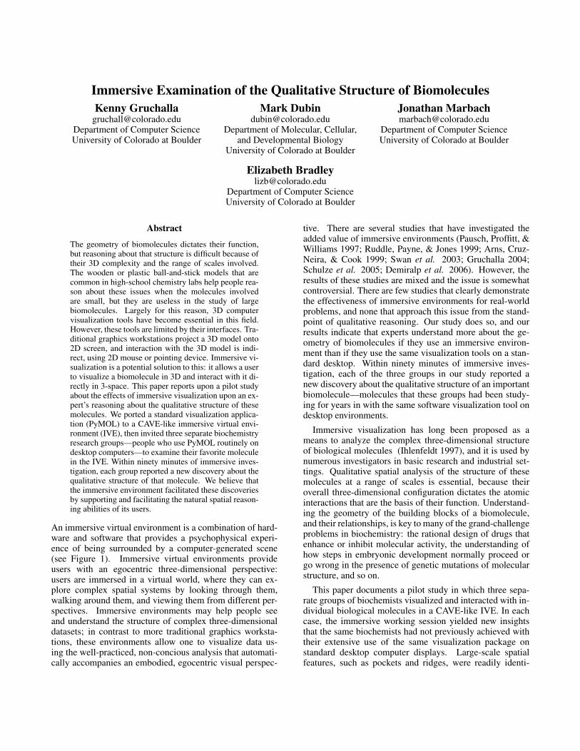
An immersive virtual environment is a combination of hard-ware and software that provides a psychophysical experi-ence of being surrounded by a computer-generated scene(see Figure 1). Immersive virtual environments provideusers with an egocentric three-dimensional perspective:users are immersed in a virtual world, where they can ex-plore complex spatial systems by looking through them,walking around them, and viewing them from different per-spectives. Immersive environments may help people seeand understand the structure of complex three-dimensionaldatasets; in contrast to more traditional graphics worksta-tions, these environments allow one to visualize data us-ing the well-practiced, non-concious analysis that automati-cally accompanies an embodied, egocentric visual perspec-
tive. There are several studies that have investigated theadded value of immersive environments (Pausch, Proffitt, &Williams 1997; Ruddle, Payne, & Jones 1999; Arns, Cruz-Neira, & Cook 1999; Swan et al. 2003; Gruchalla 2004;Schulze et al. 2005; Demiralp et al. 2006). However, theresults of these studies are mixed and the issue is somewhatcontroversial. There are few studies that clearly demonstratethe effectiveness of immersive environments for real-worldproblems, and none that approach this issue from the stand-point of qualitative reasoning. Our study does so, and ourresults indicate that experts understand more about the ge-ometry of biomolecules if they use an immersive environ-ment than if they use the same visualization tools on a stan-dard desktop. Within ninety minutes of immersive inves-tigation, each of the three groups in our study reported anew discovery about the qualitative structure of an importantbiomolecule—molecules that these groups had been study-ing for years in with the same software visualization tool ondesktop environments.
Immersive visualization has long been proposed as ameans to analyze the complex three-dimensional structureof biological molecules (Ihlenfeldt 1997), and it is used bynumerous investigators in basic research and industrial set-tings. Qualitative spatial analysis of the structure of thesemolecules at a range of scales is essential, because theiroverall three-dimensional configuration dictates the atomicinteractions that are the basis of their function. Understand-ing the geometry of the building blocks of a biomolecule,and their relationships, is key to many of the grand-challengeproblems in biochemistry: the rational design of drugs thatenhance or inhibit molecular activity, the understanding ofhow steps in embryonic development normally proceed orgo wrong in the presence of genetic mutations of molecularstructure, and so on.
This paper documents a pilot study in which three sepa-rate groups of biochemists visualized and interacted with in-dividual biological molecules in a CAVE-like IVE. In eachcase, the immersive working session yielded new insightsthat the same biochemists had not previously achieved withtheir extensive use of the same visualization package onstandard desktop computer displays. Large-scale spatialfeatures, such as pockets and ridges, were readily identi-
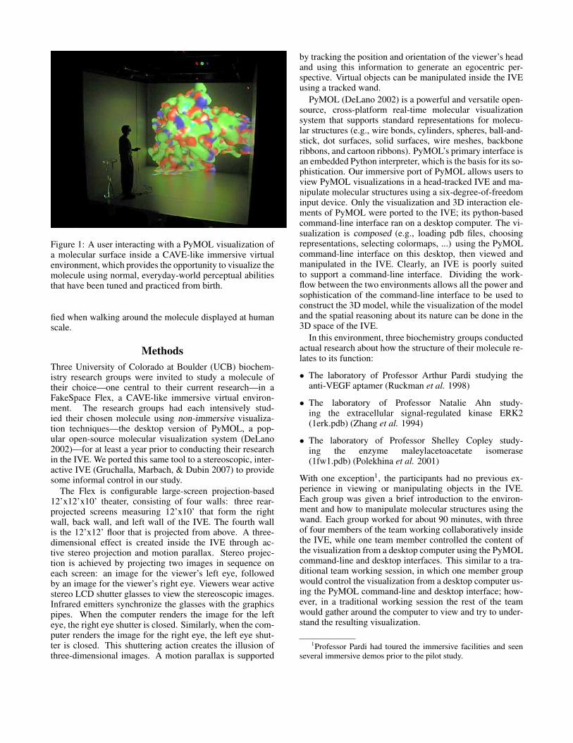
Figure 1: A user interacting with a PyMOL visualization ofa molecular surface inside a CAVE-like immersive virtualenvironment, which provides the opportunity to visualize themolecule using normal, everyday-world perceptual abilitiesthat have been tuned and practiced from birth.
fied when walking around the molecule displayed at humanscale.
MethodsThree University of Colorado at Boulder (UCB) biochem-istry research groups were invited to study a molecule oftheir choice—one central to their current research—in aFakeSpace Flex, a CAVE-like immersive virtual environ-ment. The research groups had each intensively stud-ied their chosen molecule using non-immersive visualiza-tion techniques—the desktop version of PyMOL, a pop-ular open-source molecular visualization system (DeLano2002)—for at least a year prior to conducting their researchin the IVE. We ported this same tool to a stereoscopic, inter-active IVE (Gruchalla, Marbach, & Dubin 2007) to providesome informal control in our study.
The Flex is configurable large-screen projection-based12’x12’x10’ theater, consisting of four walls: three rear-projected screens measuring 12’x10’ that form the rightwall, back wall, and left wall of the IVE. The fourth wallis the 12’x12’ floor that is projected from above. A three-dimensional effect is created inside the IVE through ac-tive stereo projection and motion parallax. Stereo projec-tion is achieved by projecting two images in sequence oneach screen: an image for the viewer’s left eye, followedby an image for the viewer’s right eye. Viewers wear activestereo LCD shutter glasses to view the stereoscopic images.Infrared emitters synchronize the glasses with the graphicspipes. When the computer renders the image for the lefteye, the right eye shutter is closed. Similarly, when the com-puter renders the image for the right eye, the left eye shut-ter is closed. This shuttering action creates the illusion ofthree-dimensional images. A motion parallax is supported
by tracking the position and orientation of the viewer’s headand using this information to generate an egocentric per-spective. Virtual objects can be manipulated inside the IVEusing a tracked wand.
PyMOL (DeLano 2002) is a powerful and versatile open-source, cross-platform real-time molecular visualizationsystem that supports standard representations for molecu-lar structures (e.g., wire bonds, cylinders, spheres, ball-and-stick, dot surfaces, solid surfaces, wire meshes, backboneribbons, and cartoon ribbons). PyMOL’s primary interface isan embedded Python interpreter, which is the basis for its so-phistication. Our immersive port of PyMOL allows users toview PyMOL visualizations in a head-tracked IVE and ma-nipulate molecular structures using a six-degree-of-freedominput device. Only the visualization and 3D interaction ele-ments of PyMOL were ported to the IVE; its python-basedcommand-line interface ran on a desktop computer. The vi-sualization is composed (e.g., loading pdb files, choosingrepresentations, selecting colormaps, ...) using the PyMOLcommand-line interface on this desktop, then viewed andmanipulated in the IVE. Clearly, an IVE is poorly suitedto support a command-line interface. Dividing the work-flow between the two environments allows all the power andsophistication of the command-line interface to be used toconstruct the 3D model, while the visualization of the modeland the spatial reasoning about its nature can be done in the3D space of the IVE.
In this environment, three biochemistry groups conductedactual research about how the structure of their molecule re-lates to its function:
• The laboratory of Professor Arthur Pardi studying theanti-VEGF aptamer (Ruckman et al. 1998)
• The laboratory of Professor Natalie Ahn study-ing the extracellular signal-regulated kinase ERK2(1erk.pdb) (Zhang et al. 1994)
• The laboratory of Professor Shelley Copley study-ing the enzyme maleylacetoacetate isomerase(1fw1.pdb) (Polekhina et al. 2001)
With one exception1, the participants had no previous ex-perience in viewing or manipulating objects in the IVE.Each group was given a brief introduction to the environ-ment and how to manipulate molecular structures using thewand. Each group worked for about 90 minutes, with threeof four members of the team working collaboratively insidethe IVE, while one team member controlled the content ofthe visualization from a desktop computer using the PyMOLcommand-line and desktop interfaces. This similar to a tra-ditional team working session, in which one member groupwould control the visualization from a desktop computer us-ing the PyMOL command-line and desktop interface; how-ever, in a traditional working session the rest of the teamwould gather around the computer to view and try to under-stand the resulting visualization.
1Professor Pardi had toured the immersive facilities and seenseveral immersive demos prior to the pilot study.
ResultsDespite having a long and extensive research history withtheir respective molecules, all three groups arrived at a newinsight from their 90-minute IVE research session. All ofthese insights were similar, and all involved qualitative rea-soning about geometry. Each group became newly awareof a large spatial feature, such as an empty space or ridge,that they had not noticed during their (considerable) previ-ous PyMOL work with the molecule on desktop computermonitors. In each case, the newly recognized feature led toinsights about the molecule’s function that follow directlyfrom geometry: how its pieces move, for instance, or howthey fit together. These are described in the following para-graphs. Each group left with the intention of exploring anew theoretical possibility based on these insights; the Ahngroup actually integrated a hypothesis concerning the struc-ture into a new grant proposal.
The Copley group recognized an empty pocket indent-ing from the surface in the enzyme maleylacetoacetate iso-merase (MAAI) (see Figure 2). MAAI is normally a dimer,in which the interface between the dimer molecules blocksthis pocket. However, the monomer of MAAI is similar to—and is used by the Copley group as—a model for anothermolecule, tetrachlorohydroquinone (TCHQ) dehalogenase,which is a monomer. This group is studying how a key com-ponent of the molecule’s active site, amino acid cysteine atposition 16 (cys16), interacts with substrate molecules thatmust be able to diffuse into TCHQ dehalogenase in orderto reach cys16. The pocket represents a large enough open-ing for such entry, with cys16 lying at its base (darkenedarea in Figures 2c and 2d). When viewing this molecule onworkstations, the researchers had discounted this region asa potential active site of TCHQ dehalogenase because theydid not judge it to be spacious enough for the substrate topenetrate to cys16. The immersive visualization gave the re-searchers the ability to stand inside the pocket, which gavethem enough information to reverse their decision.
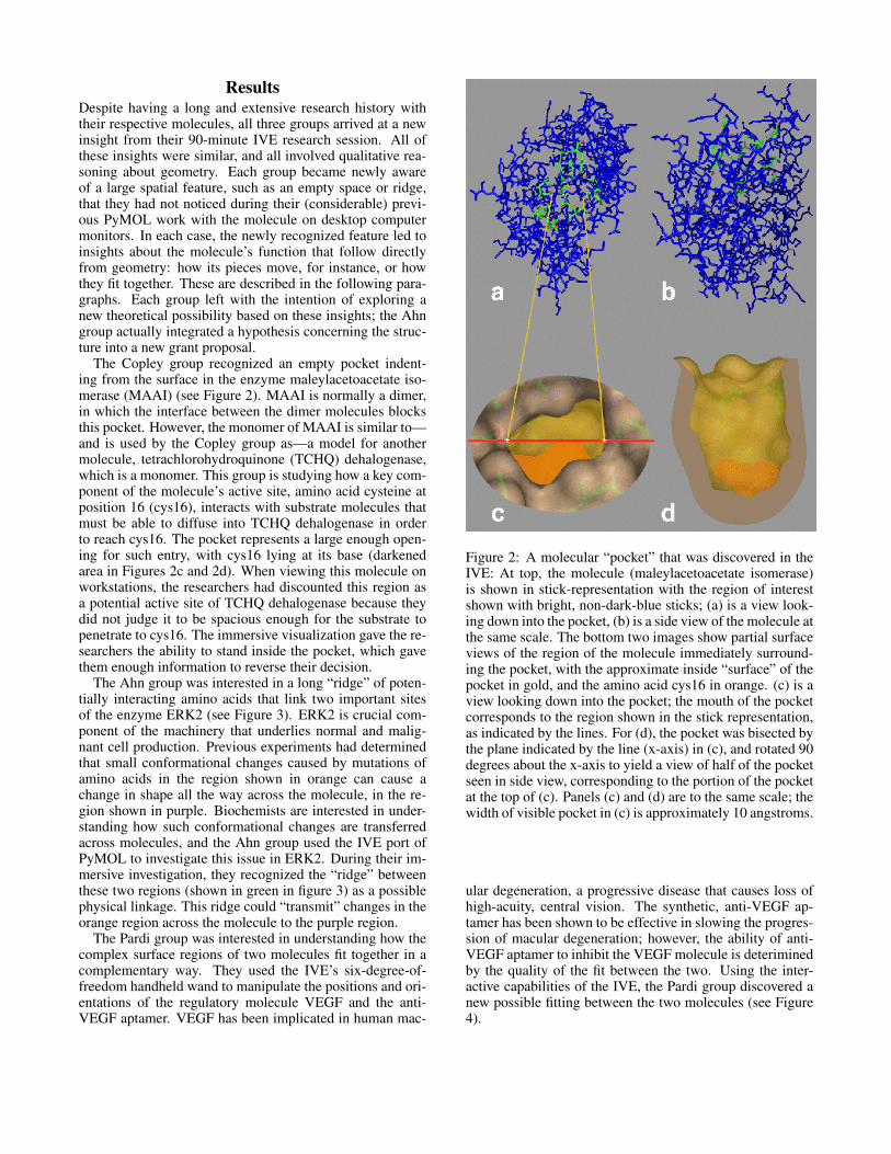
The Ahn group was interested in a long “ridge” of poten-tially interacting amino acids that link two important sitesof the enzyme ERK2 (see Figure 3). ERK2 is crucial com-ponent of the machinery that underlies normal and malig-nant cell production. Previous experiments had determinedthat small conformational changes caused by mutations ofamino acids in the region shown in orange can cause achange in shape all the way across the molecule, in the re-gion shown in purple. Biochemists are interested in under-standing how such conformational changes are transferredacross molecules, and the Ahn group used the IVE port ofPyMOL to investigate this issue in ERK2. During their im-mersive investigation, they recognized the “ridge” betweenthese two regions (shown in green in figure 3) as a possiblephysical linkage. This ridge could “transmit” changes in theorange region across the molecule to the purple region.
The Pardi group was interested in understanding how thecomplex surface regions of two molecules fit together in acomplementary way. They used the IVE’s six-degree-of-freedom handheld wand to manipulate the positions and ori-entations of the regulatory molecule VEGF and the anti-VEGF aptamer. VEGF has been implicated in human mac-
Figure 2: A molecular “pocket” that was discovered in theIVE: At top, the molecule (maleylacetoacetate isomerase)is shown in stick-representation with the region of interestshown with bright, non-dark-blue sticks; (a) is a view look-ing down into the pocket, (b) is a side view of the molecule atthe same scale. The bottom two images show partial surfaceviews of the region of the molecule immediately surround-ing the pocket, with the approximate inside “surface” of thepocket in gold, and the amino acid cys16 in orange. (c) is aview looking down into the pocket; the mouth of the pocketcorresponds to the region shown in the stick representation,as indicated by the lines. For (d), the pocket was bisected bythe plane indicated by the line (x-axis) in (c), and rotated 90degrees about the x-axis to yield a view of half of the pocketseen in side view, corresponding to the portion of the pocketat the top of (c). Panels (c) and (d) are to the same scale; thewidth of visible pocket in (c) is approximately 10 angstroms.
ular degeneration, a progressive disease that causes loss ofhigh-acuity, central vision. The synthetic, anti-VEGF ap-tamer has been shown to be effective in slowing the progres-sion of macular degeneration; however, the ability of anti-VEGF aptamer to inhibit the VEGF molecule is deteriminedby the quality of the fit between the two. Using the inter-active capabilities of the IVE, the Pardi group discovered anew possible fitting between the two molecules (see Figure4).
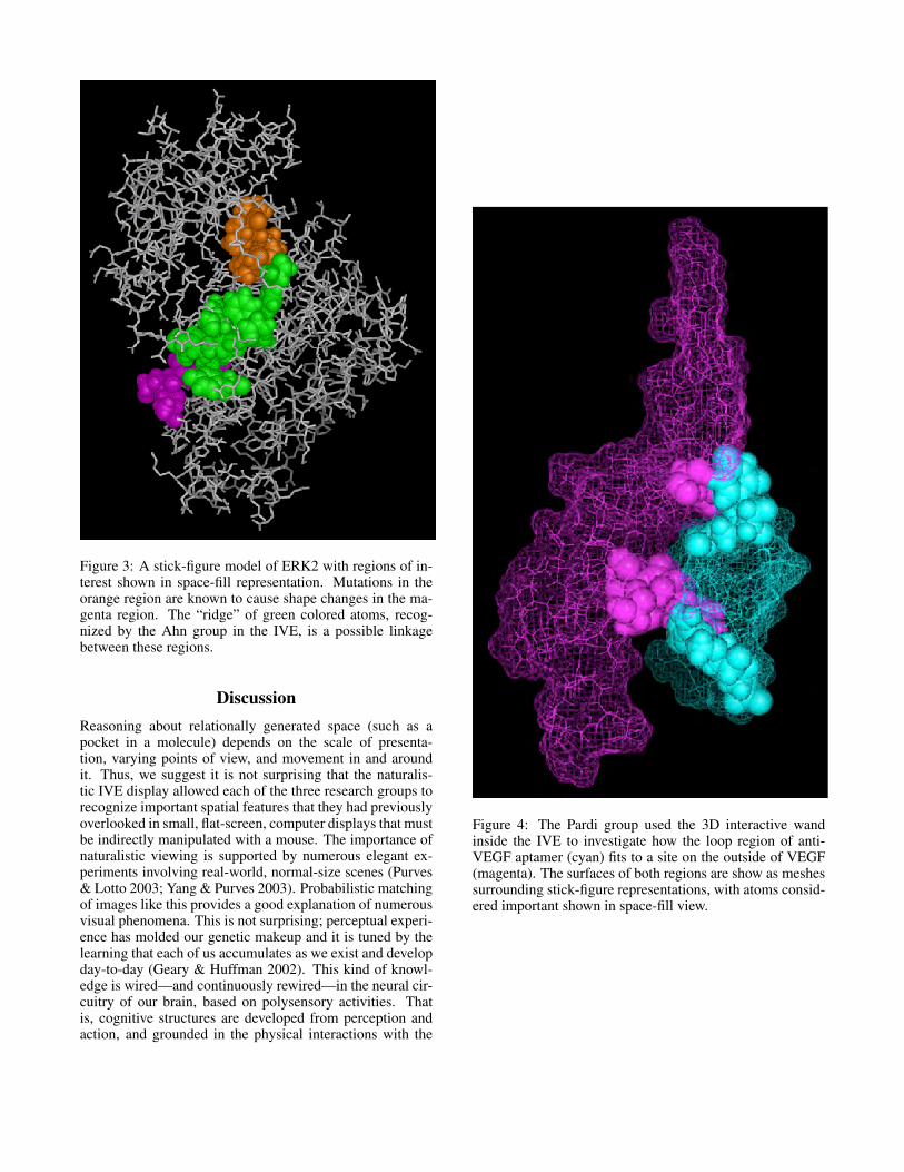
Figure 3: A stick-figure model of ERK2 with regions of in-terest shown in space-fill representation. Mutations in theorange region are known to cause shape changes in the ma-genta region. The “ridge” of green colored atoms, recog-nized by the Ahn group in the IVE, is a possible linkagebetween these regions.
DiscussionReasoning about relationally generated space (such as apocket in a molecule) depends on the scale of presenta-tion, varying points of view, and movement in and aroundit. Thus, we suggest it is not surprising that the naturalis-tic IVE display allowed each of the three research groups torecognize important spatial features that they had previouslyoverlooked in small, flat-screen, computer displays that mustbe indirectly manipulated with a mouse. The importance ofnaturalistic viewing is supported by numerous elegant ex-periments involving real-world, normal-size scenes (Purves& Lotto 2003; Yang & Purves 2003). Probabilistic matchingof images like this provides a good explanation of numerousvisual phenomena. This is not surprising; perceptual experi-ence has molded our genetic makeup and it is tuned by thelearning that each of us accumulates as we exist and developday-to-day (Geary & Huffman 2002). This kind of knowl-edge is wired—and continuously rewired—in the neural cir-cuitry of our brain, based on polysensory activities. Thatis, cognitive structures are developed from perception andaction, and grounded in the physical interactions with the
Figure 4: The Pardi group used the 3D interactive wandinside the IVE to investigate how the loop region of anti-VEGF aptamer (cyan) fits to a site on the outside of VEGF(magenta). The surfaces of both regions are show as meshessurrounding stick-figure representations, with atoms consid-ered important shown in space-fill view.
environment (Pecher & Zwaan 2005).In this context, it makes complete sense that working in an
IVE allows people to reason more effectively about the ge-ometry of biomolecules. Studies that demonstrate this effectare surprisingly rare, though, and the added value of immer-sion is controversial in the visualization community. Thisstudy is part of a larger effort that addresses this broader is-sue: a general definition of conditions under which the use offully interactive, three-dimensional, immersive visualizationadds value to research activities. As indicated in the previ-ous section, our results suggest that short, intense sessionsof IVE viewing valuably augment more extensive, non-IVEbased research of molecular function. The underlying rea-sons for this, we believe, are threefold:
• First, spatial judgments are body-relative in everyday ac-tivity (Hatfield 2003). It is easier, for example, to judgewhether you could crawl through a passageway in a caveif you are in the cave and looking at the passageway thanif you are examining a five-inch-tall rendering of it ona flat screen monitor. Our hypothesis is that examininga molecule at a human scale made it easier for the bio-chemists to reason about the different spatial structures.Because the IVE presented the biomolecules at a natu-ral and familiar scale—similar to the way many complex-shaped, everyday objects appear in the world—it facili-tated effective reasoning about there shape.
• Second, the egocentric perspective improves spatial rea-soning, object recognition, and stresses the role of actionin building knowledge. Much of this happens automati-cally: people do not stop and think about how to movetheir heads or bodies in order to get a better view of some-thing. The IVE supports this very naturally. Its uniquefeatures—natural body movements and well-practiced au-tomatic brain function as the basis for examination ofthe structure in question—is consistent with recent re-search on embodied cognition, cognition that is basedon perceptual knowledge accumulated through what wehave encountered and manipulated with our bodies aswe move within and examine the world (Wilson 2002;Wolputte 2002).
• Finally, the collaborative nature of the environment facil-itates collaborative reasoning about the data. The largescale of the environment allowed multiple biochemistryresearchers to gather inside the environment simultane-ously. All the groups commented that they found workingcollaboratively in the IVE to be much easier than crowd-ing around a small computer screen. The large scale madeit easy to see what atoms and regions another member ofthe group was referring to. Often they used bodily refer-ences to direct each other, such as, ”that group of bondsnear your left shoulder.”
Reasoning about the geometry of objects has a long andrich history in the qualitative reasoning field, and there areinteresting papers about ontologies, paradigms, techniques,and applications for this in every QR workshop—beginningwith an augmented version of Hayes’s “pieces of stuff” on-tology that was presented at QR ’87 for reasoning about
collections of molecules (Collins 1987). A few QR sys-tems have been built over the years specifically for rea-soning about molecular structure (Bandini, Cattaneo, &Stofella 1988). Most of the geometry-related work in the QRcommunity has involved mechanical devices, an application(like biomolecules) where shape and function are intimatelyinter-related. Iwasaki, Joskowicz, Nielsen, and Faltingshave made significant contributions to this over the years(Joskowicz 1987; Iwasaki 1987; Nielsen 1987; 1988; Falt-ings, Baechler, & Kun 1991; Tessler, Iwasaki, & Law 1993;Faltings 1993; Sun & Faltings 1994; Joskowicz & Sacks1997). There has also been some work in the QR commu-nity that considers the cognitive science perspective alongwith the representation and the geometry, notably from KenForbus’s group (Ferguson & Forbus 1999; Forbus, Fergu-son, & Usher 2000; Forbus, Tomai, & Usher 2003; 2005;Lovett, Dehghani, & Forbus 2006).
The study reported here has a much more complicatedapplication area than most of these papers, and much lesslofty aims. We are not trying to simulate, design, or de-duce anything. We rely on the human experts to figure outwhat’s meaningful; we want simply to understand how im-mersive environments support their reasoning about the ge-ometry that factors into that determination. Because of thecomparative nature of our study, the ontology and the modelare pre-specified. Our goal is not to figure out whether a bet-ter model or ontology exists for these purposes, as in manyinteresting QR papers, e.g., (Pacheco, Escrig, & Toledo2002) but rather to study how the presentation & interfaceaffects the spatial reasoning about the molecules. We arenot trying to generalize ideas about structure across applica-tion domains, as in (Adorni et al. 1988) nor are we tryingbuild more-abstract modelling paradigms, as in the elegantwork of Escrig (which is concerned with many of the con-cepts that arise here, like how things fit together) (Museros& Escrig 2004). There are obviously many interesting prob-lems to tackle involving the kinematics & dynamics of thebiomolecules in our study, as well as the role of geometry inthose processes, but these are “grand-challenge” problemsand outside our scope.
ConclusionThis pilot study suggests that immersive environments en-hance the ability of human experts to reason about the ge-ometry of complex biomolecules. It also contributes fur-ther evidence to the general debate about the added value oflarge-scale immersive environments in the investigation ofcomplex interactive spatial domains. The small sample sizeand lack of formal controls, however, mean that the resultsare only preliminary. The discoveries reported by the sci-entists in the study may have been facilitated by the oppor-tunity to use embodied perceptual mechanisms afforded bythe environment. Comments from the subjects suggest thatthe environment may have also provided a much improvedcollaborative atmosphere. Regardless of the specifics mech-anisms, the results are very promising: all three user testsin this study generated a new piece of science as a result oftheir improved geometric reasoning about a complex prob-lem.
AcknowledgementsWe thank Geoffrey Dorn, Gwen Pech and Mick Coady of theUniversity of Colorado-Boulder, BP Center for Visualiza-tion for their assistance, support and advice. We are gratefulto the members of the research groups who participated inthis study. Professor Pardi was especially helpful in definingthe early stages of this project and in choosing PyMOL forthis work. We thank Sara Klingenstein for assisting in theinitial research on theoretical considerations. This projectwas supported by a University of Colorado Butcher Awardto Professors Dubin and Pardi and by equipment donationsfrom NVIDIA.
ReferencesAdorni, G.; Burdese, M.; Del Grosso, A.; Loddo, R.; andZucchini, A. 1988. A qualitative approach to structuralmechanics. In Proceedings of the International Workshopon Qualitative Reasoning about Physical Systems.Arns, L.; Cruz-Neira, C.; and Cook, D. 1999. The ben-efits of statistical visualization in an immersive environ-ment. In VR ’99: Proceedings of the IEEE Virtual Reality1999 (VR’99), 88–95.Bandini, S.; Cattaneo, G.; and Stofella, P. 1988. A theoryfor molecule structures: The molecular onthology theory.In Proceedings of the International Workshop on Qualita-tive Reasoning about Physical Systems.Collins, J. W. 1987. Reasoning about fluids via molecularcollections. In Proceedings of the International Workshopon Qualitative Reasoning about Physical Systems.DeLano, W. 2002. The pymol molecular graphics system.http://www.pymol.org.Demiralp, C.; Jackson, C.; Karelitz, D.; Zhang, S.; andLaidlaw, D. H. 2006. Cave and fishtank virtual-reality dis-plays: A qualitative and quantitative comparison. IEEETransactions on Visualization and Computer Graphics12(3):323–330.Faltings, B.; Baechler, E.; and Kun, S. 1991. Efficientqualitative kinematics. In Proceedings of the InternationalWorkshop on Qualitative Reasoning about Physical Sys-tems.Faltings, B. 1993. Qualitative structural analysis usingdiagrammatic reasoning. In Proceedings of the Interna-tional Workshop on Qualitative Reasoning about PhysicalSystems.Ferguson, R., and Forbus, K. 1999. Georep: A flexibletool for spatial representation of line drawings. In Proceed-ings of the International Workshop on Qualitative Reason-ing about Physical Systems.Forbus, K. D.; Ferguson, R. W.; and Usher, J. M. 2000.Towards a computational model of sketching. In Proceed-ings of the International Workshop on Qualitative Reason-ing about Physical Systems.Forbus, K. D.; Tomai, E.; and Usher, J. 2003. Qualita-tive spatial reasoning for visual grouping in sketches. InProceedings of the International Workshop on QualitativeReasoning about Physical Systems.
Forbus, K. D.; Tomai, E.; and Usher, J. 2005. Solvingeveryday physical reasoning problems by analogy usingsketches. In Proceedings of the International Workshopon Qualitative Reasoning about Physical Systems.Geary, D., and Huffman, K. 2002. Brain and cognitiveevolution: forms of modularity and functions of mind. Psy-chol. Bull. 128(5).Gruchalla, K.; Marbach, J.; and Dubin, M. 2007. Port-ing legacy applications to immersive virtual environments:A case study. In Proceedings of 2007 International Con-ference on Computer Graphics Theory and Applications,179–184.Gruchalla, K. 2004. Immersive well-path editing: Investi-gating the added value of immersion. In VR ’04: Proceed-ings of the IEEE Virtual Reality 2004 (VR’04), 157–164.Hatfield, G. 2003. Representation and constraints: theinverse problem and the structure of visual space. ActaPsychol. (Amst.) 114:355–378.Ihlenfeldt, W. 1997. Virtual reality in chemistry. Journalof Molecular Modeling 3:368–402.Iwasaki, Y. 1987. Generating behavior equations from ex-plicit representation of mechanisms. In Proceedings of theInternational Workshop on Qualitative Reasoning aboutPhysical Systems.Joskowicz, L., and Sacks, E. 1997. Qualitative and quan-titative mechanical assembly design. In Proceedings of theInternational Workshop on Qualitative Reasoning aboutPhysical Systems.Joskowicz, L. 1987. Shape and function in mechanicaldevices. In Proceedings of the International Workshop onQualitative Reasoning about Physical Systems.Lovett, A.; Dehghani, M.; and Forbus, K. 2006. Solv-ing everyday physical reasoning problems by analogy us-ing sketches. In Proceedings of the International Workshopon Qualitative Reasoning about Physical Systems.Museros, L., and Escrig, M. T. 2004. A qualitative the-ory for shape representation and matching. In Proceed-ings of the International Workshop on Qualitative Reason-ing about Physical Systems.Nielsen, P. 1987. A qualitative approach to mechanicalconstraint. In Proceedings of the International Workshopon Qualitative Reasoning about Physical Systems.Nielsen, P. 1988. Qualitative mechanics: Envisioning theclock. In Proceedings of the International Workshop onQualitative Reasoning about Physical Systems.Pacheco, J.; Escrig, M. T.; and Toledo, F. 2002. Qualita-tive spatial reasoning on three-dimensional orientation. InProceedings of the International Workshop on QualitativeReasoning about Physical Systems.Pausch, R.; Proffitt, D.; and Williams, G. 1997. Quantify-ing immersion in virtual reality. In Proceedings of the 24thannual conference on Computer graphics and interactivetechniques, 13–18. ACM Press/Addison-Wesley Publish-ing Co.Pecher, D., and Zwaan, R. 2005. Introduction to groundingcognition. In Pecher, D., and Zwaan, R., eds., Grounding
cognition, 1–7. Cambridge, England: Cambridge Univer-sity Press.Polekhina, G.; Board, P.; Blackburn, A.; and Parker,M. 2001. Crystal structure of maleylacetoacetate iso-merase/glutathione transferase zeta reveals the molecularbasis for its remarkable catalytic promiscuity. Biochem-istry 40(6):567–576.Purves, D., and Lotto, B. 2003. In Why We See WhatWe Do: An Empirical Theory of Vision. Sunderland, MA:Sinauer.Ruckman, J.; Green, L.; Beeson, J.; Waugh, S.; Gillette,W.; Henninger, D.; Claesson-Welsh, L.; and Janjic, N.1998. 2’-fluoropyrimidine rna-based aptamers to the165-amino acid form of vascular endothelial growth fac-tor(vegf(165)) - inhibition of receptor binding and vegf-induced vascular permeability through interactions requir-ing the exon 7-encoded domain. Journal of BiologicalChemistry 273:20556–20567.Ruddle, R.; Payne, S.; and Jones, D. 1999. Navigatinglarge-scale virtual enviroments: What differences occur be-tween helmet-mounted and desk-top displays? Presence:Teleoperators and Virtual Environments 8:157–168.Schulze, J.; Forsberg, A.; Kleppe; Zeleznik, R.; and Laid-law, D. H. 2005. Characterizing the effect of level of im-mersion on a 3D marking task. In Proceedings of HCI In-ternational.Sun, K., and Faltings, B. 1994. Supporting creative me-chanical design. In Proceedings of the International Work-shop on Qualitative Reasoning about Physical Systems.Swan, J. E.; Gabbard, J. L.; Hix, D.; Schulman, R. S.; andKim, K. P. 2003. A comparative study of user performancein a map-based virtual environment. In VR ’03: Proceed-ings of IEEE Virtual Reality 2003 (VR’03), 259–266.Tessler, S.; Iwasaki, Y.; and Law, K. 1993. Qualita-tive structural analysis using diagrammatic reasoning. InProceedings of the International Workshop on QualitativeReasoning about Physical Systems.Wilson, M. 2002. Six views of embodied cognition. Psy-chon. Bull. Rev. 9(4):625–636.Wolputte, S. V. 2002. Hang on to your self: of bodies,embodiment, and selves. Ann. Rev. Anthropol. 33:251–269.Yang, Z., and Purves, D. 2003. A statistical explanation ofvisual spaces. Nat. Neurosci. 6(6):632–640.Zhang, F.; Strand, A.; Robbins, D.; Cobb, M.; and Gold-smith, E. 1994. Atomic structure of the map kinase erk2 at2.3 a resolution. Nature 367:704–711.