Embed Size (px)
Citation preview
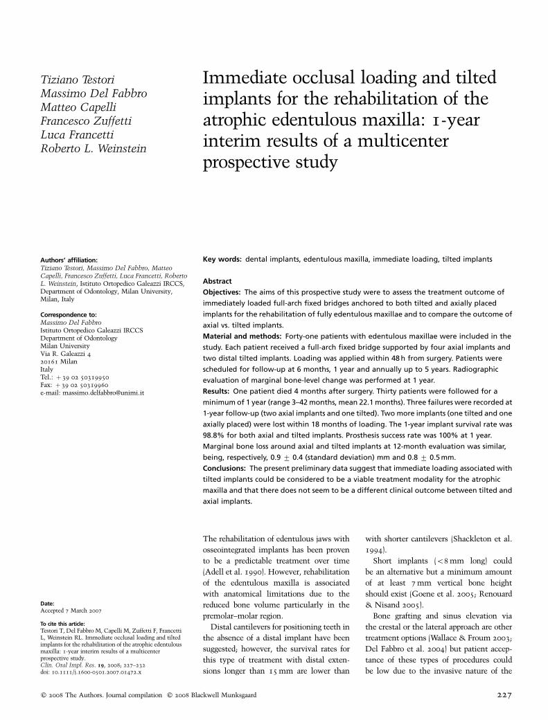
Immediate occlusal loading and tiltedimplants for the rehabilitation of theatrophic edentulous maxilla: 1-yearinterim results of a multicenterprospective study
Tiziano TestoriMassimo Del FabbroMatteo CapelliFrancesco ZuffettiLuca FrancettiRoberto L. Weinstein
Authors’ affiliation:Tiziano Testori, Massimo Del Fabbro, MatteoCapelli, Francesco Zuffetti, Luca Francetti, RobertoL. Weinstein, Istituto Ortopedico Galeazzi IRCCS,Department of Odontology, Milan University,Milan, Italy
Correspondence to:Massimo Del FabbroIstituto Ortopedico Galeazzi IRCCSDepartment of OdontologyMilan UniversityVia R. Galeazzi 420161 MilanItalyTel.: þ 39 02 50319950Fax: þ 39 02 50319960e-mail: [email protected]
Key words: dental implants, edentulous maxilla, immediate loading, tilted implants
Abstract
Objectives: The aims of this prospective study were to assess the treatment outcome of
immediately loaded full-arch fixed bridges anchored to both tilted and axially placed
implants for the rehabilitation of fully edentulous maxillae and to compare the outcome of
axial vs. tilted implants.
Material and methods: Forty-one patients with edentulous maxillae were included in the
study. Each patient received a full-arch fixed bridge supported by four axial implants and
two distal tilted implants. Loading was applied within 48 h from surgery. Patients were
scheduled for follow-up at 6 months, 1 year and annually up to 5 years. Radiographic
evaluation of marginal bone-level change was performed at 1 year.
Results: One patient died 4 months after surgery. Thirty patients were followed for a
minimum of 1 year (range 3–42 months, mean 22.1 months). Three failures were recorded at
1-year follow-up (two axial implants and one tilted). Two more implants (one tilted and one
axially placed) were lost within 18 months of loading. The 1-year implant survival rate was
98.8% for both axial and tilted implants. Prosthesis success rate was 100% at 1 year.
Marginal bone loss around axial and tilted implants at 12-month evaluation was similar,
being, respectively, 0.9 � 0.4 (standard deviation) mm and 0.8 � 0.5 mm.
Conclusions: The present preliminary data suggest that immediate loading associated with
tilted implants could be considered to be a viable treatment modality for the atrophic
maxilla and that there does not seem to be a different clinical outcome between tilted and
axial implants.
The rehabilitation of edentulous jaws with
osseointegrated implants has been proven
to be a predictable treatment over time
(Adell et al. 1990). However, rehabilitation
of the edentulous maxilla is associated
with anatomical limitations due to the
reduced bone volume particularly in the
premolar–molar region.
Distal cantilevers for positioning teeth in
the absence of a distal implant have been
suggested; however, the survival rates for
this type of treatment with distal exten-
sions longer than 15 mm are lower than
with shorter cantilevers (Shackleton et al.
1994).
Short implants (o8 mm long) could
be an alternative but a minimum amount
of at least 7 mm vertical bone height
should exist (Goene et al. 2005; Renouard
& Nisand 2005).
Bone grafting and sinus elevation via
the crestal or the lateral approach are other
treatment options (Wallace & Froum 2003;
Del Fabbro et al. 2004) but patient accep-
tance of these types of procedures could
be low due to the invasive nature of the
Date:Accepted 7 March 2007
To cite this article:Testori T, Del Fabbro M, Capelli M, Zuffetti F, FrancettiL, Weinstein RL. Immediate occlusal loading and tiltedimplants for the rehabilitation of the atrophic edentulousmaxilla: 1-year interim results of a multicenterprospective study.Clin. Oral Impl. Res. 19, 2008; 227–232doi: 10.1111/j.1600-0501.2007.01472.x
c� 2008 The Authors. Journal compilation c� 2008 Blackwell Munksgaard 227
surgical procedure associated with an
increased risk of morbidity and high costs.
Pterigoid (Balshi et al. 1999) and tuber-
osity (Bahat 1992; Khayat & Nader 1994;
Venturelli 1996) implants represent other
treatment options to restore the edentulous
maxilla; however, these treatments could
also be associated with increased morbility.
Zygomatic (Branemark et al. 2004) im-
plants in some clinical situations could
represent another possibility, especially in
extremely atrophic maxilla, but consider-
able surgical experience is needed.
The technique of tilting implants in
order to improve bone anchorage reducing
the need for bone grafting has been recently
advocated by many authors (Krekmanov
2000; Krekmanov et al. 2000; Aparicio
et al. 2001, 2002; Fortin et al. 2002;
Calandriello & Tomatis 2005) and could
provide a viable, minimally invasive treat-
ment modality, leading to high patients
acceptance.
Patients seeking replacement of a den-
ture with an implant-supported prosthesis
are mainly interested in a fixed restoration.
If, after the diagnostic phase, a fixed pros-
thesis could provide optimal lip support
esthetic and phonetics without compro-
mising oral hygiene and without the need
for bone grafting, then patient satisfaction
can be achieved at its highest level.
The aims of this study were to evaluate
the treatment outcome and patients’ satis-
faction with immediately loaded full-arch
fixed prostheses anchored to both axial and
tilted implants in the upper jaw and to
compare the clinical outcome of tilted
(test) vs. axial (control) implants in the
same patients up to 5 years.
This report presents preliminary
12-month data on the implant survival and
on peri-implant marginal bone-level changes
around tilted and axial implants. A survival
analysis is also presented considering the
overall loading time for all the patients.
Material and methods
Inclusion criteria
� Patients with totally edentulous
maxilla.
� Male and female of all races 18 years or
older.
� Patients with severely resorbed maxilla
with at least 4 mm height and 6 mm
width in the first premolar region that
would have needed bone augmentation
for placing implants in a more posterior
location (Fig. 1a and b).
� Patients for whom a decision has al-
ready been made to use dental implants
but expressed strong reluctance for any
kind of bone augmentation.
� Patients physically and psychologically
able to tolerate conventional implant
dentistry.
� Patients who agreed to sign an in-
formed consent form.
� All implants were to be seated with a
torque �30 N cm.
In case one or two axial implants could
not be inserted with a torque �30 N cm,
immediate loading was still allowed be-
cause those implants were splinted to ad-
jacent stable implants. In case either one of
the tilted implants or three or more axial
implants did not achieve the required
primary stability, immediate loading was
not applied and implants were left to heal
for at least 2 months before the prosthetic
phase.
Participants were informed about the
nature of the study and signed an informed
consent.
Exclusion criteria
� Presence of active infection or inflam-
mation in the areas intended for im-
plant placement.
� Presence of systemic diseases such as
uncontrolled diabetes.
� Patients irradiated in the head and neck
regions within 12 months before surgery.
� Presence of previous unresorbed allo-
graft at the implant site.
� Severe bruxism or clenching habits.
� Pregnancy.
� Poor oral hygiene and motivation.
Patients were recruited and treated in
three dental clinics located in North of
Italy, with specific expertize in the treat-
ment of patients by means of immediate
loading procedures. One surgeon with con-
siderable clinical experience in implant
dentistry performed all surgical procedures
at each center. For this specific type of
treatment, no randomization was possible
between the test (tilted implants) and the
control group (axially positioned implants).
Surgical procedure
Antibiotic prophylaxis was prescribed, con-
sisting of amoxicillin and clavulanic acid
(Augmentins
, Roche, Milan, Italy) 2 g 1 h
before surgery. A sedative pre-medication
[Diazapam (Valiums
) Roche] was adminis-
tered to anxious patients.
A local anesthetic agent containing arti-
caine 1 : 100 (Ultracains
D-S forte, Aventis
Pharma Deutschland GmbH, Frankfurt,
Germany) was used.
A crestal incision was made starting in
the first molar, with a vertical-releasing
incision at the midline. A mucoperiostal
buccal flap was raised and the facial bony
wall was exposed. A small antrostomy
using a piezosurgery unit with a diamond
round insert was performed to determine
the position of the anterior sinus wall. Each
patient received six implants in the max-
illa. The posterior tapered implant was
placed first (Osseotite NT Implant, 3i Im-
plant Innovations, Palm Beach, FL, USA);
it was tilted distally approximately 30–351
relative to the vertical plane parallel to the
anterior sinus wall. Visual observation
through lateral antrostomy allowed the
surgeon to insure that the implant did not
protrude into the sinus.
The two axially oriented anterior im-
plants were then placed in the pre-maxilla,
parallel to the midline. At first, the most
mesial implant was inserted at the level of
the central incisor; finally, the third im-
plant was placed about halfway between
the other two.
Careful site preparation was followed in
order to obtain high primary stability; a
30 N cm insertion torque was validated by
the drilling unit torque indication (W&H
Elcomed, W&H Dental Werk, Burmoos
GmbH, Austria). The drilling protocol for
NT-tapered implants was followed. In soft
bone, under-preparation was performed
using a shaping drill one size smaller than
the final implant diameter.
In most of the cases, the implant
shoulder was placed at the crest. All of
the posterior tilted implants required bone
contouring on the distal aspect, allowing
for proper seating of the prosthesis.
The surgical procedure was repeated in
the contra-lateral side.
At the end of the surgical phase, an
impression was taken utilizing a pick-up
technique and a novel radiopaque sterile
impression material, recently approved by
Testori et al . Immediate occlusal loading and tilted implants
228 | Clin. Oral Impl. Res. 19, 2008 / 227–232 c� 2008 The Authors. Journal compilation c� 2008 Blackwell Munksgaard
CE and FDA (Elite Implant Impression
Material, Zhermacks
, Badia Polesine,
Rovigo, Italy).
Finally, a bite registration was taken and
healing abutments were placed at 10 N cm
using a torque controller.
Restorative phase
The provisional screw-retained prosthesis
was delivered within 48 h from surgery
using temporary provisional cylinders with
fiber-reinforced acrylic teeth (Fig. 2a and b).
If the screw access hole was emerging on
the vestibular site of the prosthesis, a com-
posite resin was used to achieve an accepta-
ble esthetic appearance. The final prosthesis
was delivered 3 months later (Fig. 3a–c).
Seven final prosthesis were screw re-
tained, fabricated with a titanium frame-
work (CRESCOt Astra Tech Implant
System, Astra Tech AB, Molndal, Sweden)
with acrylic resin teeth; the remaining 23
prostheses were porcelain-cemented re-
storations with a cast mesiostructure con-
necting all the implants on each side.
The outcome measures evaluated for the
present study were:
(1) Prosthesis success: when the prosthe-
sis could be released as planned and its
function was maintained without compli-
cations, even in case of the loss of one of
more implants. Prosthesis was considered
as failed whether it was not possible to
place it as planned or whether its function
was compromised due to implant failure.
(2) Implant survival that was based on
the following criteria (Albrektsson et al.
1986): no evidence of peri-implant radiolu-
cency; no recurrent or persistent peri-im-
plant infection; no complaint of pain; and
no complaint of neuropathies or paresthe-
sia. As an adjunct to the survival criteria,
additional criteria for implant success were
also imposed. Implants were considered to
be successful if the following conditions
were met at the time of evaluation, in
conjunction with those specified for survi-
val: no crestal bone loss exceeding 1.5 mm
by the end of the first year of functional
loading, and no bone loss exceeding
0.2 mm/year in the subsequent years.
(3) Any biological or prosthetic compli-
cation: examples of possible biological
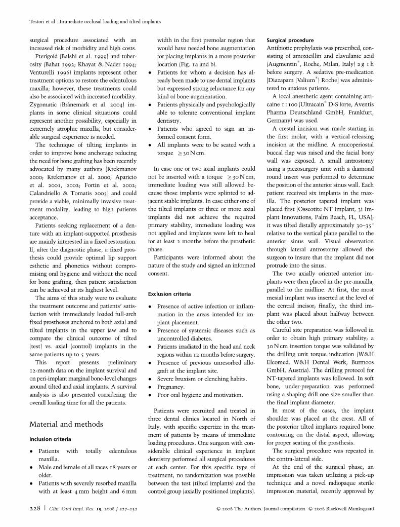
Fig. 1. (a) Pre-operative frontal view without the removable prosthesis. (b)
Pre-operative orthopantomograph of the clinical case.
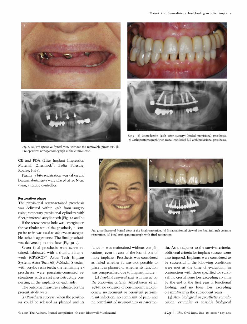
Fig. 2. (a) Immediately (48 h after surgery) loaded provisional prosthesis.
(b) Orthopantomograph with metal-reinforced full-arch provisional prosthesis.
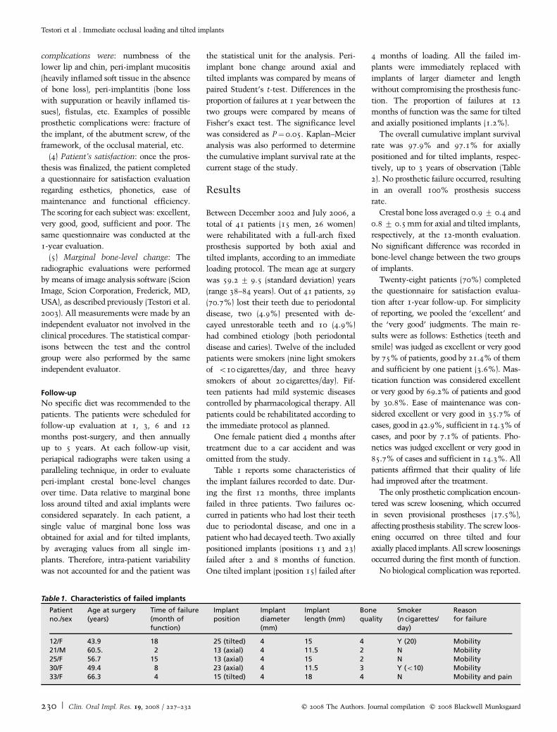
Fig. 3. (a) Extraoral frontal view of the final restoration. (b) Intraoral frontal view of the final full-arch ceramic
restoration. (c) Final orthopantomograph with final restoration.
Testori et al . Immediate occlusal loading and tilted implants
c� 2008 The Authors. Journal compilation c� 2008 Blackwell Munksgaard 229 | Clin. Oral Impl. Res. 19, 2008 / 227–232
complications were: numbness of the
lower lip and chin, peri-implant mucositis
(heavily inflamed soft tissue in the absence
of bone loss), peri-implantitis (bone loss
with suppuration or heavily inflamed tis-
sues), fistulas, etc. Examples of possible
prosthetic complications were: fracture of
the implant, of the abutment screw, of the
framework, of the occlusal material, etc.
(4) Patient’s satisfaction: once the pros-
thesis was finalized, the patient completed
a questionnaire for satisfaction evaluation
regarding esthetics, phonetics, ease of
maintenance and functional efficiency.
The scoring for each subject was: excellent,
very good, good, sufficient and poor. The
same questionnaire was conducted at the
1-year evaluation.
(5) Marginal bone-level change: The
radiographic evaluations were performed
by means of image analysis software (Scion
Image, Scion Corporation, Frederick, MD,
USA), as described previously (Testori et al.
2003). All measurements were made by an
independent evaluator not involved in the
clinical procedures. The statistical compar-
isons between the test and the control
group were also performed by the same
independent evaluator.
Follow-up
No specific diet was recommended to the
patients. The patients were scheduled for
follow-up evaluation at 1, 3, 6 and 12
months post-surgery, and then annually
up to 5 years. At each follow-up visit,
periapical radiographs were taken using a
paralleling technique, in order to evaluate
peri-implant crestal bone-level changes
over time. Data relative to marginal bone
loss around tilted and axial implants were
considered separately. In each patient, a
single value of marginal bone loss was
obtained for axial and for tilted implants,
by averaging values from all single im-
plants. Therefore, intra-patient variability
was not accounted for and the patient was
the statistical unit for the analysis. Peri-
implant bone change around axial and
tilted implants was compared by means of
paired Student’s t-test. Differences in the
proportion of failures at 1 year between the
two groups were compared by means of
Fisher’s exact test. The significance level
was considered as P¼ 0.05. Kaplan–Meier
analysis was also performed to determine
the cumulative implant survival rate at the
current stage of the study.
Results
Between December 2002 and July 2006, a
total of 41 patients (15 men, 26 women)
were rehabilitated with a full-arch fixed
prosthesis supported by both axial and
tilted implants, according to an immediate
loading protocol. The mean age at surgery
was 59.2� 9.5 (standard deviation) years
(range 38–84 years). Out of 41 patients, 29
(70.7%) lost their teeth due to periodontal
disease, two (4.9%) presented with de-
cayed unrestorable teeth and 10 (4.9%)
had combined etiology (both periodontal
disease and caries). Twelve of the included
patients were smokers (nine light smokers
of o10 cigarettes/day, and three heavy
smokers of about 20 cigarettes/day). Fif-
teen patients had mild systemic diseases
controlled by pharmacological therapy. All
patients could be rehabilitated according to
the immediate protocol as planned.
One female patient died 4 months after
treatment due to a car accident and was
omitted from the study.
Table 1 reports some characteristics of
the implant failures recorded to date. Dur-
ing the first 12 months, three implants
failed in three patients. Two failures oc-
curred in patients who had lost their teeth
due to periodontal disease, and one in a
patient who had decayed teeth. Two axially
positioned implants (positions 13 and 23)
failed after 2 and 8 months of function.
One tilted implant (position 15) failed after
4 months of loading. All the failed im-
plants were immediately replaced with
implants of larger diameter and length
without compromising the prosthesis func-
tion. The proportion of failures at 12
months of function was the same for tilted
and axially positioned implants (1.2%).
The overall cumulative implant survival
rate was 97.9% and 97.1% for axially
positioned and for tilted implants, respec-
tively, up to 3 years of observation (Table
2). No prosthetic failure occurred, resulting
in an overall 100% prosthesis success
rate.
Crestal bone loss averaged 0.9� 0.4 and
0.8� 0.5 mm for axial and tilted implants,
respectively, at the 12-month evaluation.
No significant difference was recorded in
bone-level change between the two groups
of implants.
Twenty-eight patients (70%) completed
the questionnaire for satisfaction evalua-
tion after 1-year follow-up. For simplicity
of reporting, we pooled the ‘excellent’ and
the ‘very good’ judgments. The main re-
sults were as follows: Esthetics (teeth and
smile) was judged as excellent or very good
by 75% of patients, good by 21.4% of them
and sufficient by one patient (3.6%). Mas-
tication function was considered excellent
or very good by 69.2% of patients and good
by 30.8%. Ease of maintenance was con-
sidered excellent or very good in 35.7% of
cases, good in 42.9%, sufficient in 14.3% of
cases, and poor by 7.1% of patients. Pho-
netics was judged excellent or very good in
85.7% of cases and sufficient in 14.3%. All
patients affirmed that their quality of life
had improved after the treatment.
The only prosthetic complication encoun-
tered was screw loosening, which occurred
in seven provisional prostheses (17.5%),
affecting prosthesis stability. The screw loos-
ening occurred on three tilted and four
axially placed implants. All screw loosenings
occurred during the first month of function.
No biological complication was reported.
Table 1. Characteristics of failed implants
Patientno./sex
Age at surgery(years)
Time of failure(month offunction)
Implantposition
Implantdiameter(mm)
Implantlength (mm)
Bonequality
Smoker(n cigarettes/day)
Reasonfor failure
12/F 43.9 18 25 (tilted) 4 15 4 Y (20) Mobility21/M 60.5. 2 13 (axial) 4 11.5 2 N Mobility25/F 56.7 15 13 (axial) 4 15 2 N Mobility30/F 49.4 8 23 (axial) 4 11.5 3 Y (o10) Mobility33/F 66.3 4 15 (tilted) 4 18 4 N Mobility and pain
Testori et al . Immediate occlusal loading and tilted implants
230 | Clin. Oral Impl. Res. 19, 2008 / 227–232 c� 2008 The Authors. Journal compilation c� 2008 Blackwell Munksgaard
Discussion
The clinical outcome of this prospective
study indicates that the rehabilitation of
the completely edentulous maxilla with
an immediately loaded full-arch bridge,
either screw retained or cement anchored
to tilted and axial implants, may have a
predictable outcome. Our data compare
favorably with data published by Malo
et al. (2005) concerning fixed complete-
arch immediately loaded maxillary rehabi-
litations supported by two axial and
two tilted implants. Also, immediate
loading of tilted implants in the partially
edentulous maxilla showed encouraging
success rates (Calandriello & Tomatis
2005). However, these data are not
comparable to ours because of the dif-
ferent clinical and biomechanics of the
prosthesis.
Cumulative implant survival rates of
tilted and axial implants to date are similar
up to 3 years (Table 2). These data are
consistent with other authors (Krekmanov
et al. 2000; Aparicio et al. 2001). It could
be speculated that tilted implants are
placed and anchored with greater cortical
bone contact than axial ones. In fact, the
tilted implants are placed between the
cortical bone of the crest, the mesial wall
of the maxillary sinus and the nasal floor,
achieving tricortical anchorage.
Studies in vitro analyzing the load dis-
tribution of implants connected to angu-
lated abutments discouraged their use;
however, it must be pointed out that un-
favorable results were reported for single
implants (Clelland et al. 1993), and not for
multiple implants in which the abutments
are connected together (Krekmanov et al.
2000). Furthermore, it must be kept in
mind that the external validity of in vitro
studies can be extremely low due to highly
different experimental conditions with re-
spect to the clinical field.
No increase of load transfer to the bone
with respect to axial implants was reported
in vivo for tilted implants splinted to axi-
ally positioned implants (Krekmanov et al.
2000).
Furthermore, animal studies have shown
that non-axial loading is not detrimental
for the osseointegration process (Celletti
et al. 1995; Miyata et al. 1998).
In this clinical study, the marginal bone
loss was not affected by the tilting of the
implants.
The marginal bone resorption for axial
and tilted implants showed a normal
pattern predicting normal bone response
when tilted implants are splinted, similar
to what reported in previous studies
(Calandriello & Tomatis 2005).
By tilting the posterior implants, sinus
lift procedures can be avoided by reducing
the morbidity of the surgical phase. Other
clinical advantages include (1) the possibi-
lity of placement of longer implants that
increases the bone-to-implant contact area
and the implants’ primary stability and (2)
the distance between implants can be in-
creased, reducing the cantilevers and thus
optimizing load distribution. The use of
fewer implants to support the prosthesis
and the application of the immediate
loading protocol can reduce the overall
treatment costs.
The principle of using four or six
implants instead of the maximum possible
number of implants for the rehabilitation
of fully edentulism is also supported
by long-term studies (Branemark et al.
1995).
Conclusion
The present preliminary data suggest that
immediate loading associated with tilted
implants could be considered a viable treat-
ment modality for the atrophic maxilla and
that there does not seem to be a different
clinical outcome between tilted and axial
implants. The use of tilted implants may
avoid more complex treatments, reducing
the patient’s morbidity, treatment time
and costs. These results indicate that if
the prerequisites for immediate loading
such as high primary stability (30 N cm or
more), splinting of the implants via a provi-
sional prosthesis and the use of an osteo-
conductive surface are fulfilled, tilting the
implants may not adversely affect the final
outcome.
Acknowledgements: The authors are
grateful to Dr Alan Meltzer and to Prof.
Marco Esposito for reviewing the
manuscript and to Dr Jorg M. Ritzmann
for the prosthodontic phase.
References
Adell, R., Eriksson, B., Lekholm, U., Branemark,
P.I. & Jemt, T. (1990) Long-term follow-up study
of osseointegrated implants in the treatment of
totally edentulous jaws. International Journal of
Oral & Maxillofacial Implants 5: 347–359.
Albrektsson, T., Zarb, G., Worthington, P. & Erik-
son, A.R. (1986) The long-term efficacy of cur-
rently used dental implants: a review and proposed
criteria of success. The International Journal of
Oral & Maxillofacial Implants 1: 11–25.
Aparicio, C., Arevalo, X., Ouzzani, W. & Granados,
C. (2002) A retrospective clinical and radiographic
evaluation of tilted implants used in the treatment
of the severely resorbed edentulous maxilla.
Applied Osseointegration Research 3: 17–21.
Table 2. Life table analysis of IL implants
Time interval(months)
Implants atbeginningof interval
Withdrawnimplants
Failedimplants
Intervalsurvivalrate (%)
Cumulativesurvivalrate (%)
Axial implants0–6 164 4 1 99.4 99.46–12 143 0 1 99.3 98.712–18 118 0 1 99.2 97.918–24 105 0 0 100 97.924–36 84 0 0 100 97.9436 8 0 0 100 97.9
Tilted implants0–6 82 2 1 98.8 98.86–12 71 0 0 100 98.812–18 59 0 1 98.3 97.118–24 52 0 0 100 97.124–36 42 0 0 100 97.1436 4 0 0 100 97.1
Testori et al . Immediate occlusal loading and tilted implants
c� 2008 The Authors. Journal compilation c� 2008 Blackwell Munksgaard 231 | Clin. Oral Impl. Res. 19, 2008 / 227–232
Aparicio, C., Perales, P. & Rangert, B. (2001) Tilted
implants as an alternative to maxillary sinus
grafting: a clinical, radiologic, and periotest study.
Clinical Implant Dentistry & Related Research
3: 39–49.
Bahat, O. (1992) Osseointegrated implants in the
maxillary tuberosity: report on 45 consecutive
patients. The International Journal of Oral &
Maxillofacial Implants 7: 459–467.
Balshi, T.J., Wolfinger, G.J. & Balshi, S.F. (1999)
Analysis of 356 pterygo-maxillary implants in
edentulous arches for fixed prosthesis anchorage.
The International Journal of Oral & Maxillofa-
cial Implants 14: 398–406.
Branemark, P.I., Grondahl, K., Ohrnell, L.O., Nils-
son, P., Petruson, B., Svensson, B., Engstrand, P.
& Nannmark, U. (2004) Zygoma fixture in the
management of advanced atrophy of the maxilla:
technique and long-term results. Scandinavian
Journal of Plastic and Reconstructive Surgery
and Hand Surgery 38: 70–85.
Branemark, P.I., Svensson, B. & van Steenberghe,
D. (1995) Ten-year survival rates of fixed pros-
theses on four or six implants ad modum Brane-
mark in full edentulism. Clinical Oral Implants
Research 6: 227–231.
Calandriello, R. & Tomatis, M. (2005) Simplified
treatment of the atrophic posterior maxilla via
immediate/early function and tilted implants: a
prospective 1-year clinical study. Clinical Im-
plant Dentistry & Related Research 7: 1–12.
Celletti, R., Pameijer, C.H., Bracchetti, G., Donath,
D., Persichetti, G. & Visani, I. (1995) Histologic
evaluation of osseointegrated implants restored in
nonaxial functional occlusion with preangled
abutments. The International Journal of Perio-
dontics and Restorative Dentistry 15: 563–573.
Clelland, N.L., Gilat, A., McGlumphy, E.A. &
Brantley, W.A. (1993) A photoelastic and strain
gauge analysis of angled abutments for an implant
system. The International Journal of Oral &
Maxillofacial Implants 8: 541–548.
Del Fabbro, M., Testori, T., Francetti, L. & Wein-
stein, R. (2004) Systematic review of survival
rates for implants placed in grafted maxillary
sinus. International Journal of Periodontics &
Restorative Dentistry 24: 565–577.
Fortin, Y., Sullivan, R.M. & Rangert, B. (2002) The
Marius implant bridge: surgical and prosthetic
rehabilitation for the completely edentulous upper
jaw with moderate to severe resorption: a 5-year
retrospective clinical study. Clinical Implant
Dentistry & Related Research 4: 69–77.
Goene, R., Bianchesi, C., Huerzeler, M., Del Lupo,
R., Testori, T., Davarpanah, M. & Jalbout, Z.
(2005) Performance of short implants in partial
restorations: 3-year follow-up of Osseotite im-
plants. Implant Dentistry 14: 274–280.
Khayat, P. & Nader, N. (1994) The use of osseoin-
tegrated implants in the maxillary tuberosity.
Practical Periodontics and Aesthetic Dentistry
6: 53–61.
Krekmanov, L. (2000) Placement of posterior man-
dibular and maxillary implants in patients with
severe bone deficiency: a clinical report of proce-
dure. International Journal of Oral & Maxillofa-
cial Implants 15: 722–730.
Krekmanov, L., Kahn, M., Rangert, B. & Lindstrom,
H. (2000) Tilting of posterior mandibular and
maxillary implants for improved prosthesis sup-
port. The International Journal of Oral & Max-
illofacial Implants 15: 405–414.
Malo, P., Rangert, B. & Nobre, M. (2005) All-on-4
immediate-function concept with Branemark
system implants for completely edentulous max-
illae: a 1-year retrospective clinical study. Clinical
Implant Dentistry & Related Research 7 (Suppl.
1): S88–S94.
Miyata, T., Kobayashi, Y., Araki, H., Motomura, Y.
& Shin, K. (1998) The influence of controlled
occlusal overload on peri-implant tissue: a histo-
logic study in monkeys. The International
Journal of Oral & Maxillofacial Implants 13:
677–693.
Renouard, F. & Nisand, D. (2005) Short implants in
the severely resorbed maxilla: a 2-year retrospec-
tive clinical study. Clinical Implant Dentistry &
Related Research 7: 104–110.
Shackleton, J.L., Carr, L., Slabbert, J.C. & Becker,
P.J. (1994) Survival of fixed implant-supported
prostheses related to cantilever lengths. Journal
of Prosthetic Dentistry 71: 23–26.
Testori, T., Del Fabbro, M., Szmukler-Moncler, S.,
Francetti, L. & Weinstein, R.L. (2003) Immediate
occlusal loading of Osseotites
implants in the
totally edentulous mandible. The International
Journal of Oral & Maxillofacial Implants 18:
544–551.
Venturelli, A. (1996) A modified surgical pro-
tocol for placing implants in the maxillary tuber-
osity: clinical results at 36 months after loading
with fixed partial dentures. The International
Journal of Oral & Maxillofacial Implants 11:
743–749.
Wallace, S.S. & Froum, S.J. (2003) Effect of
maxillary sinus augmentation on the survival of
endosseous dental implants as compared to
the survival of implants placed in the non-
grafted posterior maxilla: an evidence-based
literature review. Annals of Periodontology 8:
328–343.
Testori et al . Immediate occlusal loading and tilted implants
232 | Clin. Oral Impl. Res. 19, 2008 / 227–232 c� 2008 The Authors. Journal compilation c� 2008 Blackwell Munksgaard