Embed Size (px)
Citation preview

Leukemiahttps://doi.org/10.1038/s41375-019-0491-z
ARTICLE
Acute myeloid leukemia
Imipridone ONC212 activates orphan G protein-coupled receptorGPR132 and integrated stress response in acute myeloid leukemia
Takenobu Nii1 ● Varun V. Prabhu2● Vivian Ruvolo1
● Neel Madhukar3 ● Ran Zhao1● Hong Mu1
● Lauren Heese1 ●
Yuki Nishida1 ● Kensuke Kojima1,4 ● Mathew J. Garnett5 ● Ultan McDermott5 ● Cyril H. Benes6 ● Neil Charter7 ●
Sean Deacon7● Olivier Elemento3
● Joshua E. Allen2● Wolfgang Oster2 ● Martin Stogniew2
● Jo Ishizawa1 ●
Michael Andreeff1
Received: 1 August 2018 / Revised: 8 March 2019 / Accepted: 8 April 2019© Springer Nature Limited 2019
AbstractImipridones constitute a novel class of antitumor agents. Here, we report that a second-generation imipridone, ONC212,possesses highly increased antitumor activity compared to the first-generation compound ONC201. In vitro studies usinghuman acute myeloid leukemia (AML) cell lines, primary AML, and normal bone marrow (BM) samples demonstrate thatONC212 exerts prominent apoptogenic effects in AML, but not in normal BM cells, suggesting potential clinical utility.Imipridones putatively engage G protein-coupled receptors (GPCRs) and/or trigger an integrated stress response inhematopoietic tumor cells. Comprehensive GPCR screening identified ONC212 as activator of an orphan GPCR GPR132and Gαq signaling, which functions as a tumor suppressor. Heterozygous knock-out of GPR132 decreased the antileukemiceffects of ONC212. ONC212 induced apoptogenic effects through the induction of an integrated stress response, andreduced MCL-1 expression, a known resistance factor for BCL-2 inhibition by ABT-199. Oral administration of ONC212inhibited AML growth in vivo and improved overall survival in xenografted mice. Moreover, ONC212 abrogated theengraftment capacity of patient-derived AML cells in an NSG PDX model, suggesting potential eradication of AMLinitiating cells, and was highly synergistic in combination with ABT-199. Collectively, our results suggest ONC212 as anovel therapeutic agent for AML.
Introduction
Acute myeloid leukemia (AML) encompasses a group ofhighly heterogeneous hematological malignancies, some of
which can carry poor prognosis. Standard chemotherapy hasnot changed over the past 40+ years and acquired resistanceleading to disease relapse typically occurs in the majority ofAML patients. Better understanding of the disease in thelast decade has led to the development of multiple targetedtherapeutics that are being investigated in preclinical mod-els and clinical trials, and breakthrough discoveries inelderly AML treated with ABT-199 (Bcl-2 inhibition)combination.
These authors contributed equally: Takenobu Nii, Varun V. Prabhu
* Jo [email protected]
* Michael [email protected]
1 Section of Molecular Hematology and Therapy, Department ofLeukemia, The University of Texas MD Anderson Cancer Center,Houston, TX, USA
2 Oncoceutics, Inc., Philadelphia, PA, USA
3 Institute for Computational Biomedicine, Weill Cornell Medicine,New York, NY, USA
4 Division of Hematology, Respiratory Medicine and Oncology,Department of Internal Medicine, Saga University, Saga, Japan
5 Wellcome Trust Sanger Institute, Wellcome Genome Campus,Cambridge, UK
6 Department of Medicine, Massachusetts General Hospital,Boston, MA, USA
7 DiscoverX Corporation, Fremont, CA, USA
Supplementary information The online version of this article (https://doi.org/10.1038/s41375-019-0491-z) contains supplementary material,which is available to authorized users.
1234
5678
90();,:
1234567890();,:

Imipridones encompass a new class of antitumor smallmolecules that were created following the discovery of thelead compound ONC201 [1], which is in clinical trials forseveral advanced malignancies including leukemias, mye-lomas, lymphomas, and gliomas [2, 3]. ONC201 demon-strated an exceptional pharmacokinetic andpharmacodynamic safety profile and encouraging evidenceof antitumor activity [4]. ONC201 was putatively deter-mined to antagonize the G protein-coupled receptor (GPCR)superfamily dopamine receptors D2 and D3 (DRD2/3) [5],and by us and others to activate an atypical integrated stressresponse (ISR) [6, 7]. By way of this novel mechanism ofaction, ONC201 was reported to induce apoptosis innumerous human cancer types that were resistant tostandard-of-care therapeutics without causing cytotoxicityin normal cells [7, 8].
The GPCR superfamily is the largest and most diversegroup of cell surface receptors in mammals. In a patho-physiological context, GPCRs are involved in cancerinitiation, promotion, and metastasis [9], and play pivotalroles in activating major proliferative and survival signaltransduction pathways relevant to cancer cells [10, 11].Thus, GPCRs are considered promising therapeutic targetsin cancer [12].
In view of the therapeutic opportunities available withinthe GPCR superfamily, ONC201′s therapeutic index[1, 2, 7], and its novel triheterocyclic core chemical struc-ture [13], a series of second-generation analogs were gen-erated [14]. In this study, we report the effects of one suchcompound, ONC212, on AML, and a novel therapeuticmechanism targeting the orphan GPCR GPR132 and ISR.In addition, we report on unique combinatorial approachesthat extend these anticancer effects.
Materials and methods
Cell culture
Cell lines were purchased from the Leibniz-InstitutDeutsche Sammlung von Mikroorganismen und Zellk-ulturen or the American Type Culture Collection. Theauthenticity of the cell lines was confirmed by DNA fin-gerprinting with the short tandem repeat method, using aPowerPlex 16 HS System (Promega, WI, USA) within6 months before the experiments. All cell lines were testedusing the PCR Mycoplasma Detection Kit (Applied Bio-logical Materials, Richmond, BC, Canada) as directed bythe manufacturer. The mantle cell lymphoma (MCL) lineJeKo-1 was cultured in RPMI 1640 medium containing20% fetal bovine serum (FBS), AML lines (OCI-AML3,MOLM-13, MOLM-14, MV4;11, KG-1, Kasumi-1, U937,HL-60, and THP-1), T-cell leukemia (Jurkat and JurkatI9.2)
[15], DDIT3 (CHOP) knockdown cells which we pre-viously established [7], and the colorectal cancer cell lineHCT-116 were all cultured in RPMI 1640 medium con-taining 10% FBS; wild type and Bax and Bak double knockout mouse embryonic fibroblasts (MEFs) were cultured inMEMα 1640 medium containing 20% FBS. ONC201-resistant (ONC-R) cell lines were generated by chronicexposure of MCL and AML cell lines to ONC201 whoseED50s for ONC201 at 72 h were all >5 μM, as describedpreviously [7]. To generate HCT-116 GPR132 knock-outcells, we used the PITCh system gene knock-in methodpreviously established [16]. A GFP knock-in cell line at theAAVS1 locus was used as control. Details are describedin Supplemental Methods.
Reagents and drug treatment
ONC201 and ONC212 were manufactured and provided byOncoceutics Inc. (Philadelphia, PA). ABT-199 for thein vitro experiments was purchased from Selleck ChemicalsLLC (Houston, TX, USA) and for in vivo experiments fromActive Biochemku LTD (Maplewood, NJ, USA). Doxor-ubicin was purchased from Bedford Laboratories (Bedford,Ohio, USA). For 72-h drug treatment experiments, cell lineswere plated at a density of 1.5 × 10⁵/ml for JeKo-1, OCI-AML3, MOLM-13, MOLM-14, MV4;11, KG-1, U937;3.0 × 10⁵/ml for Kasumi-1, HL-60, THP-1, Jurkat, andJurkatI9.2; 5.0 × 10⁵/ml for HCT-116; and treated with theindicated compounds. For 120-h drug treatment experi-ments, cell lines were plated at a density of 5.0 × 10⁴/ml forOCI-AML3 and U937 and 1.0 × 10⁵/ml for Kasumi-1, KG-1, HL-60, and THP-1. For ISR activation, 1 μM thapsi-gargin (6 h) were used.
Flow cytometric analysis
For apoptosis analysis, annexin V (BD Pharmingen, CA, USA)and propidium iodide (PI; Sigma-Aldrich, MO, USA) wereused to assess apoptosis. annexin V- and PI-double negativecells were counted as live cells. To determine absolute cellnumbers, CountBright beads (Thermo Fisher Scientific, MA,USA) were added to each sample. For cell cycle analysis, DNAcontent analysis was performed by staining with PI and 5-ethynyl-2′-deoxyuridine (EdU) incorporation was analyzed bythe Click-iT EdU Alexa Fluor 647 Flow Cytometry Assay Kit(Thermo Fisher Scientific), as per manufacturer’s instructions.To measure mitochondrial membrane potential (MMP), weloaded cells with MitoTracker Red CMXRos (300 nM) andMitoTracker Green (100 μM, both from Molecular Probes,Eugene, OR, USA) for 1 h at 37 °C. The MMP was thenassessed by measuring CMXRos retention (red fluorescence),whereas simultaneously adjusting for mitochondrial mass(green fluorescence) [17].
T. Nii et al.

Statistical analyses
Differences among groups were analyzed for statisticalsignificance with GraphPad Prism 6 software (Graph-PadSoftware, San Diego, CA, USA) using a Student’s t-test orANOVA followed by Tukey’s post hoc test. A p < 0.05 wasconsidered statistically significant. Unless otherwise indi-cated, values are expressed as the mean ± SD calculated byperforming three or more independent experiments. Fordrug combination analysis, synergistic effects of drugs wereanalyzed by CompuSyn software (ComboSyn Inc., Para-mus, NJ, USA). The combination index (CI) providesquantitative determination for synergism (CI < 1), additiveeffect (CI= 1), and antagonism (CI > 1) [18, 19].
Results
ONC212 exhibits potent antitumor effects via theactivation of orphan GPCR GPR132
We recently reported that the first generation imipridoneONC201 putatively antagonizes DRD2/3 [5]. Therefore, wespeculated that imipridones in general could selectively targetthe GPCR superfamily of proteins. Experimental GPCR pro-filing using the PathHunter β-arrestin assay [20] determined thatONC212 (Fig. 1a) was highly selective for activating theorphan GPCR GPR132 (Fig. 1b). Although six hits wereidentified from the initial screen with this single high con-centration of ONC212, multi-dose validation studies confirmedthat ONC212 only engaged GPR132 at nanomolar concentra-tions (EC50 ~400 nM) (Fig. 1c and Table S3). We furtherperformed the BioSensAll assays to determine which Gα iso-form was engaged downstream of GPR132, and demonstratedthat Gαq family proteins (Gαq, Gα14, and Gα15/16) are spe-cifically activated via GPR132 by ONC212 (Figs. 1d and S1)while Gαs family proteins were not (Fig. S1). The activation ofGαq was blocked by the Gαq inhibitor UBO (Fig. 1d), indi-cating the specificity of detected bioluminescence resonanceenergy transfer (BRET) signals. Interestingly, Gαq activationvaried with pH, consistent with GPR132 being a pH sensitiveGPCR [21] (Fig. S2).
To further assess the impacts of GPR132 on cytotoxiceffects of ONC212 in tumor cells, we generated HCT-116GPR132 knock-out cells by gene knock-in method usingCRISPR/Cas9 [16] (Fig. S3A). We identified one hetero-zygous knock-out clone, which has a knock-in allele and in-frame 27 base-pair deletion on the other allele (Figs. 1eand S3B, S3C). The GPR132 heterozygous knock-outdecreased ONC212′s apoptogenic effects (Fig. 1f, g), fur-ther supporting a role for GPR132 in this process. Interest-ingly, GPR132 knock-down by shRNA decreased targetlevels by approximately 50%, but ONC212 increased
GPR132 levels above baseline; hence this system could notbe used to determine impact of GPR132 on activity (data notshown).
RNA-seq data available in The Cancer Genome Atlas(TCGA) revealed that GPR132 is expressed in a wide rangeof tumor types, with highest expression in leukemias andlymphomas (http://www.cbioportal.org/) without any repor-ted loss-of-function gene mutations (Fig. S4). Previous stu-dies have implicated GPR132 as tumor suppressor [22, 23],suggesting that activation of GPR132 by ONC212 couldexert growth inhibitory and apoptotic effects. We analyzedthe in vitro antitumor effects of ONC212 in the GDSCcollection of cell lines (i.e., 1,088 human cancer cell lines)and found that leukemias and lymphomas were most sen-sitive (Fig. S5A). When we further analyzed types of leu-kemias, AML, acute lymphoblastic leukemia, and chronicmyeloid leukemia lines were all similarly sensitive toONC212 (Fig. S5B, S5E). In support of the therapeuticrelevance of GPR132 for ONC212 antitumor activity,expression of GPR132 mRNA was positively correlatedwith sensitivity to ONC212 (Fig. 1h), based on geneexpression profiling data in the GDSC panel. Moreover, wecompared GPR132 expression between human primaryAML and normal hematopoietic samples in the RNA-seqdata from TCGA and The Genotype-Tissue Expressionprojects using Gene Expression Profiling Interactive Ana-lysis [24]. The AML patient samples significantly higherlevels of GPR132 expression compared to normal samples(Fig. 1i). Since a threshold level for normal hematopoieticcells has not been established, the lack of observed toxicitycould be related to lower levels of GRP132. These datasuggest that GPR132 could be a potential therapeutic target,particularly in AML.
ONC212 induces apoptosis in AML cells
We assessed ONC212′s efficacy and mechanisms in AMLcells. We first tested the apoptogenic effects of ONC212 in aseries of AML cell lines (Fig. 2a, b). Compared with theED50s of ONC201 that we reported previously [7], ONC212exerted markedly enhanced apoptogenic effects [ED50s ofONC201 [7] versus ONC212 (Fig. 2a) at 72 h; 6.4 μM versus258.7 nM for OCI-AML3, 3.6 μM versus 105.7 nM forMOLM13 cells, respectively], indicating that this agent was>30-fold more potent than the parent compound.
Several cell lines were relatively insensitive 72 h afterONC212 exposure (Fig. 2a), but became an apoptotic at120 h (Figs. 2b and S6A). Time course analysis of OCI-AML3 cells showed that induction of apoptosis took morethan 36 h and gradually increased in a time-dependentmanner (Fig. 2c, d). ONC212 reduced MMP, suggestingmitochondrial apoptosis (Fig. S7A, S7B), consistent withpreviously reported GPR132-mediated mitochondrial
Imipridone ONC212 activates orphan G protein-coupled receptor GPR132 and integrated stress response in. . .

Fig. 1 Target screening of G protein-coupled receptors (GPCRs) identifiesGPR132 as a receptor of ONC212. a Chemical structure of ONC212. bONC212 (10 µM) target screening in GPCRs performed with the Path-Hunter β-Arrestin assay. Dotted line indicates a 20% cut-off to selecttargets for subsequent validation. c ONC212 dose-response curve in thePathHunter β-Arrestin assay for GPR132. EC50= 405 nM (n= 2). dCells were pre treated for 1 h with UBO-QIC (100 nM). Experimental datawere produced in singleton and curves were fitted using the 4-parameterlogistic nonlinear regression model (GraphPad 6). Data in graphs representa 10min incubation with ONC212 and expressed as uBRET. e qRT-PCRanalysis of GPR132 expression. GFP heterozygous knock-in at GPR132
locus (GPR132+/−; GPR132 heterozygous knock-out) and at AAVS1locus (AAVS1+/−; gene knock-in control). f The percentage of apoptoticcells (Annexin V+ cells) induced by ONC212 treatment for 72 h inGPR132+/− and AAVS1+/−. g The percentage of live cell numbers after72-h treatment with ONC212 in GPR132+/− and AAVS1+/−. h AverageONC212 response as measured by the area under the dose-response curve(AUC) in GDSC cell lines with high versus low GPR132 expression.****P < 0.001. i comparison of GPR132 expression in AML and normalsamples using Gene expression profiling interactive analysis (GEPIA).AML (n= 173) and Normal (n= 70) *p < 0.01
T. Nii et al.

apoptosis [25]. In addition, Bax and Bak double knockoutMEFs showed resistance to ONC212, compared to wild-type MEFs (Fig. S7C, S7D). Of note, ONC-R cell lines
[7] showed cross-resistance to ONC212 (Figs. 2e, fand S6A–S6D), suggesting common resistance mechanismsfor the two compounds. Also, as reported for ONC201
Fig. 2 Assessment of ONC212 potency in AML cells. a Summary dataof ONC212′s effects on several AML cell lines at the indicated con-centration for 72 h. b The percentage of apoptotic cells induced byONC212 treatment for 120 h in KG-1, HL-60, U937, Kasumi-1, andTHP-1 cell lines. c Time course analysis of apoptosis induced byONC212 or ONC201 in OCI-AML3 cells. d Time course analysis ofthe percentage of live cells following treatment with ONC212 orONC201. Annexin V- and PI-negative cells were counted as live cells.e The percentage of apoptotic cells induced by ONC212 treatment for
72 h in parental or ONC201-resistant (ONC-R) OCI-AML3 cells.f The percentage of live cell numbers after 72-h treatment withONC212 in parental or ONC-R OCI-AML3 cells. The percentage ofAnnexin V+ cells of MV4;11 (g) and MOLM13 (h) cells treated withONC212 (250 nM) for 4, 8, 24, 36, and 72 h. ONC212 medium wasreplaced by fresh medium at these time points and cell viability wasdetermine at 72 h for all samples. Data are shown as the mean ± SD (n= 3). **p < 0.01, ***p < 0.005
Imipridone ONC212 activates orphan G protein-coupled receptor GPR132 and integrated stress response in. . .

[7], ONC212 increased γH2A.X only to the extent seenin Nutlin-3a treated cell, which was selected as acompound that induces only minimal DNA damage [26](Fig. S6E). This finding suggests that ONC212 is non-genotoxic, which is a potential advantage for its clinicalimplementation.
We also performed wash-out experiments of ONC212 toinvestigate how long this agent is required to be present toinduce irreversible apoptosis. Unlike ONC201, in which atleast a 72 h exposure is required for apoptosis induction, only12 h of exposure to ONC212 was sufficient (Fig. 2g, h).
GPR132 mRNA expression was detected in all testedAML cell lines (Fig. S8A). There was a statistically sig-nificant correlation between the level of apoptosis inductionby ONC212 and GPR132 mRNA expression in the AMLcell lines tested (Fig. S8B). However, THP-1 and Kasumi-1
cells, which respond only slowly and display the lowestsensitivity to ONC212, were outliers (Fig. 2b) [7]. In addi-tion, ONC212 treatment transcriptionally increased GPR132expression (Fig. S8C), suggesting that the apoptogeniceffects of ONC212 reflect not only functional activation ofGPR132 but also on its transcriptional induction.
ONC212 efficiently induces cell death in patient-derived AML, but not normal hematopoietic cells
Next, we examined the effects of ONC212 on human pri-mary AML and normal bone marrow (BM) cells (Fig. S9A).ONC212 treatment inhibited blast colony formation in adose-dependent manner (Figs. 3a, b, and S9B). Importantly,in normal BM cells there were no marked effects byONC212 on colony formation (Fig. 3c) and differentiation
Fig. 3 ONC212 treatmentinhibits the clonogenic growthof human AML blasts but notnormal hematopoietic cells. Fortreatment with or withoutONC212 (100 or 200 nM), a thepercentage of blast colonyformation from three patientsamples were examined. **p <0.01, ***p < 0.005. b Thenumber of blast colonies fromAML patients without ONC212treatment. c Hematopoieticcolony formation from twohealthy donor BM samplesfollowing the above-mentionedtreatment. d Mean of each typeof hematopoietic colonygenerated in percentage.ONC212 did not affect thefrequency of each colonies. Dataare shown as the mean ± SD(n= 3). N.S.= not significant
T. Nii et al.

potential of hematopoietic progenitor cells (Figs. 3dand S9C). ONC212 also did not affect cell viability ofnormal BM and lung fibroblasts at doses that were effica-cious in this regard in malignant cells (Fig. S10). Theseresults suggested that ONC212 exerts selective toxicity inleukemia as compared to nonmalignant cells.
ONC212 dysregulates cell cycle progressionresulting in antiproliferative effects in AML cells
To examine the cell cycle effects of ONC212, we analyzedthe incorporation of EdU and DNA content in AML celllines. ONC212 treatment increased the percentage of G0/G1and decreased S and G2/M cells and EdU incorporation in Sphase in MOLM13 and HL-60 cells (Fig. S11A–S11E). Inparticular, ONC212 treatment induced a more prominentG0/G1 arrest in MOLM13 than in HL-60 cells (Fig. S11A).To confirm the growth inhibitory effects of ONC212, wecounted the number of viable cells at early time pointsbefore apoptosis was induced and indeed, the proliferationof viable cells was inhibited by ONC212 (Fig. S11F, S11G)at 24 h indicating a cytostatic effect on AML cells.
ONC212 exhibits antileukemia activity in vivo
For assessing ONC212′s antileukemia effects on leukemia-initiating cells (LICs), we utilized an established PDXmouse model [7]. Bulk AML cells [t(9;11)(p22; q23),CEBPA and ATM mutants] from secondarily engraftedmice (i.e., LIC-enriched PDX cells) were incubated ex vivofor 36 h with or without 250 nM of ONC212. We theninjected identical numbers of viable treated and untreatedcells into recipient NSG mice. After 1 month, the percen-tage of human CD45+ cells in the peripheral blood, spleen,and BM was significantly decreased in the ONC212-treatedgroup compared to the untreated control (Figs. 4aand S12A). Spleens collected from the treated mice weresignificantly smaller than those obtained from the controls(Fig. S12B, S12C). These results suggested that ONC212impairs the engraftment capacity of LICs.
We next administered ONC212 orally to mice bearingtwo different AML xenografts. In the first model, MV4,11 cells were subcutaneously injected into the right and leftflank of mice. Mice were treated with cytarabine, ONC201,or ONC212 when the tumors reached a detectable volume.ONC212 significantly reduced leukemia xenograft growth,as well as the cytarabine treatment, while ONC201 was notefficacious in this model at similar doses (Fig. 4b). Of note,more frequent dosing with lower doses of ONC212 (5 or 25mg/kg) did not inhibit leukemia progression (Figs. 4band S13).
In the second model, we utilized systemic AML xeno-grafts. OCI-AML3 cells expressing luciferase were injected
into NSG-S mice via tail vein and monitored by biolumi-nescence imaging (BLI). After confirmation of AMLengraftment after 7 days (Fig. 4c), mice were treated withoral administration of ONC212, serial BLI images showedthat ONC212 significantly inhibited AML growth as soonas during the week of therapy (Fig. 4c). Moreover, ONC212treatment decreased spleen leukemia burden (Fig. 4d) andprolonged overall survival compared with controls (p=0.0003; Fig. 4e). The median survival increased from 43 to49 days (+14%). Consistently, AML cell infiltration in BMand spleens of the ONC212-treated mice was reduced, asindicated by spleen size (Fig. 4d) and flow cytometricanalysis (Fig. S14). These results indicated that ONC212exerts antileukemia activity in vivo. The most effectivedose/schedule remains to be established.
ONC212 activates an ISR in AML cells and synergizedwith the BCL-2 inhibitor ABT-199 in vitro and in vivo
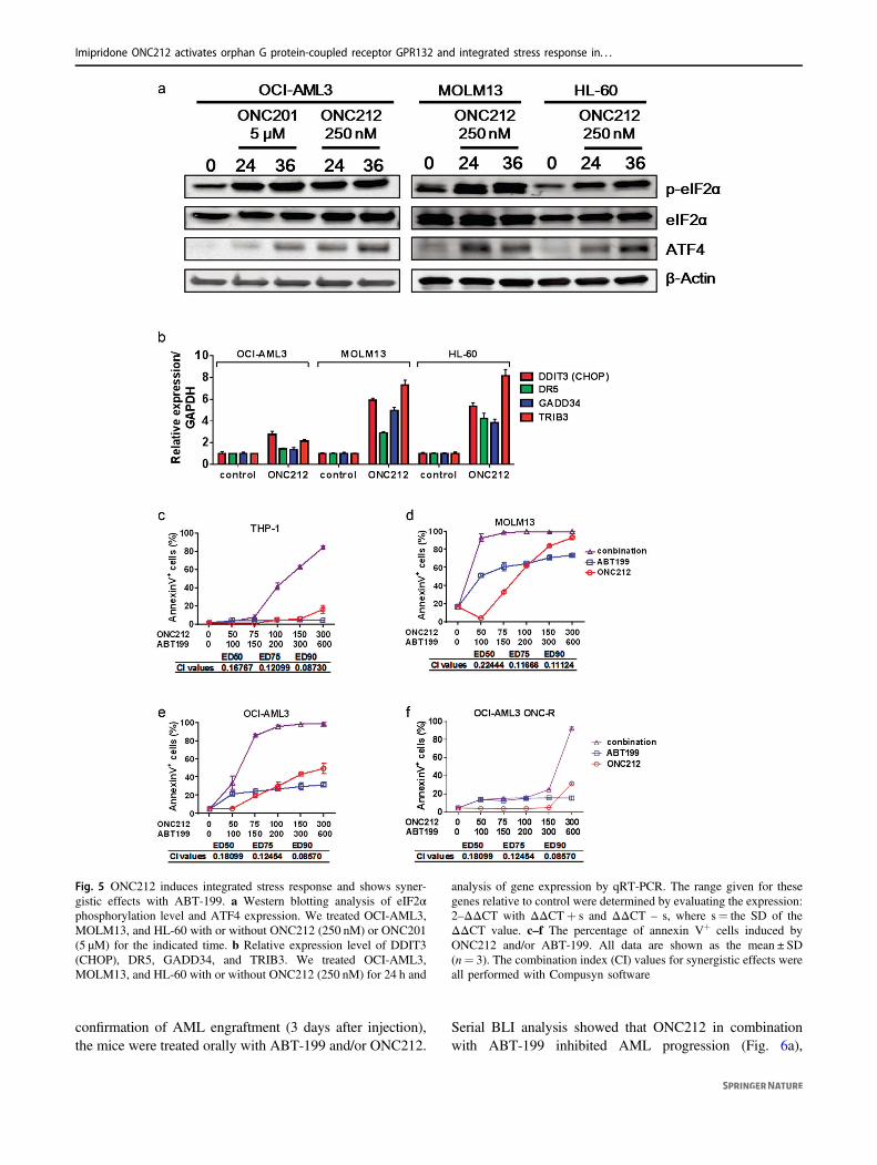
We and others previously reported that an ISR-like pathwayinvolving ATF4 was one of the major molecular mechan-isms involved in apoptosis induced by ONC201 in solid andhematologic tumors [6, 7], and comparable induction of ISRby ONC212 was recently also reported in pancreatic cancercells [27]. Similarly, 24 and 36 h treatment with ONC212increased phosopho-eIF2α and ATF4 protein abundance(Fig. 5a). DDIT3 (CHOP) mRNA, a transcriptional target ofATF4, and its target genes GADD34, DR5, and TRIB3 werealso upregulated in ONC212-treated AML cells by a 24-hexposure to ONC212 (Fig. 5b).
To investigate the potential link of the ISR pathway withGPR132-Gαq signaling, we analyzed key effector mole-cules of ISR in the heterozygous GPR132 knockout HCT-116 cells. DDIT3 (CHOP) induction appeared lower, whileresultant induction of the CHOP downstream genes (DR5and GADD34) was observed at a similar level compared tothose in control HCT-116 cells (Fig. S15A). This suggeststhat CHOP signaling is perhaps independent of GPR132-Gαq signaling activated by ONC212. Next, to investigatethe involvement of ISR in ONC212-induced apoptosis, wetested the role of DR5 and CHOP the two major moleculeswhich could function as an apoptogenic effectors. We firsttested the DR5-related extrinsic apoptosis pathway by usingJurkatI9.2 cells carrying inactivated mutant caspase 8 [15].The mutant Jurkat cells were only slightly less sensitivethan parental Jurkat cells, showing 60–80% apoptosis atnanomolar concentrations of ONC212 (Fig. S15B), sug-gesting that DR5 and its downstream apoptotic signaling areonly minimally involved in ONC212-induced apoptosis.We also tested CHOP knock down (KD) OCI-AML3 cellsthat we previously established [7]. The CHOP KD cellswere more sensitive to ONC212 than control cells(Fig. S15C, S15D), indicating that CHOP plays an
Imipridone ONC212 activates orphan G protein-coupled receptor GPR132 and integrated stress response in. . .

antiapoptotic role in ONC212-induced apoptosis, a resultsimilar to our previous finding with ONC201 [7]. Morecomprehensive screening of ISR-related molecules wouldbe required to determine which ISR effectors contributemost to ONC212-induced apoptosis. Of note, in ONC212-resistant (ONC-R) OCI-AML3 cells, an increase of ATF4by ONC212 was not observed (Fig. S15E). There was nochange in baseline GPR132 expression levels betweenparental versus ONC-R cells (Fig. S8A). Therefore, theobserved resistance in OCI-AML3 ONC-R cells occursperhaps upstream of ISR induction, but is not caused byreduction in basal expression levels of GPR132.
ONC201 was reported to transcriptionally induce TRAILin solid tumors [1], but this mechanism was found by us notto be operational in hematological malignancies [7]. Simi-larly, ONC212 did not increase TRAIL mRNA in OCI-
AML3 cells, and rather decreased it in MOLM13 and HL-60 cells (Fig. S15F).
BCL-2 has been implicated as a protective factor in cellsunder ISR [7, 28–33]. Also importantly, ONC212 markedlyreduced MCL-1 protein levels (Fig. S16), is the key resis-tance factor for BCL-2 inhibition in AML [34]. Conse-quently, apoptosis induction was significantly increased bycombined treatment with ONC212 and the BCL-2 inhibitorABT-199 compared to either drug alone, even in THP-1cells, which were the most resistant cells to both ONC212and ABT-199, as well as in ONC-R OCI-AML3 cells (Figs.5c–f and S17, S18), suggesting that this combination couldpotentially overcome resistance to either agent alone.
We further investigated combinatorial regimen in vivousing a systemic AML model employing MOLM13-Luccells intravenously injected into NSG-S mice. After
Fig. 4 ONC212 treatment inhibits the leukemia stem and progenitorcells. a, b Ex vivo treatment of primary AML patient-derived xeno-graft (pdx) cells with ONC212. One month post injection of pdx-AMLcells with or without ONC212 (250 nM), we analyzed human CD45+
cells in peripheral blood (PB), spleen, and BM. a Quantification dataof human CD45+ cells. Data are shown as the mean ± SD (n= 3). bMV4;11 cells were subcutaneously implanted in the flanks of athymicnude. Tumor volume of the AML xenograft model treated withONC201 (50 mg/kg/wk), ONC212 [(1) 5 mg/kg/wk; (2) 5 mg/kg,twice/wk; (3) 25 mg/kg, twice/wk; (4) 50 mg/kg/wk] and cytarabine
(100 mg/kg, 5 times/wk). n= 10, *p < 0.05 relative to vehicle control.d, e NSG-S mice injected OCI-AML3-Luc were treated with orwithout ONC212 (treatment started on day 7, 50 mg/kg/mice, twice/wk for 7 wk). c Serial bioluminescence intensity of mice bearing OCI-AML3-Luc treated with the vehicle or ONC212. ONC212 treatmentdecreased OCI-AML3 propagation. d Picture of spleen from miceinjected OCI-AML3-Luc with or without ONC212 treatment after45 days of OCI-AML3-Luc cells injection. e Kaplan–Meier survivalcurves for mice (control: n= 8, ONC212: n= 11)
T. Nii et al.
confirmation of AML engraftment (3 days after injection),the mice were treated orally with ABT-199 and/or ONC212.
Serial BLI analysis showed that ONC212 in combinationwith ABT-199 inhibited AML progression (Fig. 6a),
Fig. 5 ONC212 induces integrated stress response and shows syner-gistic effects with ABT-199. a Western blotting analysis of eIF2αphosphorylation level and ATF4 expression. We treated OCI-AML3,MOLM13, and HL-60 with or without ONC212 (250 nM) or ONC201(5 µM) for the indicated time. b Relative expression level of DDIT3(CHOP), DR5, GADD34, and TRIB3. We treated OCI-AML3,MOLM13, and HL-60 with or without ONC212 (250 nM) for 24 h and
analysis of gene expression by qRT-PCR. The range given for thesegenes relative to control were determined by evaluating the expression:2–ΔΔCT with ΔΔCT+ s and ΔΔCT – s, where s= the SD of theΔΔCT value. c–f The percentage of annexin V+ cells induced byONC212 and/or ABT-199. All data are shown as the mean ± SD(n= 3). The combination index (CI) values for synergistic effects wereall performed with Compusyn software
Imipridone ONC212 activates orphan G protein-coupled receptor GPR132 and integrated stress response in. . .

resulting in highly significant prolongation of survivalcompared to the effects exerted by either drug alone (Fig. 6b,c). Median survival increased from 20–21 days (+5% forONC212 and ABT-199, respectively) to 30 days, a 50%increase for the combination, p < 0.0001). Of note, this AMLmodel of MOLM13 is highly aggressive with only ~20-daymedian survival, which may have reduced the potentialsurvival benefit of the monotherapy groups of ONC212 orABT-199, compared to the OCI-AML3 model shown inFig. 4e. This also implicates that the combination therapy isable to exert a prominent anti-AML effects in such anaggressive type of the AML model, where single treatmentswith ABT-199 or OCN212 was completely ineffective.
Discussion
In this study, we investigated a novel, second-generationimipridone ONC212, which is a derivative of ONC201.ONC201 is now in multiple clinical trials for leukemias,lymphomas, myelomas, and solid tumors. We show thatONC212 activates the orphan GPCR GPR132, a noveltarget in hematological malignancies. ONC212 exerts anti-tumor activities in a broad range of cancers and our detailedstudies in AML demonstrate that ONC212 potently inducesapoptosis in AML cell lines expressing high levels ofGPR132, with significant in vivo efficacy. These anti-AMLeffects were much more potent than those observed forONC201 and as potent as those reported by us and othersfor the BCL-2 inhibitor ABT-199 (venetoclax). Collec-tively, we propose a novel therapeutic strategy by activatingGPR132, using the novel GPR132 activator ONC212.
The functions of GPR132 in cancer biology are poorlyunderstood, but Lin and Ye [25] demonstrated that forcedexpression of GPR132 results in mitochondrial apoptosis.Activation of GPR132 was shown to inhibit BCR-ABL-induced acute B-cell lymphoblastic leukemia [23]. We heredemonstrate that ONC212 directly and transcriptionallyactivates GPR132, efficiently induces apoptosis in AMLcells that highly express GPR132. Moreover, heterozygousknockout of GPR132 decreases the apoptogenicity ofONC212. Given these results, this orphan GPCR representsa potential therapeutic target for cancer therapy.
The link between GPR132 and ISR induced by ONC212is unclear. Considering that GPR132 heterozygous KO inHCT-116 did not reduce ATF4-CHOP signaling byONC212, we speculate that these two pathways are inde-pendent. However, since ISRs were not induced byONC212 in OCI-AML3 ONC-R cells which are highlyresistant to ONC212-induced (namely, GPR132-mediated)apoptosis, we also speculate activation of a commonupstream target for ISR and GPR132 activation. A bettermechanistic understanding of how GPR132-Gaq signalinginduces apoptosis has yet to be developed in future studies.
ONC212 induced the apoptosis as we reported forONC201 [7]. Moreover, by targeting BCL-2, a prosurvivalfactor against apoptosis [28, 30–33], the combinationtreatment of ONC212 and ABT-199 was found highlysynergistic, as validated in our AML xenograft model.ABT-199 and related BCL-2 inhibitors currently in clinicaltrials have shown antileukemia activity as monotherapy [35]and pronounced synergy with standard chemotherapy anddemethylating agents [36]. This synergism could be alsoexplained by our finding that ONC212 reduces protein
Fig. 6 ONC212 and ABT-199combination treatmentsynergistically decreases AMLpropagation in vivo. a Serialbioluminescence images of micebearing MOLM13-Luc treatedwith the solvent, ONC212 (50mg/kg; 3 times/wk), ABT-199(100 mg/kg; daily), andcombination treatment(treatment started on day 3administered by oral gavage).b Kaplan–Meier survival curvesfor mice as described above (n= 10). The survival of the micewas remarkably prolonged byONC212 and ABT-199 oralgavage treatment. c Body weightof mice bearing MOLM13-Luctreated with the solvent,ONC212, ABT-199, andcombination treatment (n= 10)
T. Nii et al.

abundance of MCL-1, which is known to be the mostimportant resistance factor for ABT-199 in AML [34].Therefore, ONC212 and ABT-199 are efficiently targetingeach other’s resistance factors. Considering the lowresponse rate to therapy with ABT-199 and high relapse rateeven after ABT-199 and azacitidine or Cytarabine [37–39],the concept of combining ONC212 with BH3 mimetics,including Bcl-2 and Mcl-1 inhibitors, could provide excel-lent rationale for highly effective and nongenotoxic noveldrug combinations in AML. This concept is supported bythe strategy combining Bcl-2 and Mcl-1 targeted agents[40–42], or Bcl-2 and MDM2 inhibition [43] in preclinicalsurvival extension similar to the ones shown here for thecombination of ONC212 and ABT-199 [43], which ispresently validated in a clinical trial [44] with 50% CR inrelapsed/refractory AML the proposed combinatorial treat-ment appears promising.
Taken together, selectively targeting GPR132 by imi-pridone ONC212 maybe a promising therapeutic strategyfor AML. The present study provides the first reportedevidence of therapeutically targeting GPR132 in oncology.Based on our preclinical findings, further development ofONC212 is in progress to enable the clinical evaluation ofthis novel agent in patients with advanced hematologicmalignancies.
Acknowledgements This work was supported in part by grants fromthe National Institutes of Health (P01CA055164), Cancer PreventionResearch Institute of Texas (CPRIT, RP121010), the Paul and MaryHaas Chair in Genetics (to MA), the MD Anderson’s Cancer CenterSupport Grant (CA016672) (to MA); the Japan Heart Foundation/Bayer Yakuhin Research Grant Abroad and International ResearchFund for Subsidy of Kyushu University School of Medicine Alumni(to TN); NIH Leukemia SPORE Career Enhancement Programs (toJI); and Oncoceutics, Inc. GDSC screening was supported by a grantfrom the Wellcome Trust (102696).
Author contributions TN, JI, VVP, KK, JEA, WO, MS, and MAconceived and designed the study and wrote, reviewed, and/or revisedthe manuscript. TN, JI, VVP, NM, VR, LH, RZ, YN, SD, JEA, andHM acquired the data. TN, JI, VVP, YN, NM, KK, MJG, UM, CHB,NC, OE, JEA, MS, and MA analyzed and interpreted the data.
Compliance with ethical standards
Conflict of interest VVP, JEA, WO, and MS are employees andstockholders of Oncoceutics. MA is a member of the scientific advi-sory board of Oncoceutics and stock holder.
Publisher’s note: Springer Nature remains neutral with regard tojurisdictional claims in published maps and institutional affiliations.
References
1. Allen JE, Krigsfeld G, Mayes PA, Patel L, Dicker DT, Patel AS,et al. Dual inactivation of Akt and ERK by TIC10 signals Foxo3a
nuclear translocation, TRAIL gene induction, and potent anti-tumor effects. Sci Transl Med. 2013;5:171ra17. https://doi.org/10.1126/scitranslmed.3004828
2. Allen JE, Kline CLB, Prabhu VV, Wagner J, Ishizawa J, Mad-hukar N, et al. Discovery and clinical introduction of first-in-classimipridone ONC201. Oncotarget. 2016;7:74380–92. https://doi.org/10.18632/oncotarget.11814
3. Arrillaga-Romany I, Chi AS, Allen JE, Oster W, Wen PY,Batchelor TT. A phase 2 study of the first imipridone ONC201, aselective DRD2 antagonist for oncology, administered every threeweeks in recurrent glioblastoma. Oncotarget. 2017;8:79298–304.https://doi.org/10.18632/oncotarget.17837
4. Stein MN, Bertino JR, Kaufman HL, Mayer T, Moss R, Silk A,et al. First-in-human clinical trial of oral ONC201 in patients withrefractory solid tumors. Clin Cancer Res. 2017;23:4163–9. https://doi.org/10.1158/1078-0432.ccr-16-2658
5. Kline CLB, Ralff MD, Lulla AR, Wagner JM, Abbosh PH, DickerDT. et al. Role of dopamine receptors in the anticancer activity ofONC201. Neoplasia. 2018;20:80–91. https://doi.org/10.1016/j.neo.2017.10.002
6. Kline CL, Van den Heuvel AP, Allen JE, Prabhu VV, Dicker DT,El-Deiry WS. ONC201 kills solid tumor cells by triggering anintegrated stress response dependent on ATF4 activation by spe-cific eIF2alpha kinases. Sci Signal. 2016;9:ra18. https://doi.org/10.1126/scisignal.aac4374
7. Ishizawa J, Kojima K, Chachad D, Ruvolo P, Ruvolo V, JacamoRO, et al. ATF4 induction through an atypical integrated stressresponse to ONC201 triggers p53-independent apoptosis inhematological malignancies. Sci Signal. 2016;9:ra17. https://doi.org/10.1126/scisignal.aac4380
8. Allen JE, Crowder R, El-Deiry WS. First-in-class small moleculeONC201 induces DR5 and cell death in tumor but not normalcells to provide a wide therapeutic index as an anti-cancer agent.PLoS ONE 2015;10:e0143082. https://doi.org/10.1371/journal.pone.0143082
9. Dorsam RT, Gutkind JS. G-protein-coupled receptors and cancer.Nat Rev Cancer. 2007;7:79–94. https://doi.org/10.1038/nrc2069
10. Lynch JR, Wang JY. G protein-coupled receptor signaling in stemcells and cancer. Int J Mol Sci. 2016;17:707. https://doi.org/10.3390/ijms17050707
11. Lappano R, Maggiolini M. G protein-coupled receptors: noveltargets for drug discovery in cancer. Nat Rev Drug Discov.2011;10:47–60. https://doi.org/10.1038/nrd3320
12. Jones LH, Bunnage ME. Applications of chemogenomic libraryscreening in drug discovery. Nat Rev Drug Discov.2017;16:285–96. https://doi.org/10.1038/nrd.2016.244
13. Wagner J, Kline CL, Pottorf RS, Nallaganchu BR, Olson GL,Dicker DT, et al. The angular structure of ONC201, a TRAILpathway-inducing compound, determines its potent anti-canceractivity. Oncotarget. 2014;5:12728–37. https://doi.org/10.18632/oncotarget.2890
14. Wagner J, Kline CL, Ralff MD, Lev A, Lulla A, Zhou L, et al.Preclinical evaluation of the imipridone family, analogues ofclinical stage anti-cancer small molecule ONC201, reveals potentanti-cancer effects of ONC212. Cell Cycle. 2017;16:1790–9.https://doi.org/10.1080/15384101.2017.1325046
15. Juo P, Woo MSA, Kuo CJ, Signorelli P, Biemann HP, HannunYA, et al. FADD is required for multiple signaling eventsdownstream of the receptor Fas. Cell Growth Differ.1999;10:797–804.
16. Sakuma T, Nakade S, Sakane Y, Suzuki KT, Yamamoto T.MMEJ-assisted gene knock-in using TALENs and CRISPR-Cas9with the PITCh systems. Nat Protoc. 2016;11:118–33. https://doi.org/10.1038/nprot.2015.140
17. Kojima K, Konopleva M, Tsao T, Andreeff M, Ishida H, ShiotsuY, et al. Selective FLT3 inhibitor FI-700 neutralizes Mcl-1 and
Imipridone ONC212 activates orphan G protein-coupled receptor GPR132 and integrated stress response in. . .

enhances p53-mediated apoptosis in AML cells with activatingmutations of FLT3 through Mcl-1/Noxa axis. Leukemia.2010;24:33–43. https://doi.org/10.1038/leu.2009.212
18. Chou TC, Motzer RJ, Tong Y, Bosl GJ. Computerized quantitationof synergism and antagonism of taxol, topotecan, and cisplatinagainst human teratocarcinoma cell growth: a rational approach toclinical protocol design. J Natl Cancer Inst. 1994;86:1517–24.
19. Chou TC. Preclinical versus clinical drug combination studies.Leuk Lymphoma. 2008;49:2059–80. https://doi.org/10.1080/10428190802353591
20. Southern C, Cook JM, Neetoo-Isseljee Z, Taylor DL, Ket-tleborough CA, Merritt A, et al. Screening beta-arrestin recruit-ment for the identification of natural ligands for orphan G-protein-coupled receptors. J Biomol Screen. 2013;18:599–609. https://doi.org/10.1177/1087057113475480
21. Justus CR, Dong L, Yang LV. Acidic tumor microenvironmentand pH-sensing G protein-coupled receptors. Front Physiol.2013;4:354. https://doi.org/10.3389/fphys.2013.00354
22. Weng Z, Fluckiger AC, Nisitani S, Wahl MI, Le LQ, Hunter CA,et al. DNA damage and stress inducible G protein-coupledreceptor blocks cells in G2/M. Proc Natl Acad Sci USA.1998;95:12334–9. https://doi.org/10.1073/pnas.95.21.12334
23. Le LQ, Kabarowski JH, Wong S, Nguyen K, Gambhir SS, WitteON. Positron emission tomography imaging analysis of G2A as anegative modifier of lymphoid leukemogenesis initiated by theBCR-ABL oncogene. Cancer Cell. 2002;1:381–91.
24. Tang Z, Li C, Kang B, Gao G, Li C, Zhang Z. GEPIA: a webserver for cancer and normal gene expression profiling andinteractive analyses. Nucleic Acids Res. 2017;45:W98–W102.https://doi.org/10.1093/nar/gkx247
25. Lin P, Ye RD. The lysophospholipid receptor G2A activates a spe-cific combination of G proteins and promotes apoptosis. J Biol Chem.2003;278:14379–86. https://doi.org/10.1074/jbc.M209101200
26. Verma R, Rigatti MJ, Belinsky GS, Godman CA, Giardina C.DNA damage response to the Mdm2 inhibitor nutlin-3. BiochemPharmacol. 2010;79:565–74. https://doi.org/10.1016/j.bcp.2009.09.020
27. Lev A, Lulla AR, Wagner J, Ralff MD, Kiehl JB, Zhou Y, et al.Anti-pancreatic cancer activity of ONC212 involves the unfoldedprotein response (UPR) and is reduced by IGF1-R and GRP78/BIP. Oncotarget. 2017;8:81776–93. https://doi.org/10.18632/oncotarget.20819
28. Akl H, Vervloessem T, Kiviluoto S, Bittremieux M, Parys JB, DeSmedt H, et al. A dual role for the anti-apoptotic Bcl-2 protein incancer: mitochondria versus endoplasmic reticulum. BiochimBiophys Acta. 2014;1843:2240–52. https://doi.org/10.1016/j.bbamcr.2014.04.017
29. Cheng EH, Wei MC, Weiler S, Flavell RA, Mak TW, Lindsten T,et al. BCL-X(L) sequester BH3 domain-only molecules prevent-ing BAX- and BAK-mediated mitochondrial apoptosis. Mol Cell.2001;8:705–11.
30. Sano R, Reed JC. ER stress-induced cell death mechanisms.Biochim Biophys Acta. 2013;1833:3460–70. https://doi.org/10.1016/j.bbamcr.2013.06.028
31. Kornblau SM, Thall PF, Estrov Z, Walterscheid M, Patel S,Theriault A, et al. The prognostic impact of BCL2 proteinexpression in acute myelogenous leukemia varies with cytoge-netics. Clin Cancer Res. 1999;5:1758–66.
32. Ishizawa J, Kojima K, McQueen T, Ruvolo V, Chachad D,Nogueras-Gonzalez GM, et al. Mitochondrial profiling of acutemyeloid leukemia in the assessment of response to apoptosismodulating drugs. PLoS One. 2015;10:e0138377 https://doi.org/10.1371/journal.pone.0138377
33. Cory S, Adams JM. The BCL2 family: regulators of the cellularlife-or-death switch. Nat Rev Cancer. 2002;2:647–56. https://doi.org/10.1038/nrc883
34. Pan R, Hogdal LJ, Benito JM, Bucci D, Han L, Borthakur G, et al.Selective BCL-2 inhibition by ABT-199 causes on-target celldeath in acute myeloid leukemia. Cancer Discov. 2014;4:362–75.https://doi.org/10.1158/2159-8290.cd-13-0609
35. Konopleva M, Pollyea DA, Potluri J, Chyla B, Hogdal L, BusmanT, et al. Efficacy and biological correlates of response in a phase IIstudy of venetoclax monotherapy in patients with acute myelo-genous leukemia. Cancer Discov. 2016;6:1106–17. https://doi.org/10.1158/2159-8290.cd-16-0313
36. DiNardo C, Pollyea D, Pratz K, Thirman MJ, Letai A, Frattini M,et al. A phase 1b study of venetoclax (ABT-199/GDC-0199) incombination with decitabine or azacitidine in treatment-naivepatients with acute myelogenous leukemia who are ≥ to 65 yearsand not eligible for standard induction therapy. Blood.2015;126:327.
37. Pollyea DA, Stevens BM, Jones CL, Winters A, Pei S, Minha-juddin M, et al. Venetoclax with azacitidine disrupts energymetabolism and targets leukemia stem cells in patients with acutemyeloid leukemia. Nat Med. 2018;24:1859–66. https://doi.org/10.1038/s41591-018-0233-1
38. Pollyea DA, Jordan CT. Why are hypomethylating agents or low-dose cytarabine and venetoclax so effective? Curr Opin Hematol.2019;26:71–6. https://doi.org/10.1097/moh.0000000000000485
39. Nakada D. Venetolax with azacitidine drains fuel from AML stemcells. Cell Stem Cell. 2019;24:7–8. https://doi.org/10.1016/j.stem.2018.12.005
40. Grundy M, Balakrishnan S, Fox M, Seedhouse CH, Russell NH.Genetic biomarkers predict response to dual BCL-2 and MCL-1targeting in acute myeloid leukaemia cells. Oncotarget.2018;9:37777–89. https://doi.org/10.18632/oncotarget.26540
41. Bate-Eya LT, den Hartog IJ, van der Ploeg I, Schild L, Koster J,Santo EE, et al. High efficacy of the BCL-2 inhibitor ABT199(venetoclax) in BCL-2 high-expressing neuroblastoma cell linesand xenografts and rational for combination with MCL-1 inhibi-tion. Oncotarget. 2016;7:27946–58. https://doi.org/10.18632/oncotarget.8547
42. Liu T, Wan Y, Liu R, Ma L, Li M, Fang H. Design, synthesis andpreliminary biological evaluation of indole-3-carboxylic acid-based skeleton of Bcl-2/Mcl-1 dual inhibitors. Bioorg Med Chem.2017;25:1939–48. https://doi.org/10.1016/j.bmc.2017.02.014
43. Pan R, Ruvolo V, Mu H, Leverson JD, Nichols G, Reed JC, et al.Synthetic lethality of combined Bcl-2 inhibition and p53 activa-tion in AML: mechanisms and superior antileukemic efficacy.Cancer Cell. 2017;32:748–60. e6. https://doi.org/10.1016/j.ccell.2017.11.003
44. Daver N, Pollyea DA, Yee KWL, Fenaux P, Brandwein JM, VeyN, et al. Preliminary results from a phase ib study evaluating BCL-2 inhibitor venetoclax in combination with MEK inhibitor cobi-metinib or MDM2 inhibitor idasanutlin in patients with relapsedor refractory (R/R) AML. Blood. 2017;130:813.
T. Nii et al.