Embed Size (px)
Citation preview
1498 Biochemical Society Transactions (2014) Volume 42, part 6
Imaging tumour heterogeneity of theconsequences of a PKCα–substrate interactionin breast cancer patientsGregory Weitsman*1, Katherine Lawler*†1, Muireann T. Kelleher*‡1, James E. Barrett†, Paul R. Barber§,Eamon Shamil*, Frederic Festy‖, Gargi Patel*¶, Gilbert O. Fruhwirth*,**, Lufei Huang§, Iain D.C. Tullis§,Natalie Woodman††, Enyinnaya Ofo*, Simon M. Ameer-Beg*, Sheeba Irshad‡‡, John Condeelis§§,Cheryl E. Gillett††, Paul A. Ellis¶, Borivoj Vojnovic§‖‖, Anthony C.C. Coolen† and Tony Ng*‡‡¶¶2
*Richard Dimbleby Department of Cancer Research, Randall Division & Division of Cancer Studies, Kings College London, Guy’s Medical School Campus,
London SE1 1UL, U.K.
†Department of Mathematics, King’s College London, Strand Campus, London WC2R 2LS, U.K.
‡Department of Medical Oncology, St George’s NHS Trust, London SW17 0QT, U.K.
§Gray Institute for Radiation Oncology & Biology, University of Oxford, Old Road Campus Research Building, Roosevelt Drive, Oxford OX3 7DQ, U.K.
‖Biomaterials, Biomimetics and Biophotonics Division, King’s College London Dental Institute, London SE1 9RT, U.K.
¶Department of Medical Oncology, Guy’s and St. Thomas Foundation Trust, London SE1 9RT, U.K.
**Division of Imaging Science and Biomedical Engineering, King’s College London, London SE1 7EH, U.K.
††Guy’s & St. Thomas’ Breast Tissue & Data Bank, King’s College London, Guy’s Hospital, London SE1 9RT, U.K.
‡‡Breakthrough Breast Cancer Research Unit, Department of Research Oncology, Guy’s Hospital King’s College London School of Medicine, London, SE1 9RT,
U.K.
§§Tumor Microenvironment and Metastasis Program, Albert Einstein Cancer Center, New York, NY 10461, U.S.A.
‖‖Randall Division of Cell & Molecular Biophysics, King’s College London, London, U.K
¶¶UCL Cancer Institute, Paul O’Gorman Building, University College London, London WC1E 6DD, U.K.
AbstractBreast cancer heterogeneity demands that prognostic models must be biologically driven and recent clinicalevidence indicates that future prognostic signatures need evaluation in the context of early compared withlate metastatic risk prediction. In pre-clinical studies, we and others have shown that various protein–protein interactions, pertaining to the actin microfilament-associated proteins, ezrin and cofilin, mediatebreast cancer cell migration, a prerequisite for cancer metastasis. Moreover, as a direct substrate for proteinkinase Cα, ezrin has been shown to be a determinant of cancer metastasis for a variety of tumourtypes, besides breast cancer; and has been described as a pivotal regulator of metastasis by linkingthe plasma membrane to the actin cytoskeleton. In the present article, we demonstrate that our tissueimaging-derived parameters that pertain to or are a consequence of the PKC–ezrin interaction can beused for breast cancer prognostication, with inter-cohort reproducibility. The application of fluorescencelifetime imaging microscopy (FLIM) in formalin-fixed paraffin-embedded patient samples to probe proteinproximity within the typically <10 nm range to address the oncological challenge of tumour heterogeneity, isdiscussed.
IntroductionBreast cancer is the most common malignancy in women.Despite improving survival rates, the global burden of breastcancer remains high with approximately half a million breast
Key words: breast cancer, cofilin, ezrin, Forster resonance energy transfer (FRET)/fluorescence-
lifetime imaging microscopy (FLIM), protein kinase Cα (PKCα).
Abbreviations: AIS, automated image segmentation; CK, cytokeratin; EGFR, epidermal growth
factor receptor; ER, oestrogen receptor; ERM, ezrin/radixin/moesin; FFPE, formalin-fixed paraffin-
embedded; FLIM, fluorescence-lifetime imaging microscopy; HER2, human epidermal receptor
2; PKC, protein kinase C; SVM, support vector machine.1These authors made an equal contribution to this article.
2To whom correspondence should be addressed (email [email protected]).
cancer related deaths reported worldwide annually. A numberof prognostic tools predicting the risk of metastatic relapse areused by oncologists to guide clinical decision-making [1,2].The accuracy of these prognostic models is far from perfect,and recent clinical evidence indicates that the traditionalclinicopathological parameters (e.g. tumour size, lymph nodestatus) used in prognostic models such as Adjuvant Online[1] or the St. Gallen’s Consensus [2] may not correlatewell with clinical outcome in some breast cancer subtypes[3,4]. Similarly, although prognostic models using multigenesignatures such as Mammaprint [5] or Oncotype Dx [6] havebeen shown to outperform clinicopathological parameter-based tools in predicting distant metastases [7], a number
C©The Authors Journal compilation C©2014 Biochemical Society Biochem. Soc. Trans. (2014) 42, 1498–1505; doi:10.1042/BST20140165Bio
chem
ical
So
ciet
y T
ran
sact
ion
s
ww
w.b
ioch
emso
ctra
ns.
org

Protein Kinase C Signalling in Health and Disease 1499
of studies have also highlighted their shortcomings. Despitemolecular estimation of high-risk disease in node-negativebreast cancer patients by Oncotype Dx and Mammaprint,69 % and 44 % of these patients, respectively, experiencedlong-term disease-free survival.
In recent years, next-generation sequencing approacheshave demonstrated the cellular heterogeneity of tumours,comprising distinct subpopulations of cancer cells character-ized by specific genomic profiles, and thereby representingthe clonal evolution of that tumour [8–10]. In the presentarticle, we focus on protein expression, post-translationalmodification and protein–protein interaction, and theirfunctional consequences, which offer complementary in-formation to the transcriptome, copy number variation(CNV) and mutational profiles in human breast cancer[11,12]. Currently, the presence/absence of tissue proteinmarkers such as oestrogen receptor (ER), human epidermalreceptor 2 [HER2 (ErbB2)] and progesterone receptor (PgR);plus epidermal growth factor receptor (EGFR) status and atleast one basal marker [cytokeratin (CK) 5/6], are used topredict cancer progression and guide treatment strategy [13].Despite the use of these markers to guide stratification oftreatment (e.g. anti-ER, HER2 or EGFR targeting inhibitors),the need for improved prognostic and predictive biomarkersremain. For instance, Santagata et al. [14] have recentlysuggested a new classification based on tissue quantificationby multiplex immunofluorescence imaging-based detectionof ER, vitamin D receptor (VDR), androgen receptor (AR),CK5 and the proliferation marker Ki67. They showed thatthis new classification, which is based on defining tumoursubtypes according to their similarities with specific normalcell origin subtypes, can be used for disease prognostication.
In the present article, we describe a set of key opticalproteomic parameters [15] pertaining to a protein subnetworkwhich is involved in regulating cancer cell motility, forpredicting the time to cancer metastasis among heterogeneousbreast cancer patient populations.
Protein kinase Cα (PKCα) in cancerdevelopment and metastasisPKCα (a conventional PKC isoform) belongs to thefamily of protein kinases initially identified as phospholipidand calcium-dependent kinases [16], which are involvedin tumour promotion and progression as a response tostimulation with phorbol ester PMA [17]. More recently,this PKC isoform has been found to be important formaintaining the breast cancer stem cell population [18].Downstream targets include Raf1 [19] which in turn activatesextracellular-signal-regulated kinase 1/2 (ERK1/2), c-JunN-terminal kinase (JNK) and nuclear factor κB (NF-κB)leading to increased transcription of metalloproteinase-9 andtumour cell migration [20–23]. Among other targets for activePKCα, we have identified β1 integrin, fascin and ezrin [24–26], which form signal complexes on the cell membraneand propagate the signal to the cytoskeleton, triggering a
migratory response. Many other PKC targets exist withinthe motility pathway [27] but are outside the scope of thepresent article due to space constraints.
Ezrin and cofilin in cancer cell migrationEzrin [belonging to the ezrin/radixin/moesin (ERM) familyof proteins] and cofilin are actin-remodelling proteins playingdifferent roles in reorganization of actin cytoskeleton whichresults in directional motility of the cell. ERM proteins andcofilin are linked in one gene/signalling network [28,29] andtheir function depends on the presence of each other [30].Ezrin expression was found to be necessary for metastasis[31] and its cytoplasmic or nuclear localization correlatedwith aggressiveness and lymph node positivity in humanbreast cancer [32,33]. In addition to being a substrate forPKC [25], it can also be activated by ER signalling via the c-Srcpathway [34]. The phosphorylation/dephosphorylation cycleof cofilin also plays an important role in actin remodellingwhich is required for tumour cell protrusion [35], andtherefore cell invasive potential [36–38]. In addition to theERM–cofilin association at a transcriptional level, ERMand the sodium/hydrogen exchanger 1 (NHE-1) have beenshown to localize to cofilin-positive invadopodia in a talin-dependent manner to promote invadopodium maturation[39]. This physical association via talin therefore links thesetwo important actin-remodelling proteins in a pathway thatcan trigger cancer invasiveness. A combined assessment ofthe activation of these two classes of proteins should providesynergistic information for clinical assessment of the risk ofmetastasis.
Development and application of imagingassays for prediction of clinical outcomeIn pre-clinical studies, we and others have shown thatvarious protein–protein interactions, pertaining to the actinmicrofilament-associated proteins, ezrin and cofilin, mediatebreast cancer cell migration, a prerequisite for cancermetastasis [25,35,36,38,40,41]. There is no robust platform tomeasure these interactions in large-scale clinical sample sets.Our automated imaging platform [42] measures FRET, via thedecrease in donor lifetime (reviewed in [43]), by fluorescencelifetime imaging microscopy (FLIM), to directly monitorvalidated protein–protein interactions [24,26,44,45] andpost-translational modifications that include conformationalchanges, in cultured cells [24,46–50]. A two antibodyFRET/FLIM assay to measure endogenous protein–proteininteractions (PKC–ezrin) in archived pathological materialwas developed together with new fluorescence-based assaysfor measuring the phosphorylation and subcellular localiza-tion of ezrin and cofilin (Figures 1 and 2). We hypothesizedthat these protein interaction/localization-based assays cangenerate useful information for predicting the likelihood ofmetastasis due to the biological function pertaining to thesecytoskeleton-remodelling molecules.
C©The Authors Journal compilation C©2014 Biochemical Society
1500 Biochemical Society Transactions (2014) Volume 42, part 6
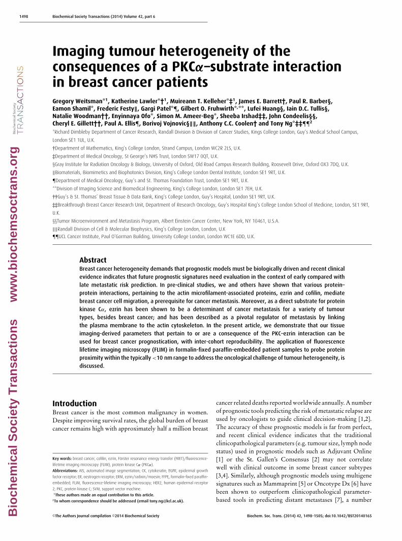
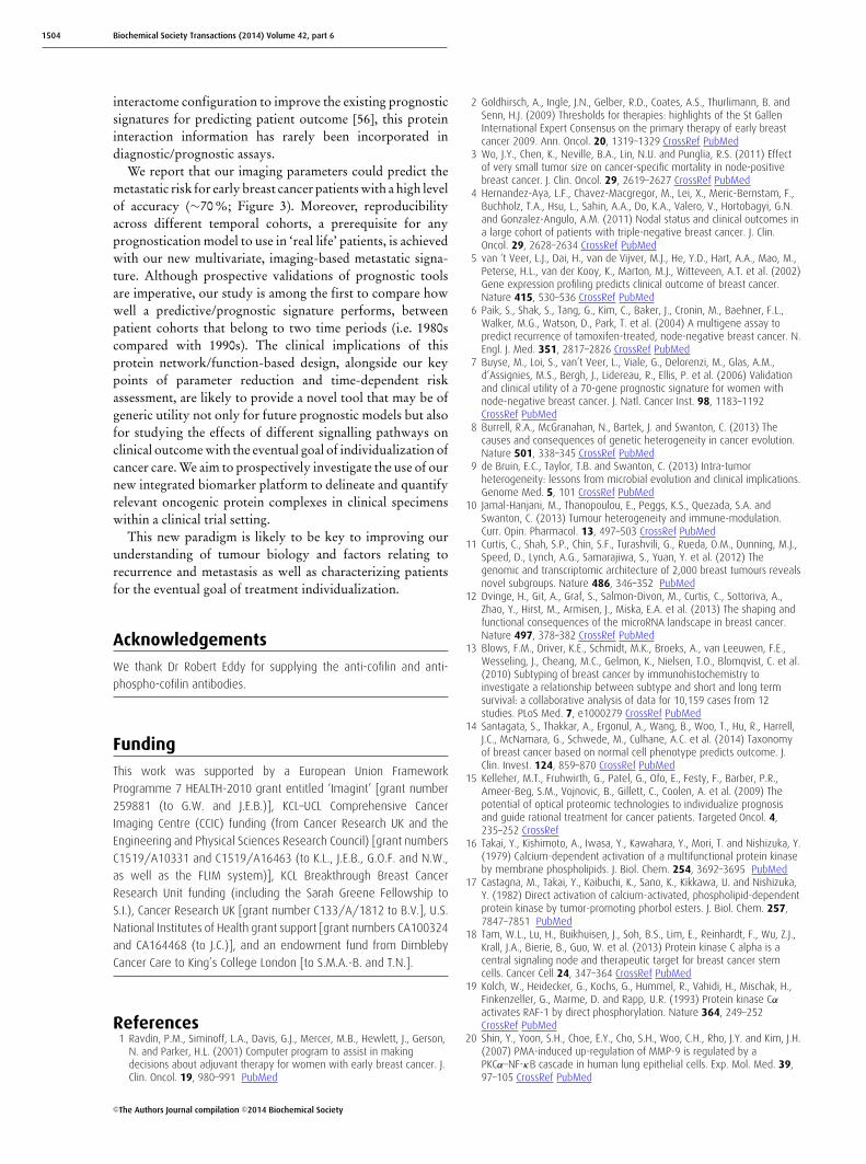
Figure 1 Imaging PKCα–ezrin interaction in FFPE samples
Representative images of breast cancer tissue stained with anti-ezrin IgG [labelled with Cy2 (A, C and D) or Alexa Fluor® 546
(B)] and anti-PKCα IgG [labelled with Cy3 (A, C and D) or Cy5 (B)]. (A and B) FRET/FLIM images show interaction between
proteins (decrease in lifetime, indicated by red pixels in the pseudocolour tumour map). (C and D) Utilization of images for
AIS algorithm to generate imaging parameters shown to the right of the images.
C©The Authors Journal compilation C©2014 Biochemical Society
Protein Kinase C Signalling in Health and Disease 1501
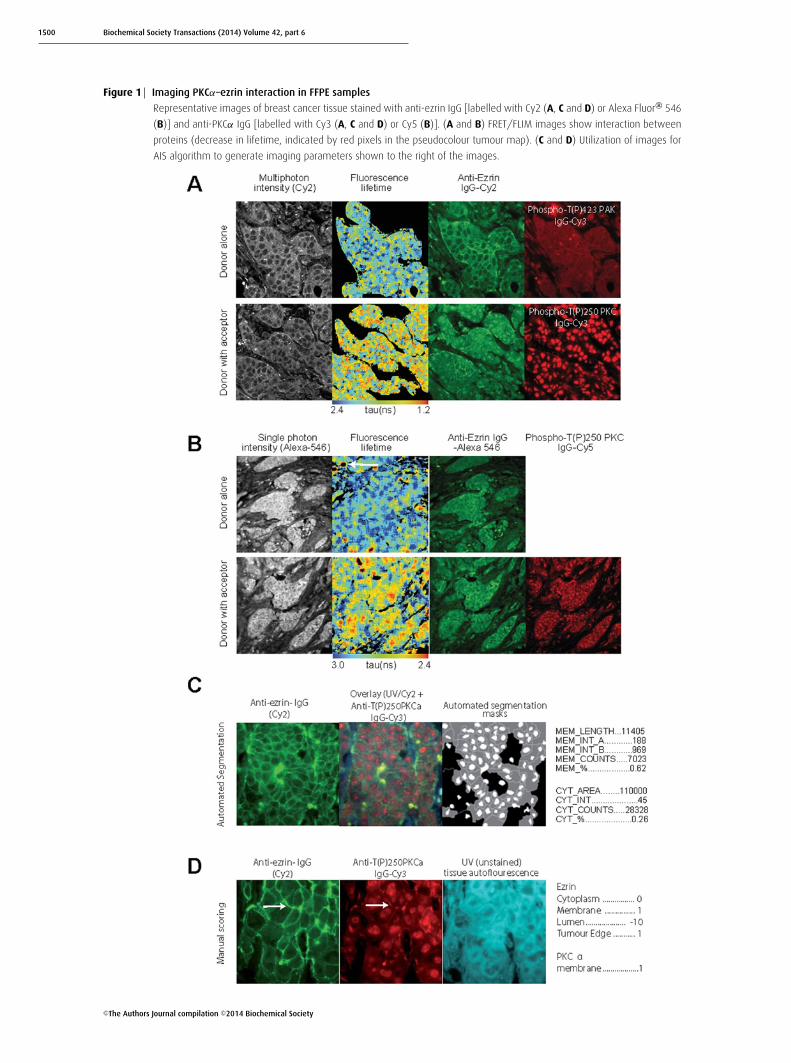
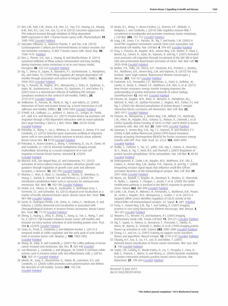
Figure 2 Imaging activation status of ezrin and cofilin in FFPE samples
Representative images of breast cancer tissue stained with anti-ezrin IgG–Cy2 and anti-phospho-ERM IgG–Cy3 (A); and with
anti-cofilin IgG–Cy2 and anti-phospho-cofilin IgG–Cy3 (B). Pseudocolour maps show higher co-localization intensities in one
sample (upper panel) and lower in another sample (low panel).
Specific FLIM-based ezrin–PKCα–proteininteraction detected in formalin-fixedparaffin-embedded (FFPE) tissuesTwo- (Figure 1A) and single- (Figure 1B) photon excitation-based acquisition of intermolecular FRET efficiency detectedspecific protein–protein interactions between ezrin and PKC(see the FLIM/FRET images of invasive breast carcinomasamples that were labelled with fluorescently conjugatedanti-ezrin IgG and anti-activated PKCα IgG). Comparisonof the corresponding FRET efficiencies measured with
either two- or single-photon excitation found no significantdifference between the two lifetime acquisition methods andtherefore single-photon excitation was chosen to acquire thesubsequent FLIM data. Although the immune/inflammatorycell infiltrate (see white arrow in Figure 1B) was autofluores-cent, this contributed little (since the number of pixels/areawas small proportionally) to the overall mean fluorescencelifetime per tumour. Similarly, the non-specific nuclearstaining of the acceptor fluorophore-labelled antibody (anti-activated PKCα IgG) did not interfere with the determination
C©The Authors Journal compilation C©2014 Biochemical Society
1502 Biochemical Society Transactions (2014) Volume 42, part 6
of FRET by FLIM [51,52], which is based on the short-ening of donor fluorescence lifetime of the donor fluorophoreused to label the anti-ezrin IgG.
Subcellular protein localization and/orphosphorylation quantificationEzrin–PKCα protein complex formation should result indownstream molecular events such as ezrin phosphorylation,redistribution and stabilization at the membrane [25].Figures 1 and 2 show ezrin stabilization at the membrane(Figures 1C and 1D), with concomitant ERM phosphoryla-tion (Figure 2A) and activation of PKCα (as shown byThr250 phosphorylation [53], Figure 1D), preferentially atthe membrane of invasive breast carcinoma cells (see whitearrow). The subcellular localization of proteins in tissuemicroarray cores was further quantified by automated imagesegmentation (AIS) and a manual scoring system. Nineimage parameters for the subcellular distribution of ezrin(Figure 1C) across heterogeneous breast tumours weregenerated in less than 5 s by AIS. The parallel ‘manual’ scoringsystem (Figure 1D) generated five parameters, describing thesubcellular compartment expression levels of both ezrin andPKCα in each tissue core. Further automated co-localizationanalyses demonstrated an increase in the total ezrin/phospho-ERM and cofilin/phospho-cofilin co-localization intensity atthe cell–cell borders and/or edges of invasive tumour cells(Figures 2A and 2B).
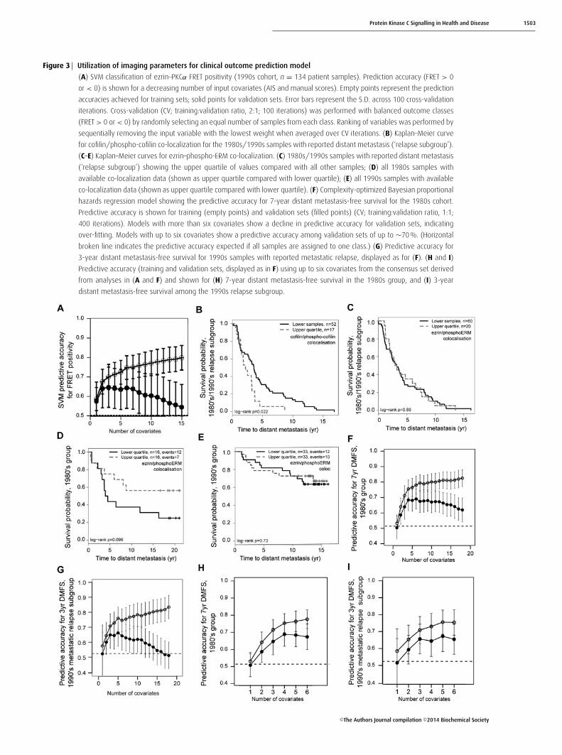
Selection of a consensus set ofimaging-based covariates for a metastaticpredictive modelWe next established that the non-FRET-based image para-meters (AIS or manual score) pertaining to ezrin and PKCwere associated with FRET positivity, which is a measureof ezrin–PKCα interaction. Ezrin–PKCα protein complexformation influences downstream molecular events such asezrin phosphorylation, redistribution and stabilization at themembrane, which are measured by the non-FRET-basedimage parameters [25]. Using single-photon FLIM-derivedFRET positivity (>0 %) to define a binary outcome, a supportvector machine (SVM) [54] predicted FRET positivity using acombination of the ezrin distribution (AIS and manual score)and phosphorylation parameters to an accuracy of ∼65 %(Figure 3A). A degree of overfitting was apparent when >
four covariates were used to build the SVM.Next, two independent breast cancer cohorts were imaged
for cofilin/phospho-cofilin and ezrin/phospho-ezrin co-localization, along with ezrin localization analysis using AIS(88 patient samples from the 1980s series and a total of134 patient samples from the 1990s series were availablefor evaluation). There is a high degree of heterogeneitybetween the two cohorts (the adjuvant treatments for thetwo cohorts differed significantly: 1980s compared with1990s; chemotherapy 0 % compared with 30 %; endocrine
36 % compared with 76 %, respectively). In exploratoryunivariate analysis, the upper quartile of cofilin/phospho-cofilin co-localization intensities was associated with earlydistant metastasis among patients with reported relapse[Figure 3B; upper quartile (n = 17) compared with lowervalues (n = 52); P = 0.022, log-rank test]. This was not thecase for ezrin/phospho-ezrin co-localization (Figure 3C);furthermore, exploratory analysis of lower and upperquartiles indicated that lower quartile ezrin/phospho-ERMco-localization may be associated with poorer distantmetastasis-free survival in the 1980s series (Figure 3D;P = 0.098, not significant, log-rank test) but not the 1990sseries (Figure 3E; P = 0.73, not significant, log-rank test).
The predictive accuracy of all 18 covariates (16 imagingparameters, Figures 1C and 1D, and whether or not thepatient had received treatments: tamoxifen or chemotherapy)was assessed for distant metastasis-free survival using aBayesian proportional hazards regression model with cross-validation for the two temporal cohorts (1980s and 1990s).A step-down procedure was used to iteratively reduce thenumber of covariates in the model, and over-fitting wasobserved when greater than six covariates were used to buildthe model. The top six covariates were identified for the 1980sand 1990s (relapse) cohorts. The predictive accuracy (bothtraining and validation) for 7-year distant metastasis-freesurvival in the 1980s cohort is shown in Figures 3(F) and 3(G).
There was an overlap between the top four covariates inthe SVM analysis for FRET positivity (Figure 3A), and thetwo separate lists of the top six covariates for predictingmetastatic relapse. On the basis of this overlap with SVManalysis, a final consensus set of six covariates (averagemembrane intensity, relative and absolute; cytoplasm averageintensity and number of pixels inside cytoplasm, manualezrin cytoplasm score and membrane length) was selectedand confirmed by re-running the Bayesian proportionalhazards regression against each of the two patient cohorts.For the 1980s cohort, the model predicted 7-year distantmetastasis-free survival to an accuracy of up to ∼70 %(Figure 3H). The same consensus set of covariates was foundto be predictive for early relapse (within 3 years) amongpatients with reported distant metastases in the 1990s cohort(Figure 3I).
ConclusionsThe remarkable diversity in breast cancer dictates thatprognostic models must be biologically driven. We describethe first optical imaging-based tumour metastatic signature,measuring underlying biological variables which are pertinentin tumour metastases. Our semi-automated tissue imagingplatform is capable of performing an integrated analysisof protein phosphorylation, protein–protein interactionand subcellular protein expression/distribution, using FFPEtissue microarrays. Incorporation of protein interaction datawas shown to also improve the predictive performance ofprognostic gene expression signatures [55,56]. Despite theimportance of adjunct information supplied by the protein
C©The Authors Journal compilation C©2014 Biochemical Society
Protein Kinase C Signalling in Health and Disease 1503
Figure 3 Utilization of imaging parameters for clinical outcome prediction model
(A) SVM classification of ezrin–PKCα FRET positivity (1990s cohort, n = 134 patient samples). Prediction accuracy (FRET > 0
or < 0) is shown for a decreasing number of input covariates (AIS and manual scores). Empty points represent the prediction
accuracies achieved for training sets; solid points for validation sets. Error bars represent the S.D. across 100 cross-validation
iterations. Cross-validation (CV; training:validation ratio, 2:1; 100 iterations) was performed with balanced outcome classes
(FRET > 0 or < 0) by randomly selecting an equal number of samples from each class. Ranking of variables was performed by
sequentially removing the input variable with the lowest weight when averaged over CV iterations. (B) Kaplan–Meier curve
for cofilin/phospho-cofilin co-localization for the 1980s/1990s samples with reported distant metastasis (‘relapse subgroup’).
(C–E) Kaplan–Meier curves for ezrin–phospho-ERM co-localization. (C) 1980s/1990s samples with reported distant metastasis
(‘relapse subgroup’) showing the upper quartile of values compared with all other samples; (D) all 1980s samples with
available co-localization data (shown as upper quartile compared with lower quartile); (E) all 1990s samples with available
co-localization data (shown as upper quartile compared with lower quartile). (F) Complexity-optimized Bayesian proportional
hazards regression model showing the predictive accuracy for 7-year distant metastasis-free survival for the 1980s cohort.
Predictive accuracy is shown for training (empty points) and validation sets (filled points) (CV; training:validation ratio, 1:1;
400 iterations). Models with more than six covariates show a decline in predictive accuracy for validation sets, indicating
over-fitting. Models with up to six covariates show a predictive accuracy among validation sets of up to ∼70 %. (Horizontal
broken line indicates the predictive accuracy expected if all samples are assigned to one class.) (G) Predictive accuracy for
3-year distant metastasis-free survival for 1990s samples with reported metastatic relapse, displayed as for (F). (H and I)
Predictive accuracy (training and validation sets, displayed as in F) using up to six covariates from the consensus set derived
from analyses in (A and F) and shown for (H) 7-year distant metastasis-free survival in the 1980s group, and (I) 3-year
distant metastasis-free survival among the 1990s relapse subgroup.
C©The Authors Journal compilation C©2014 Biochemical Society
1504 Biochemical Society Transactions (2014) Volume 42, part 6
interactome configuration to improve the existing prognosticsignatures for predicting patient outcome [56], this proteininteraction information has rarely been incorporated indiagnostic/prognostic assays.
We report that our imaging parameters could predict themetastatic risk for early breast cancer patients with a high levelof accuracy (∼70 %; Figure 3). Moreover, reproducibilityacross different temporal cohorts, a prerequisite for anyprognostication model to use in ‘real life’ patients, is achievedwith our new multivariate, imaging-based metastatic signa-ture. Although prospective validations of prognostic toolsare imperative, our study is among the first to compare howwell a predictive/prognostic signature performs, betweenpatient cohorts that belong to two time periods (i.e. 1980scompared with 1990s). The clinical implications of thisprotein network/function-based design, alongside our keypoints of parameter reduction and time-dependent riskassessment, are likely to provide a novel tool that may be ofgeneric utility not only for future prognostic models but alsofor studying the effects of different signalling pathways onclinical outcome with the eventual goal of individualization ofcancer care. We aim to prospectively investigate the use of ournew integrated biomarker platform to delineate and quantifyrelevant oncogenic protein complexes in clinical specimenswithin a clinical trial setting.
This new paradigm is likely to be key to improving ourunderstanding of tumour biology and factors relating torecurrence and metastasis as well as characterizing patientsfor the eventual goal of treatment individualization.
Acknowledgements
We thank Dr Robert Eddy for supplying the anti-cofilin and anti-
phospho-cofilin antibodies.
Funding
This work was supported by a European Union Framework
Programme 7 HEALTH-2010 grant entitled ‘Imagint’ [grant number
259881 (to G.W. and J.E.B.)], KCL–UCL Comprehensive Cancer
Imaging Centre (CCIC) funding (from Cancer Research UK and the
Engineering and Physical Sciences Research Council) [grant numbers
C1519/A10331 and C1519/A16463 (to K.L., J.E.B., G.O.F. and N.W.,
as well as the FLIM system)], KCL Breakthrough Breast Cancer
Research Unit funding (including the Sarah Greene Fellowship to
S.I.), Cancer Research UK [grant number C133/A/1812 to B.V.], U.S.
National Institutes of Health grant support [grant numbers CA100324
and CA164468 (to J.C.)], and an endowment fund from Dimbleby
Cancer Care to King’s College London [to S.M.A.-B. and T.N.].
References1 Ravdin, P.M., Siminoff, L.A., Davis, G.J., Mercer, M.B., Hewlett, J., Gerson,
N. and Parker, H.L. (2001) Computer program to assist in makingdecisions about adjuvant therapy for women with early breast cancer. J.Clin. Oncol. 19, 980–991 PubMed
2 Goldhirsch, A., Ingle, J.N., Gelber, R.D., Coates, A.S., Thurlimann, B. andSenn, H.J. (2009) Thresholds for therapies: highlights of the St GallenInternational Expert Consensus on the primary therapy of early breastcancer 2009. Ann. Oncol. 20, 1319–1329 CrossRef PubMed
3 Wo, J.Y., Chen, K., Neville, B.A., Lin, N.U. and Punglia, R.S. (2011) Effectof very small tumor size on cancer-specific mortality in node-positivebreast cancer. J. Clin. Oncol. 29, 2619–2627 CrossRef PubMed
4 Hernandez-Aya, L.F., Chavez-Macgregor, M., Lei, X., Meric-Bernstam, F.,Buchholz, T.A., Hsu, L., Sahin, A.A., Do, K.A., Valero, V., Hortobagyi, G.N.and Gonzalez-Angulo, A.M. (2011) Nodal status and clinical outcomes ina large cohort of patients with triple-negative breast cancer. J. Clin.Oncol. 29, 2628–2634 CrossRef PubMed
5 van ‘t Veer, L.J., Dai, H., van de Vijver, M.J., He, Y.D., Hart, A.A., Mao, M.,Peterse, H.L., van der Kooy, K., Marton, M.J., Witteveen, A.T. et al. (2002)Gene expression profiling predicts clinical outcome of breast cancer.Nature 415, 530–536 CrossRef PubMed
6 Paik, S., Shak, S., Tang, G., Kim, C., Baker, J., Cronin, M., Baehner, F.L.,Walker, M.G., Watson, D., Park, T. et al. (2004) A multigene assay topredict recurrence of tamoxifen-treated, node-negative breast cancer. N.Engl. J. Med. 351, 2817–2826 CrossRef PubMed
7 Buyse, M., Loi, S., van’t Veer, L., Viale, G., Delorenzi, M., Glas, A.M.,d’Assignies, M.S., Bergh, J., Lidereau, R., Ellis, P. et al. (2006) Validationand clinical utility of a 70-gene prognostic signature for women withnode-negative breast cancer. J. Natl. Cancer Inst. 98, 1183–1192CrossRef PubMed
8 Burrell, R.A., McGranahan, N., Bartek, J. and Swanton, C. (2013) Thecauses and consequences of genetic heterogeneity in cancer evolution.Nature 501, 338–345 CrossRef PubMed
9 de Bruin, E.C., Taylor, T.B. and Swanton, C. (2013) Intra-tumorheterogeneity: lessons from microbial evolution and clinical implications.Genome Med. 5, 101 CrossRef PubMed
10 Jamal-Hanjani, M., Thanopoulou, E., Peggs, K.S., Quezada, S.A. andSwanton, C. (2013) Tumour heterogeneity and immune-modulation.Curr. Opin. Pharmacol. 13, 497–503 CrossRef PubMed
11 Curtis, C., Shah, S.P., Chin, S.F., Turashvili, G., Rueda, O.M., Dunning, M.J.,Speed, D., Lynch, A.G., Samarajiwa, S., Yuan, Y. et al. (2012) Thegenomic and transcriptomic architecture of 2,000 breast tumours revealsnovel subgroups. Nature 486, 346–352 PubMed
12 Dvinge, H., Git, A., Graf, S., Salmon-Divon, M., Curtis, C., Sottoriva, A.,Zhao, Y., Hirst, M., Armisen, J., Miska, E.A. et al. (2013) The shaping andfunctional consequences of the microRNA landscape in breast cancer.Nature 497, 378–382 CrossRef PubMed
13 Blows, F.M., Driver, K.E., Schmidt, M.K., Broeks, A., van Leeuwen, F.E.,Wesseling, J., Cheang, M.C., Gelmon, K., Nielsen, T.O., Blomqvist, C. et al.(2010) Subtyping of breast cancer by immunohistochemistry toinvestigate a relationship between subtype and short and long termsurvival: a collaborative analysis of data for 10,159 cases from 12studies. PLoS Med. 7, e1000279 CrossRef PubMed
14 Santagata, S., Thakkar, A., Ergonul, A., Wang, B., Woo, T., Hu, R., Harrell,J.C., McNamara, G., Schwede, M., Culhane, A.C. et al. (2014) Taxonomyof breast cancer based on normal cell phenotype predicts outcome. J.Clin. Invest. 124, 859–870 CrossRef PubMed
15 Kelleher, M.T., Fruhwirth, G., Patel, G., Ofo, E., Festy, F., Barber, P.R.,Ameer-Beg, S.M., Vojnovic, B., Gillett, C., Coolen, A. et al. (2009) Thepotential of optical proteomic technologies to individualize prognosisand guide rational treatment for cancer patients. Targeted Oncol. 4,235–252 CrossRef
16 Takai, Y., Kishimoto, A., Iwasa, Y., Kawahara, Y., Mori, T. and Nishizuka, Y.(1979) Calcium-dependent activation of a multifunctional protein kinaseby membrane phospholipids. J. Biol. Chem. 254, 3692–3695 PubMed
17 Castagna, M., Takai, Y., Kaibuchi, K., Sano, K., Kikkawa, U. and Nishizuka,Y. (1982) Direct activation of calcium-activated, phospholipid-dependentprotein kinase by tumor-promoting phorbol esters. J. Biol. Chem. 257,7847–7851 PubMed
18 Tam, W.L., Lu, H., Buikhuisen, J., Soh, B.S., Lim, E., Reinhardt, F., Wu, Z.J.,Krall, J.A., Bierie, B., Guo, W. et al. (2013) Protein kinase C alpha is acentral signaling node and therapeutic target for breast cancer stemcells. Cancer Cell 24, 347–364 CrossRef PubMed
19 Kolch, W., Heidecker, G., Kochs, G., Hummel, R., Vahidi, H., Mischak, H.,Finkenzeller, G., Marme, D. and Rapp, U.R. (1993) Protein kinase Cαactivates RAF-1 by direct phosphorylation. Nature 364, 249–252CrossRef PubMed
20 Shin, Y., Yoon, S.H., Choe, E.Y., Cho, S.H., Woo, C.H., Rho, J.Y. and Kim, J.H.(2007) PMA-induced up-regulation of MMP-9 is regulated by aPKCα–NF-κB cascade in human lung epithelial cells. Exp. Mol. Med. 39,97–105 CrossRef PubMed
C©The Authors Journal compilation C©2014 Biochemical Society
Protein Kinase C Signalling in Health and Disease 1505
21 Kim, J.M., Noh, E.M., Kwon, K.B., Kim, J.S., You, Y.O., Hwang, J.K., Hwang,B.M., Kim, B.S., Lee, S.H., Lee, S.J. et al. (2012) Curcumin suppresses theTPA-induced invasion through inhibition of PKCα-dependentMMP-expression in MCF-7 human breast cancer cells. Phytomedicine 19,1085–1092 CrossRef PubMed
22 Gonzalez-Villasana, V., Gutierrez-Puente, Y. and Tari, A.M. (2013)Cyclooxygenase-2 utilizes Jun N-terminal kinases to induce invasion, butnot tamoxifen resistance, in MCF-7 breast cancer cells. Oncol. Rep. 30,1506–1510 PubMed
23 Kim, J., Thorne, S.H., Sun, L., Huang, B. and Mochly-Rosen, D. (2011)Sustained inhibition of PKCα reduces intravasation and lung seedingduring mammary tumor metastasis in an in vivo mouse model.Oncogene 30, 323–333 CrossRef PubMed
24 Ng, T., Shima, D., Squire, A., Bastiaens, P.I., Gschmeissner, S., Humphries,M.J. and Parker, P.J. (1999) PKCα regulates β1 integrin-dependent cellmotility through association and control of integrin traffic. EMBO J. 18,3909–3923 CrossRef PubMed
25 Ng, T., Parsons, M., Hughes, W.E., Monypenny, J., Zicha, D., Gautreau, A.,Arpin, M., Gschmeissner, S., Verveer, P.J., Bastiaens, P.I. and Parker, P.J.(2001) Ezrin is a downstream effector of trafficking PKC–integrincomplexes involved in the control of cell motility. EMBO J. 20,2723–2741 CrossRef PubMed
26 Anilkumar, N., Parsons, M., Monk, R., Ng, T. and Adams, J.C. (2003)Interaction of fascin and protein kinase Cα: a novel intersection in celladhesion and motility. EMBO J. 22, 5390–5402 CrossRef PubMed
27 O’Neill, A.K., Gallegos, L.L., Justilien, V., Garcia, E.L., Leitges, M., Fields,A.P., Hall, R.A. and Newton, A.C. (2011) Protein kinase Cα promotes cellmigration through a PDZ-dependent interaction with its novel substratediscs large homolog 1 (DLG1). J. Biol. Chem. 286, 43559–43568CrossRef PubMed
28 Patsialou, A., Wang, Y., Lin, J., Whitney, K., Goswami, S., Kenny, P.A. andCondeelis, J.S. (2012) Selective gene-expression profiling of migratorytumor cells in vivo predicts clinical outcome in breast cancer patients.Breast Cancer Res. 14, R139 CrossRef PubMed
29 Patsialou, A., Bravo-Cordero, J., Wang, Y, Entenberg, D., Liu, H., Clarke, M.and Condeelis, J.C. (2013) Intravital multiphoton imaging revealsmulticellular streaming as a crucial component of in vivo cellmigration in human breast tumors. Intravital 2, e25294CrossRef PubMed
30 Marsick, B.M., San Miguel-Ruiz, J.E. and Letourneau, P.C. (2012)Activation of ezrin/radixin/moesin mediates attractive growth coneguidance through regulation of growth cone actin and adhesionreceptors. J. Neurosci. 32, 282–296 CrossRef PubMed
31 Khanna, C., Wan, X., Bose, S., Cassaday, R., Olomu, O., Mendoza, A.,Yeung, C., Gorlick, R., Hewitt, S.M. and Helman, L.J. (2004) Themembrane–cytoskeleton linker ezrin is necessary for osteosarcomametastasis. Nat. Med. 10, 182–186 CrossRef PubMed
32 Arslan, A.A., Silvera, D., Arju, R., Giashuddin, S., Belitskaya-Levy, I.,Formenti, S.C. and Schneider, R.J. (2012) Atypical ezrin localization as amarker of locally advanced breast cancer. Breast Cancer Res. Treat. 134,981–988 CrossRef PubMed
33 Sarrio, D., Rodriguez-Pinilla, S.M., Dotor, A., Calero, F., Hardisson, D. andPalacios, J. (2006) Abnormal ezrin localization is associated withclinicopathological features in invasive breast carcinomas. Breast CancerRes. Treat. 98, 71–79 CrossRef PubMed
34 Zheng, S., Huang, J., Zhou, K., Zhang, C., Xiang, Q., Tan, Z., Wang, T. andFu, X. (2011) 17β-Estradiol enhances breast cancer cell motility andinvasion via extra-nuclear activation of actin-binding protein ezrin. PLoSONE 6, e22439 CrossRef PubMed
35 Tania, N., Prosk, E., Condeelis, J. and Edelstein-Keshet, L. (2011) Atemporal model of cofilin regulation and the early peak of actin barbedends in invasive tumor cells. Biophys. J. 100, 1883–1892CrossRef PubMed
36 Wang, W., Eddy, R. and Condeelis, J. (2007) The cofilin pathway in breastcancer invasion and metastasis. Nat. Rev. 7, 429–440 CrossRef
37 van Rheenen, J., Condeelis, J. and Glogauer, M. (2009) A common cofilinactivity cycle in invasive tumor cells and inflammatory cells. J. Cell Sci.122, 305–311 CrossRef PubMed
38 Ghosh, M., Song, X., Mouneimne, G., Sidani, M., Lawrence, D.S. andCondeelis, J.S. (2004) Cofilin promotes actin polymerization and definesthe direction of cell motility. Science 304, 743–746CrossRef PubMed
39 Beaty, B.T., Wang, Y., Bravo-Cordero, J.J., Sharma, V.P., Miskolci, V.,Hodgson, L. and Condeelis, J. (2014) Talin regulates moesin–NHE-1recruitment to invadopodia and promotes mammary tumor metastasis.J. Cell Biol. 205, 737–751 CrossRef PubMed
40 Legg, J.W., Lewis, C.A., Parsons, M., Ng, T. and Isacke, C.M. (2002) Anovel PKC-regulated mechanism controls CD44 ezrin association anddirectional cell motility. Nat. Cell Biol. 4, 399–407 CrossRef PubMed
41 Prag, S., Parsons, M., Keppler, M.D., Ameer-Beg, S.M., Barber, P., Hunt, J.,Beavil, A.J., Calvert, R., Arpin, M., Vojnovic, B. and Ng, T. (2007) Activatedezrin promotes cell migration through recruitment of the GEF Dbl to lipidrafts and preferential downstream activation of Cdc42. Mol. Biol. Cell 18,2935–2948 CrossRef PubMed
42 Barber, P.R., Tullis, I.D., Pierce, G.P., Newman, R.G., Prentice, J., Rowley,M.I., Matthews, D.R., Ameer-Beg, S.M. and Vojnovic, B. (2013) The GrayInstitute ‘open’ high-content, fluorescence lifetime microscopes. J.Microsc. 251, 154–167 CrossRef PubMed
43 Fruhwirth, G.O., Fernandes, L.P., Weitsman, G., Patel, G., Kelleher, M.,Lawler, K., Brock, A., Poland, S.P., Matthews, D.R., Keri, G. et al. (2011)How Forster resonance energy transfer imaging improves theunderstanding of protein interaction networks in cancer biology.ChemPhysChem 12, 442–461 CrossRef PubMed
44 Parsons, M., Keppler, M.D., Kline, A., Messent, A., Humphries, M.J.,Gilchrist, R., Hart, I.R., Quittau-Prevostel, C., Hughes, W.E., Parker, P.J. andNg, T. (2002) Site-directed perturbation of protein kinase C–integrininteraction blocks carcinoma cell chemotaxis. Mol. Cell. Biol. 22,5897–5911 CrossRef PubMed
45 Parsons, M., Monypenny, J., Ameer-Beg, S.M., Millard, T.H., Machesky,L.M., Peter, M., Keppler, M.D., Schiavo, G., Watson, R., Chernoff, J. et al.(2005) Spatially distinct binding of Cdc42 to PAK1 and N-WASP in breastcarcinoma cells. Mol. Cell. Biol. 25, 1680–1695 CrossRef PubMed
46 Ganesan, S., Ameer-Beg, S.M., Ng, T.T., Vojnovic, B. and Wouters, F.S.(2006) A dark yellow fluorescent protein (YFP)-based resonanceenergy-accepting chromoprotein (REACh) for Forster resonance energytransfer with GFP. Proc. Natl. Acad. Sci. U.S.A. 103, 4089–4094CrossRef PubMed
47 Dadke, S., Cotteret, S., Yip, S.C., Jaffer, Z.M., Haj, F., Ivanov, A., Rauscher,III, F., Shuai, K., Ng, T., Neel, B.G. and Chernoff, J. (2007) Regulation ofprotein tyrosine phosphatase 1B by sumoylation. Nat. Cell Biol. 9, 80–85CrossRef PubMed
48 Makrogianneli, K., Carlin, L.M., Keppler, M.D., Matthews, D.R., Ofo, E.,Coolen, A., Ameer-Beg, S.M., Barber, P.R., Vojnovic, B. and Ng, T. (2009)Integrating receptor signal inputs that influence small Rho GTPaseactivation dynamics at the immunological synapse. Mol. Cell. Biol. 29,2997–3006 CrossRef PubMed
49 Morris, J.R., Boutell, C., Keppler, M., Densham, R., Weekes, D., Alamshah,A., Butler, L., Galanty, Y., Pangon, L., Kiuchi, T. et al. (2009) The SUMOmodification pathway is involved in the BRCA1 response to genotoxicstress. Nature 462, 886–890 CrossRef PubMed
50 Carlin, L.M., Evans, R., Milewicz, H., Fernandes, L., Matthews, D.R., Perani,M., Levitt, J., Keppler, M.D., Monypenny, J., Coolen, T. et al. (2011) Atargeted siRNA screen identifies regulators of Cdc42 activity at thenatural killer cell immunological synapse. Sci. Signal. 4, ra81 PubMed
51 Festy, F., Ameer-Beg, S.M., Ng, T. and Suhling, K. (2007) Imagingproteins in vivo using fluorescence lifetime microscopy. Mol. Biosyst. 3,381–391 CrossRef PubMed
52 Wouters, F.S., Verveer, P.J. and Bastiaens, P.I. (2001) Imagingbiochemistry inside cells. Trends Cell Biol. 11, 203–211 CrossRef PubMed
53 Ng, T., Squire, A., Hansra, G., Bornancin, F., Prevostel, C., Hanby, A.,Harris, W., Barnes, D., Schmidt, S., Mellor, H. et al. (1999) Imaging proteinkinase Cα activation in cells. Science 283, 2085–2089 CrossRef PubMed
54 Chang, C.C. and Lin, C.J. (2001) Training nu-support vector classifiers:theory and algorithms. Neural Comput. 13, 2119–2147 CrossRef PubMed
55 Chuang, H.Y., Lee, E., Liu, Y.T., Lee, D. and Ideker, T. (2007)Network-based classification of breast cancer metastasis. Mol. Syst. Biol.3, 140 CrossRef PubMed
56 Taylor, I.W., Linding, R., Warde-Farley, D., Liu, Y., Pesquita, C., Faria, D.,Bull, S., Pawson, T., Morris, Q. and Wrana, J.L. (2009) Dynamic modularityin protein interaction networks predicts breast cancer outcome. Nat.Biotechnol. 27, 199–204 CrossRef PubMed
Received 12 June 2014doi:10.1042/BST20140165
C©The Authors Journal compilation C©2014 Biochemical Society