Embed Size (px)
Citation preview

Imaging Surveillance in Women with a History of Treated Breast Cancer
Wei Tse Yang, M.D.

Breast Cancer
1. Extent 2. Response 3. Recurrence

Surveillance Breast Cancer
1. Extent 2. Response


Surveillance Breast Cancer
1. Extent 2. Response 3. Recurrence

Courtesy Wendie Berg, MD, PhD
62-year-old woman Left BCT T2N0 ER(+) IDC + chemo 1997 New right ILC 2011

Courtesy Wendie Berg, MD, PhD Courtesy Wendie Berg, MD, PhD
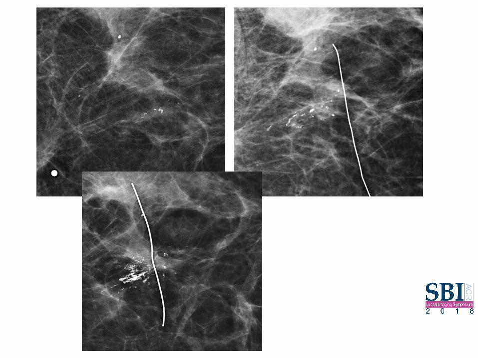
Followup at 18 months post APBI treatment (same patient) right breast
Grade 2 IDC-ILC, ER/PR(+), HER2 eq
Women with PHBC
Population increasing • Gains life expectancy • Population breast screening • Improved cancer Tx
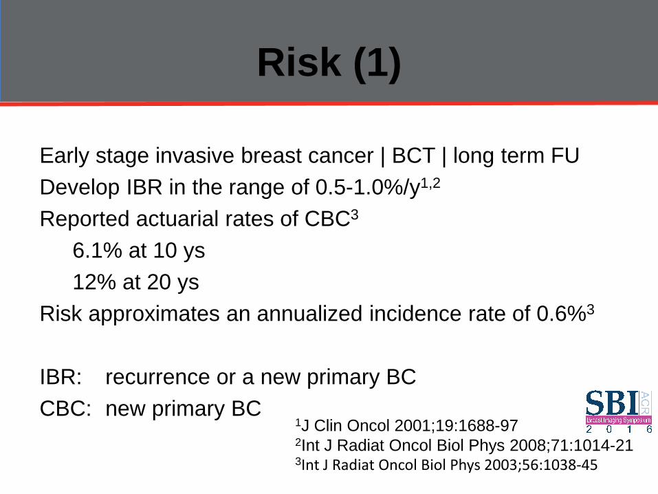
Risk (1)
Early stage invasive breast cancer | BCT | long term FU Develop IBR in the range of 0.5-1.0%/y1,2
Reported actuarial rates of CBC3
6.1% at 10 ys 12% at 20 ys Risk approximates an annualized incidence rate of 0.6%3
IBR: recurrence or a new primary BC CBC: new primary BC
1J Clin Oncol 2001;19:1688-97 2Int J Radiat Oncol Biol Phys 2008;71:1014-21 3Int J Radiat Oncol Biol Phys 2003;56:1038-45

Risk (2)
The risk of developing a further breast cancer in the treated/previously unaffected breast varies according to: • Tumor • Therapeutic variables Associated with the first breast cancer
Int J Radiat Oncol Biol Phys. 56(4):1038-45, 2003 Breast Cancer Res Treat. 129(3):963-9, 2011

Surveillance women with PHBC
Identify and manage health and QOL issues related to BC and its therapy of women previously treated (non-metastatic) initial dx stage I-II BC General consensus • Should have FU & screening mammography • Frequency and duration of mammographic
surveillance varies in guidelines and practice
J Clin Oncol 2004;22:4010-8 Br J Cancer 2007;96:1625-32 Br J Cancer 2007;96:1632-41

Women with PHBC
Screening mammography Breast density Frequency surveillance Screening MRI Alternate strategies

Abstract Context—Women with a personal history of breast cancer (PHBC) are at risk of developing another breast cancer and are recommended for screening mammography. Few high-quality data exist on screening performance in PHBC women. Objective—To examine the accuracy and outcomes of mammography screening in PHBC women relative to screening of similar women without PHBC. Design and Setting—Cohort of PHBC women, mammogram matched to non-PHBC women, screened through facilities (1996–2007) affiliated with the Breast Cancer Surveillance Consortium. Participants—There were 58 870 screening mammograms in 19 078 women with a history of early-stage (in situ or stage I-II invasive) breast cancer and 58 870 matched (breast density, age group, mammography year, and registry) screening mammograms in 55 315 non-PHBC women. Main Outcome Measures—Mammography accuracy based on final assessment, cancer detection rate, interval cancer rate, and stage at diagnosis. Results—Within 1 year after screening, 655 cancers were observed in PHBC women (499 invasive, 156 in situ) and 342 cancers (285 invasive, 57 in situ) in non-PHBC women. Screening accuracy and outcomes in PHBC relative to non-PHBC women were cancer rates of 10.5 per 1000 screens (95%CI, 9.7–11.3) vs 5.8 per 1000 screens (95%CI, 5.2–6.4), cancer detection rate of 6.8 per 1000 screens (95%CI, 6.2–7.5) vs 4.4 per 1000 screens (95% CI, 3.9–5.0), interval cancer rate of 3.6 per 1000 screens (95%CI, 3.2–4.1) vs 1.4 per 1000 screens (95% CI, 1.1–1.7), sensitivity 65.4% (95% CI, 61.5%–69.0%) vs 76.5% (95% CI, 71.7%–80.7%), specificity 98.3% (95% CI, 98.2%–98.4%) vs 99.0% (95% CI, 98.9%–99.1%), abnormal mammogram results in 2.3% (95% CI, 2.2%–2.5%) vs 1.4% (95% CI, 1.3%–1.5%) (all comparisons P <.001). Screening sensitivity in PHBC women was higher for detection of in situ cancer (78.7%;95% CI, 71.4%–84.5%) than invasive cancer (61.1%; 95% CI, 56.6%–65.4%), P<.001; lower in the initial 5 years (60.2%; 95% CI, 54.7%–65.5%) than after 5 years from first cancer (70.8%;95% CI, 65.4%–75.6%), P =.006; and was similar for detection of ipsilateral cancer (66.3%; 95% CI, 60.3%–71.8%) and contralateral cancer (66.1%; 95% CI, 60.9%–70.9%), P=.96. Screen-detected and interval cancers in women with and without PHBC were predominantly early stage. Conclusion—Mammography screening in PHBC women detects early-stage second breast cancers but has lower sensitivity and higher interval cancer rate, despite more evaluation and higher underlying cancer rate, relative to that in non-PHBC women.
JAMA. 2011 February 23; 305(8): 790–799. © 2011 American Medical Association. All rights reserved.

Screening Mammography PHBC
Cohort study: Breast Cancer Surveillance Consortium (1996-2007) breast cancer diagnosis, diagnosis date, cancer characteristics PHBC women – mammogram matched to non-PHBC women
JAMA 2011 Feb 23; 305(8): 790-799
Screening Mammography PHBC
Outcome measures accuracy • final assessment • interval cancer detection rate • stage at diagnosis
Screening Mammography PHBC
58 870 screening mammograms 19 078 women early, Tis Stage I-II invasive BC 58 870 matched screening mammograms [breast density, age, mammography year] 55 315 non-PHBC women

Results (1)
PHBC: 655 cancers (499 inv, 156 is) Non-PHBC: 342 cancers (285 inv, 57 is) All results PHBC vs non-PHBC (P<.001) Cancer rates: 10.5 per 1000 screens (95% CI, 9.7-11.3) vs 5.8 per 1000 screens (95% CI, 5.2-6.4) Cancer detection rates: 6.8 per 1000 screens (95% CI, 6.2-7.5) vs 4.4 per 1000 screens (95% CI, 3.9-5.0)

Results (2)
Sensitivity: 65.4% (95%CI, 61.5%-69%) vs 76.5% (95% CI, 71.7%-80.7%) Interval cancer rate: 3.6 per 1 000 screens (95% CI, 3.2-4.1) vs 1.4 per 1 000 screens (95% CI, 1.1-1.7)

Screening Mammography PHBC
• detects early-stage second breast cancers • lower sensitivity • higher interval cancer rate despite more evaluation and higher underlying cancer rate relative to that in non- PHBC women
Results (3)
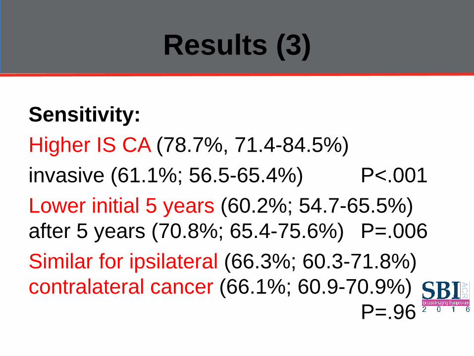
Sensitivity: Higher IS CA (78.7%, 71.4-84.5%) invasive (61.1%; 56.5-65.4%) P<.001 Lower initial 5 years (60.2%; 54.7-65.5%) after 5 years (70.8%; 65.4-75.6%) P=.006 Similar for ipsilateral (66.3%; 60.3-71.8%) contralateral cancer (66.1%; 60.9-70.9%) P=.96

Meta-analyses
2263 subjects (13 studies) with recurrences Pooled analyses: IBR CBC regional distant mets Effect early detection potentially underestimated 58% recurrences detected early Early detection: improved survival of women who experienced BC recurrence Hazard ratio for late vs early detection of relapse: 1.68 (1.48-1.91)
Breast Cancer Res Treat 2009;114:403-12
Screening Mammography PHBC
Evidence based only studies on early detection of IBR/CBC (n=10) **various surveillance strategies that include mammography Study-specific HR: 0.1-0.86 Beneficial effect in the range of 90%-14% relative reduction in the hazard of BC death
Breast Cancer Res Treat 2009;114:403-12

Screening Mammography PHBC
Nested case-control; 65 years and older 1846 | Stage I-II invasive BC Surveillance & mortality Recurrence (B/N/DM) who died of BC (first 5ys of FU) compared with women who did not die Protective association with M:OR = 0.69; 0.53-0.92 Each additional screening M 0.7x decrease in conditional odds for BC mortality *Effect most evident stage I
J Clin Oncol 2007;25:3001-6

Screening Mammography PHBC
Houssami 1044 asymptomatic second BCs a/w • Earlier detection • More favorable stage • Smaller IBR (P<0.001), CBC (P<0.001) • Fewer nodal metastases (P=0.0001) • HR: 0.53
Ann Oncol 2009;20:1505-10

Screening Mammography PHBC
Variability in how the accuracy of mammography interpretation is rendered • Type clinical events considered in analysis IBR/CBC/Both/Regional nodal recurrences • Data for M-only detection or any method
detection
Screening M Surveillance PHBC
Early detection IBC/CBC may not confer the anticipated benefit if the risk of mortality is large, or partly determined by the first breast cancer Specificity data sparse (unselected women with PHBC) Sensitivity over-estimated

Assumption of benefit
No RCTs examining the impact on mortality Non-randomized studies: lead-time and length-time bias (over-estimate the benefit from screening) Measured survival/FU time from the time of diagnosis of the 1st and 2nd BC Estimates for the early detection of IBR/CBC are affected by length-time bias

Women with PHBC
Screening mammography Breast density Frequency surveillance Screening MRI Alternate strategies
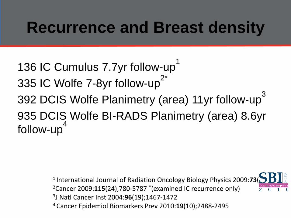
Recurrence and Breast density
136 IC Cumulus 7.7yr follow-up1
335 IC Wolfe 7-8yr follow-up2* 392 DCIS Wolfe Planimetry (area) 11yr follow-up3 935 DCIS Wolfe BI-RADS Planimetry (area) 8.6yr follow-up4
1 International Journal of Radiation Oncology Biology Physics 2009:73(1);75-79 2Cancer 2009:115(24);780-5787 *(examined IC recurrence only) 3J Natl Cancer Inst 2004:96(19);1467-1472
4 Cancer Epidemiol Biomarkers Prev 2010:19(10);2488-2495
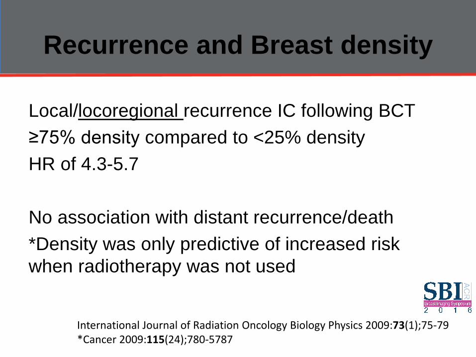
Recurrence and Breast density
Local/locoregional recurrence IC following BCT ≥75% density compared to <25% density HR of 4.3-5.7 No association with distant recurrence/death *Density was only predictive of increased risk when radiotherapy was not used
International Journal of Radiation Oncology Biology Physics 2009:73(1);75-79 *Cancer 2009:115(24);780-5787

Recurrence and Breast density
≥75% density compared to <25% density RR of local recurrence of any cancer of 3.0 No significant risk increase for contralateral breast Increased risk for cancer recurrence—in the contralateral breast (HR 3, 1.4 and 5 respectively). No significant risk increase for ipsilateral breast.
J Natl Cancer Inst 2004:96(19);1467-1472
Cancer Epidemiol Biomarkers Prev 2010:19(10);2488-2495

Women with PHBC
Screening mammography Breast density Frequency surveillance Screening MRI Alternate strategies

Purpose To provide recommendations on the follow-up and management of patients with breast cancer who have completed primary therapy with curative intent. Methods To update the 2006 guideline of the American Society of Clinical Oncology (ASCO), a systematic review of the literature published from March 2006 through March 2012 was completed using MEDLINE and the Cochrane Collaboration Library. An Update Committee reviewed the evidence to determine whether the recommendations were in need of updating. Results There were 14 new publications that met inclusion criteria: nine systematic reviews (three included meta-analyses) and five randomized controlled trials. After its review and analysis of the evidence, the Update Committee concluded that no revisions to the existing ASCO recommendations were warranted. Recommendations Regular history, physical examination, and mammography are recommended for breast cancer follow-up. Physical examinations should be performed every 3 to 6 months for the first 3 years, every 6 to 12 months for years 4 and 5, and annually thereafter. For women who have undergone breast-conserving surgery, a post-treatment mammogram should be obtained 1 year after the initial mammogram and at least 6 months after completion of radiation therapy. Thereafter, unless otherwise indicated, a yearly mammographic evaluation should be performed. The use of complete blood counts, chemistry panels, bone scans, chest radiographs, liver ultrasounds, pelvic ultrasounds, computed tomography scans, [18F]fluorodeoxyglucose–positron emission tomography scans, magnetic resonance imaging, and/or tumor markers (carcinoembryonic antigen, CA 15-3, and CA 27.29) is not recommended for routine follow-up in an otherwise asymptomatic patient with no specific findings on clinical examination.
J Clin Oncol 31:961-965. © 2012 by American Society of Clinical Oncology

THE BOTTOM LINE
ASCO GUIDELINE UPDATE Breast Cancer Follow-Up and Management After Primary Treatment: American Society of Clinical Oncology Clinical Practice Guideline Update Intervention • Modes of surveillance for patients with breast cancer who have completed primary therapy with curative intent Target Audience • Medical oncologists, primary care providers, oncology nurses, surgical oncologists, pathologists, and nuclear medicine
specialists Key Recommendations • Regular history, physical examination, and mammography are recommended • Examinations should be performed every 3 to 6 months for the first 3 years, every 6 to 12 months for years 4 and 5,
and annually thereafter • For women who have undergone breast-conserving surgery, a post-treatment mammogram should be obtained 1 year
after the initial mammogram and at least 6 months after completion of radiation therapy; thereafter, unless otherwise indicated, a yearly mammographic evaluation should be performed
• Use of CBCs, chemistry panels, bone scans, chest radiographs, liver ultrasounds, computed tomography scans, [18F]fluorodeoxyglucose–positron emission tomography scanning, magnetic resonance imaging, or tumor markers (carcinoembryonic antigen, CA 15-3, and CA 27.29) is not recommended for routine breast cancer follow-up in an otherwise symptomatic patient with no specific findings on clinical examination
Methods • A comprehensive systematic review of the literature was conducted, and an Update Committee was convened to
review the evidence and develop guideline recommendations A Data Supplement (including evidence tables) and clinical tools and resources can be found at http://www.asco.org/guidelines/breastfollowup
J Clin Oncol 31:961-965. © 2012 by American Society of Clinical Oncology

Key Recommendations • Regular history, physical examination, and mammography
are recommended • Examinations should be performed every 3 to 6 months for
the first 3 years, every 6 to 12 months for years 4 and 5, and annually thereafter
• For women who have undergone breast-conserving surgery, a post-treatment mammogram should be obtained 1 year after the initial mammogram and at least 6 months after completion of radiation therapy; thereafter, unless otherwise indicated, a yearly mammographic evaluation should be performed
J Clin Oncol 31:961-965. © 2012 by American Society of Clinical Oncology

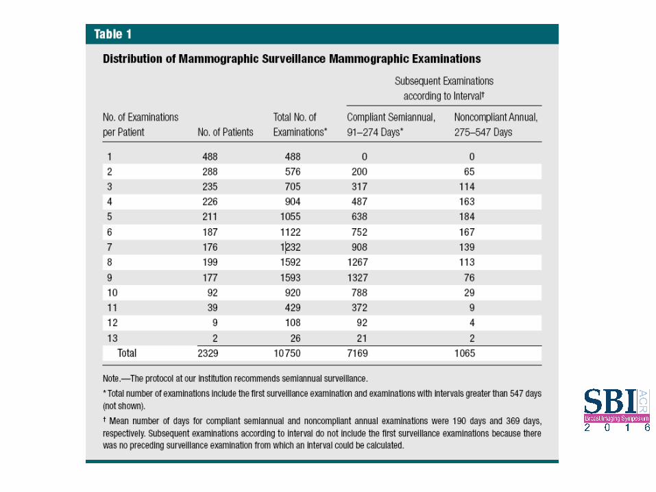
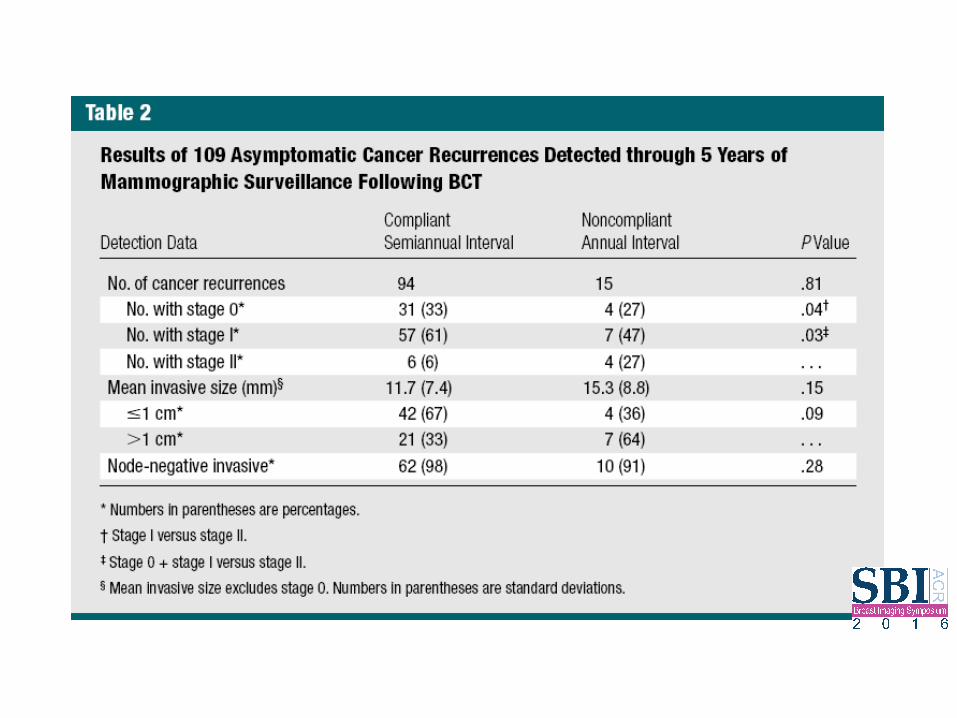
Purpose: To compare cancer recurrence outcomes on the basis of compliant semiannual versus noncompliant annual ipsilateral mammographic surveillance following breast conservation therapy (BCT). Materials and Methods: A HIPAA-compliant retrospective review was performed of post-BCT examinations from 1997 through 2008 by using a deidentified database. The Committee on Human Research did not require institutional review board approval for this study, which was considered quality assurance. Groups were classified according to compliance with institutional post-BCT protocol, which recommends semiannual mammographic examinations of the ipsilateral breast for 5 years. A compliant semiannual examination was defined as an examination with an interval of 0–9 months, although no examination had intervals less than 3 months. A noncompliant annual examination was defined as an examination with an interval of 9–18 months. Cancer recurrence outcomes were compared on the basis of the last examination interval leading to diagnosis. Results: Initially, a total of 10 750 post-BCT examinations among 2329 asymptomatic patients were identified. Excluding initial mammographic follow-up, there were 8234 examinations. Of these, 7169 examinations were semiannual with 94 recurrences detected and 1065 examinations were annual with 15 recurrences detected. There were no differences in demographic risk factors or biopsy rates. Recurrences identified at semiannual intervals were significantly less advanced than those identified at annual intervals (stage I vs stage II, P = .04; stage 0 + stage I vs stage II, P = .03). Nonsignificant findings associated with semiannual versus annual intervals included smaller tumor size (mean, 11.7 vs 15.3 mm; P = .15) and node negativity (98% vs 91%, P = .28). Conclusion: Results suggest that a semiannual interval is preferable for ipsilateral mammographic surveillance, allowing detection of a significantly higher proportion of cancer recurrences at an earlier stage than noncompliant annual surveillance. ©RSNA, 2012
Radiology: Volume 264: Number 2—August 2012 ▪ radiology.rsna.org

Semiannual Ipsilateral Mammography
Retrospective Post BCT mammography Semiannual ipsilateral 5 years 1997-2008 Compliant : 0-9, not < 3 Non-compliant: 9-18 Cancer recurrences compared basis last exam interval leading to outcome
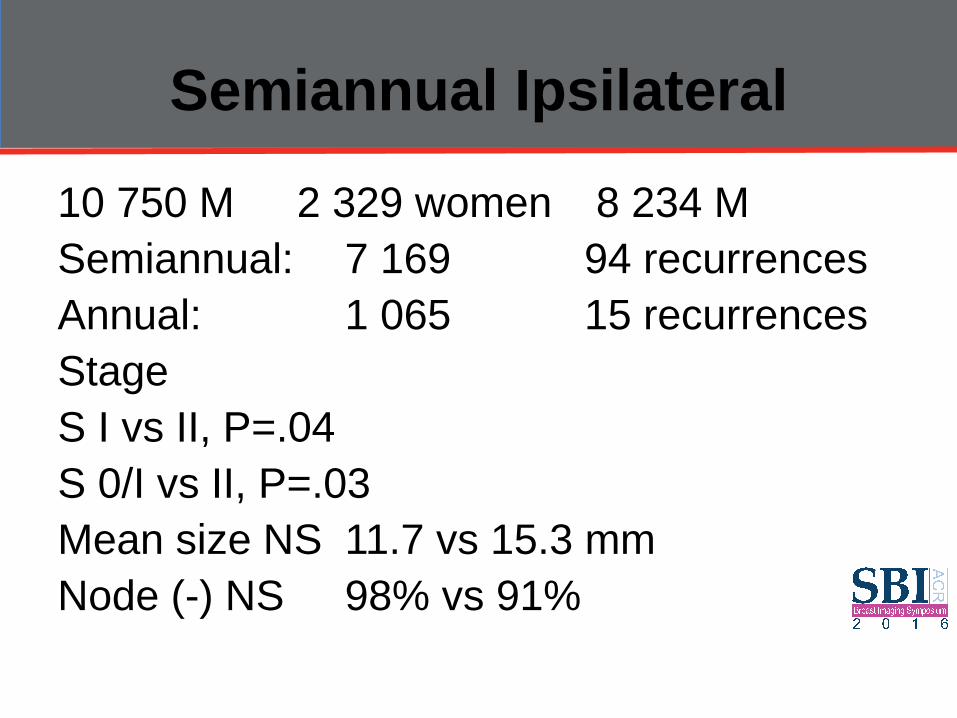
Semiannual Ipsilateral
10 750 M 2 329 women 8 234 M Semiannual: 7 169 94 recurrences Annual: 1 065 15 recurrences Stage S I vs II, P=.04 S 0/I vs II, P=.03 Mean size NS 11.7 vs 15.3 mm Node (-) NS 98% vs 91%

Women with PHBC
Screening mammography Breast density Frequency surveillance Screening MRI Alternate strategies

(CA Cancer J Clin 2007;57:75–89.) © American Cancer Society, Inc., 2007.

TABLE 1 Recommendations for Breast MRI Screening as an Adjunct to Mammography
*Evidence from nonrandomized screening trials and observational studies. †Based on evidence of lifetime risk for breast cancer. ‡Payment should not be a barrier. Screening decisions should be made on a case-by-case basis, as there may be particular factors to support MRI. More data on these groups is expected to be published soon.

Insufficient Evidence to Recommend for or Against MRI Screening‡
Lifetime risk 15–20%, as defined by BRCAPRO or other models that are largely dependent on family history
Lobular carcinoma in situ (LCIS) or atypical lobular hyperplasia (ALH)
Atypical ductal hyperplasia (ADH)
Heterogeneously or extremely dense breast on mammography
Women with a personal history of breast cancer, including ductal carcinoma in situ (DCIS)
‡Payment should not be a barrier. Screening decisions should be made on a case-by-case basis, as there may be particular factors to support MRI. More data on these groups is expected to be published soon.

TABLE 2 Published Breast MRI Screening Study Results
n/a = not applicable.
(CA Cancer J Clin 2007;57:75–89.) © American Cancer Society, Inc., 2007.

Radiology: Volume 272: Number 2—August 2014 ▪ radiology.rsna.org
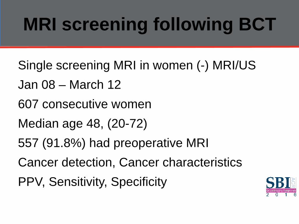
Purpose: To retrospectively investigate the outcomes of single-screening breast magnetic resonance (MR) imaging in women who had a history of breast conservation therapy (BCT) for breast cancers and who had previous negative mammography and ultrasonographic (US) findings. Materials and Methods: This study was institutional review board–approved and informed consent was waived. Between January 2008 and March 2012, 607 consecutive women (median age, 48 years; age range, 20–72 years) who underwent BCT for breast cancer, had negative mammography and US findings, and underwent subsequent screening breast MR imaging were studied. Of the study population, 91.8% (557 of 607) patients underwent preoperative MR examinations. Cancer detection rate, characteristics of detected cancers, positive predictive value (PPV), sensitivity, and specificity were assessed. Multivariate logistic regression analysis was performed to identify independent clinicalpathologic factors associated with women with cancers detected by using MR imaging. Results: Eleven cancers (eight invasive, three ductal carcinoma insitu; median invasive size, 0.8 cm; range, 0.4–1.4 cm; all node negative) were additionally detected with MR imaging in 607 women (18.1 cancers per 1000 women). PPV for recall, PPV for biopsy, sensitivity, and specificity were 9.4% (11 of 117 examinations), 43.5% (10 of 23 examinations), 91.7% (11 of 12 examinations), and 82.2% (489 of 595 examinations), respectively. At multivariate analysis, the independent factors associated with women with Mrdetected cancers were age younger than 50 years at initial diagnosis (P , .001) and more than a 24-month interval between initial surgery and screening MR imaging (P = .011). Conclusion: Single-screening MR imaging depicted 18.1 additional cancers per 1000 women with a history of BCT. Multivariate analysis revealed age younger than 50 years at initial younger than 50 years © RSNA, 2014
MRI screening following BCT
Single screening MRI in women (-) MRI/US Jan 08 – March 12 607 consecutive women Median age 48, (20-72) 557 (91.8%) had preoperative MRI Cancer detection, Cancer characteristics PPV, Sensitivity, Specificity

MRI screening following BCT
11 additional cancers 8 invasive, 3 DCIS Median size 0.8 cm (0.4 – 1.4) Node (-) 18.1 cancers per 1 000 women


MRI screening following BCT
PPV Recurrence 9.4% (11/117) PPV Biopsy 43.5% (10/23) Sensitivity 91.7% (11/12) Specificity 82.2% (489/595)


JNCI J Natl Cancer Inst (2016) 108(3): djv349
Screening MRI PHBC
High risk for BC based on genetic risk or family history (GFH)1
Compared screening MRI in women with PH and GFH of breast cancer JNCI 2016;108(3): djv349

Screening MRI PHBC
Case series registry data Collected at time of MRI & at 12 month FU Age, PH MRI, clinical indication MRI (chi-sq) Combined contribution of these variables in predicting risk of a FP (logistic regression)

Screening MRI PHBC – Results 1
1521 women, 7/2004-11/2011 915 PH vs 606 GFH Overall sensitivity 79.4% all cancers 88.5% invasive cancers FP: 12.3% vs 21.6%, P < .001 Specificity: 94.0% vs 86.0%, P < .001 Sensitivity, Cancer detection NS, P > .99

Screening MRI PHBC – Results 2
Age (P < .001) Prior MRI (P < .001) Clinical indication (P < .001) Individually associated with initial false-positive rate (P = .001) Age and prior MRI remained statistically significant in multivariate modeling (P < .001)

Screening MRI PHBC
Superior performance vs GFH

AJR 2010; 195:510–516

Screening MRI PHBC
Retrospective chart review 1999-2001 144 women PHBC (no FHBC) 1699 breast MRIs Determine the cancer detection & bx rate
Brennan AJR 2010

Screening MRI PHBC
44 women (31%, 95%CI 15-29%): MRI Bxs Malignant: 17 (12%; 7-18%) Benign: 27 (19%; 13-26%) Total: 18 Malignant lesions, 1 metachronous
Brennan AJR 2010

Screening MRI PHBC
MRI Bx: 61; PPV: 39%; 95%CI 27-53% 18 Malignancies: 17 CAs, 1 lipoSA 17 CAs: 12 Invasive (71%); 5 DCIS (29%) 10 minimal CAs 10 cancers detected by MRI-only DCIS: 4/10 (40%) vs 1/7 (14%) Minimal CAs: 7/10 (70%) vs 3/7 (43%)
Brennan AJR 2010
Screening MRI PHBC
Benefit certain subsets of patients • Have not had preoperative MRI at initial dx • Have not taken hormonal therapy • RCT prospective to best determine the
cost-effectiveness
Brennan AJR 2010

ABSTRACT Background. Diagnosis of breast cancer recurrence can be difficult as a result of the presence of scar tissue in the breast. Magnetic resonance imaging (MRI) may be superior to traditional imaging in diagnosis of recurrence because of its ability to differentiate malignancy from scarring. Current guidelines on investigation of suspected breast cancer recurrence recommend MRI when other investigations have equivocal findings. We performed the first systematic review on this topic. Methods. Literature search revealed 35 potentially relevant studies; 10 were included in final analysis. Included were clinical studies comparing MRI with another diagnostic modality for diagnosis of breast cancer recurrence, with at least 10 patients, in the English language. Data extraction focused on sensitivity and specificity of standard diagnostic modalities and MRI for diagnosis of local disease recurrence. Results. In total 494 patients were assessed across 10 studies; all were case series. Sensitivity of MRI for detection of recurrence ranged 75–100 %, while specificity ranged 66.6–100 %. Both sensitivity and specificity increased when MRI was performed after a longer time interval from the original surgery, although the longest follow-up reported was only 36 months. A negative MRI can avoid the need for further biopsy. Conclusions. Available data are based on clinically heterogeneous case series and superiority over standard triple assessment for breast cancer recurrence has not been proven. At present, MRI cannot be recommended in the routine diagnostic assessment for breast cancer recurrence but has a potentially useful role as a second-line investigation. A negative MRI is more useful than a positive MRI as positive MRIs require further investigation.
Ann Surg Oncol (2012) 19:3035–3041. © Society of Surgical Oncology 2012

Screening MRI PHBC
35 clinical studies 1993-2006 Comparing MRI with another diagnostic modality for breast recurrence diagnosis Sensitivity and specificity
Quinn Ann Surg Oncol 2012
Screening MRI PHBC
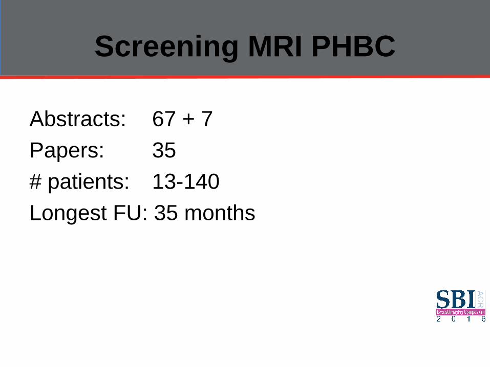
Abstracts: 67 + 7 Papers: 35 # patients: 13-140 Longest FU: 35 months

Screening MRI PHBC
Increase interventions in breast CA? Increase radical Sx Specificity (Breast CA recurrence): 66-100% Mandates Preoperative Bx Sensitivity (BCT or MX): 75-100% Negative MRI conclusive Omit need for repeat bx scarred tissue Not first line diagnosis for breast recurrence
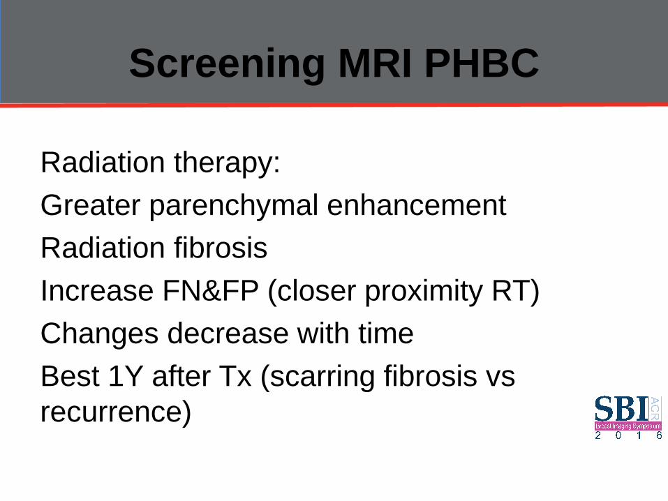
Screening MRI PHBC
Radiation therapy: Greater parenchymal enhancement Radiation fibrosis Increase FN&FP (closer proximity RT) Changes decrease with time Best 1Y after Tx (scarring fibrosis vs recurrence)
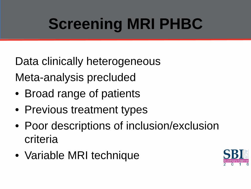
Screening MRI PHBC
Data clinically heterogeneous Meta-analysis precluded • Broad range of patients • Previous treatment types • Poor descriptions of inclusion/exclusion
criteria • Variable MRI technique

Screening MRI PHBC
MRI: Expensive, resource intensive, delay time to further Tx Cost analysis MRI use in the assessment breast cancer recurrence Long term FU data on patients with (-) MRI

Alternate Imaging Strategies

Molecular Breast Imaging
Gamma-ray emitting radiotracers 99mTc-sestamibi and 99mTc-tetrofosmin Post-therapy changes scar tissue Limitation: low sensitivity for small BCs High-resolution specific breast cameras detection sub-cm malignant lesions Radiation
Q J Nucl Med Mol Imaging 2013 Dec;57(4):340-51

Conclusion (1)
No RCT that examines the impact of breast cancer mortality achieved in the use of annual mammography No other imaging modality (MRI) sufficient evidence to justify inclusion in the recommended surveillance of breast cancer survivors

Conclusion (2)
Screening mammography: some suggestion of benefit systematic review small data sets BC survivors tend to be younger (~20% new cases in the U.S. are diagnosed in women under 50 ys)

Conclusion (3)
Screening mammography Modality of choice Breast density Risk factor Frequency surveillance Annual vs 6 monthly Screening MRI Warrants further investigation


![Diagnostic and quantitative imaging of knee osteoarthritis5.3 Imaging techniques ... found that arthritis decreased quality of life measure, EQ-5D [7], even more ... Earliest stage](https://img.pdfslide.us/doc/110x75/5ed567fbbfdf2a2eb564662f/diagnostic-and-quantitative-imaging-of-knee-osteoarthritis-53-imaging-techniques.jpg)