Embed Size (px)
Citation preview
1
Imaging of the
Musculoskeletal System
Dr. Lindsay Davidson
Dr. Paul Fenton
Craig Goldie
Overview
Imaging is an essential tool for the modern physician. It allows us to analyze anatomy, detect
pathologies and monitor disease healing or progression. In the musculoskeletal system,
imaging is invaluable to detect injuries and other pathologies.
Objectives
Upon completion of this module, the learner will be able to:
1. Describe the imaging modalities and techniques commonly used in the evaluation of the
musculoskeletal system,
2. Describe common indications for the use of each modality,
3. Quantify in broad terms the relative radiation exposure associated with each imaging
modality,
4. Demonstrate an organized approach to the analysis of plain radiographs of the
musculoskeletal system.
Reference
Read Rheumatology 3: Getting the most out of radiology by Graham Reid and John M.
Esdaile in CMAJ • May 2, 2000; 162 (9)
2
Fundamentals of Imaging
All imaging modalities have the same principle components:
1. An object to analyze (i.e. the patient)
2. An energy source (i.e. light or sound waves)
3. A detector (i.e. x-ray film, MRI detector)
Each modality has advantages and disadvantages and therefore is best suited to the
investigation of particular clinical scenarios. Clinical examples will be used to illustrate the
indications for each type of imaging described in this module.
Introduction
Fundamentals of Imaging – continued…
Types of imaging
1. Plain radiographs (x-rays)
2. Computed Tomography (CT)
3. Magnetic Resonance Imaging (MRI)
4. Ultrasound (USS)
5. Bone Scan
The most common types of imaging used in the investigation of the musculoskeletal system
will be discussed. These are:
3
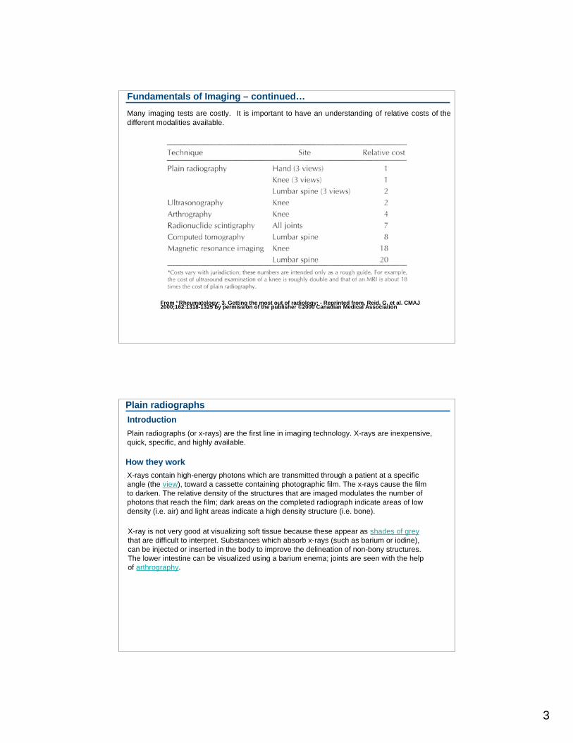
From “Rheumatology: 3. Getting the most out of radiology: - Reprinted from, Reid, G. et al. CMAJ2000;162:1318-1325 by permission of the publisher ©2000 Canadian Medical Association
Many imaging tests are costly. It is important to have an understanding of relative costs of the
different modalities available.
Fundamentals of Imaging – continued…
Plain radiographs
Plain radiographs (or x-rays) are the first line in imaging technology. X-rays are inexpensive,
quick, specific, and highly available.
How they work
X-rays contain high-energy photons which are transmitted through a patient at a specific
angle (the view), toward a cassette containing photographic film. The x-rays cause the film
to darken. The relative density of the structures that are imaged modulates the number of
photons that reach the film; dark areas on the completed radiograph indicate areas of low
density (i.e. air) and light areas indicate a high density structure (i.e. bone).
X-ray is not very good at visualizing soft tissue because these appear as shades of grey
that are difficult to interpret. Substances which absorb x-rays (such as barium or iodine),
can be injected or inserted in the body to improve the delineation of non-bony structures.
The lower intestine can be visualized using a barium enema; joints are seen with the help
of arthrography.
Introduction
4
Plain radiographs - continued…
Advantages
X-rays are inexpensive, widely, and they provide an excellent initial evaluation of bone detail and
anatomic relationships. They are the most specific imaging modality.
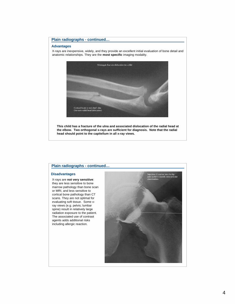
This child has a fracture of the ulna and associated dislocation of the radial head at
the elbow. Two orthogonal x-rays are sufficient for diagnosis. Note that the radial
head should point to the capitellum in all x-ray views.
Plain radiographs - continued…
Disadvantages
X-rays are not very sensitive:
they are less sensitive to bone
marrow pathology than bone scan
or MRI, and less sensitive to
cortical bone pathology than CT
scans. They are not optimal for
evaluating soft tissue. Some x-
ray views (e.g. pelvis; lumbar
spine) result in relatively large
radiation exposure to the patient.
The associated use of contrast
agents adds additional risks
including allergic reaction.
5
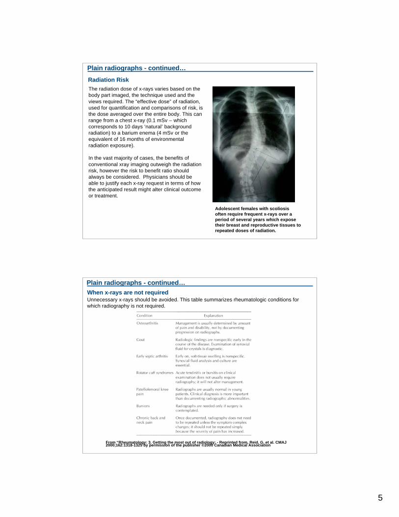
Radiation Risk
The radiation dose of x-rays varies based on the
body part imaged, the technique used and the
views required. The “effective dose” of radiation,
used for quantification and comparisons of risk, is
the dose averaged over the entire body. This can
range from a chest x-ray (0.1 mSv – which
corresponds to 10 days ‘natural’ background
radiation) to a barium enema (4 mSv or the
equivalent of 16 months of environmental
radiation exposure).
In the vast majority of cases, the benefits of
conventional xray imaging outweigh the radiation
risk, however the risk to benefit ratio should
always be considered. Physicians should be
able to justify each x-ray request in terms of how
the anticipated result might alter clinical outcome
or treatment.
Plain radiographs - continued…
Adolescent females with scoliosis
often require frequent x-rays over a
period of several years which expose
their breast and reproductive tissues to
repeated doses of radiation.
When x-rays are not requiredUnnecessary x-rays should be avoided. This table summarizes rheumatologic conditions for
which radiography is not required.
Plain radiographs - continued…
From “Rheumatology: 3. Getting the most out of radiology: - Reprinted from, Reid, G. et al. CMAJ2000;162:1318-1325 by permission of the publisher ©2000 Canadian Medical Association
6
CT
Introduction
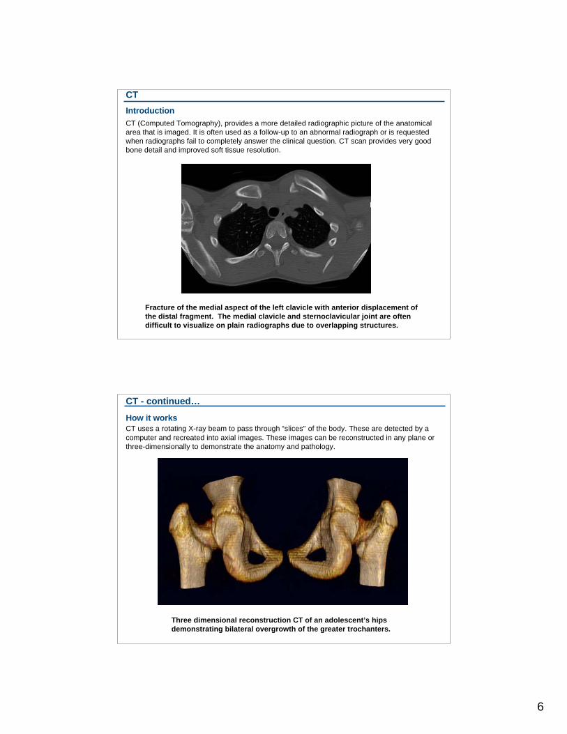
CT (Computed Tomography), provides a more detailed radiographic picture of the anatomical
area that is imaged. It is often used as a follow-up to an abnormal radiograph or is requested
when radiographs fail to completely answer the clinical question. CT scan provides very good
bone detail and improved soft tissue resolution.
Fracture of the medial aspect of the left clavicle with anterior displacement of
the distal fragment. The medial clavicle and sternoclavicular joint are often
difficult to visualize on plain radiographs due to overlapping structures.
CT - continued…
How it works
CT uses a rotating X-ray beam to pass through “slices” of the body. These are detected by a
computer and recreated into axial images. These images can be reconstructed in any plane or
three-dimensionally to demonstrate the anatomy and pathology.
Three dimensional reconstruction CT of an adolescent’s hips
demonstrating bilateral overgrowth of the greater trochanters.
7
CT - continued…
Radiation Risk
CT scans deliver the highest radiation exposure used in current medical imaging practice. This
occurs because multiple x-ray scans are used to construct the images. A CT scan generates
approximately 10 mSv, or the equivalent of 3 years 'background' radiation. CT scans require a
more careful risk vs benefit evaluation than conventional radiographs.
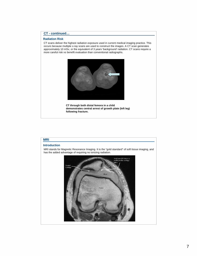
CT through both distal femora in a child
demonstrates central arrest of growth plate (left leg)
following fracture.
MRI
Introduction
MRI stands for Magnetic Resonance Imaging. It is the “gold standard” of soft tissue imaging, and
has the added advantage of requiring no ionizing radiation.
8
MRI - continued…
How it worksThe MRI machine creates a large external magnetic field, which causes the protons (H+) in
the body to polarize and align with the magnet. A special radio-frequency pulse is applied
and causes the protons to 'tip' out of alignment into a higher energy state (excitation
phase). The pulse is relaxed, the protons remove to the lower energy state, during which
they emit energy. Each tissue has slightly different characteristics due to the proton
environment (relaxation phase). This differential is used to generate the MRI image.
Each tissue-type has a unique appearances on MRI which helps the radiologist to identify
anatomy and pathology such as trauma, infection, inflammation or tumour.
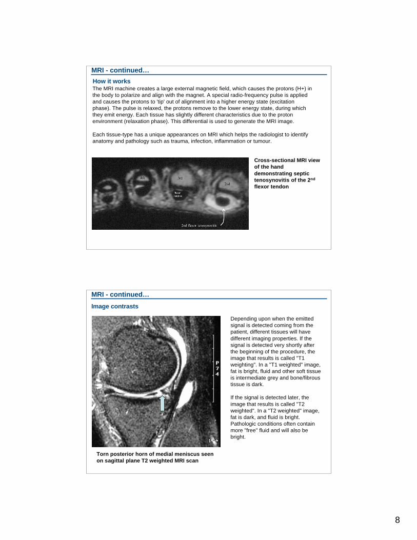
Cross-sectional MRI view
of the hand
demonstrating septic
tenosynovitis of the 2nd
flexor tendon
MRI - continued…
Image contrasts
Depending upon when the emitted
signal is detected coming from the
patient, different tissues will have
different imaging properties. If the
signal is detected very shortly after
the beginning of the procedure, the
image that results is called "T1
weighting". In a "T1 weighted" image,
fat is bright, fluid and other soft tissue
is intermediate grey and bone/fibrous
tissue is dark.
If the signal is detected later, the
image that results is called "T2
weighted". In a "T2 weighted" image,
fat is dark, and fluid is bright.
Pathologic conditions often contain
more "free" fluid and will also be
bright.
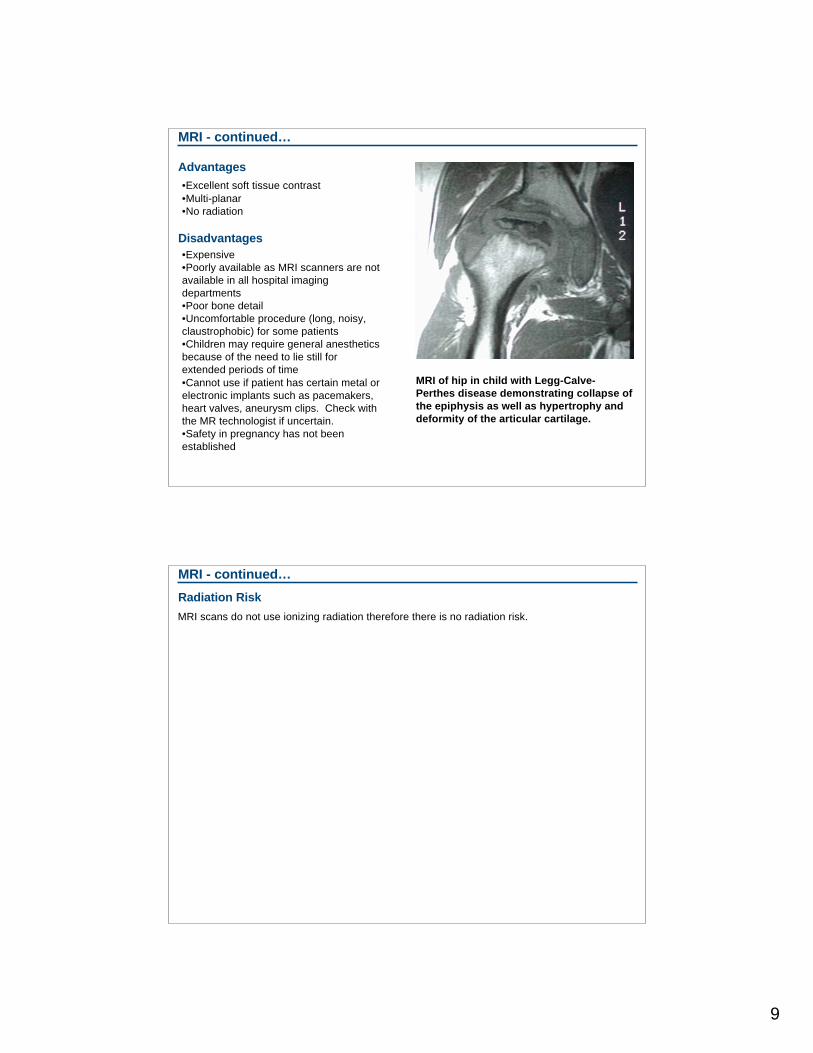
Torn posterior horn of medial meniscus seen
on sagittal plane T2 weighted MRI scan
9
MRI - continued…
Advantages
•Excellent soft tissue contrast
•Multi-planar
•No radiation
Disadvantages
•Expensive
•Poorly available as MRI scanners are not
available in all hospital imaging
departments
•Poor bone detail
•Uncomfortable procedure (long, noisy,
claustrophobic) for some patients
•Children may require general anesthetics
because of the need to lie still for
extended periods of time
•Cannot use if patient has certain metal or
electronic implants such as pacemakers,
heart valves, aneurysm clips. Check with
the MR technologist if uncertain.
•Safety in pregnancy has not been
established
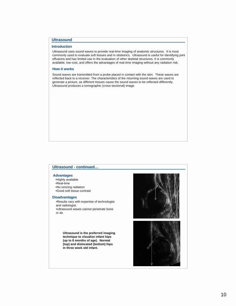
MRI of hip in child with Legg-Calve-
Perthes disease demonstrating collapse of
the epiphysis as well as hypertrophy and
deformity of the articular cartilage.
MRI - continued…
Radiation Risk
MRI scans do not use ionizing radiation therefore there is no radiation risk.
10
Ultrasound
Introduction
Ultrasound uses sound waves to provide real-time imaging of anatomic structures. It is most
commonly used to evaluate soft tissues and in obstetrics. Ultrasound is useful for identifying joint
effusions and has limited use in the evaluation of other skeletal structures. It is commonly
available, low cost, and offers the advantages of real-time imaging without any radiation risk.
How it works
Sound waves are transmitted from a probe placed in contact with the skin. These waves are
reflected back to a receiver. The characteristics of the returning sound waves are used to
generate a picture, as different tissues cause the sound waves to be reflected differently.
Ultrasound produces a tomographic (cross-sectional) image.
Ultrasound - continued…
Advantages•Highly available
•Real-time
•No ionizing radiation
•Good soft tissue contrast
Disadvantages
•Results vary with expertise of technologist
and radiologist.
•Ultrasound waves cannot penetrate bone
or air.
Ultrasound is the preferred imaging
technique to visualize infant hips
(up to 8 months of age). Normal
(top) and dislocated (bottom) hips
in three week old infant.
11
Ultrasound - continued…
Radiation Risk
Ultrasound scans do not use ionizing radiation therefore there is no radiation risk.
Bone Scan
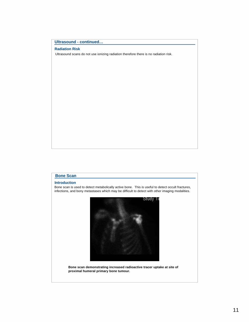
IntroductionBone scan is used to detect metabolically active bone. This is useful to detect occult fractures,
infections, and bony metastases which may be difficult to detect with other imaging modalities.
Bone scan demonstrating increased radioactive tracer uptake at site of
proximal humeral primary bone tumour.
12
Bone Scan - continued…
How it works
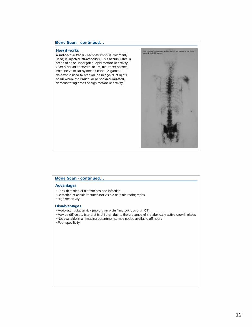
A radioactive tracer (Technetium 99 is commonly
used) is injected intravenously. This accumulates in
areas of bone undergoing rapid metabolic activity.
Over a period of several hours, the tracer passes
from the vascular system to bone. A gamma-
detector is used to produce an image. “Hot spots”
occur where the radionuclide has accumulated,
demonstrating areas of high metabolic activity.
Bone Scan - continued…
Advantages
•Early detection of metastases and infection
•Detection of occult fractures not visible on plain radiographs
•High sensitivity
Disadvantages•Moderate radiation risk (more than plain films but less than CT)
•May be difficult to interpret in children due to the presence of metabolically active growth plates
•Not available in all imaging departments; may not be available off-hours
•Poor specificity
13
Bone Scan - continued…
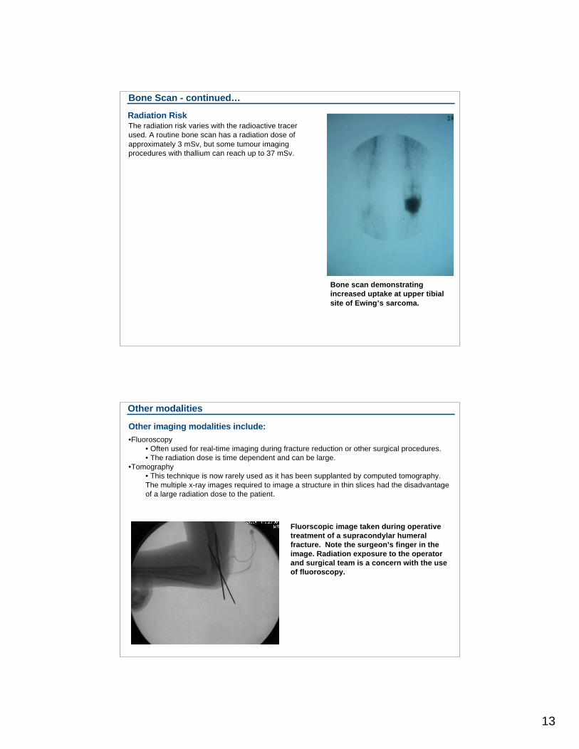
Radiation RiskThe radiation risk varies with the radioactive tracer
used. A routine bone scan has a radiation dose of
approximately 3 mSv, but some tumour imaging
procedures with thallium can reach up to 37 mSv.
Bone scan demonstrating
increased uptake at upper tibial
site of Ewing’s sarcoma.
Other modalities
•Fluoroscopy
• Often used for real-time imaging during fracture reduction or other surgical procedures.
• The radiation dose is time dependent and can be large.
•Tomography
• This technique is now rarely used as it has been supplanted by computed tomography.
The multiple x-ray images required to image a structure in thin slices had the disadvantage
of a large radiation dose to the patient.
Other imaging modalities include:
Fluorscopic image taken during operative
treatment of a supracondylar humeral
fracture. Note the surgeon’s finger in the
image. Radiation exposure to the operator
and surgical team is a concern with the use
of fluoroscopy.
14
Uses of imaging
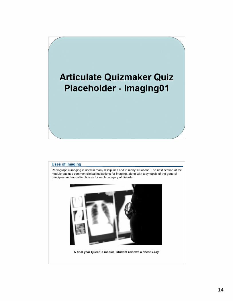
Radiographic imaging is used in many disciplines and in many situations. The next section of the
module outlines common clinical indications for imaging, along with a synopsis of the general
principles and modality choices for each category of disorder.
A final year Queen’s medical student reviews a chest x-ray
15
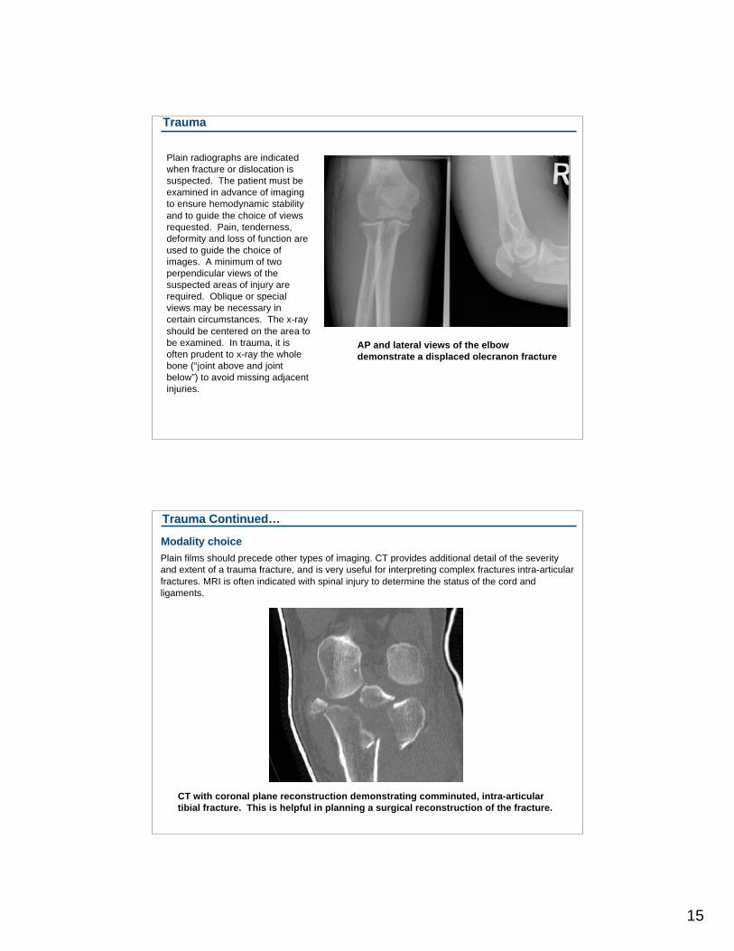
Trauma
Plain radiographs are indicated
when fracture or dislocation is
suspected. The patient must be
examined in advance of imaging
to ensure hemodynamic stability
and to guide the choice of views
requested. Pain, tenderness,
deformity and loss of function are
used to guide the choice of
images. A minimum of two
perpendicular views of the
suspected areas of injury are
required. Oblique or special
views may be necessary in
certain circumstances. The x-ray
should be centered on the area to
be examined. In trauma, it is
often prudent to x-ray the whole
bone (“joint above and joint
below”) to avoid missing adjacent
injuries.
AP and lateral views of the elbow
demonstrate a displaced olecranon fracture
Trauma Continued…
Plain films should precede other types of imaging. CT provides additional detail of the severity
and extent of a trauma fracture, and is very useful for interpreting complex fractures intra-articular
fractures. MRI is often indicated with spinal injury to determine the status of the cord and
ligaments.
Modality choice
CT with coronal plane reconstruction demonstrating comminuted, intra-articular
tibial fracture. This is helpful in planning a surgical reconstruction of the fracture.
16
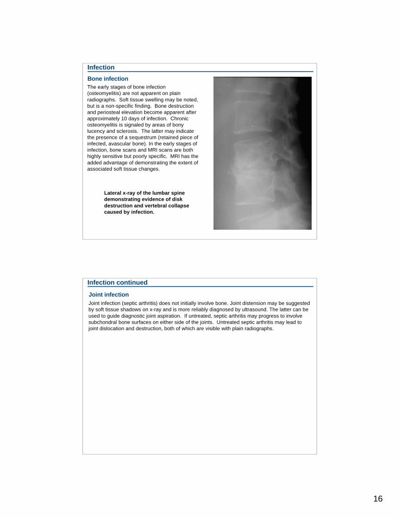
Infection
The early stages of bone infection
(osteomyelitis) are not apparent on plain
radiographs. Soft tissue swelling may be noted,
but is a non-specific finding. Bone destruction
and periosteal elevation become apparent after
approximately 10 days of infection. Chronic
osteomyelitis is signaled by areas of bony
lucency and sclerosis. The latter may indicate
the presence of a sequestrum (retained piece of
infected, avascular bone). In the early stages of
infection, bone scans and MRI scans are both
highly sensitive but poorly specific. MRI has the
added advantage of demonstrating the extent of
associated soft tissue changes.
Bone infection
Lateral x-ray of the lumbar spine
demonstrating evidence of disk
destruction and vertebral collapse
caused by infection.
Infection continued
Joint infection
Joint infection (septic arthritis) does not initially involve bone. Joint distension may be suggested
by soft tissue shadows on x-ray and is more reliably diagnosed by ultrasound. The latter can be
used to guide diagnostic joint aspiration. If untreated, septic arthritis may progress to involve
subchondral bone surfaces on either side of the joints. Untreated septic arthritis may lead to
joint dislocation and destruction, both of which are visible with plain radiographs.
17
Infection
Soft tissue infection is usually imaged via MRI or CT. This is particularly useful for deep
collections of pus in the epidural or retroperitoneal space. Plain film are not usually helpful in
diagnosis of soft tissue infections, however they can be useful to quickly detect the presence of
gas, such as in a clostridial gas gangrene.
Soft tissue infection
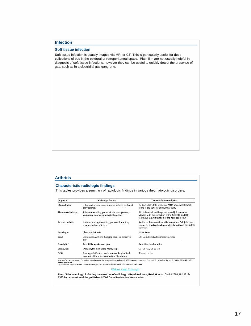
Arthritis
Characteristic radiologic findings
This tables provides a summary of radiologic findings in various rheumatologic disorders.
From “Rheumatology: 3. Getting the most out of radiology: - Reprinted from, Reid, G. et al. CMAJ 2000;162:1318-
1325 by permission of the publisher ©2000 Canadian Medical Association
Click on image to enlarge
18
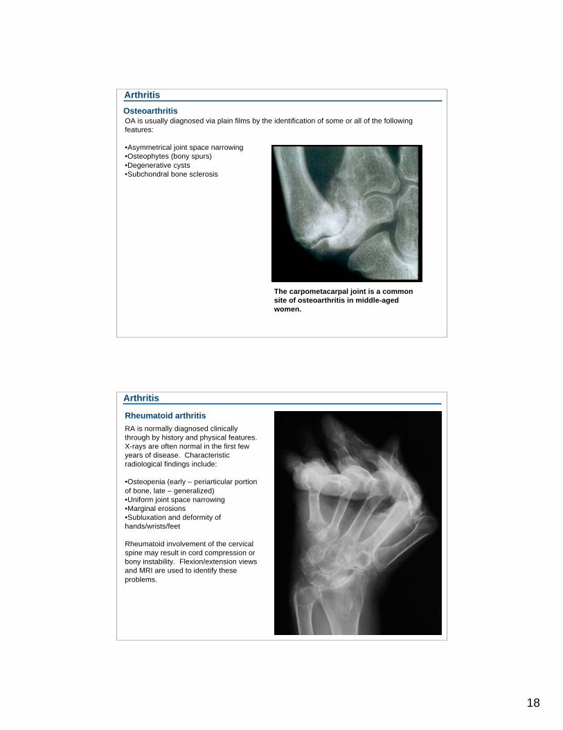
Arthritis
OA is usually diagnosed via plain films by the identification of some or all of the following
features:
•Asymmetrical joint space narrowing
•Osteophytes (bony spurs)
•Degenerative cysts
•Subchondral bone sclerosis
Osteoarthritis
The carpometacarpal joint is a common
site of osteoarthritis in middle-aged
women.
Arthritis
Rheumatoid arthritis
RA is normally diagnosed clinically
through by history and physical features.
X-rays are often normal in the first few
years of disease. Characteristic
radiological findings include:
•Osteopenia (early – periarticular portion
of bone, late – generalized)
•Uniform joint space narrowing
•Marginal erosions
•Subluxation and deformity of
hands/wrists/feet
Rheumatoid involvement of the cervical
spine may result in cord compression or
bony instability. Flexion/extension views
and MRI are used to identify these
problems.
19
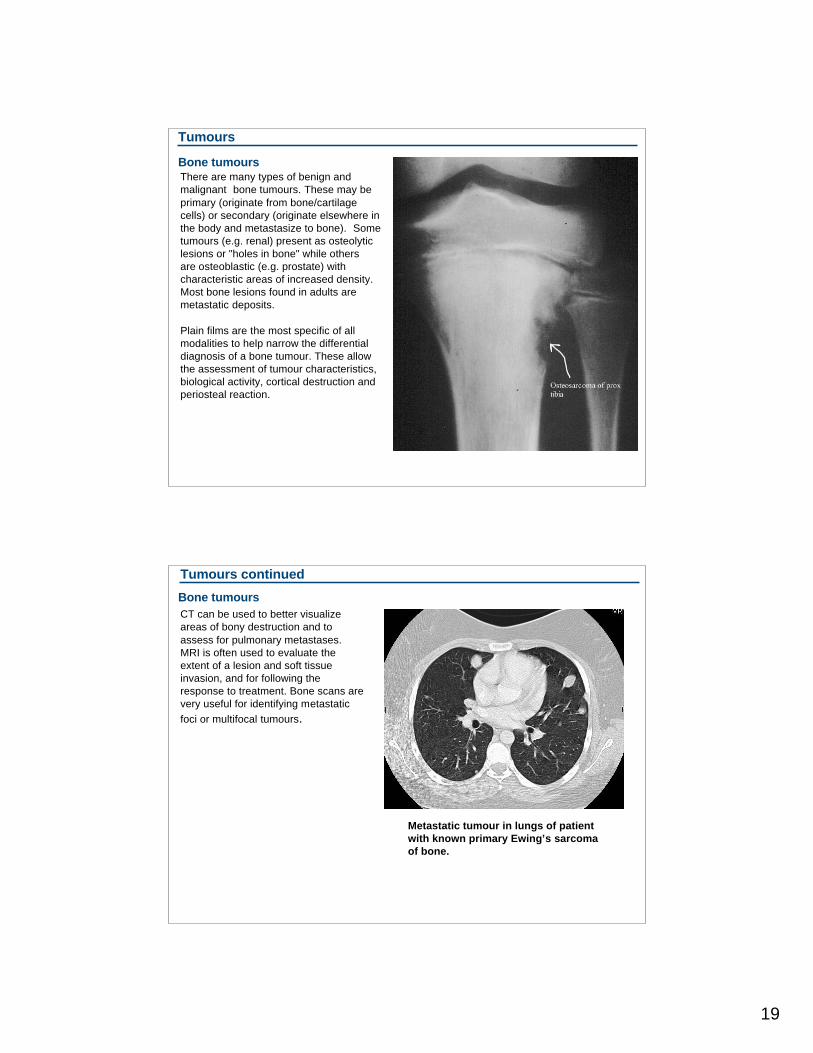
Tumours
There are many types of benign and
malignant bone tumours. These may be
primary (originate from bone/cartilage
cells) or secondary (originate elsewhere in
the body and metastasize to bone). Some
tumours (e.g. renal) present as osteolytic
lesions or "holes in bone" while others
are osteoblastic (e.g. prostate) with
characteristic areas of increased density.
Most bone lesions found in adults are
metastatic deposits.
Plain films are the most specific of all
modalities to help narrow the differential
diagnosis of a bone tumour. These allow
the assessment of tumour characteristics,
biological activity, cortical destruction and
periosteal reaction.
Bone tumours
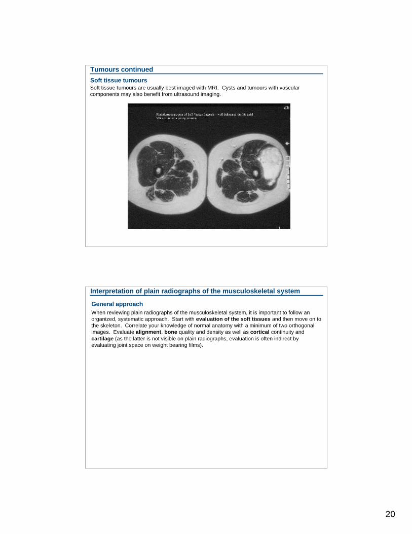
Tumours continued
CT can be used to better visualize
areas of bony destruction and to
assess for pulmonary metastases.
MRI is often used to evaluate the
extent of a lesion and soft tissue
invasion, and for following the
response to treatment. Bone scans are
very useful for identifying metastatic
foci or multifocal tumours.
Bone tumours
Metastatic tumour in lungs of patient
with known primary Ewing’s sarcoma
of bone.
20
Tumours continued
Soft tissue tumoursSoft tissue tumours are usually best imaged with MRI. Cysts and tumours with vascular
components may also benefit from ultrasound imaging.
Interpretation of plain radiographs of the musculoskeletal system
General approach
When reviewing plain radiographs of the musculoskeletal system, it is important to follow an
organized, systematic approach. Start with evaluation of the soft tissues and then move on to
the skeleton. Correlate your knowledge of normal anatomy with a minimum of two orthogonal
images. Evaluate alignment, bone quality and density as well as cortical continuity and
cartilage (as the latter is not visible on plain radiographs, evaluation is often indirect by
evaluating joint space on weight bearing films).
21
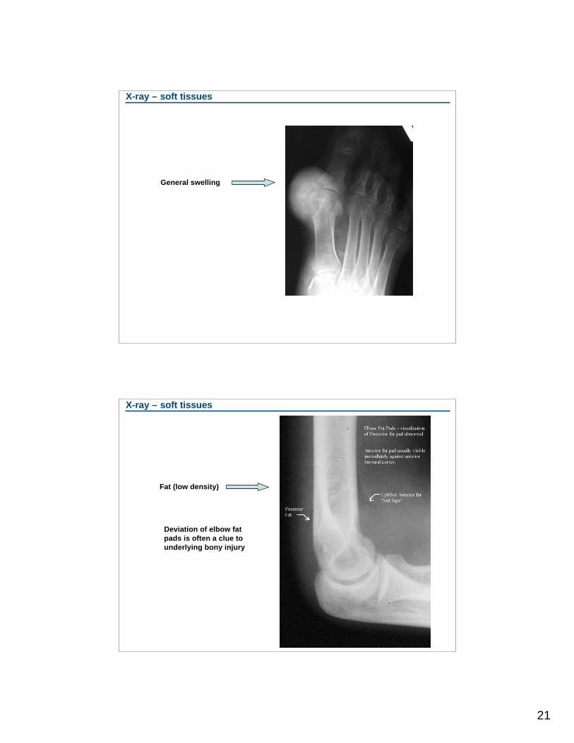
X-ray – soft tissues
General swelling
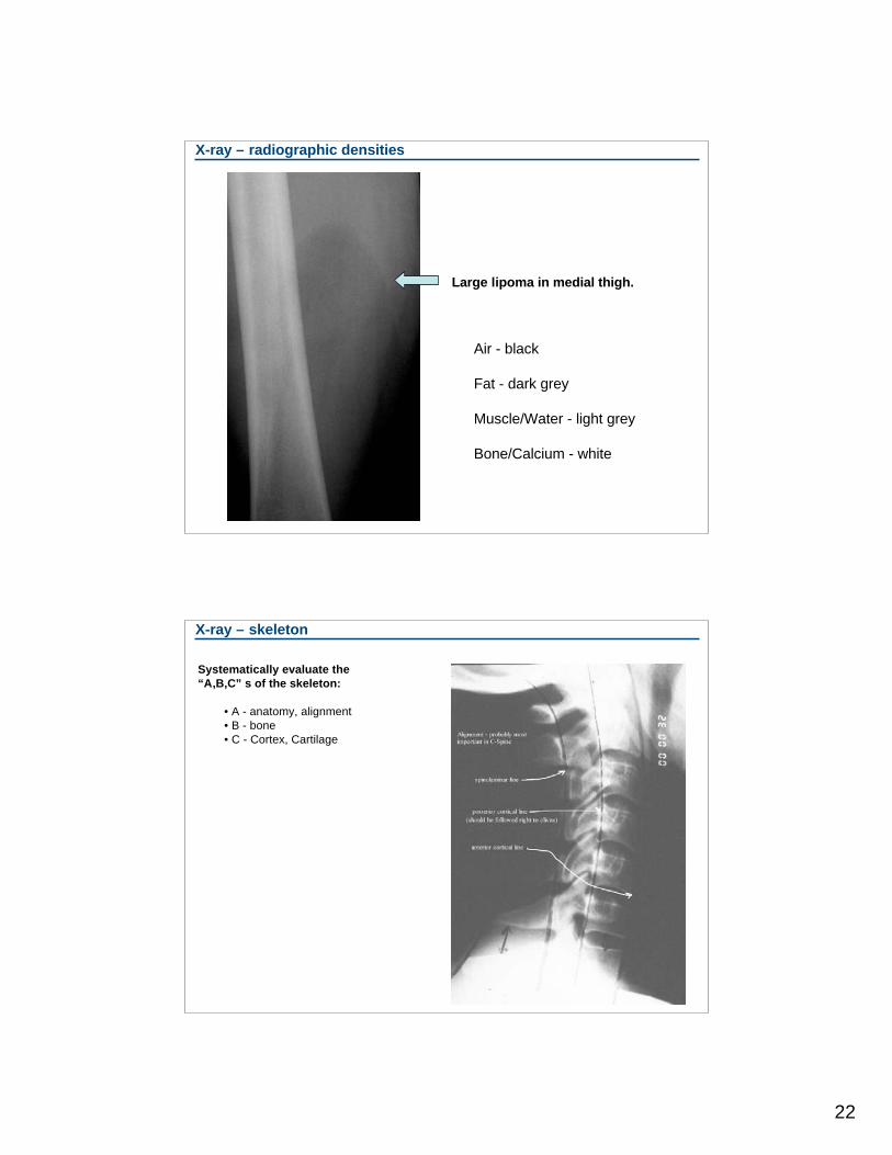
X-ray – soft tissues
Fat (low density)
Deviation of elbow fat
pads is often a clue to
underlying bony injury
22
X-ray – radiographic densities
Air - black
Fat - dark grey
Muscle/Water - light grey
Bone/Calcium - white
Large lipoma in medial thigh.
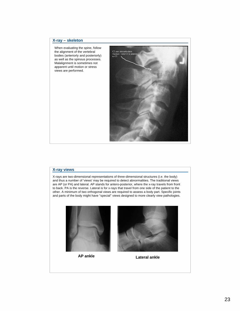
X-ray – skeleton
Systematically evaluate the
“A,B,C” s of the skeleton:
• A - anatomy, alignment
• B - bone
• C - Cortex, Cartilage
23
X-ray – skeleton
When evaluating the spine, follow
the alignment of the vertebral
bodies (anteriorly and posteriorly)
as well as the spinous processes.
Malalignment is sometimes not
apparent until motion or stress
views are performed.
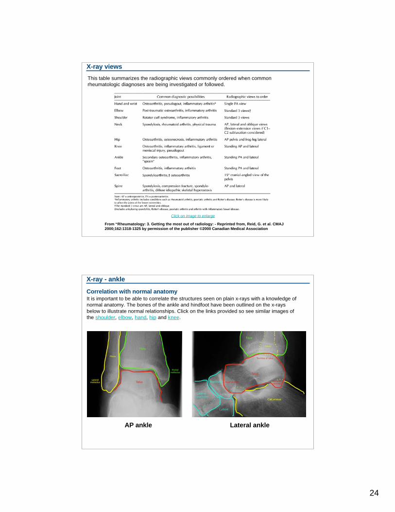
X-ray views
X-rays are two-dimensional representations of three-dimensional structures (i.e. the body)
and thus a number of 'views' may be required to detect abnormalities. The traditional views
are AP (or PA) and lateral. AP stands for antero-posterior, where the x-ray travels from front
to back. PA is the reverse. Lateral is for x-rays that travel from one side of the patient to the
other. A minimum of two orthogonal views are required to assess a body part. Specific joints
and parts of the body might have “special” views designed to more clearly view pathologies.
AP ankle Lateral ankle
24
X-ray views
This table summarizes the radiographic views commonly ordered when common
rheumatologic diagnoses are being investigated or followed.
From “Rheumatology: 3. Getting the most out of radiology: - Reprinted from, Reid, G. et al. CMAJ
2000;162:1318-1325 by permission of the publisher ©2000 Canadian Medical Association
Click on image to enlarge
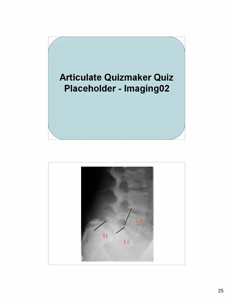
X-ray - ankle
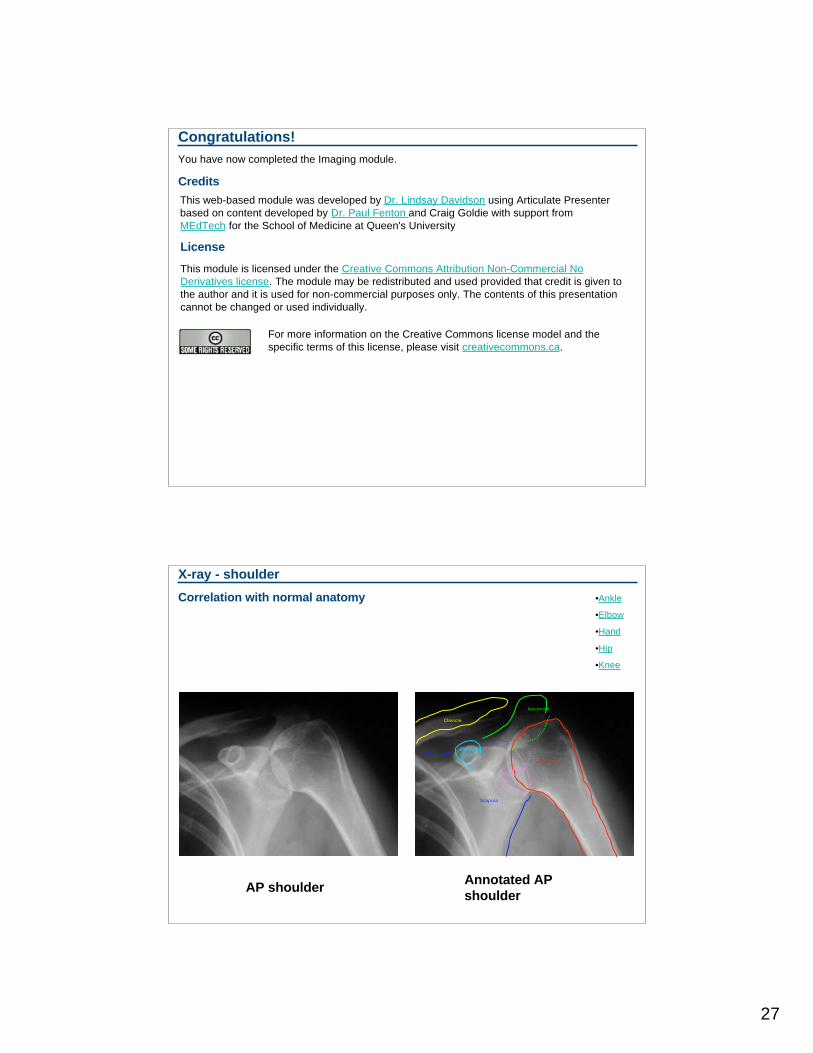
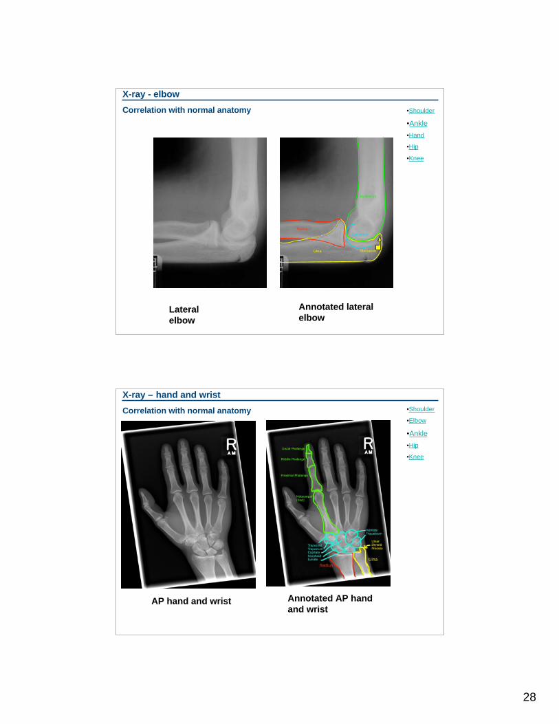
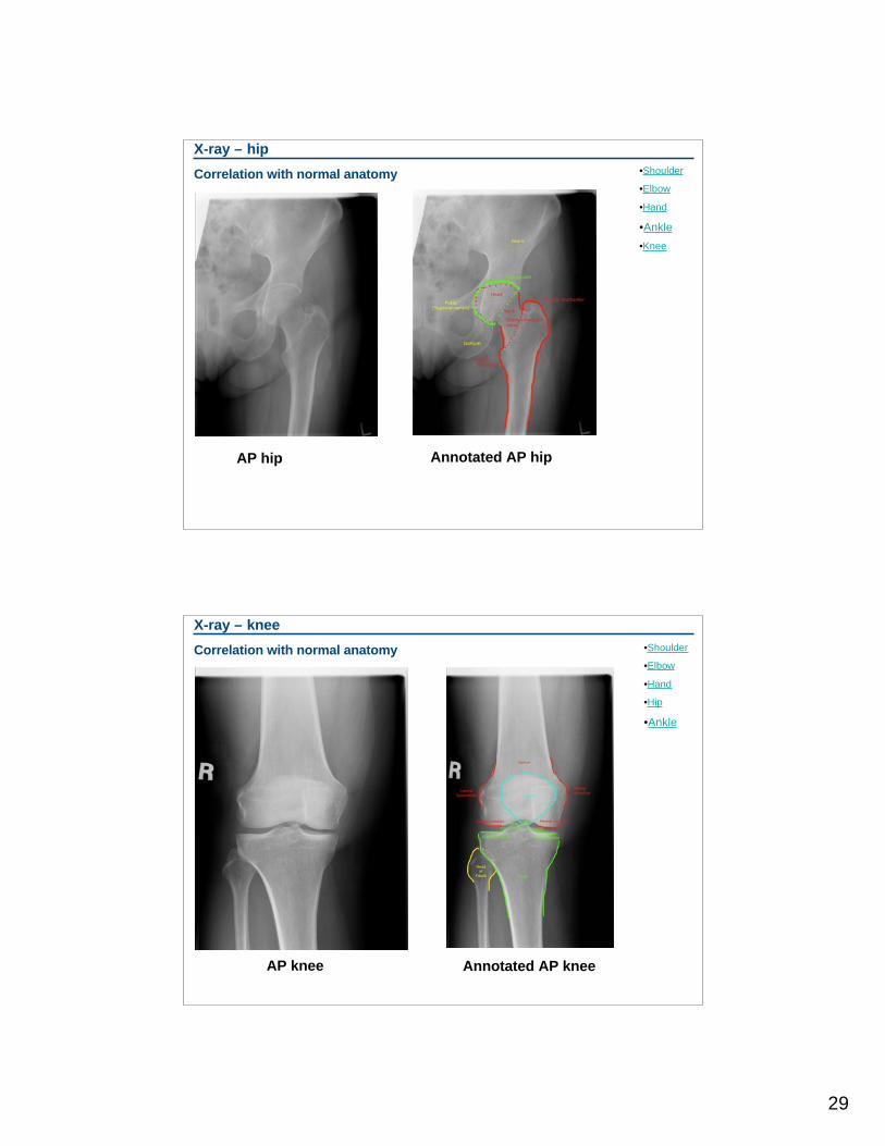
Correlation with normal anatomyIt is important to be able to correlate the structures seen on plain x-rays with a knowledge of
normal anatomy. The bones of the ankle and hindfoot have been outlined on the x-rays
below to illustrate normal relationships. Click on the links provided so see similar images of
the shoulder, elbow, hand, hip and knee.
AP ankle Lateral ankle
25
L5L5S1S1
L4L4
26
The AP view of theThe AP view of the
lumbar spine furtherlumbar spine further
demonstrates thedemonstrates the
severesevere
malalignment of L5malalignment of L5
which effectively lieswhich effectively lies
perpendicular to theperpendicular to the
x-ray beam. Thisx-ray beam. This
has been likened tohas been likened to
the shape ofthe shape of
NapoleonNapoleon
BonaparteBonaparte’’s hat.s hat.
““NapoleonNapoleon’’s hats hat””
L5L5
Summary
Final thoughts
Now that you have worked through the entire module, you should be familiar with the most
common imaging modalities and their potential application to the investigation of
musculoskeletal conditions. In particular, make sure that you are comfortable with an
organized approach to the analysis of standard views of the major anatomic regions in the
body. While you may not have the expertise of a trained radiologist, a systematic approach,
combined with a good understanding of normal anatomy is an important first step in
becoming comfortable with the interpretation of imaging studies. Practice this at every
opportunity as you enter the clinical portion of your training.
27
Congratulations!
You have now completed the Imaging module.
This web-based module was developed by Dr. Lindsay Davidson using Articulate Presenter
based on content developed by Dr. Paul Fenton and Craig Goldie with support from
MEdTech for the School of Medicine at Queen's University
This module is licensed under the Creative Commons Attribution Non-Commercial No
Derivatives license. The module may be redistributed and used provided that credit is given to
the author and it is used for non-commercial purposes only. The contents of this presentation
cannot be changed or used individually.
For more information on the Creative Commons license model and the
specific terms of this license, please visit creativecommons.ca.
Credits
License
X-ray - shoulder
Correlation with normal anatomy
AP shoulderAnnotated AP
shoulder
Clavicle
Humerus
Acromion
Coracoidprocess
Glenoidfossa
Scapula
•Ankle
•Elbow
•Hand
•Hip
•Knee
28
X-ray - elbow
Correlation with normal anatomy
Lateral
elbow
Annotated lateral
elbow
Ulna
Radius
Ulna
Humerus
Capitellum
Olecranon
•Shoulder
•Ankle
•Hand
•Hip
•Knee
X-ray – hand and wrist
Correlation with normal anatomy
AP hand and wrist Annotated AP hand
and wrist
•Shoulder
•Elbow
•Ankle
•Hip
•Knee
29
X-ray – hip
Correlation with normal anatomy
AP hip Annotated AP hip
•Shoulder
•Elbow
•Hand
•Ankle
•Knee
X-ray – knee
Correlation with normal anatomy
AP knee Annotated AP knee
•Shoulder
•Elbow
•Hand
•Hip
•Ankle


![Enema Administration[1]](https://img.pdfslide.us/doc/110x75/55289a1f49795921048b4a43/enema-administration1.jpg)