Embed Size (px)
Citation preview

MINISYMPOSIUM: JUVENILE IDIOPATHIC ARTHRITIS
Imaging of the knee in juvenile idiopathic arthritis
Robert Hemke1 & Nikolay Tzaribachev2& Anouk M. Barendregt1,3 & J. Merlijn van den
Berg3 & Andrea S. Doria4 & Mario Maas1
Received: 29 August 2017 /Accepted: 11 October 2017# The Author(s) 2017. This article is an open access publication
Abstract In juvenile idiopathic arthritis (JIA), imaging is in-creasingly used in clinical practice. In this paper we discussimaging of the knee, the clinically most commonly affectedjoint in JIA. In the last decade, a number of important stepshave been made in the development of imaging outcome mea-sures in children with JIA knee involvement. Ultrasound isundergoing a fast validation process, which should be accom-plished within the next few years. The validation processes ofMRI as an imaging biomarker for clinical trials in the JIA kneeare at an advanced stage, with important data available on thefeasibility, reliability and validity of the Juvenile ArthritisMRI Scoring system. Moreover, both US and MRI data areemerging on the normal appearance of the growing knee joint.
Keywords Children . Juvenile idiopathic arthritis . Knee .
Magnetic resonance imaging . Radiography . Ultrasound
Introduction
The knee joint is the most commonly affected joint in juvenileidiopathic arthritis (JIA) [1]. It can, therefore, be considered anindex joint for evaluation of disease and for monitoring re-sponse to therapy. As in other joints, knee involvement in thescope of JIA is characterized by a swollen, painful, warm jointwith loss of function.
Conventional radiography has played an important role inthe management of JIA, but imaging techniques such as ultra-sonography (US) and MRI are now considered more helpfulfor several reasons. First, the trend towards early suppressionof inflammation to prevent irreversible damage of cartilageand bone has shifted the emphasis from detecting damage(using radiography) to detecting early joint changes of JIA.This drives the need for imaging techniques that are moresensitive than radiography in the evaluation of inflammatoryprocesses as well as early osteochondral changes. In this re-gard, MRI and US play an increasingly important role in eval-uating and monitoring disease activity [2]. Second, the phys-ical examination remains the reference standard for identify-ing disease activity in both daily practice and in clinical trials.However, physical examination has limited reliability, even ifperformed by an experienced observer [3], underpinning thepotential of imaging in helping clinical decision-making.Moreover, advances in therapies have increased the numberof children who reach clinically inactive disease. Further,knowledge about subclinical inflammation and its influenceon the child’s outcome is rising [2].
Conventional radiography
In the clinical setting, conventional radiography still plays animportant role, especially in narrowing the differential
* Robert [email protected]
1 Department of Radiology and Nuclear MedicineAcademic Medical Center, University of AmsterdamMeibergdreef 9, 1105AZ Amsterdam, The Netherlands
2 Pediatric Rheumatology Research InstituteBad Bramstedt, Germany
3 Department of Pediatric Hematology, Immunology,Rheumatology and Infectious Disease,Emma Children’s Hospital AMCUniversity of AmsterdamAmsterdam, The Netherlands
4 Department of Diagnostic Imaging, The Hospital for Sick Children,University of TorontoToronto, ON, Canada
Pediatr Radiol (2018) 48:818–827https://doi.org/10.1007/s00247-017-4015-6
/ Published online: 2013 May 8

diagnosis and in establishing a baseline for disease follow-up.Although radiography provides important information ongrowth disturbances and damage to cartilage and bone, it doesnot show early changes suggestive of active inflammation [4].No validated scoring methods are available for evaluating JIAknee disease activity using radiography. There is little infor-mation on reliability and validity of scores for use in childrenor on the potential limitations of radiographic scoring systemsfor assessing growing joints because of the measurementproperties of the scales [5].
Albeit nonspecific, the presence of joint fluid and synovialthickening can be seen as increased density in the infrapatellarfat pad and suprapatellar region. In the Western population,bone erosions in knee joints in children with JIA are relativelyrare. Because of the availability of more effective treatmentoptions and the relatively large amount of epiphyseal cartilagein knees in growing children, erosive damage is usually onlyseen as a late complication of JIA. When present, bone cystsand bone erosions can be seen. Moreover, loss of articularcartilage can cause gradual joint space narrowing. Jointmalalignment and ankyloses in the knee are extremely rare.
Radiography does have an advantage over ultrasound andMRI in the determination of growth disturbances. Due tohyperaemia, overgrowth of extremities and disturbance ofepiphyseal bone formation can be observed. Moreover, it iseasier to compare bilateral joints using radiography comparedto, for instance, MRI.
Ultrasound
In paediatric rheumatology, ultrasound plays an important rolein narrowing the differential diagnosis and can be useful fortreatment monitoring as well as for guidance for joint injec-tions [6]. It is superior to clinical examination in diagnosingdisease activity and in detecting subclinical disease [7].Because of its relatively low cost and wide accessibility, itallows for assessment of multiple joints within scanning timesthat vary according to the level of detail required for the ex-amination. Limitations to the method include its inability toexamine bone marrow or to reliably detect central erosivechanges given the low penetration of the ultrasound beam tothe central aspect of the joint with high-frequency transducers[8].
Acquisition techniques and ultrasound definitions
For the application of US in paediatric rheumatology, bothgrey-scale B-mode and power/colour Doppler modes shouldbe used in every examination. Also, standard scanning posi-tions should be considered. For standard clinical paediatricultrasound examination of the knee, the child is placed insupine position with a slightly flexed knee. The standard
sagittal view includes the patella inferiorly, the quadricepstendon, and the suprapatellar recess [9]. An axial view in thepopliteal fossa can be considered to exclude a Baker cyst [9].In the scope of research a more extensive ultrasound protocolof the knee can be considered (Table 1).
The main US features characterizing pathology in JIA atthis stage of the development of a US-based outcome measureare synovial thickening and synovial effusion [10]. Synovialthickening (Fig. 1) is defined as abnormal, intra-articular,hypoechoic material that is non-displaceable. Synovial effu-sion (Fig. 1) is defined as an abnormal, intra-articular, anecho-ic or hypoechoic material that is displaceable. When lookingfor a synovial effusion, one should be aware that a change ofpatient position can influence the level of effusion detected.Moreover, a scanning technique avoiding excessive pressureis warranted because joint fluid can shift to another synovialrecess when too much pressure is used [11].
Power/colour Doppler is useful to acquire informationabout tissue perfusion. It is important to clarify that synovitiscan be detected on the basis of B-mode findings (synovialthickening or synovial effusion) alone. Nevertheless powerand colour Doppler have proved to be useful in differentiatingactive (hypervascular) from fibrotic (hypovascular) pannus[11]. In growing children, the sole detection of mildly in-creased vascularity does not allow for the diagnosis of syno-vitis because of the different levels of joint vascularity accord-ing to a child’s age and level of physical activity of the joint[12]. It is important to recognize the presence of such normalvessels in paediatric joints from normal maturation and theseshould not be considered pathological [13].
In children with JIA, loss of cartilage thickness has beendescribed. US has a strong correlation with MRI in the eval-uation of cartilage thickness of the medial and lateral condyleand the intercondylar region, with a rho correlation of 0.70–0.86 [14].
Scoring system
We are currently developing a consensus-based US scoringsystem. The preliminary scoring system consists of threegrades separated for synovitis and synovial perfusion(Table 2). Its reliability will be tested in a patient-basedexercise.
Normal values
Age-related US findings have been described in healthy chil-dren [13, 15]. There are considerable age-related variations inchildren’s joints, including the knee examined by US, whereDoppler depicts vascularity, particularly within the epiphysealcartilage of the children at a younger age. Also, age- andgender-related differences and standard reference values ofthe cartilage thickness of the knee in healthy children have
Pediatr Radiol (2018) 48:818–827 819
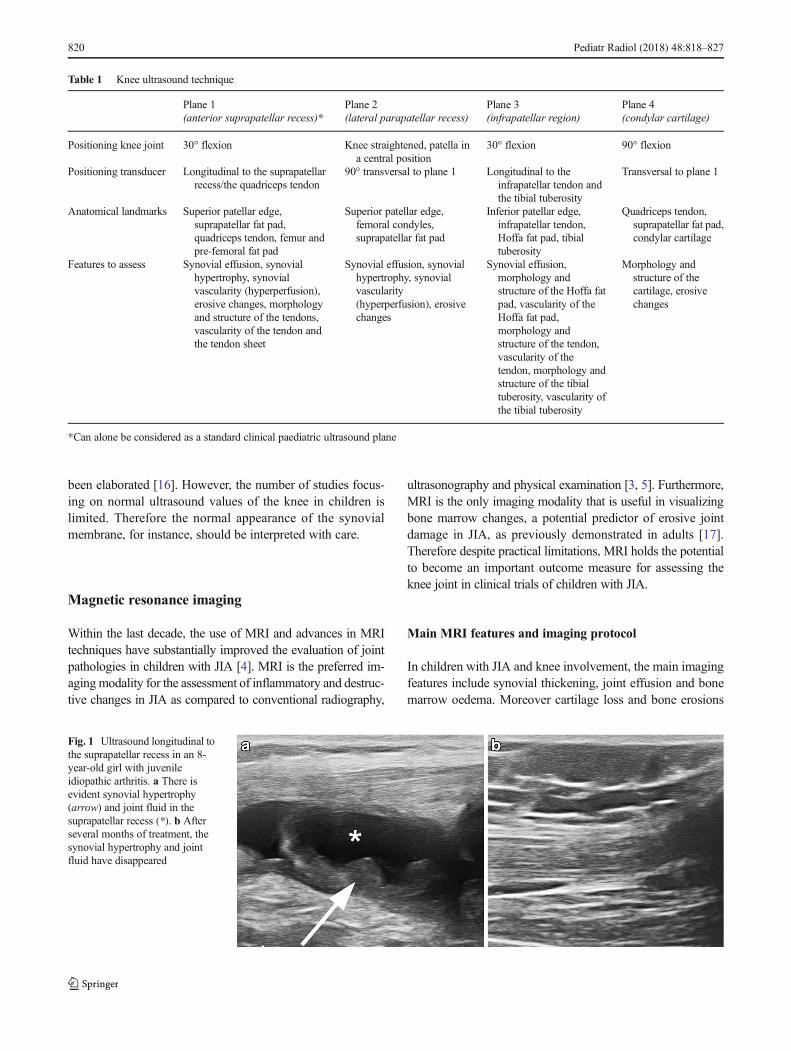
been elaborated [16]. However, the number of studies focus-ing on normal ultrasound values of the knee in children islimited. Therefore the normal appearance of the synovialmembrane, for instance, should be interpreted with care.
Magnetic resonance imaging
Within the last decade, the use of MRI and advances in MRItechniques have substantially improved the evaluation of jointpathologies in children with JIA [4]. MRI is the preferred im-agingmodality for the assessment of inflammatory and destruc-tive changes in JIA as compared to conventional radiography,
ultrasonography and physical examination [3, 5]. Furthermore,MRI is the only imaging modality that is useful in visualizingbone marrow changes, a potential predictor of erosive jointdamage in JIA, as previously demonstrated in adults [17].Therefore despite practical limitations, MRI holds the potentialto become an important outcome measure for assessing theknee joint in clinical trials of children with JIA.
Main MRI features and imaging protocol
In children with JIA and knee involvement, the main imagingfeatures include synovial thickening, joint effusion and bonemarrow oedema. Moreover cartilage loss and bone erosions
Table 1 Knee ultrasound technique
Plane 1(anterior suprapatellar recess)*
Plane 2(lateral parapatellar recess)
Plane 3(infrapatellar region)
Plane 4(condylar cartilage)
Positioning knee joint 30° flexion Knee straightened, patella ina central position
30° flexion 90° flexion
Positioning transducer Longitudinal to the suprapatellarrecess/the quadriceps tendon
90° transversal to plane 1 Longitudinal to theinfrapatellar tendon andthe tibial tuberosity
Transversal to plane 1
Anatomical landmarks Superior patellar edge,suprapatellar fat pad,quadriceps tendon, femur andpre-femoral fat pad
Superior patellar edge,femoral condyles,suprapatellar fat pad
Inferior patellar edge,infrapatellar tendon,Hoffa fat pad, tibialtuberosity
Quadriceps tendon,suprapatellar fat pad,condylar cartilage
Features to assess Synovial effusion, synovialhypertrophy, synovialvascularity (hyperperfusion),erosive changes, morphologyand structure of the tendons,vascularity of the tendon andthe tendon sheet
Synovial effusion, synovialhypertrophy, synovialvascularity(hyperperfusion), erosivechanges
Synovial effusion,morphology andstructure of the Hoffa fatpad, vascularity of theHoffa fat pad,morphology andstructure of the tendon,vascularity of thetendon, morphology andstructure of the tibialtuberosity, vascularity ofthe tibial tuberosity
Morphology andstructure of thecartilage, erosivechanges
*Can alone be considered as a standard clinical paediatric ultrasound plane
Fig. 1 Ultrasound longitudinal tothe suprapatellar recess in an 8-year-old girl with juvenileidiopathic arthritis. a There isevident synovial hypertrophy(arrow) and joint fluid in thesuprapatellar recess (*). b Afterseveral months of treatment, thesynovial hypertrophy and jointfluid have disappeared
820 Pediatr Radiol (2018) 48:818–827

— although relatively rare in paediatric patients — can beobserved. Tendinopathy, enthesopathy, inhomogeneity of theinfra-patellar fat pad and bone cysts can be seen, as well, butthese abnormalities are relatively uncommon in the knee joint.Moreover, the reliability of scoring these secondary features isunsatisfactory [18]. Table 3 shows an MRI protocol for eval-uating the JIA knee joint.
Synovial inflammation leading to synovial thickening isthe principal pathological process in JIA, and the presenceof synovial thickening on knee MRI is associated with theclinical onset of JIA [19]. On MRI, the inflamed synovialmembrane is thickened and irregular, and its outline maybe wavy. In the knee, a synovial thickness of >2 mm isconsidered pathological. The central locations in the knee(around the cruciate ligaments and retropatellar andsuprapatellar areas, Fig. 2) are most commonly affected[20]. The signal intensity of this thickened synovial mem-brane is low to intermediate on T1-weighted images andhigh on T2-weighted images, similar to joint effusion. T1-weighted images after administration of gadolinium-basedcontrast agents provide better differentiation between jointeffusion and synovial thickening [21]. Omitting intrave-nous gadolinium-based contrast agents in the MRI assess-ment of joints in JIA is therefore not advised.
Although not specific for JIA, joint effusion (Fig. 2) canfrequently be found in children with JIA [22]. On MRI, effu-sion demonstrates high signal intensity on fluid-sensitive im-ages and low signal intensity on T1-weighted images. Jointeffusions are predominantly located in the suprapatellar andcentral joint recesses.
MRI is the state-of-the-art imaging technique to visualizechanges in bone marrow suggestive of bone marrow oedema(Fig. 3). Bone marrow oedema can be seen as lesions withpoorly definedmargins within trabecular bone. It is marked byhigh signal intensity on T2-weighted fat-saturated images andlow signal intensity on T1-weighted images. In rheumatoidarthritis, longitudinal studies have shown that the presenceof bone marrow oedema is a key predictor of early erosivejoint damage in adults with rheumatoid arthritis [17].Therefore bone marrow oedema and synovial thickening areconsidered to be the most sensitive MRI features for monitor-ing disease activity in rheumatoid arthritis [23–25]. Howeverto our knowledge there are a paucity of longitudinal studies, ifany, focused on the prognostic value of bone marrow oedemain children with JIA.
Cartilage loss may be seen as areas of increased watercontent (proton-density/T2 hyperintense) in the articular car-tilage or contour abnormalities, defects or thinning of the
Table 2 Suggested scoringsystem of ultrasound for the kneejoint
Feature Definition Locations Scale
Synovitis B mode Comprises synovial hypertrophyand synovial effusion
Suprapatellar recess,parapatellar recess
Grade 1 = mild
Grade 2 = moderate
Grade 3 = severe
Synovitis power/colourDoppler mode
Comprises synovial hypertrophyand hypervascularity*
Suprapatellar recess,parapatellar recess
Grade 1 = mild
Grade 2 = moderate
Grade 3 = severe
*Note that hypervascularity without synovial hypertrophy does not count for synovitis
Synovitis = synovial hypertrophy and/or synovial effusion. Synovial hypertrophy is defined as abnormal, intra-articular, hypoechoic material that is non-displaceable. Synovial effusion is defined as abnormal, intra-articular, orhypoechoic material that is displaceable
Table 3 Knee MRI protocol
Sequence Plane Goal Required or optional
T2 FS or STIR (mDixon)* Sagittal Joint effusion, bone marrow oedema, bone erosions Required
T2 FS or STIR (mDixon)* Coronal Bone marrow oedema, bone erosions Required
T1 (mDixon)* Coronal Bone marrow oedema, bone erosions Required
Gradient echo / PD Sagittal Cartilage loss Required
T1 FS post-Gd Axial Synovial thickening, joint effusion Required
Gradient echo (3-D) Axial Cartilage loss Optional
T1 FS pre-Gd Axial Synovial thickening, joint effusion Optional
T1 FS post-Gd Sagittal Synovial thickening, joint effusion Optional
*mDixon best option if available
FS fat-suppressed, Gd gadolinium, PD proton density, STIR short tau inversion recovery, T1 T1-weighted spin echo, T2 T2-weighted
Pediatr Radiol (2018) 48:818–827 821

cartilage. Gradient-echo and proton-density-weighted se-quences provide good contrast in the cartilage structure andcan be used to evaluate the cartilage–synovial fluid interfaceand the subchondral bone.
MRI clearly shows the difference between changes in thearticular cartilage and bone erosions. Bone erosions can beseen on T1-weighted images as loss of the normal low signalintensity of cortical bone and loss of the normal high signalintensity of trabecular bone (Fig. 3). Typically they have sharpmargins. On T2-weighted images bone erosions appear ashypointense lesions, in contrast to subchondral cysts, whichshow as hyperintense signal on fluid-sensitive images.
Scoring methods
Although MRI is the preferred imaging modality for detect-ing both inflammatory as well as destructive changes in JIA,international experiences with the use of MRI as an outcome
measure in JIA are limited. Subsequently, this technique isunderused in both clinical practice and research. In order tofurther internationally validate MRI as an outcome measurein JIA, a special interest group on Outcome Measures inRheumatology on MRI was created in 2011 [26]. Withinthis special interest group on MRI in JIA, three workinggroups were initiated for further assessment of MRI scoringsystems: one group focuses on temporomandibular joints,another on small joints and the third on large joints (primar-ily the knee joint).
Reliability
In the last decade, two MRI scoring systems have been devel-oped for the assessment of the paediatric knee joint: (1) theJuvenile Arthritis MRI Scoring system for use in JIA [27] and(2) the International Prophylaxis Study Group MRI Scoringsystem focusing on haemophilic arthropathy [28].
Fig. 2 Axial T1-weighted MRI of the knee with fat suppression aftergadolinium-based contrast administration in three patients with juvenileidiopathic arthritis shows synovial hypertrophy in the central locations ofthe knee. a Enhancing synovial hypertrophy in the retropatellar region(arrow) in an 9-year old girl. b Enhancing synovial hypertrophy in the
suprapatellar region (arrow) in an 11-year old boy. Notice the non-enhancing low-signal-intensity joint fluid in thesuprapatellar recess (*).c Enhancing synovial hypertrophy around the cruciate ligaments (arrow)in a 15-year old girl
Fig. 3 Sagittal MR images of theknee in a 16-year-old girl withpoly-articular juvenile idiopathicarthritis. a T2-weighted sagittalMRI with fat suppression showsbone marrow oedema in thefemur (arrow) and tibial plateau.b T1-weighted sagittal MRIshows bone erosions in the tibialplateau (arrow), with irregularsignal intensity of the corticalbone and loss of the normal highsignal intensity of trabecular bone
822 Pediatr Radiol (2018) 48:818–827

Recently the Outcome Measures in Rheumatology(OMERACT) special interest group onMRI in JIA conducteda reliability study evaluating both scoring methods [18]. Thisstudy showed good inter-observer reliability for MRI scoresfocusing on active disease (e.g., synovial thickening, jointeffusion and bone marrow changes) and moderate to substan-tial inter-observer reliability for scores focusing on damage(e.g., cartilage lesions, bone erosions). Finally, the group pro-posed a combined juvenile arthritis MRI scoring system(JAMRIS)— shown in Table 4— including items from boththe original Juvenile Arthritis MRI Scoring and theInternational Prophylaxis Study Group systems [18].
Besides this international reliability study, the intra-observer and inter-observer reliability of the originalJuvenile Arthritis MRI Scoring system has been evaluated,and both proved to be reliable [27]. These data are trustworthy,especially regarding MRI features focusing on active disease.Caution is needed with regard to MRI characteristics focusingon damage because these features are relatively uncommonlyobserved in JIA patients.
Feasibility
Although MRI has some practical limitations such as limitedavailability in some regions/countries and the challenge ofmaintaining the same position for a prolonged period of time,it has proved feasible to perform contrast-enhanced knee MRIin children with JIA as young as 5 years old without the use ofanaesthesia or sedation [29]. Moreover the use of the originalJuvenile Arthritis MRI Scoring system proved feasible be-cause the scoring takes an acceptable median of 6.6 min perpatient [21].
Construct and clinical validity
Within the last few years, two studies have focused on thesensitivity to changes in disease activity using the originalJuvenile Arthritis MRI Scoring system [27, 30]. In both stud-ies, with a follow-up period of 1 year, improvement of clinicalJIA disease activity scores was associated with a significantdecrease in MRI-based synovial thickening scores. These re-sults represent good responsiveness of the Juvenile ArthritisMRI Scoring system, which is an important measure of valid-ity. Moreover the Juvenile Arthritis MRI Scoring synovialthickening score proved useful in discriminating clinically ac-tive from clinically inactive JIA patients [31], indicating gooddiscriminant validity.
Next to the responsiveness and discriminant validity, theclinical validity has also been evaluated. In a study focusingon whether clinical, laboratory or MRI measures were ableto differentiate JIA with active arthritis from other causes ofnon-infectious arthritis in a group of patients with clinicalsigns of early arthritis, multivariate analysis showed that
MRI-based synovial thickening was independently associat-ed with JIA (odds ratio [OR] 6.58, 95% confidence interval2.36–18.33) [19].
Normal values
Because synovial inflammation is the hallmark of disease ac-tivity in JIA, it is important to determine the normal appear-ance and thickness of the synovial membrane in children. Asstated, an intravenous injection of a gadolinium-based contrastagent is warranted for reliable evaluation of the synovial mem-brane. Therefore data on normal values for the synovial mem-brane in healthy children are sparse. Two studies that focusedon the contrast-enhanced appearance of the synovial mem-brane in children have been performed: (1) A study byNusman et al. [32] looked at the synovial membrane of theknee in children with inflammatory bowel disease who wereclinically unaffected by arthritis; (2) Hemke et al. [33] deter-mined the synovial membrane in knees of healthy children,using a standardized imaging protocol with post-contrast im-ages obtained in the early phase (<5 min). The latter studyshowed that the normal synovial membrane thickness mea-sures a maximum of 1.8 mm. The membrane was thickestaround the cruciate ligaments, and retropatellar andsuprapatellar recesses. The study by Nusman et al. showed athickened synovial membrane (>2 mm) in more than half ofpatients with inflammatory bowel disease; however they didnot observe any synovial thickness >4 mm. In all probability,the results from Hemke et al. are a more valid reflection of thetrue normal appearance of the synovial membrane becausethis study included healthy children (compared to patientswith inflammatory bowel disease) and obtained post-contrastimages in the early phase. Therefore, the Juvenile ArthritisMRI Scoring cut-off value of >2 mm for synovial thickeningcan be considered a valid measure.
Some joint fluid in the knee can be seen in the majority ofhealthy children. The largest pockets of normal joint fluid inhealthy children are located in the central locations of the knee— around the cruciate ligaments and retropatellar region [33].The mean diameter of the largest pocket of joint fluid in kneesof healthy children is about 3 mm [15, 33].
Bone marrow changes suggestive of bone marrow oedemain knees of healthy children are relatively uncommon. In astudy of 57 healthy children, bonemarrow changes suggestiveof bone marrow oedema were observed in 3 healthy childrenonly. In all three children, the bone marrow changes werelocated in the apex patellae [33]. The presence of bone mar-row changes suggestive of bone marrow oedema in the apexpatellae in children with JIA should, therefore, be interpretedwith care.
Moreover, zones of hematopoietic red bone marrow in thedistal diaphysis and metaphysis of the femur might be seen asflame-like regions with signal characteristics fitting with bone
Pediatr Radiol (2018) 48:818–827 823

marrow oedema. Typically, these marrow flames originatefrom the physis and have straight vertical margins [34].These hematopoietic bone marrow flames are a normal find-ing in growing children and should not be mistaken for bonemarrow oedema. So-called speckled bone marrow is anothernormal variant that might be mistaken for pathology. Smallspots with bone marrow oedema signal characteristics are
predominantly located in the feet and ankles of children youn-ger than 15 years, although the spots can be seen in the tibiaplateau and the distal epiphysis of the femur as well [35]. Thespeckled appearance might be caused by focal regions of re-sidual hematopoietic bone marrow or physiological stress,possibly related to weight-bearing or altered biomechanicsduring normal growth.
Table 4 Combined juvenile arthritis MRI scoring system
Feature Definition Locations Scale
Synovial thickeninga An area of the synovial compartment thatshows a thickened synovial membraneand which can show enhancement afterintravenous gadolinium administration
Six locations:patellofemoral area, suprapatellar recesses,
infrapatellar fat pad, adjacent to theanterior and posterior cruciateligaments, medial posterior condyle,and lateral posterior condyle
(0) normal, ≤2 mm(1) mild, >2 mm to ≤4 mm(2) moderate/severe, >4 mmTotals result in a minimum
score of 0 and a maximumscore of 12
Joint effusionb An increased amount of fluid within thesynovial compartment with high signalintensity on T2-weighted images andlow signal intensity on T1-weightedimages. Joint effusion has nopost-gadolinium enhancement
The maximal diameter of the largestpocket of joint effusion is scored
(0) normal, ≤3 mm(1) mild, >3 mm to ≤5 mm(2) moderate/severe, >5 mmTotals result in a minimum
score of 0 and a maximumscore of 2
Bone marrow oedemaa An abnormality within the trabecular boneof the epiphysis, with ill-definedmargins and high signal intensity onT2-weighted fat-saturated images andlow signal intensity on T1-weightedimages
Eight locations:lateral patella, medial patella, medial femur
condyle, lateral femur condyle, medialweight-bearing region of the femur,lateral weight-bearing region of thefemur, medial tibia plateau, lateral tibiaplateau
Presence of bone marrowedema is scoredsemi-quantitatively basedon the subjectivelyestimated percentage ofinvolved bone volume ateach site as follows:
(0) none(1) <10% of the whole bone
volume(2) ≥10–25% of the whole
bone volume(3) >25% of the whole bone
volumeTotals result in a minimum
score of 0 and a maximumscore of 24
Cartilage lossb Loss of cartilaginous tissue either focally(superficial or deep) or diffusely
Scored at the most severely affectedlocation
(0) none(1) any loss(2) >50% volume loss(3) full-thickness loss(4) full-thickness loss >50% of
surfaceTotals result in a minimum
score of 0 and a maximumscore of 4
Bone erosionsb A sharply marginated bone lesion withcorrect juxta-articular localization,typical signal characteristics and visiblein two planes with a cortical break in atleast one plane. On T1-weighted imagesthere is a loss of the normal low signalintensity of cortical bone and loss of thenormal high signal intensity oftrabecular bone
Scored at the most severely affectedlocation
(0) none(1) mild, any loss(2) moderate/severe, >50%
surface involvementTotals result in a minimum
score of 0 and a maximumscore of 2
a Original Juvenile Arthritis MRI Scoring itembOriginal International Prophylaxis Study Group item
824 Pediatr Radiol (2018) 48:818–827

The thickness of the normal articular cartilage in knees ofgrowing children differs with age. As expected, the cartilage isnormally thicker in younger children compared to older chil-dren. A study performed by Keshava et al. [15] clearly dem-onstrates these differences among different age groups inhealthy boys. Moreover they showed that the normal thick-ness of the articular cartilage differs per location within theknee joint [15]. In the distal femur, ossification usually startsin the centre of the cartilaginous epiphysis. Because the newlyformed ossification centre contains hematopoietic bone mar-row, its signal intensity is the same as that of red marrow in theadjacent distal femoral metaphysis [36]. The ossification cen-tre enlarges from endochondral bone development. Adjacentcartilage cells undergo hypertrophy during endochondral os-sification, which results in increased signal intensity on T2-weighted images [34]. This area of normal high signal inten-sity is most obvious in the posterior part of the distal femoralepiphysis and can be quite discrete [37]. Furthermore, it isimportant to be aware of the high prevalence of ossificationvariants of the femoral condyle among boys ages 2–12 yearsand girls ages 2–10 years; this should not be considered path-ological [37].
Future work
The validation process of US in children with JIA is mov-ing forward. Despite the relatively easy applicability ofUS, until now there has been no specific US knee scoringsystem. Rather the US knee examination is part of a moreglobal US score covering multiple joints with respect todisease activity. The Outcome Measures in Rheumatologypaediatric ultrasound working sub-group has performed anumber of steps towards the validation of US as an out-come measure in paediatric rheumatology. Next importantsteps of the work of the sub-group include multicentrereliability testing of the newly created score tested in asingle-centre prospective study. In this case, sensitivity tochange will also be tested. The sub-group has started itswork in tendons and entheses and should follow theOutcome Measures in Rheumatology Filter 2.0 criteriafor the validation process. Another important step willbe to develop scores incorporating osteochondral changes,where US is expected to be beneficial in evaluating pe-ripheral cartilage. Its utility in detecting erosive changesmight also be tested, and compared to that of MRI andradiography. This underlines the importance of close col-laboration between US and MRI working groups, whichis planned to start in 2018 with a joint symposium.
Although the first steps towards developing evidence-based guidelines for MRI data acquisition and interpretationhave beenmade, further cooperation is necessary. The use of ajuvenile arthritis MRI scoring system for the knee as an
outcome measure in daily practice and clinical trials is prom-ising. Thus far, juvenile arthritis MRI scoring has only beeninternationally tested in JIA patients visiting academic paedi-atric rheumatology centres in the Netherlands and Canadawith full access to high-quality treatment. This has resultedin a population of studied JIA patients with only mild to mod-erate disease activity. Consequently, the presence of destruc-tive changes of cartilage and bone has been relatively low inthe studies performed until now. To evaluate the value ofjuvenile arthritis MRI scoring as a sensitive measure regardingdestructive changes, further international collaboration is war-ranted, especially among research centres with access to moreseverely affected JIA patients. Furthermore, collaborationshould focus on developing an MRI atlas of healthy joints inchildren, obtaining agreement on an optimal imaging protocolfor the knee and further validating scoring methods.Interaction between researchers and health professionals inJIA imaging is essential to obtain international consensusand continuous improvement of MRI outcome measures.Such collaboration is expected to be very fruitful under theumbrella of an international, well-accepted collaborative in-ternational group such as the Outcome Measures inRheumatology working group.
According to the Quantification Imaging BiomarkerAlliance of the Radiological Society of North America, quan-titative imaging techniques covering the full spectrum of im-aging have to be developed and validated to provide moreobjective tools to measure disease activity throughout allfields of medicine [38]. With respect to this, theQuantification Imaging Biomarker Alliance has formed theContrast-enhanced Ultrasound Working Group to exploreUS contrast agent applications and use specific quantificationsoftware to evaluate disease activity and increase objectivityof the results of the US technique.
Forthcoming research is expected to shed more light on thesuitability of advanced quantitative MRI techniques for eval-uating inflammatory and destructive changes in the JIA knee,including dynamic contrast-enhanced MRI, T2-mapping anddiffusion-weighted imaging [39–44]. Currently these ad-vanced imaging techniques are used particularly in the contextof research and to a lesser extent in daily practice. The exactvalue of advanced MRI techniques in children with JIA has tobe determined in larger prospective studies. To be viable indaily practice, these imaging techniques should be sensitive tochange on which evidence is limited [45]. It is important tofurther develop and implement advanced imaging techniquesin clinical practice. For example, the contrast-free approach ofdiffusion-weighted imaging is highly desirable in clinicalpractice because it could substantially improve patient careby optimizing MRI feasibility in paediatric JIA patients;moreover, establishing normal values for the MRI atlas ofhealthy joints is less ethically compromising if contrast-freeimaging is available.
Pediatr Radiol (2018) 48:818–827 825

Conclusion
In this paper, we discussed the status of imaging the JIAknee. In the last decade a number of important steps havebeen made in the development of imaging outcome mea-sures in children with JIA knee involvement. Ultrasoundis undergoing a fast validation process, which should beaccomplished within the next few years. The validationprocesses of MRI as an imaging biomarker for clinicaltrials in the JIA knee are at an advanced stage, with im-portant data forthcoming from both single-centre as wellas international multi-centre studies on the feasibility, re-liability and validity of the Juvenile Arthritis MRI Scoringsystem. Moreover, data are emerging in both US and MRIon the normal appearance of the growing knee joint.However, future research is clearly needed, especiallywithin the scope of evaluating the value of JuvenileArthritis MRI Scoring as a sensitive measure for assessingdestructive changes. The ultrasound score needs furthervalidation, as well. Moreover, further validation is neededfor promising advanced quantitative US and MRI tech-niques for the evaluation of inflammatory and destructivechanges in JIA.
References
1. Hemke R, Nusman CM, van der Heijde DM et al (2015) Frequencyof joint involvement in juvenile idiopathic arthritis during a 5-yearfollow-up of newly diagnosed patients: implications for MR imag-ing as outcome measure. Rheumatol Int 35:351–357
2. Colebatch-Bourn AN, Edwards CJ, Collado P et al (2015) EULAR-PReS points to consider for the use of imaging in the diagnosis andmanagement of juvenile idiopathic arthritis in clinical practice. AnnRheum Dis 74:1946–1957
3. Guzman J, Burgos-Vargas R, Duarte-Salazar C et al (1995)Reliability of the articular examination in children with juvenilerheumatoid arthritis: interobserver agreement and sources of dis-agreement. J Rheumatol 22:2331–2336
4. Miller E, Uleryk E, Doria AS (2009) Evidence-based outcomes ofstudies addressing diagnostic accuracy ofMRI of juvenile idiopath-ic arthritis. AJR Am J Roentgenol 192:1209–1218
5. Doria AS, Babyn PS, Feldman B (2006) A critical appraisal ofradiographic scoring systems for assessment of juvenile idiopathicarthritis. Pediatr Radiol 36:759–772
6. Magni-Manzoni S, Collado P, Jousse-Joulin S et al (2014) Currentstate of musculoskeletal ultrasound in paediatric rheumatology: re-sults of an international survey. Rheumatology 53:491–496
7. Rebollo-Polo M, Koujok K, Weisser C et al (2011) Ultrasoundfindings on patients with juvenile idiopathic arthritis in clinicalremission. Arthritis Care Res 63:1013–1019
8. Malattia C, Damasio MB, Magnaguagno F et al (2008) Magneticresonance imaging, ultrasonography, and conventional radiographyin the assessment of bone erosions in juvenile idiopathic arthritis.Arthritis Rheum 59:1764–1772
9. Carmichael J, Rosendahl K (2016) Musculoskeletal imaging. In:Beek E, Rijn RR (eds) Diagnostic pediatric ultrasound. GeorgThieme Verlag, Stuttgart
10. Roth J, Jousse-Joulin S, Magni-Manzoni S et al (2015) Definitionsfor the sonographic features of joints in healthy children. ArthritisCare Res 67:136–142
11. Martinoli C, Bianchi S (2007) Knee. In: Bianchi S, Martinoli C(eds) Ultrasound of the musculoskeletal system. Springer, Berlin
12. Roth J, Ravagnani V, Backhaus M et al (2016) Preliminary defini-tions for the sonographic features of synovitis in children. ArthritisCare Res 69:1217–1223
13. Collado P, Vojinovic J, Nieto JC et al (2016) Toward standardizedmusculoskeletal ultrasound in pediatric rheumatology: normal age-related ultrasound findings. Arthritis Care Res 68:348–356
14. Pradsgaard DO, Fiirgaard B, Spannow AH et al (2015) Cartilagethickness of the knee joint in juvenile idiopathic arthritis: compar-ative assessment by ultrasonography and magnetic resonance im-aging. J Rheumatol 42:534–540
15. Keshava SN, Gibikote SV, Mohanta A et al (2015) Ultrasound andmagnetic resonance imaging of healthy paediatric ankles and knees:a baseline for comparison with haemophilic joints. Haemophilia 21:e210–e222
16. Spannow AH, Pfeiffer-Jensen M, Andersen NT et al (2010)Ultrasonographic measurements of joint cartilage thickness inhealthy children: age- and sex-related standard reference values. JRheumatol 37:2595–2601
17. McQueen FM (2007) A vital clue to deciphering bone pathology:MRI bone oedema in rheumatoid arthritis and osteoarthritis. AnnRheum Dis 66:1549–1552
18. Hemke R, Tzaribachev N, Nusman CM et al (2017) Magnetic res-onance imaging (MRI) of the knee as an outcome measure in juve-nile idiopathic arthritis: an OMERACT reliability study on MRIscales. J Rheumatol 44:1224–1230
19. Hemke R, Kuijpers TW, Nusman CM et al (2015) Contrast-enhanced MRI features in the early diagnosis of juvenile idiopathicarthritis. Eur Radiol 25:3222–3229
20. Nusman CM, Hemke R, Schonenberg D et al (2014) Distributionpattern of MRI abnormalities within the knee and wrist of juvenileidiopathic arthritis patients: signature of disease activity. AJR Am JRoentgenol 202:W439–W446
21. Hemke R, Kuijpers TW, van den Berg JM et al (2013) Thediagnostic accuracy of unenhanced MRI in the assessment ofjoint abnormalities in juvenile idiopathic arthritis. Eur Radiol23:1998–2004
22. Gylys-Morin VM, Graham TB, Blebea JS et al (2001) Knee in earlyjuvenile rheumatoid arthritis: MR imaging findings. Radiology220:696–706
23. Haavardsholm EA, Bøyesen P, Østergaard M et al (2008) Magneticresonance imaging findings in 84 patients with early rheumatoidarthritis: bone marrow oedema predicts erosive progression. AnnRheum Dis 67:794–800
24. Conaghan PG, O'Connor P, McGonagle D et al (2003) Elucidationof the relationship between synovitis and bone damage: a random-ized magnetic resonance imaging study of individual joints in pa-tients with early rheumatoid arthritis. Arthritis Rheum 48:64–71
826 Pediatr Radiol (2018) 48:818–827
Compliance with ethical standards
Conflicts of interest None
Open Access This article is distributed under the terms of the CreativeCommons At t r ibut ion 4 .0 In te rna t ional License (h t tp : / /creativecommons.org/licenses/by/4.0/), which permits unrestricted use,distribution, and reproduction in any medium, provided you give appro-priate credit to the original author(s) and the source, provide a link to theCreative Commons license, and indicate if changes were made.

25. Hetland ML, Ejbjerg B, Horslev-Petersen K et al (2009) MRI boneoedema is the strongest predictor of subsequent radiographic pro-gression in early rheumatoid arthritis. Results from a 2-yearrandomised controlled trial (CIMESTRA). Ann Rheum Dis 68:384–390
26. Hemke R,Doria AS, Tzaribachev N et al (2014) Selecting magneticresonance imaging (MRI) outcomemeasures for juvenile idiopathicarthritis (JIA) clinical trials: first report of the MRI in JIA specialinterest group. J Rheumatol 41:354–358
27. Hemke R, van Rossum MA, van Veenendaal M et al (2013)Reliability and responsiveness of the juvenile arthritis MRI scoring(JAMRIS) system for the knee. Eur Radiol 23:1075–1083
28. Feldman BM, Funk S, Lundin B et al (2008) Musculoskeletal mea-surement tools from the international prophylaxis study group(IPSG). Haemophilia 14:162–169
29. Hemke R, van Veenendaal M, Kuijpers TWet al (2012) Increasingfeasibility and patient comfort of MRI in children with juvenileidiopathic arthritis. Pediatr Radiol 42:440–448
30. Hemke R, van Veenendaal M, van den Berg JM et al (2014) One-year followup study on clinical findings and changes in magneticresonance imaging-based disease activity scores in juvenile idio-pathic arthritis. J Rheumatol 41:119–127
31. Hemke R, Maas M, van Veenendaal M et al (2014) Contrast-enhancedMRI compared with the physical examination in the eval-uation of disease activity in juvenile idiopathic arthritis. Eur Radiol24:327–334
32. Nusman CM, Hemke R, Benninga MA et al (2016) Contrast-enhanced MRI of the knee in children unaffected by clinical arthri-tis compared to clinically active juvenile idiopathic arthritis pa-tients. Eur Radiol 26:1141–1148
33. Hemke R, van den Berg JM, Nusman CM et al (2017) Contrast-en-hanced MRI findings of the knee in healthy children; establishingnormal values. Eur Radiol. https://doi.org/10.1007/s00330-017-5067-6
34. Laor T, Jaramillo D (2009) MR imaging insights into skeletal mat-uration: what is normal? Radiology 250:28–38
35. Shabshin N, SchweitzerME,MorrisonWB et al (2006) High-signalT2 changes of the bone marrow of the foot and ankle in children:red marrow or traumatic changes? Pediatr Radiol 36:670–676
36. Waitches G, Zawin JK, Poznanski AK (1994) Sequence and rate ofbone marrow conversion in the femora of children as seen on MRimaging: are accepted standards accurate? AJR Am J Roentgenol162:1399–1406
37. Jans L, Jaremko J, Ditchfield M et al (2012) Ossification variants ofthe femoral condyles are not associated with osteochondritisdissecans. Eur J Radiol 81:3384–3389
38. Radiological Society of North America (2017) QuantitativeImaging Biomarkers Alliance web page. https://www.rsna.org/QIBA/. Accessed 28 Sept 2017
39. Hilbert F, Holl-Wieden A, Sauer A et al (2017) Intravoxel incoher-ent motionmagnetic resonance imaging of the knee joint in childrenwith juvenile idiopathic arthritis. Pediatr Radiol 47:681–690
40. Barendregt AM, Nusman CM, Hemke R et al (2015) Feasibility ofdiffusion-weighted magnetic resonance imaging in patients withjuvenile idiopathic arthritis on 1.0-T open-bore MRI. SkeletRadiol 44:1805–1811
41. Hemke R, Lavini C, Nusman CM et al (2014) Pixel-by-pixel anal-ysis of DCE-MRI curve shape patterns in knees of active and inac-tive juvenile idiopathic arthritis patients. Eur Radiol 24:1686–1693
42. Nusman CM, Hemke R, Lavini C et al (2017) Dynamic contrast-enhanced magnetic resonance imaging can play a role in predictingflare in juvenile idiopathic arthritis. Eur J Radiol 88:77–81
43. Nusman CM, Lavini C, Hemke R et al (2017) Dynamic contrast-enhanced magnetic resonance imaging of the wrist in children withjuvenile idiopathic arthritis. Pediatr Radiol 47:205–213
44. Barendregt AM, van Gulik EC, Lavini C et al (2017) Diffusion-weighted imaging for assessment of synovial inflammation in juve-nile idiopathic arthritis: a promising imaging biomarker as an alter-native to gadolinium-based contrast agents. Eur Radiol. https://doi.org/10.1007/s00330-017-4876-y
45. Workie DW, Graham TB, Laor T et al (2007) Quantitative MRcharacterization of disease activity in the knee in children withjuvenile idiopathic arthritis: a longitudinal pilot study. PediatrRadiol 37:535–543
Pediatr Radiol (2018) 48:818–827 827