Embed Size (px)
Citation preview
Imaging of HeadacheWhat to Do if I Cannot Find
Any Clue?
Prof. Siriporn Hirunpat, MD
Prince of Songkla University
Case Based Discussion
Case 1
• A 38-year-old man presented with a sudden onset of severe headache & feeling of almost syncope (presyncope).
• The patient was previously healthy and had no underlying disease.
• On PE, he had full consciousness with BP 130/80 mm Hg.
• No evidence of neurological deficit was found and all basic investigations revealed normal results.
• He was sent for an MRI and MRA in order to search for the cause of his severe headache, such as ruptured aneurysm, and to exclude the possibility of minor stroke.
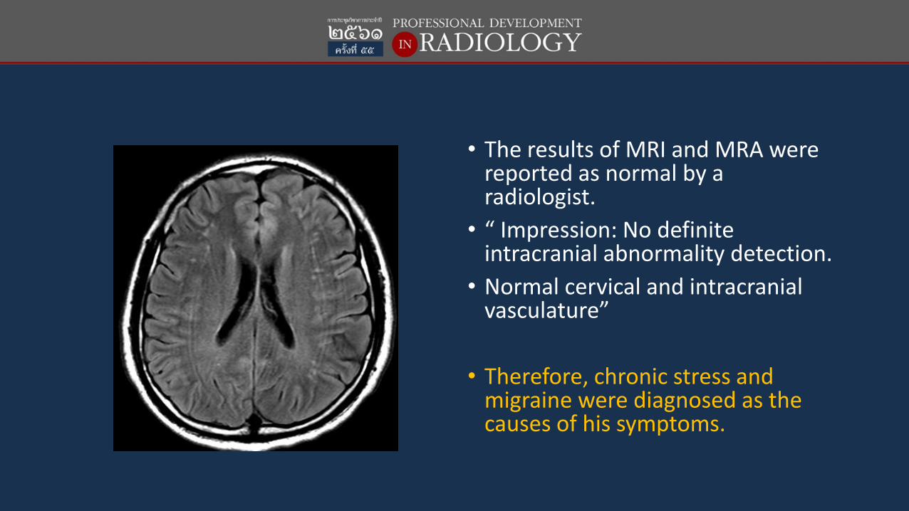
• The results of MRI and MRA were reported as normal by a radiologist.
• “ Impression: No definite intracranial abnormality detection.
• Normal cervical and intracranial vasculature”
• Therefore, chronic stress and migraine were diagnosed as the causes of his symptoms.
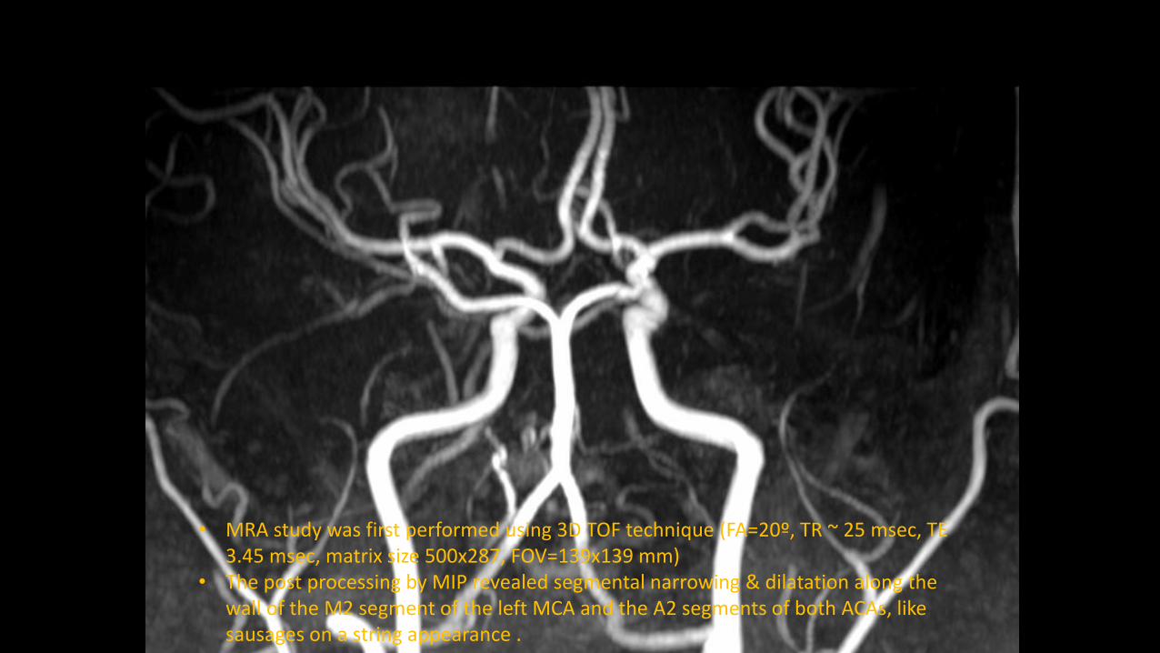
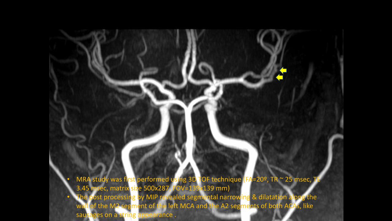
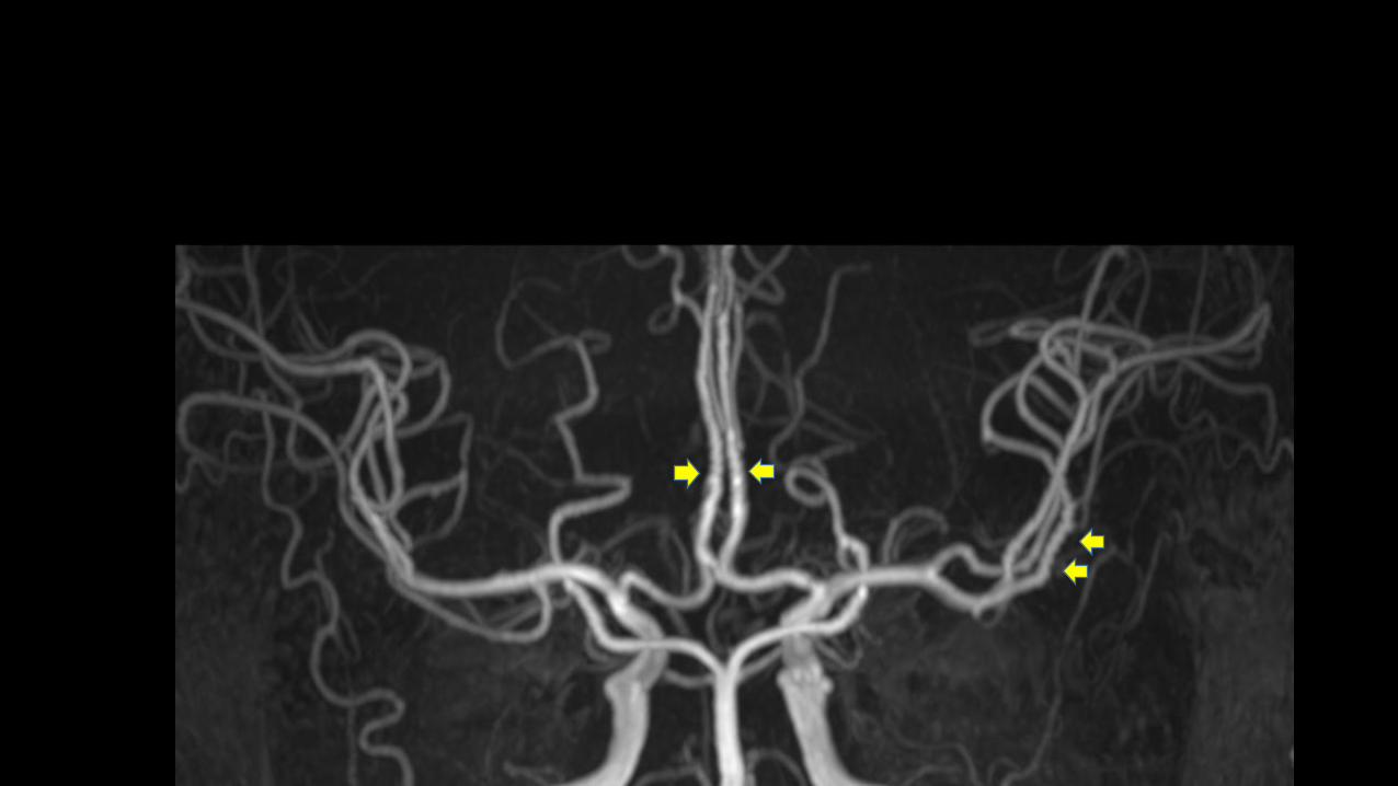
• MRA study was first performed using 3D TOF technique (FA=20º, TR ~ 25 msec, TE 3.45 msec, matrix size 500x287, FOV=139x139 mm)
• The post processing by MIP revealed segmental narrowing & dilatation along the wall of the M2 segment of the left MCA and the A2 segments of both ACAs, like sausages on a string appearance .
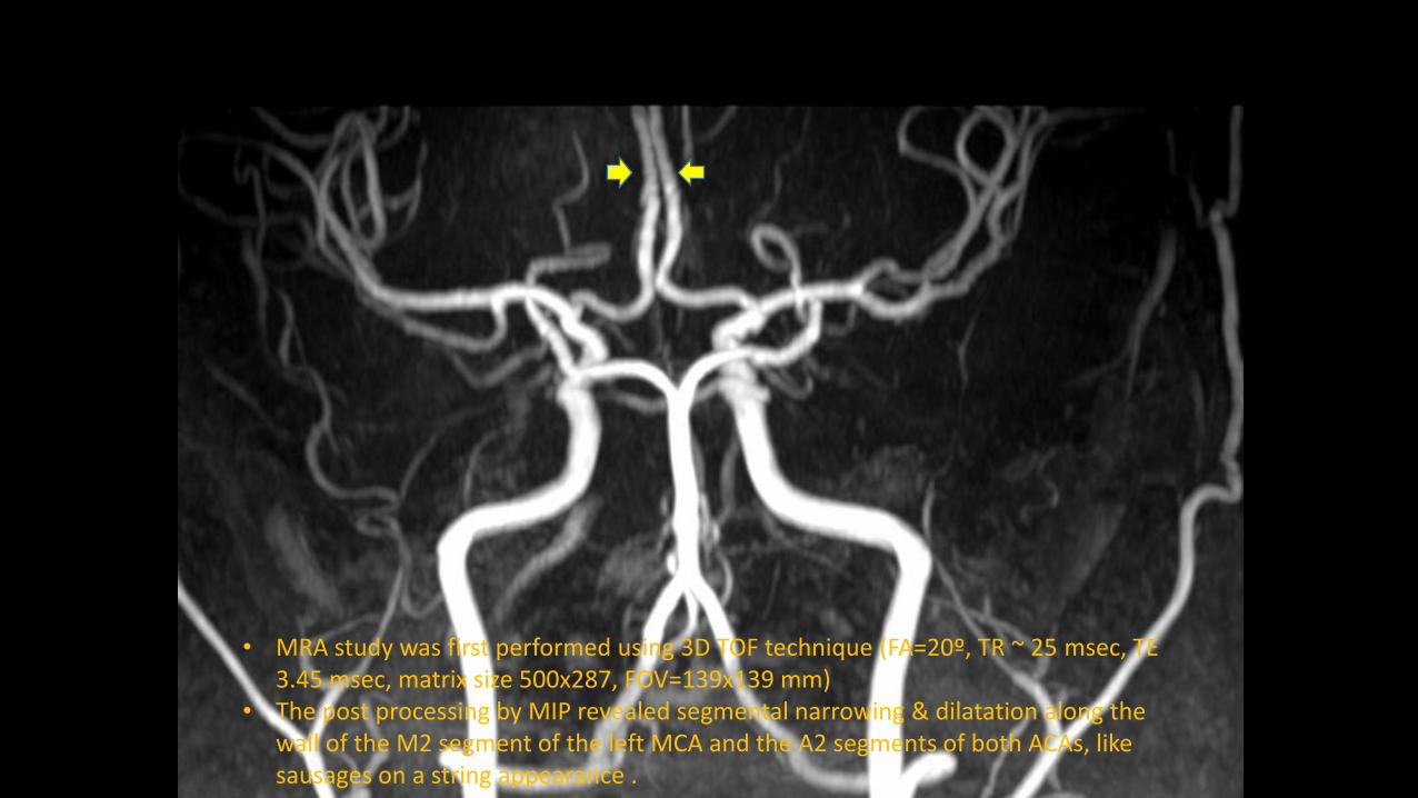
• With regard to the possibility of artifacts mimicking a lesion, the patient underwent MRA on the following day using many MRA techniques:• 3D TOF
• 3D PC (venc 45 cm/sec)
• Post contrast enhanced MRA
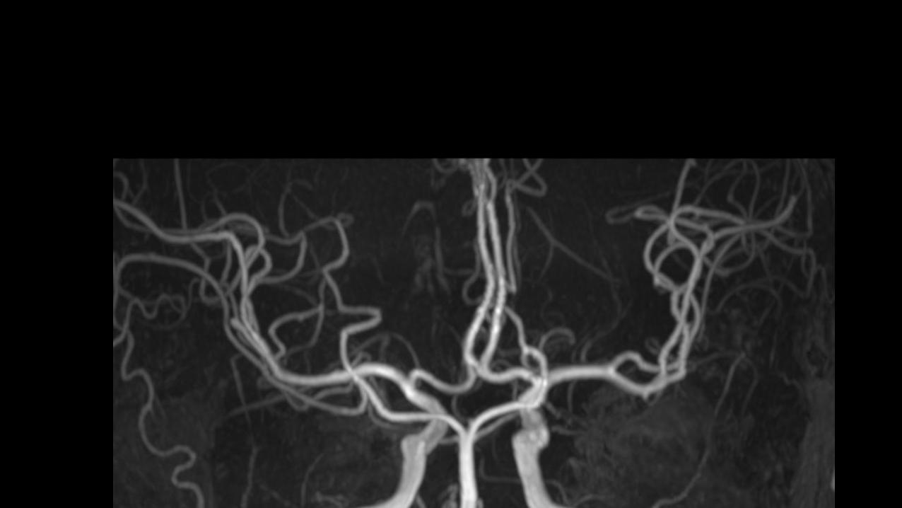
3D TOF (FA=20º, TR ~ 25 msec, TE 3.45 msec, matrix size 328x572, FOV=152x152 mm )
Persisting irregularities along the wall of the M2 segment of left MCA and the A2 segments of both
ACAs were noted in all studies.
3D TOF (FA=20º, TR ~ 25 msec, TE 3.45 msec, matrix size 328x572, FOV=152x152 mm )
Persisting irregularities along the wall of the M2 segment of left MCA and the A2 segments of both
ACAs were noted in all studies.
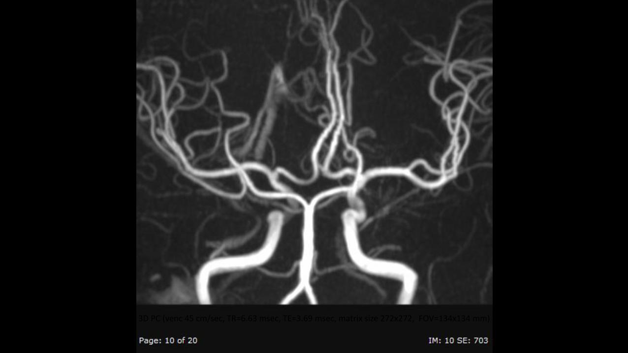
3D PC (venc 45 cm/sec, TR=6.63 msec, TE=3.69 msec, matrix size 272x272, FOV=134x134 mm)
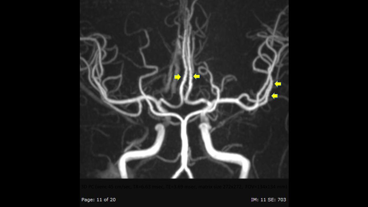
3D PC (venc 45 cm/sec, TR=6.63 msec, TE=3.69 msec, matrix size 272x272, FOV=134x134 mm)
Post contrast enhanced MRA ( FA=30º, TR=4.86 msec, TE 1.67 msec, matrix size 250x400, FOV=200x200 mm ).
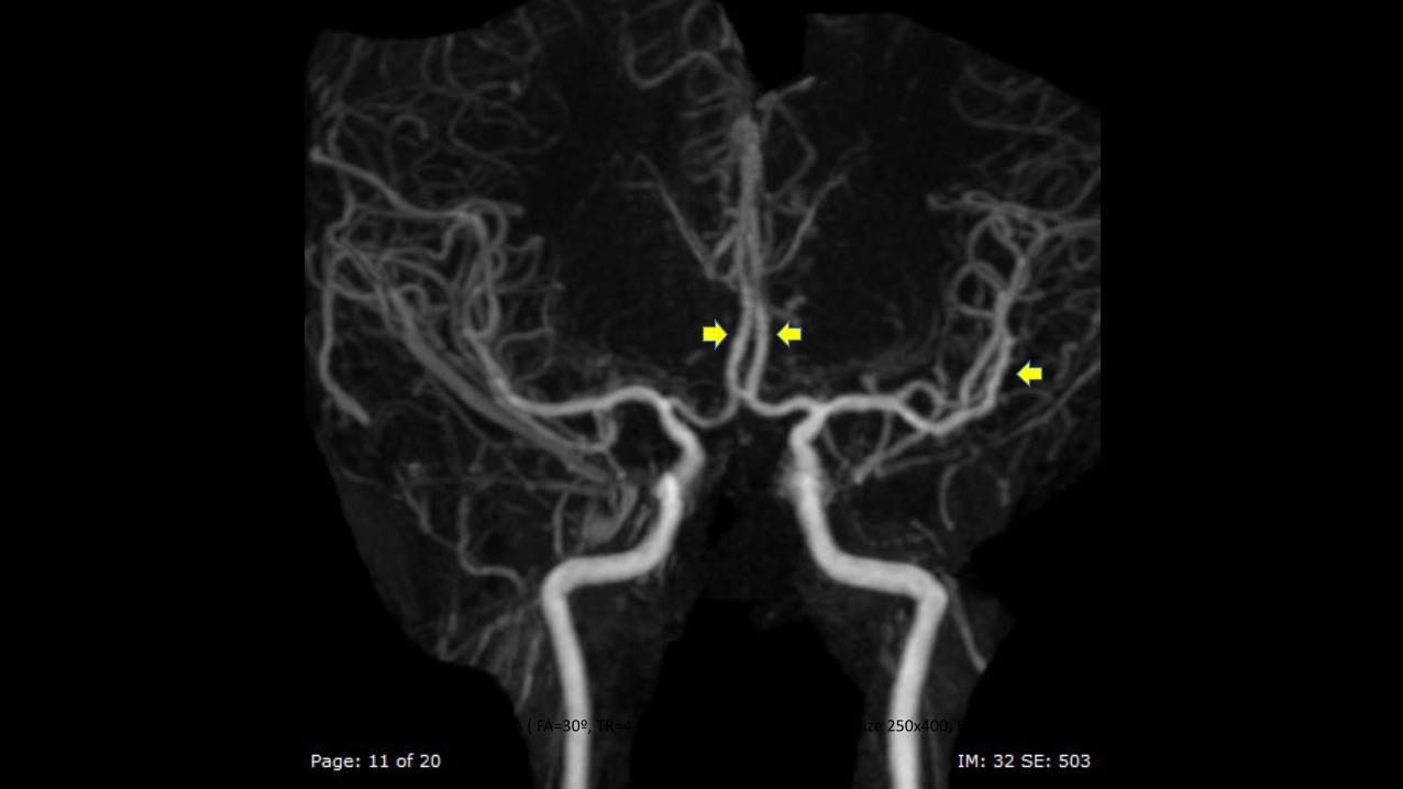
Post contrast enhanced MRA ( FA=30º, TR=4.86 msec, TE 1.67 msec, matrix size 250x400, FOV=200x200 mm ).
• Since there was no definite cause of vasculopathy or vasculitis can be found upon further investigations, the patient was diagnosed as complicated migraine.
• Symptomatic treatments were provided and the patient's severe headache disappeared.
3D TOF (FA 20, TR ~25 TE=3.45matrix size 572x287 FOV 159x159 mm)
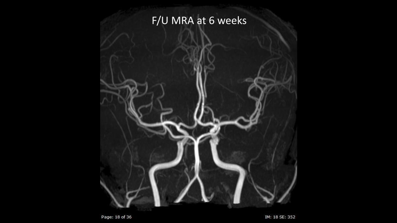
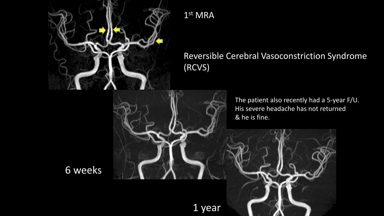
• F/U MRA at 6 weeks
F/U MRA at 6 weeks
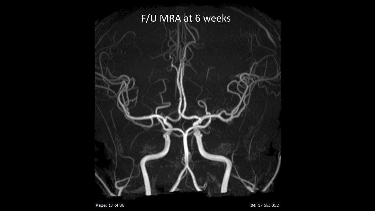
• F/U MRA at 6 weeks
F/U MRA at 6 weeks
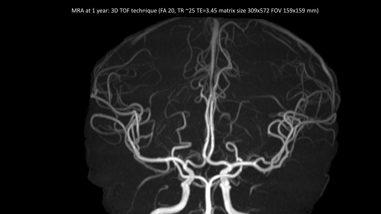
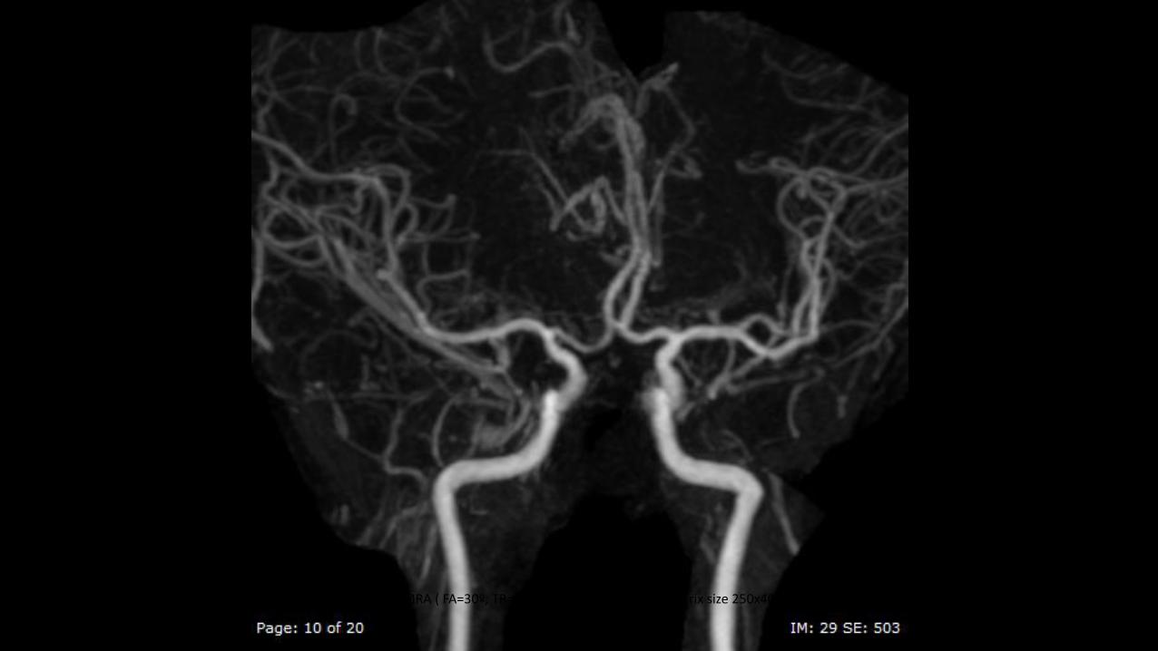
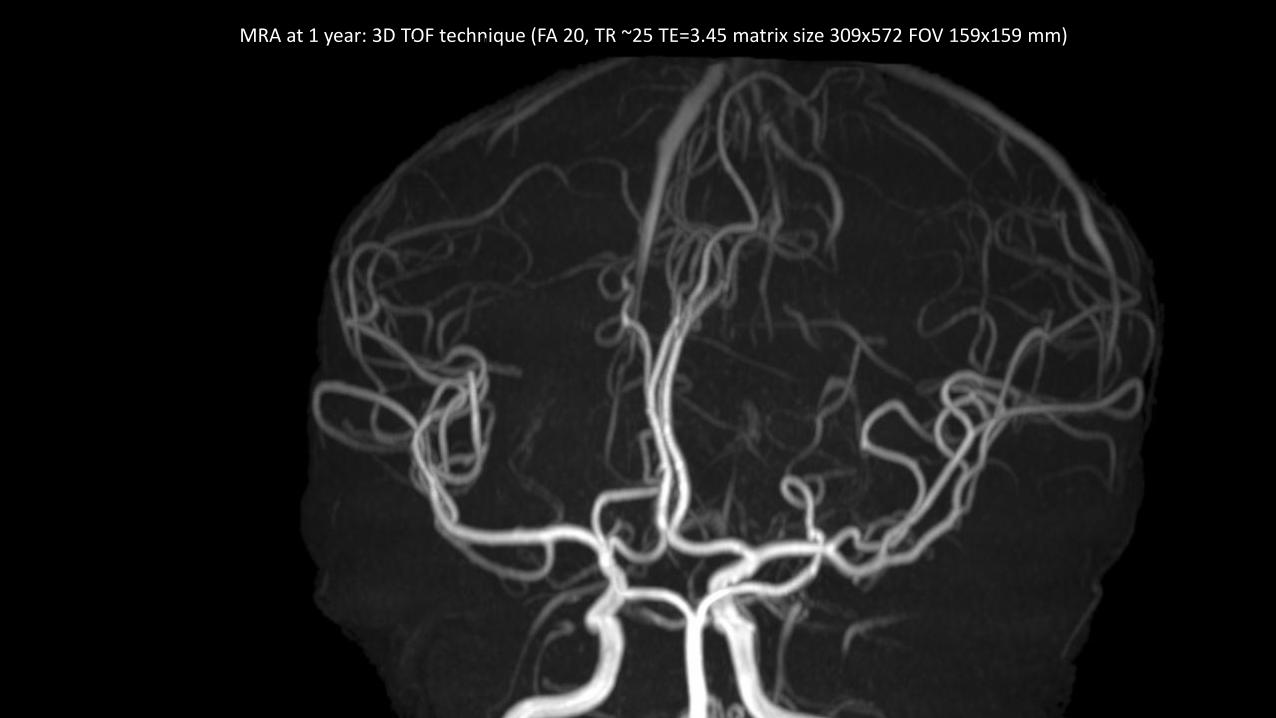
MRA at 1 year: 3D TOF technique (FA 20, TR ~25 TE=3.45 matrix size 309x572 FOV 159x159 mm) • &1 year revealed full recovery of the vasoconstriction and RCVS
was diagnosed
MRA at 1 year: 3D TOF technique (FA 20, TR ~25 TE=3.45 matrix size 309x572 FOV 159x159 mm) • &1 year revealed full recovery of the vasoconstriction and RCVS
was diagnosed
6 weeks
1 year
1st MRA
The patient also recently had a 5-year F/U. His severe headache has not returned & he is fine.
Reversible Cerebral Vasoconstriction Syndrome (RCVS)
Reversible Cerebral Vasoconstriction Syndrome (RCVS)
• The syndrome is characterized by severe headache due to vasoconstriction of cerebral arteries which resolve spontaneously.
• Screaming, crying, agitation, confusion and collapse are common because of the excruciating pain.
• Associated with photophobia, nausea/vomiting and focal neurological deficit secondary to ischemia can sometimes occur.
• F > M
• It has been reported in people age from 10-76 years but occur peaks at around 42 yrs.
**Ducros A. Reversible cerebral vasoconstriction syndrome. Lancet Neurol 2012;11:906 - 917.
RCVS
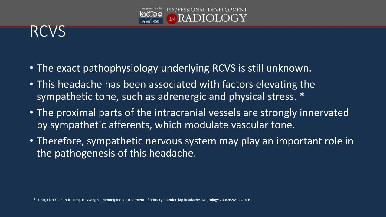
• The exact pathophysiology underlying RCVS is still unknown.
• This headache has been associated with factors elevating the sympathetic tone, such as adrenergic and physical stress. *
• The proximal parts of the intracranial vessels are strongly innervated by sympathetic afferents, which modulate vascular tone.
• Therefore, sympathetic nervous system may play an important role in the pathogenesis of this headache.
* Lu SR, Liao YC, Fuh JL, Lirng JF, Wang SJ. Nimodipine for treatment of primary thunderclap headache. Neurology 2004;62(8):1414-6.
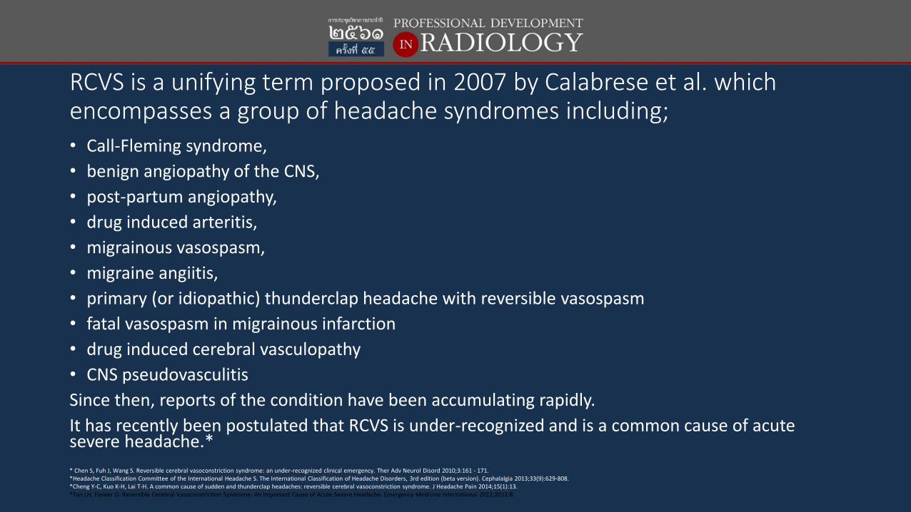
RCVS is a unifying term proposed in 2007 by Calabrese et al. which encompasses a group of headache syndromes including;• Call-Fleming syndrome,
• benign angiopathy of the CNS,
• post-partum angiopathy,
• drug induced arteritis,
• migrainous vasospasm,
• migraine angiitis,
• primary (or idiopathic) thunderclap headache with reversible vasospasm
• fatal vasospasm in migrainous infarction
• drug induced cerebral vasculopathy
• CNS pseudovasculitis
Since then, reports of the condition have been accumulating rapidly.
It has recently been postulated that RCVS is under-recognized and is a common cause of acute severe headache.*
* Chen S, Fuh J, Wang S. Reversible cerebral vasoconstriction syndrome: an under-recognized clinical emergency. Ther Adv Neurol Disord 2010;3:161 - 171.*Headache Classification Committee of the International Headache S. The International Classification of Headache Disorders, 3rd edition (beta version). Cephalalgia 2013;33(9):629-808.*Cheng Y-C, Kuo K-H, Lai T-H. A common cause of sudden and thunderclap headaches: reversible cerebral vasoconstriction syndrome. J Headache Pain 2014;15(1):13.*Tan LH, Flower O. Reversible Cerebral Vasoconstriction Syndrome: An Important Cause of Acute Severe Headache. Emergency Medicine International 2012;2012:8.
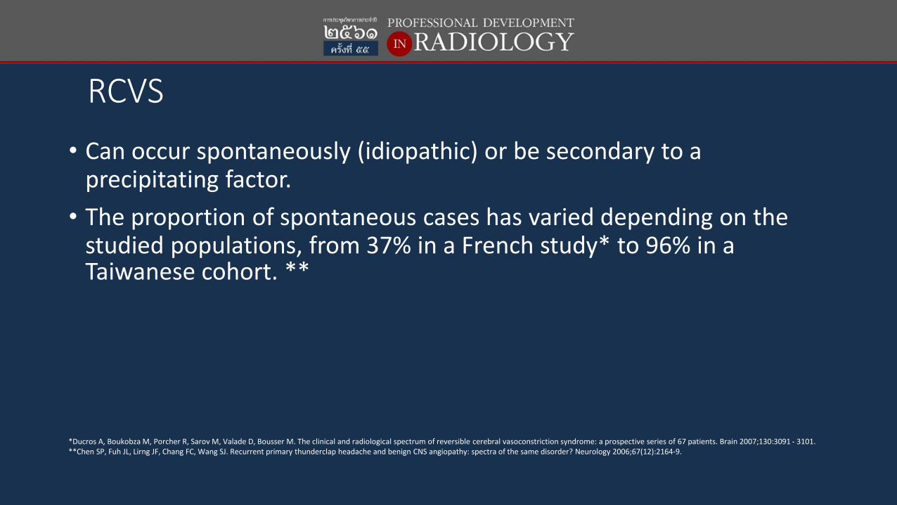
• Can occur spontaneously (idiopathic) or be secondary to a precipitating factor.
• The proportion of spontaneous cases has varied depending on the studied populations, from 37% in a French study* to 96% in a Taiwanese cohort. **
*Ducros A, Boukobza M, Porcher R, Sarov M, Valade D, Bousser M. The clinical and radiological spectrum of reversible cerebral vasoconstriction syndrome: a prospective series of 67 patients. Brain 2007;130:3091 - 3101.**Chen SP, Fuh JL, Lirng JF, Chang FC, Wang SJ. Recurrent primary thunderclap headache and benign CNS angiopathy: spectra of the same disorder? Neurology 2006;67(12):2164-9.
RCVS
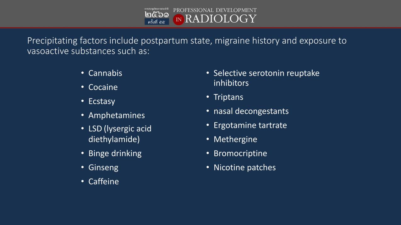
Precipitating factors include postpartum state, migraine history and exposure to vasoactive substances such as:
• Cannabis
• Cocaine
• Ecstasy
• Amphetamines
• LSD (lysergic acid diethylamide)
• Binge drinking
• Ginseng
• Caffeine
• Selective serotonin reuptake inhibitors
• Triptans
• nasal decongestants
• Ergotamine tartrate
• Methergine
• Bromocriptine
• Nicotine patches
RCVS
• Despite recently accepted to be a common cause of acute severe headaches, RCVS is still commonly missed due to its recent description in literature.
• Furthermore, the degree of vasoconstriction may be too subtle to be definitely detected, or the detected lesion may be missed classified as vasculitis or other causes of vasculopathy.
RCVS
• Likely to be one of the most common vasculopathies that is confused with vasculitis.
• Vasculopathy is any disorder of blood vessels while vasculitis implies inflammation of blood vessels which often requires brain biopsy for definite diagnosis.
RCVS
• Do not require brain biopsy but can be suggested by the following triad:• Appropriate clinical history (acute severe or thunderclap
headache)
• Complete resolution of angiographic vasoconstriction within
3 months
• Absence of inflammatory/infection etiology.
RCVS
• Although angiogram is the best imaging modality for diagnosis, CTA or MRA could be good alternative option since angiography is not a convenient tool for frequent F/U.
• CTA&MRA have been reported to be valuable for clinical evaluation in patients with RCVS.*
*Chen SP, Fuh JL, Wang SJ, Chang FC, Lirng JF, Fang YC, et al. Magnetic resonance angiography in reversible cerebral vasoconstriction syndromes. Ann Neurol 2010;67(5):648-56.*Marder CP, Donohue MM, Weinstein JR, Fink KR. Multimodal imaging of reversible cerebral vasoconstriction syndrome: a series of 6 cases. AJNR Am J Neuroradiol 2012;33(7):1403-10.
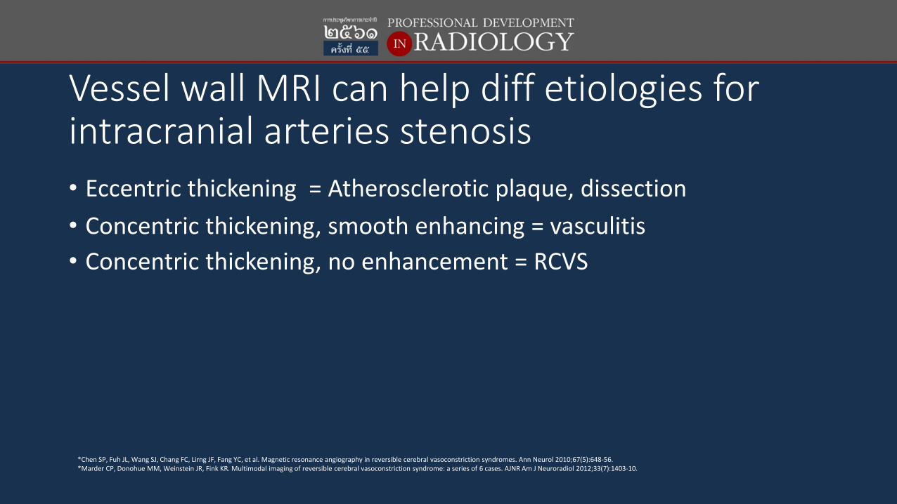
Vessel wall MRI can help diff etiologies for intracranial arteries stenosis
• Eccentric thickening = Atherosclerotic plaque, dissection
• Concentric thickening, smooth enhancing = vasculitis
• Concentric thickening, no enhancement = RCVS
*Chen SP, Fuh JL, Wang SJ, Chang FC, Lirng JF, Fang YC, et al. Magnetic resonance angiography in reversible cerebral vasoconstriction syndromes. Ann Neurol 2010;67(5):648-56.*Marder CP, Donohue MM, Weinstein JR, Fink KR. Multimodal imaging of reversible cerebral vasoconstriction syndrome: a series of 6 cases. AJNR Am J Neuroradiol 2012;33(7):1403-10.
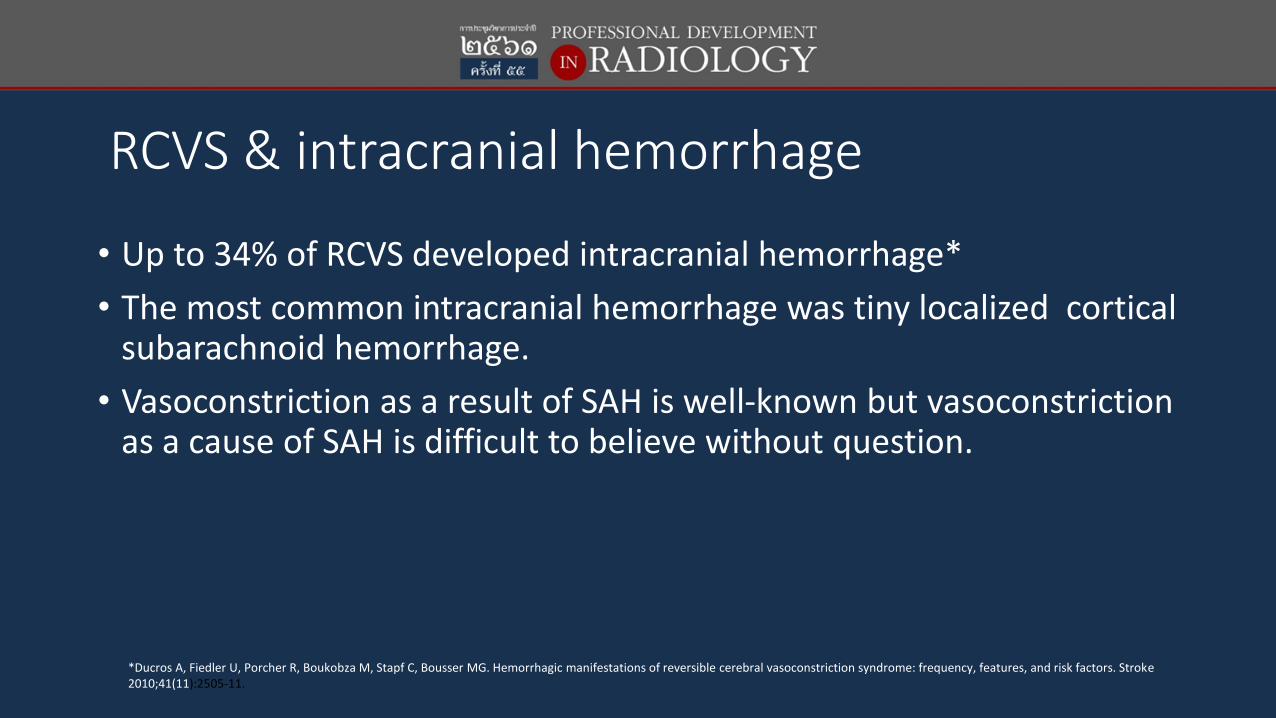
RCVS & intracranial hemorrhage
• Up to 34% of RCVS developed intracranial hemorrhage*
• The most common intracranial hemorrhage was tiny localized cortical subarachnoid hemorrhage.
• Vasoconstriction as a result of SAH is well-known but vasoconstriction as a cause of SAH is difficult to believe without question.
*Ducros A, Fiedler U, Porcher R, Boukobza M, Stapf C, Bousser MG. Hemorrhagic manifestations of reversible cerebral vasoconstriction syndrome: frequency, features, and risk factors. Stroke 2010;41(11):2505-11.
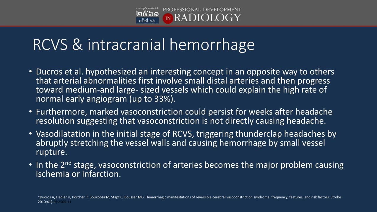
RCVS & intracranial hemorrhage
• Ducros et al. hypothesized an interesting concept in an opposite way to others that arterial abnormalities first involve small distal arteries and then progress toward medium-and large- sized vessels which could explain the high rate of normal early angiogram (up to 33%).
• Furthermore, marked vasoconstriction could persist for weeks after headache resolution suggesting that vasoconstriction is not directly causing headache.
• Vasodilatation in the initial stage of RCVS, triggering thunderclap headaches by abruptly stretching the vessel walls and causing hemorrhage by small vessel rupture.
• In the 2nd stage, vasoconstriction of arteries becomes the major problem causing ischemia or infarction.
*Ducros A, Fiedler U, Porcher R, Boukobza M, Stapf C, Bousser MG. Hemorrhagic manifestations of reversible cerebral vasoconstriction syndrome: frequency, features, and risk factors. Stroke 2010;41(11):2505-11.
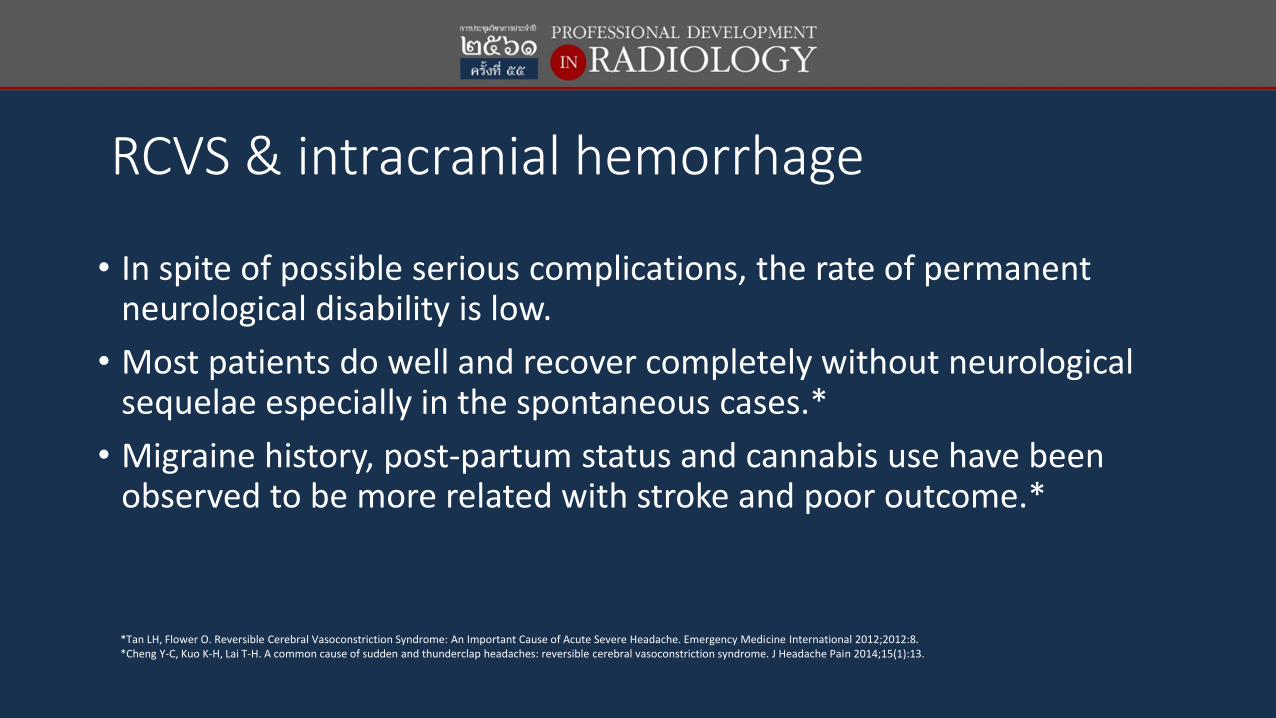
RCVS & intracranial hemorrhage
• In spite of possible serious complications, the rate of permanent neurological disability is low.
• Most patients do well and recover completely without neurological sequelae especially in the spontaneous cases.*
• Migraine history, post-partum status and cannabis use have been observed to be more related with stroke and poor outcome.*
*Tan LH, Flower O. Reversible Cerebral Vasoconstriction Syndrome: An Important Cause of Acute Severe Headache. Emergency Medicine International 2012;2012:8.*Cheng Y-C, Kuo K-H, Lai T-H. A common cause of sudden and thunderclap headaches: reversible cerebral vasoconstriction syndrome. J Headache Pain 2014;15(1):13.
Tx of RCVS
• There is no established therapy in RCVS.
• Good outcomes have been reported with symptomatic Tx, discontinuation of any possible related substances, triggers and rest.
• Calcium channel blockers such as Nimodipine has been shown to terminate the headache in 63-84% of patients* in spite of no outcome benefit over symptomatic alone in a large case series report.**
*Dodick DW. Reversible segmental cerebral vasoconstriction (Call-Fleming syndrome): the role of calcium antagonists. Cephalalgia 2003;23(3):163-5.**Singhal A, Hajj-Ali R, Topcuoglu M, Fok J, Bena J, Yang D, et al. Reversible cerebral vasoconstriction syndromes: analysis of 139 cases. Arch Neurol 2011;68:1005 - 1012
Conclusion
• RCVS is still under-recognized cause of acute severe headache.
• The primary diagnostic dilemma is distinguishing RCVS from primary CNS arteritis.
• Complete resolution of vasoconstriction of cerebral arteries within 3 months without evidence of inflammatory/infection etiology should suggest the Dx.
• RCBV is a possible cause of spontaneous SAH, esp. tiny cortical SAH
Case 2
• 36-year-old-male developed headache, diplopia and blurred vision, for 2 weeks
• PE: bilateral papilledema and 6th nerve palsy
• Request MRI brain to search for the cause of increase intracranial pressure:
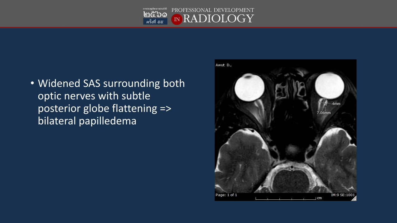
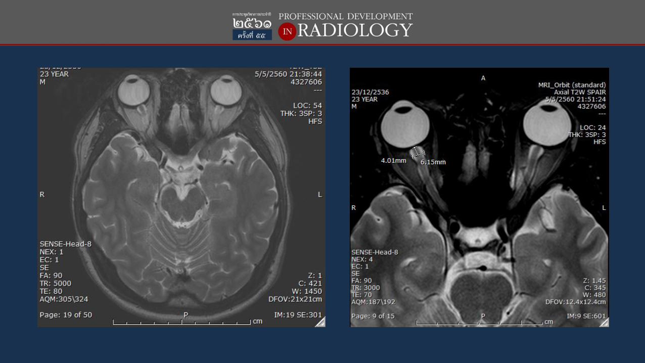
• Widened SAS surrounding both optic nerves with subtle posterior globe flattening => bilateral papilledema
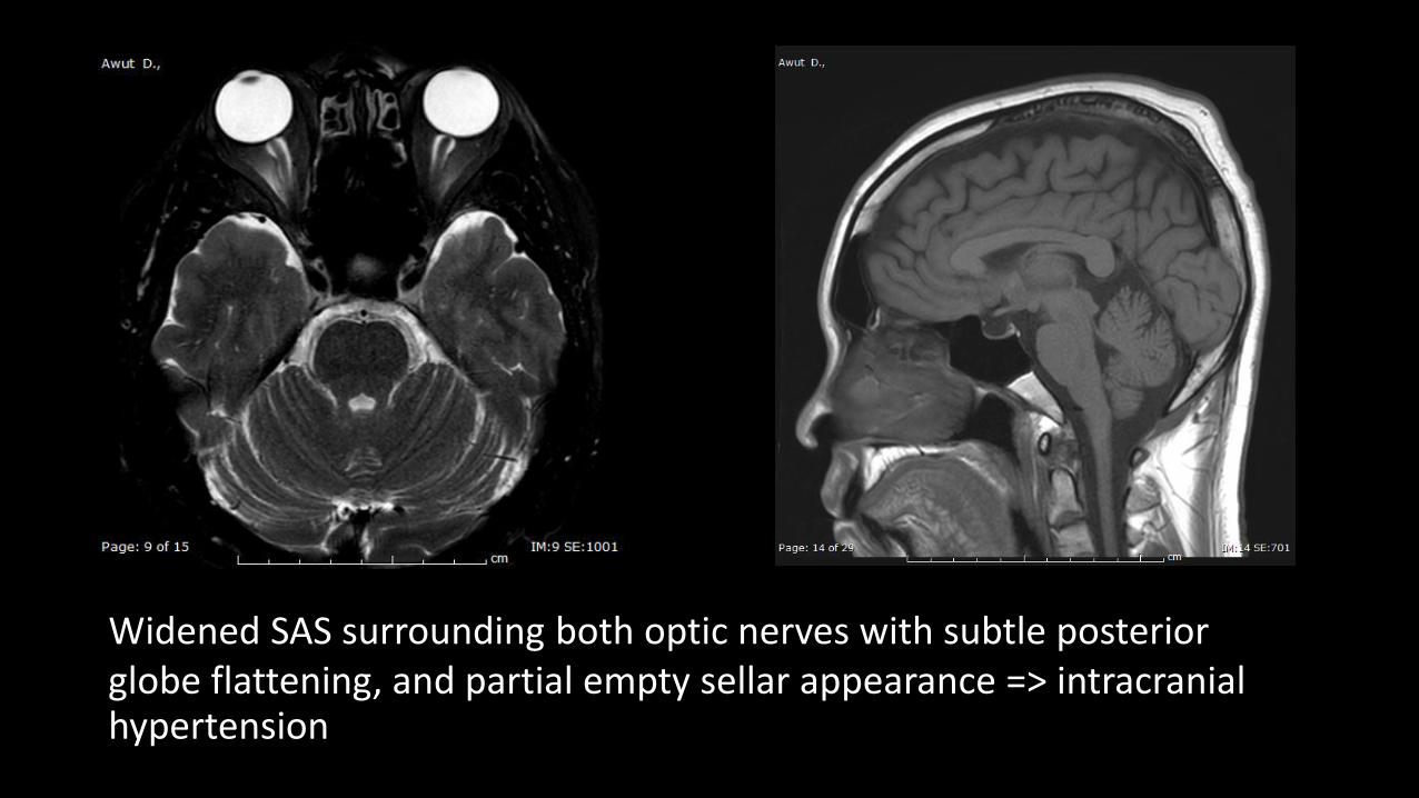
Widened SAS surrounding both optic nerves with subtle posterior globe flattening, and partial empty sellar appearance => intracranial hypertension
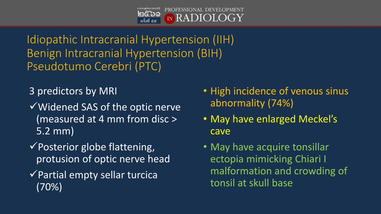
Idiopathic Intracranial Hypertension (IIH)Benign Intracranial Hypertension (BIH)Pseudotumo Cerebri (PTC)
3 predictors by MRI
Widened SAS of the optic nerve (measured at 4 mm from disc > 5.2 mm)
Posterior globe flattening, protusion of optic nerve head
Partial empty sellar turcica(70%)
• High incidence of venous sinus abnormality (74%)
• May have enlarged Meckel’scave
• May have acquire tonsillarectopia mimicking Chiari I malformation and crowding of tonsil at skull base
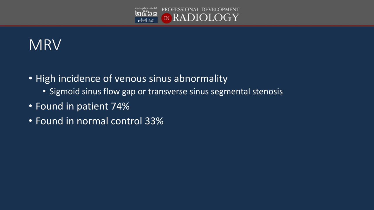
MRV
• High incidence of venous sinus abnormality• Sigmoid sinus flow gap or transverse sinus segmental stenosis
• Found in patient 74%
• Found in normal control 33%
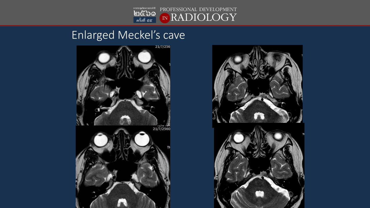
Enlarged Meckel’s cave
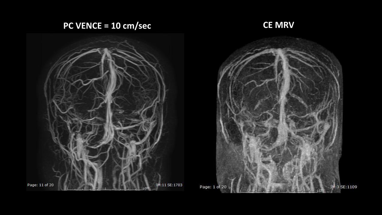
PC VENCE = 10 cm/sec CE MRV
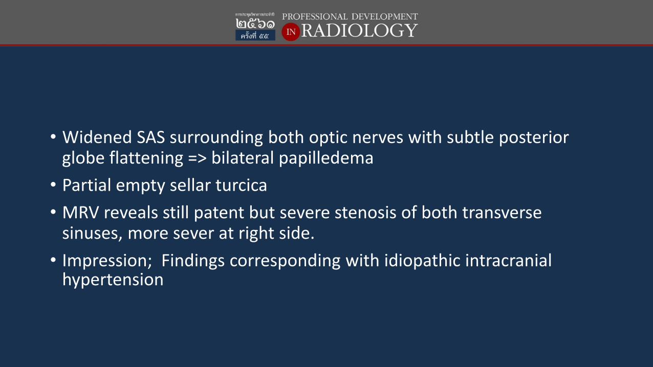
• Widened SAS surrounding both optic nerves with subtle posterior globe flattening => bilateral papilledema
• Partial empty sellar turcica
• MRV reveals still patent but severe stenosis of both transverse sinuses, more sever at right side.
• Impression; Findings corresponding with idiopathic intracranial hypertension
Idiopathic intracranial hypertension (IIH)Benign intracranial hypertension (BIH)Pseudotumo cerebri (PTC)
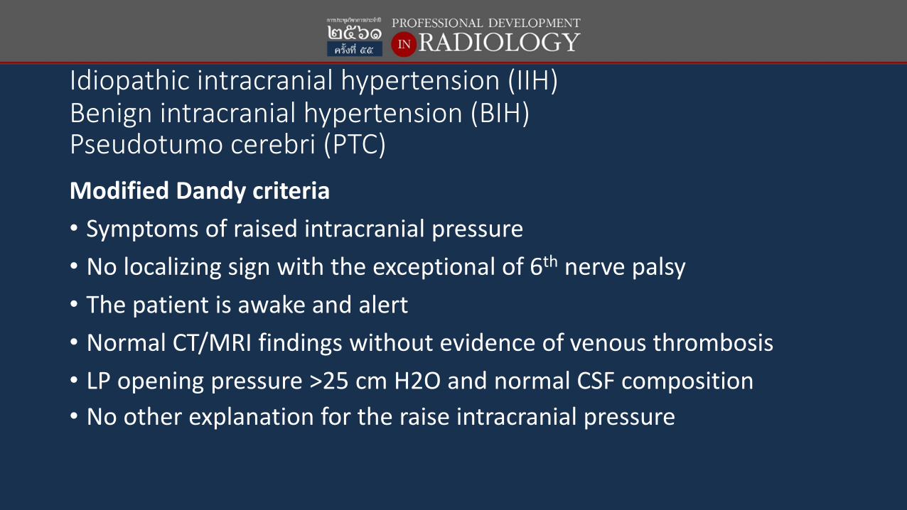
Modified Dandy criteria
• Symptoms of raised intracranial pressure
• No localizing sign with the exceptional of 6th nerve palsy
• The patient is awake and alert
• Normal CT/MRI findings without evidence of venous thrombosis
• LP opening pressure >25 cm H2O and normal CSF composition
• No other explanation for the raise intracranial pressure
Idiopathic intracranial hypertension (IIH)Benign intracranial hypertension (BIH)Pseudotumo cerebri (PTC)
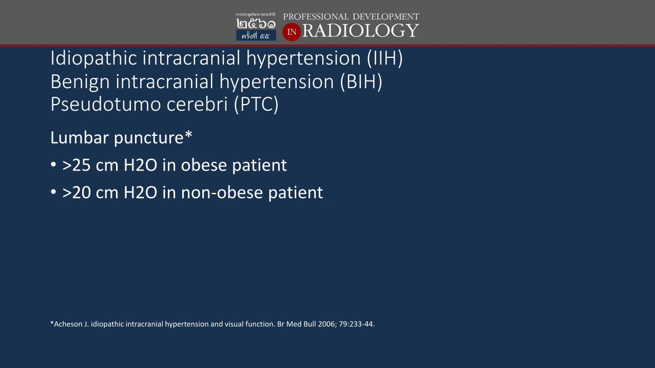
Lumbar puncture*
• >25 cm H2O in obese patient
• >20 cm H2O in non-obese patient
*Acheson J. idiopathic intracranial hypertension and visual function. Br Med Bull 2006; 79:233-44.
• LP => OP 26 CP 23 • Clear color CSF no abnormal cell• หลงัการวินิจฉยั idiopathic intracranial hypertension
• ประวตัิเพ่ิมเติมคือ ผู้ ป่วยมีน า้หนกั เพ่ิมขึน้มากถงึ 10 kg ใน 2 เดือนนี ้คือวา่อ้วนขึน้เร็วมาก
• Start treatment • ลดน า้หนกั • Acetazolamide (Diamox) • ลดได้ 5 kg ใน 1 เดือน • => Improve headache and blur vision but still have small visual filed defect and
diplopia• F/U at 4 months ลดน า้หนกั ได้ 17 kg จาก 89 kg => 72 kg หายปวดหวั ไมเ่ห็นภาพซ้อน และmuch
improve tiny residual visual filed defect
• Over 90% of IIH are obese or overweight
• Non-obese patients also are at greater risk of IIH if they have a recent weight grain of 5% or more
• The pathophysiology of IIH is unclear and the mechanism by which obesity contributes to the development of IIH is poorly understood.
• Obesity elevates intra-abdominal pressure => increase pleural and cardiac filling pressure and impedes venous return from the brain. The resulting increase in intracranial venous pressure may reduce CSF absorption*
• Weight loss is an effective treatment
Subramaniam S, Fletcher WA. Obesity & Weight loss in Idiopathic Intracranial Hyperention: A narrative Review: J Neuro-Ophthalmo2017; 37:197-205.
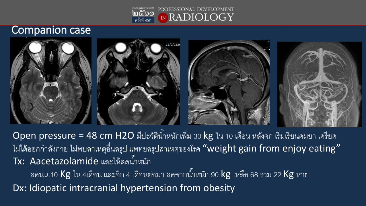
Companion case
Open pressure = 48 cm H2O มีปะวตัิน า้หนกัเพิ่ม 30 kg ใน 10 เดือน หลงัจก เร่ิมเรียนดมยา เครียด ไมไ่ด้ออกก าลงักาย ไมพ่บสาเหตอ่ืุนสรุป แพทยสรุปสาเหตขุองโรค “weight gain from enjoy eating”Tx: Aacetazolamide และให้ลดน า้หนกั
ลดนน.10 Kg ใน 4เดือน และอีก 4 เดือนตอ่มา ลดจากน า้หนกั 90 kg เหลือ 68 รวม 22 Kg หาย Dx: Idiopatic intracranial hypertension from obesity
Compared case
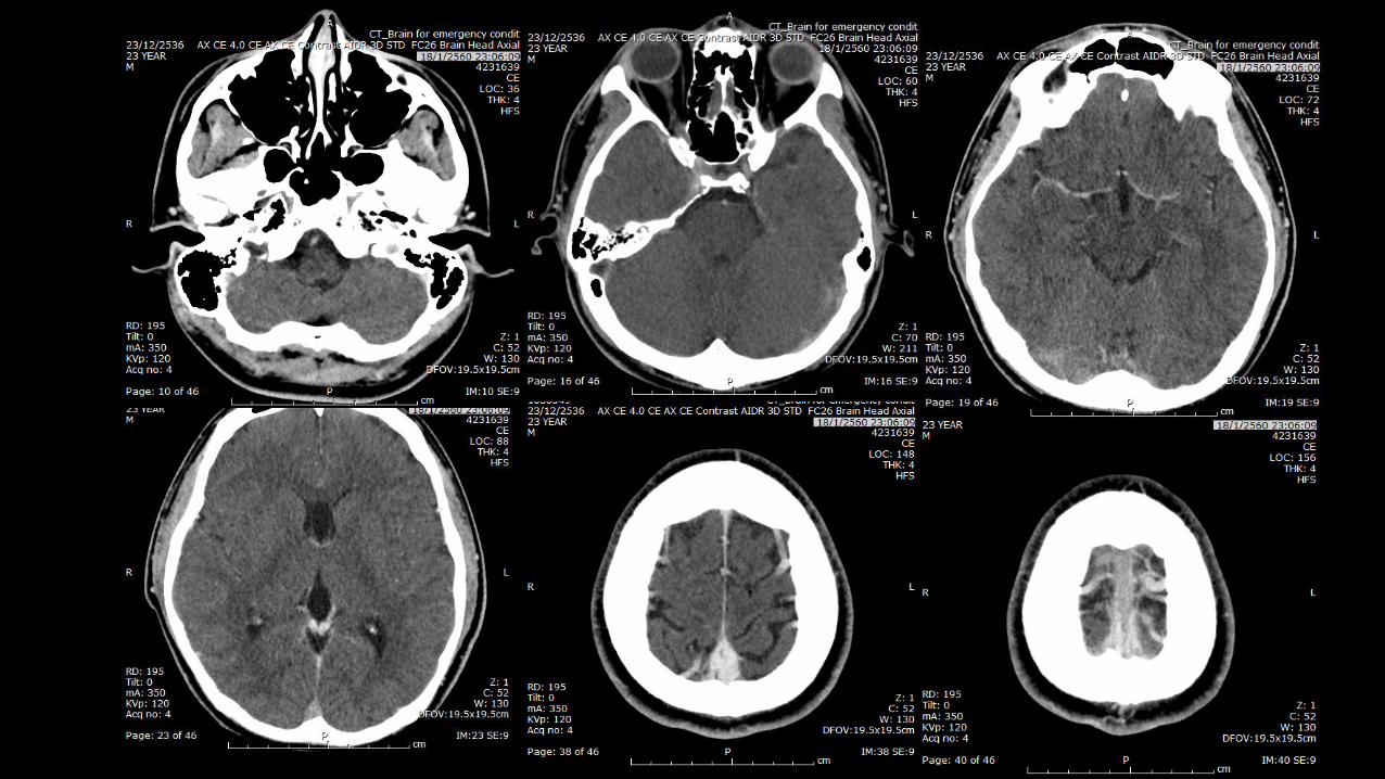
A 23-year-old man presented with progressive severe headache
• Headache for 1 month but much more severe in 2 days => private clinic Dx Migraine => Tx not improved and even more severe=> patient come to ER
• On PE, BP 130/90 mm Hg without evidence of neurological deficit
Tx: Diclofenac 1Amp => not improve
• Film sinus=> not seen air fluid level
• CT emergency
Take Home Messages
I. RCVS is a common cause of acute severe headaches and is still commonly missed due to its recent description in literature.
II. The incidence of IIH is increasing, attributable to the rise of obesity High incidence of venous sinus abnormality (74%)
Venous sinus thrombosis should always be excluded, esp. in non-obese patient
Thank you for your attention