Embed Size (px)
Citation preview

Imaging of Disorders of
Cortical Formation Neuronal Migration Disorders
By Mamdouh Mahfouz MD
Professor of Radiology Cairo University
ssregypt.com

Cortical formation
7th week gestational age
Mitotic activity in the subependymal layer of the ventricular wall, cells begin to migrate to form the cerebral cortex.
[ Atlas SW, 1996, MR imaging of brain and spine]

Any insult during the first two trimesters will lead to migrational anomalies:
Infections
Metabolic
Ischemia
Genetic
Chromosomal
Patients usually present with:
Seizures
Developmental delay
Mental retardation

Disorders of Cortical Formation
Abnormal organization
Abnormal migration
Abnormal cell proliferation

Disorders of Cortical Formation
Malformation
Hemimegalencephaly
Cortical dysplasia
Hamartomatous
Tuberous sclerosis
[Forme furste]
Neoplastic
Dysembryoblastic NET
Gangliogliomas
Abnormal cell
proliferation

Isolated form
Commonest
One hemisphere is affected
Syndromic form • One cerebral hemisphere • Hemihypertrophy of a part or all one side of the body
Total form
• Least common • Cerebellar+ cerebral Hemispheres + Brainstem on one side
Hemimegalencephaly
Enlargement of all or part of the to cerebral hemisphere and ventricle.
Focal or extensive calcification in both white and grey matter.
Gliosis of the white matter
Straightening of the frontal horn of the enlarged ventricle.
Agyria, pachygyria, polymicrogyria, heterotopia.
MRS: NAA , creatine, choline, myoinsitol (glial proliferation)
Hemimegalencephaly

Hemimegalencephaly
Imaging Findings: Enlarged hemisphere
Lateral ventricle is often enlarged
Dysplastic cortex [ thick, calcified,..]
Heterotopia
Loss of gray- white matter interface.
Hamartomatous overgrowth of a part or all one
hemisphere
Broumandi DD et al ., Radio graphics , 2004

M 3M Hemimegalencephaly

M 3M Hemimegalencephaly

Hemimegalencephaly
Broumandi DD et al ., Radiographics , 2004
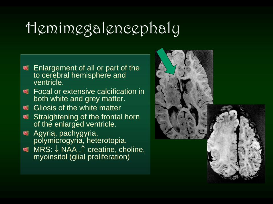
Hemimegalencephaly
Blaser & Jay,Neuroimaging Clinics , 1999

Focal Cortical dysplasia (FCD)
Presence of abnormal neurons and glia arranged abnormally in focal areas in the cerebral cortex
The incidence of FCD is 5- 15% in patients with seizures.
MRI findings
Abnormal high signal intensity in the gray matter on T2- weighted Images
Blurring of the gray/ white matter junction
Thickening of the affected cortex

Focal Cortical dysplasia (FCD)
M 16Y

Supratentorial [ temporal lobe] 60%
Well defined wedge shaped
Extends from the cortex to the ventricle
calcium
No edema
enhancement
Multinodular cystic appearance
Low T1 High T2
May remodel the calverium
No recurrence after removal
DD Infarct
Actual neoplasm DNET Cortical dysplasia
Dysembryoblastic Epithelial Tumors DNET
Old names [ Hamartoma, mixed glioma] [ WHO I ]
Blaser & Jay,Neuroimaging Clinics , 1999

Ganglioglioma
Temporal lobe 85% of cases
80% occur before the age of 30 Y
Calcifications 40% of cases
Benign with calverial remodeling
A solid enhancing part is the rule in all cases
Non specific MR findings
Blaser & Jay,Neuroimaging Clinics , 1999
In children < 10Y the tumor is
large & more cystic with more
edema around .The lesion may
convert into the aggressive form
, anaplastic ganglioglioma
which is suspected by the degree
of edema

Anaplastic Ganglioglioma

Disorders of migration Migration occurs during 8th week along radial glial fibers [RGF]
Damage of RGF Migration disorders
Ischemia
Infection
Trauma
metabolic errors

Three layers
The cortex may be normal or
pachygyric
Seizures with mild-moderate
mental retardation
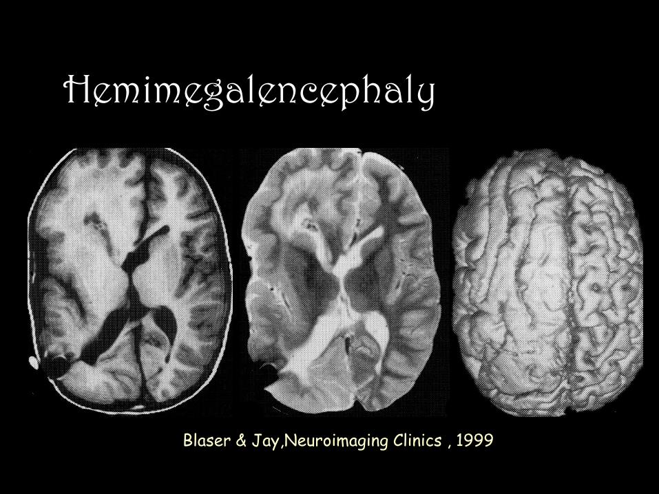
Band Heterotopia [double cortex]

The severity of symptoms correlates with
Degree of cortical disorganization
Thickness of the heterotopic band
High signal on T2 WIs
Degree of ventriculomegaly
Band Heterotopia
Band heterotopia
[ Anne Osborn, Hand book of Neuroradiology, 1996]

Lissencephaly
A rare congenital brain
malformation that means
Smooth brain (agyria),
represents a spectrum of
disorders ranging from total
agyria to mixed agyria and
pachygyria
Exact cause is not known
F 5M

Complete form
Some gyral formation along the inferior frontal and temporal lobes
Imaging
● broad flat gyri with thickened cortex
● primitive sylvian fissures hourglass
configuration
Lissencephaly

Lissencephaly MRI findings
Agyric brain with or without areas of pachygyria
Hour glass configuration of the brain
Primitive vertical Sylvian fissures
Small temporal lobes
Associated anomalies:
•Corpus callosum hypoplasia
• Small brain stem
• Gray matter heterotopia

Lissencephaly

Pachygyria /Lissencephaly
Normal

PACHYGYRIA
F 1Y
F 5Y

Focal gyral thickening
Surface reconstruction from a 3D data set [ TR / TE =15 / 8 MSEC ]
Blaser & Jay,Neuroimaging Clinics , 1999

Gray matter Heterotopia
Neurons in abnormal location due to arrest of radial migration to the surface of the brain
Types:
• Subependymal heterotopia
• Focal heterotopia
• Diffuse band heterotopia
[ Double cortex syndrome]

Late-onset seizures with
normal milestones
Focal isolated lesions or
diffuse form
Diffuse bilateral form is
associated with cerebellar
hypoplasia
DD tuberous sclerosis
Subependymal heterotopia

Subependymal heterotopia F 8Y

Subependymal heterotopia F 20Y

Subependymal heterotopia
Tuberous sclerosis

Focal Heterotopia MRI findings
Focal masses within the deep white matter
Isointense to the cortex in all sequences
No perilesional edema
No mass effect
Ipsilateral dysmorphology of the lateral ventricle

Focal heterotopia F 13Y

14-20 weeks Sylvian and parieto- occipiral sulci
32-33 week Large number of sulci are formed
38-40 week normal adult sulcal pattern is reached
Disruption of the process of gyral formation
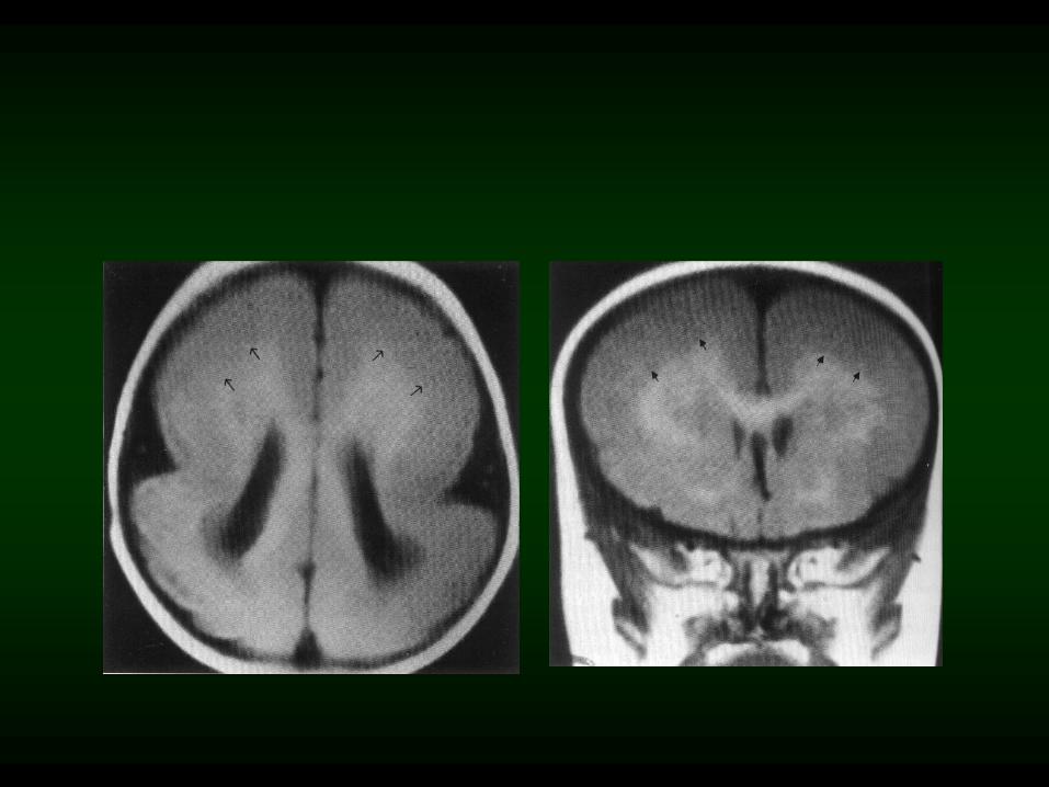
Polymicrogyria
Schizencephaly
Disorders of cortical organization
Abnormal organization

Thickened cortex with many small gayri
Gayri may be so small to be identified on imaging Flat thick cortex similar to pachygyria or agyria
3D reformatted images
Polymicrogyria PMG
Bilateral symmetric PMG of the operculum
Primitive sylvian fissures

Polymicrogyria F 20Y

Small clefts with coapted walls [Closed lip type]
Large clefts with free communication between the ventricle and subarachnoid spaces [ Open lip type]
Schizencephaly [ gray matter-lined clefts]
Atlas SW , MRI of the Brain and Spine ,2nd Edition ,1996

Schizencephaly Originally described as bilateral symmetrical full thickness cleft within the cerebral hemispheres.
The cleft is lined by gray matter and commonly involves the parasylvian regions.
The cause is unknown
The severity of disease correlates with the extent of schizencephaly.
Type I: [closed lip]
• Fused cleft lined with
gray matter
Type II:[open lip]
• Large true
hemispheric cleft lined by a membrane of two layers of gray matter
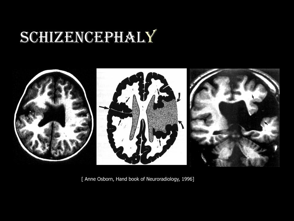
Schizencephaly
[ Anne Osborn, Hand book of Neuroradiology, 1996]

Type II Schizencephaly
[ Robertson SJ, Wolpert SM:Brain: congenital.In Runge VM, editor: clinical MRI, Philadelphia, 1990]
Porencephaly

surface reconstruction from a 3-
D data set
Open lip schizencephaly
Blaser & Jay,Neuroimaging Clinics , 1999

F3Y

M 21Y

Pachygyria

THANK YOU
MAMDOUH MAHFOUZ MD

M 8Y


M 9M F 25Y

Migrational Disorders of the Brain
Gyral abnormalities
• Agyria [Lissencephaly]
• Pacchygyria
• Polymicrogyria
Schizencephaly
Gray matter heterotopia
Unilateral megalencephaly

Subependymal heterotopia

Pachygyria
Broad flat gyria
Commonly coexist with agyria
May be focal or diffuse


venous angioma

Type I Schizencephaly


Megalencephaly

Megalencephaly

Megalencephaly

Megalencephaly

Megalencephaly

Megalencephaly

Hemimegalencephaly

Subependymal heterotopia

Polymicrogyria PMG

Schizencephaly

Schizencephaly