Embed Size (px)
Citation preview
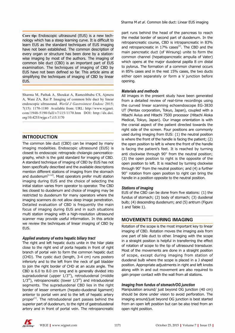
Imaging of common bile duct by linear endoscopic ultrasound
Malay Sharma, Amit Pathak, Abid Shoukat, Chittapuram Srinivasan Rameshbabu, Akash Ajmera, Zeeshn Ahamad Wani, Praveer Rai
Malay Sharma, Amit Pathak, Abid Shoukat, Department of Gastroenterology, Jaswant Rai Speciality Hospital, Meerut, Uttar Pradesh 250001, India
Chittapuram Srinivasan Rameshbabu, Department of Anatomy, Muzaffarnagar Medical College, Muzaffarnagar, Uttar Pradesh 251203, India
Akash Ajmera, Department of Medicine, Marshall University, Huntington, WV 25504, United States
Zeeshn Ahamad Wani, Jaswant Rai Speciality Hospital, Meerut, Uttar Pradesh 250001, India
Praveer Rai, Sanjay Gandhi Post Graduate Institute, Lucknow, Uttar Pradesh 226014, India
Author contributions: Sharma M, Pathak A and Rai P performed EUS; Pathak A contributed to conceptualizing the manuscript; Rameshbabu CS contributed to making sketches; Ajmera A and Wani ZA contributed to proof reading of the manuscript. Shoukat A contributed to critical revision of the manuscript.
Conflict-of-interest statement: The authors have no conflict of interest.
Open-Access: This article is an open-access article which was selected by an in-house editor and fully peer-reviewed by external reviewers. It is distributed in accordance with the Creative Commons Attribution Non Commercial (CC BY-NC 4.0) license, which permits others to distribute, remix, adapt, build upon this work non-commercially, and license their derivative works on different terms, provided the original work is properly cited and the use is non-commercial. See: http://creativecommons.org/licenses/by-nc/4.0/
Correspondence to: Dr. Malay Sharma, MD, DM, Gastroen-terologist, Director, Department of Gastroenterology, Jaswant Rai Speciality Hospital, Mawana Road, Meerut, Uttar Pradesh 250001, India. [email protected]: +91-98-37031148Fax: +91-12-12657154
Received: March 21, 2015 Peer-review started: March 22, 2015
First decision: May 19, 2015Revised: July 30, 2015 Accepted: August 16, 2015Article in press: August 21, 2015Published online: October 25, 2015
AbstractImaging of common bile duct (CBD) can be done by many techniques. Endoscopic retrograde cholangio-pancreaticography is considered the gold standard for imaging of CBD. A standard technique of imaging of CBD by endoscopic ultrasound (EUS) has not been specifically described. The available descriptions mention different stations of imaging from the stomach and duodenum. The CBD lies closest to duodenum and choice of imaging may be restricted to duodenum for many operators. Generally most operators prefer multi station imaging during EUS and the choice of selecting the initial station varies from operator to operator. Detailed evaluation of CBD is frequently the main focus of imaging during EUS and in such situations multi station imaging with a high-resolution ultrasound scanner may provide useful information. Examination of the CBD is one of the primary indications for doing an EUS and it can be done from five stations: (1) the fundus of stomach; (2) body of stomach; (3) duodenal bulb; (4) descending duodenum; and (5) antrum. Following down the upper 1/3rd of CBD can do imaging of entire CBD from the liver window and following up the lower 1/3rd of CBD can do imaging of entire CBD from the pancreatic window. This article aims at simplifying the techniques of imaging of CBD by linear EUS.
Key words: Endoscopic ultrasound; Common bile duct; Pancreas; Pancreatic duct; Portal vein
© The Author(s) 2015. Published by Baishideng Publishing Group Inc. All rights reserved.
REVIEW
1170 October 25, 2015|Volume 7|Issue 15|WJGE|www.wjgnet.com
Submit a Manuscript: http://www.wjgnet.com/esps/Help Desk: http://www.wjgnet.com/esps/helpdesk.aspxDOI: 10.4253/wjge.v7.i15.1170
World J Gastrointest Endosc 2015 October 25; 7(15): 1170-1180ISSN 1948-5190 (online)
© 2015 Baishideng Publishing Group Inc. All rights reserved.
Core tip: Endoscopic ultrasound (EUS) is a new tech-nology which has a steep learning curve. It is difficult to learn EUS as the standard techniques of EUS imaging have not been established. The common description of every organ or structure has been done by a station-wise imaging by most of the authors. The imaging of common bile duct (CBD) is an important part of EUS examination. The techniques of imaging of CBD by EUS have not been defined so far. This article aims at simplifying the techniques of imaging of CBD by linear EUS.
Sharma M, Pathak A, Shoukat A, Rameshbabu CS, Ajmera A, Wani ZA, Rai P. Imaging of common bile duct by linear endoscopic ultrasound. World J Gastrointest Endosc 2015; 7(15): 1170-1180 Available from: URL: http://www.wjgnet.com/1948-5190/full/v7/i15/1170.htm DOI: http://dx.doi.org/10.4253/wjge.v7.i15.1170
INTRODUCTIONThe common bile duct (CBD) can be imaged by many imaging modalities. Endoscopic ultrasound (EUS) is closest to endoscopic retrograde cholangio pancreaticography, which is the gold standard for imaging of CBD. A standard technique of imaging of CBD by EUS has not been specifically described and the available descriptions mention different stations of imaging from the stomach and duodenum[112]. Most operators prefer multi station imaging during EUS and the choice of selecting the initial station varies from operator to operator. The CBD lies closest to duodenum and choice of imaging may be restricted to duodenum for many operators where the imaging scanners do not allow deep image penetration. Detailed evaluation of CBD is frequently the main focus of imaging during EUS and in such situations multi station imaging with a highresolution ultrasound scanner may provide useful information. In this article we review the techniques of linear imaging of CBD by EUS.
Applied anatomy of extra hepatic biliary tractThe right and left hepatic ducts unite in the hilar plate close to the right end of porta hepatis in front of right branch of portal vein to form the common hepatic duct (CHD). The cystic duct (length, 34 cm) runs postero inferiorly and to the left from the neck of gall bladder to join the right border of CHD at an acute angle. The CBD is 6.0 to 8.0 cm long and is generally divided into supraduodenal (upper 1/3rd), retroduodenal (middle 1/3rd), retropancreatic (lower 1/3rd) and intraduodenal segments. The supraduodenal CBD lies in the right border of lesser omentum (hepatoduodenal ligament) anterior to portal vein and to the left of hepatic artery proper[1]. The retroduodenal part passes behind the superior part of duodenum, to the right of gastroduodenal artery and in front of portal vein. The retropancreatic
part runs behind the head of the pancreas to reach the medial border of second part of duodenum. In the retropancreatic course, CBD is intrapancreatic in 83% and retropancreatic in 17% cases[2]. The CBD and the main pancreatic duct (of Wirsung) unite to form the common channel (hepatopancreatic ampulla of Vater) which opens at the major duodenal papilla 8 cm distal to pylorus. The formation of a common channel occurs in 85% cases and in the rest 15% cases, the two ducts either open separately or form a V junction before opening.
Materials and methodsAll images in the present study have been generated from a detailed review of real-time recordings using the curved linear scanning echoendoscope EG-3830 UT (Pentax corporation, Tokyo, Japan), coupled with a Hitachi Avius and Hitachi 7500 processor (Hitachi Aloka Medical, Tokyo, Japan). Our image orientation is with the cranial aspect of the patient directed towards the right side of the screen. Four positions are commonly used during imaging from EUS: (1) the neutral position is where the front of the handle is facing the patient; (2) the open position to left is where the front of the handle is facing the patient’s feet. It is reached by turning anti clockwise through 90° from the neutral position; (3) the open position to right is the opposite of the open position to left. It is reached by turning clockwise through 90° from the neutral position; and (4) a further 90° rotation from open position to right can bring the handle in a position opposite to the neutral position.
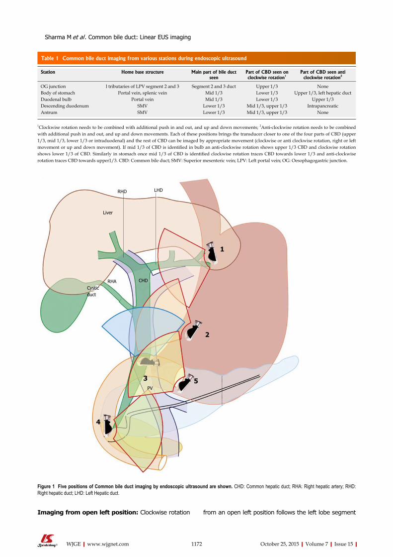
Stations of imaging EUS of the CBD can be done from five stations: (1) the fundus of stomach; (2) body of stomach; (3) duodenal bulb; (4) descending duodenum; and (5) antrum (Figure 1 and Table 1).
MOVEMENTS DURING IMAGING Rotation of the scope is the most important key to linear imaging of CBD. Rotation moves the imaging axis from one part of bile duct to other. Imaging with the scope in a straight position is helpful in transferring the effect of rotation of scope to the tip of ultrasound transducer. Most of the movements are done in a straight position of scope, except during imaging from station of duodenal bulb where the scope is placed in a J shaped position. Appropriate adjustments in right and left knobs along with in and out movement are also required to gain proper contact with the wall from all stations.
Imaging from fundus of stomach/OG junctionManipulation around/ just beyond OG junction (40 cm) should be done under vision to avoid perforation. The imaging around/just beyond OG junction is best started from an open left position but can be also tried from an open right position.
Sharma M et al . Common bile duct: Linear EUS imaging
1171WJGE|www.wjgnet.com October 25, 2015|Volume 7|Issue 15|
Imaging from open left position: Clockwise rotation from an open left position follows the left lobe segment
1172WJGE|www.wjgnet.com October 25, 2015|Volume 7|Issue 15|
Station Home base structure Main part of bile duct seen
Part of CBD seen on clockwise rotation1
Part of CBD seen anti clockwise rotation2
OG junction l tributaries of LPV segment 2 and 3 Segment 2 and 3 duct Upper 1/3 None Body of stomach Portal vein, splenic vein Mid 1/3 Lower 1/3 Upper 1/3, left hepatic ductDuodenal bulb Portal vein Mid 1/3 Lower 1/3 Upper 1/3Descending duodenum SMV Lower 1/3 Mid 1/3, upper 1/3 IntrapancreaticAntrum SMV Lower 1/3 Mid 1/3, upper 1/3 None
Table 1 Common bile duct imaging from various stations during endoscopic ultrasound
1Clockwise rotation needs to be combined with additional push in and out, and up and down movements; 2Anti-clockwise rotation needs to be combined with additional push in and out, and up and down movements. Each of these positions brings the transducer closer to one of the four parts of CBD (upper 1/3, mid 1/3, lower 1/3 or intraduodenal) and the rest of CBD can be imaged by appropriate movement (clockwise or anti clockwise rotation, right or left movement or up and down movement). If mid 1/3 of CBD is identified in bulb an anti-clockwise rotation shows upper 1/3 CBD and clockwise rotation shows lower 1/3 of CBD. Similarly in stomach once mid 1/3 of CBD is identified clockwise rotation traces CBD towards lower 1/3 and anti-clockwise rotation traces CBD towards upper1/3. CBD: Common bile duct; SMV: Superior mesenteric vein; LPV: Left portal vein; OG: Oesophagogastric junction.
RHD LHD
Liver
CHDRHACysticduct
PV
1
2
53
4
Figure 1 Five positions of Common bile duct imaging by endoscopic ultrasound are shown. CHD: Common hepatic duct; RHA: Right hepatic artery; RHD: Right hepatic duct; LHD: Left Hepatic duct.
Sharma M et al . Common bile duct: Linear EUS imaging
imaging of right lobe of liver is not possible from OG junction, as the right lobe ducts generally lies farther away from the probe. However the GHL does not inter-fere in imaging of right hepatic duct and with suitable adjustments of focus and frequency the right lobe and ducts of segment 4/5 (if dilated) can be identified and followed towards the upper CBD near the hilum by anticlockwise rotation.
Imaging from body of stomachFollowing down the upper 1/3rd of CBD can do imaging
2 and 3 ducts to left hepatic duct and further rotation traces the left hepatic duct towards the liver hilum. If the intrahepatic biliary radicles (IHBR’s) are dilated it is easy to follow the course of ducts by clockwise rotation. If the IHBR’s are not dilated the segmental portal vein radicles should be followed. The gastrohepatic ligament (GHL), which come between the EUS probe and left lobe of liver, interferes with the imaging during rotation (Figures 24).
Imaging from open right position: Generally
1173WJGE|www.wjgnet.com October 25, 2015|Volume 7|Issue 15|
Bumbilical segmentof LPV
LHDafter union ofsegment 2 and 3
LHVLHD
US LPV
diaphragm
23
A
segment 1
LHD
segment 4 ductRPV
4
4
MPV
C
RPV
segment 1
segment 4
segment 5
segment 4 veinMPV
44
LHD
D
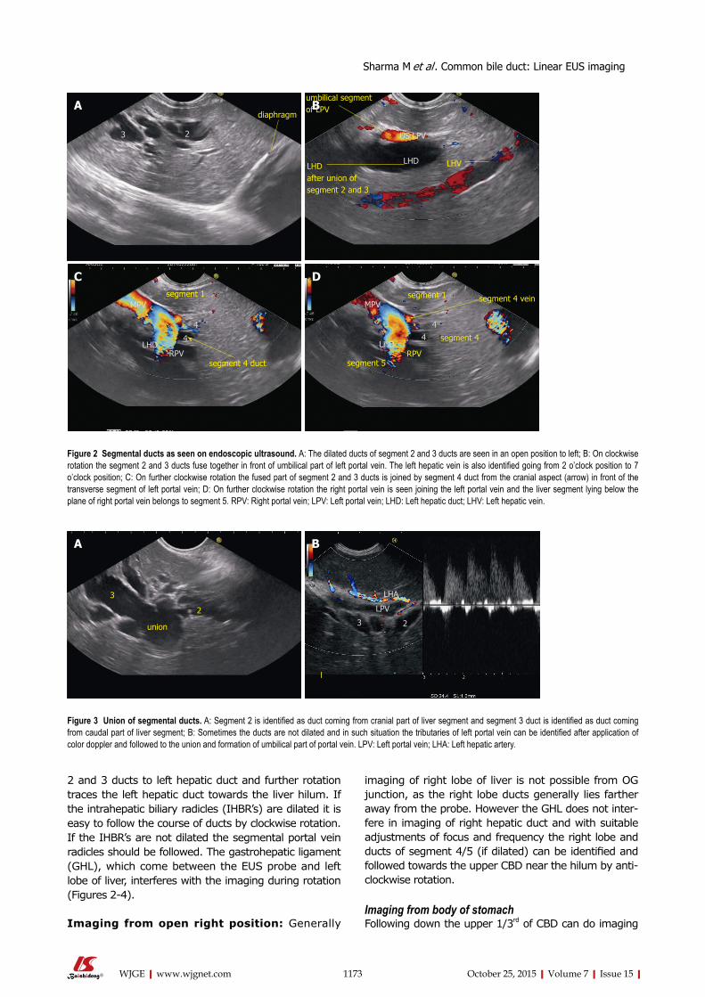
Figure 2 Segmental ducts as seen on endoscopic ultrasound. A: The dilated ducts of segment 2 and 3 ducts are seen in an open position to left; B: On clockwise rotation the segment 2 and 3 ducts fuse together in front of umbilical part of left portal vein. The left hepatic vein is also identified going from 2 o’clock position to 7 o’clock position; C: On further clockwise rotation the fused part of segment 2 and 3 ducts is joined by segment 4 duct from the cranial aspect (arrow) in front of the transverse segment of left portal vein; D: On further clockwise rotation the right portal vein is seen joining the left portal vein and the liver segment lying below the plane of right portal vein belongs to segment 5. RPV: Right portal vein; LPV: Left portal vein; LHD: Left hepatic duct; LHV: Left hepatic vein.
2
3
union
A
LHA
LPV
23
B
Figure 3 Union of segmental ducts. A: Segment 2 is identified as duct coming from cranial part of liver segment and segment 3 duct is identified as duct coming from caudal part of liver segment; B: Sometimes the ducts are not dilated and in such situation the tributaries of left portal vein can be identified after application of color doppler and followed to the union and formation of umbilical part of portal vein. LPV: Left portal vein; LHA: Left hepatic artery.
Sharma M et al . Common bile duct: Linear EUS imaging
of entire CBD from the liver window and following up the lower 1/3rd of CBD can do imaging of entire CBD from the pancreatic window.
Following down from liver window: Imaging of CBD while following it down from the fundus towards body of stomach requires a movement of the EUS probe along lesser curvature. This movement can be easily executed under vision after distension of stomach with air but the presence of air usually creates interference with ultrasound imaging. To avoid this interference due to air, a smooth combination of three movements: (1) push in of about 25 to 30 cm. from fundus; (2) clockwise rotation of 90 degree; and (3) up movement of up and down knob for about 90 degree is generally preferred. This movement allows a relative blind slide of the transducer along lesser curvature with nil or minimum distension of air and follows down the CBD from upper 1/3rd towards the lower 1/3rd. Once the movement is completed the scope comes to lie in a position near the
antrum and the left hand comes to lie close to the chest of the operator (Figure 5).
Following up from pancreatic window: A reversal of the movement described above can be done under vision by initially proceeding towards antrum after air inflation and subsequently coming back after air suction from antrum towards the fundus. This reversal movement follows up the CBD from the lower 1/3rd towards the upper 1/3rd. If it is difficult to trace the course of CBD by this movement, the home base of portal venous confluence of splenic vein with superior mesenteric vein is initially located in the neck of pan-creas. The lower 1/3rd of CBD is easily identified behind the portal venous confluence (Figure 6).
Imaging from bulbThe pylorus is located by “setting sun sign” and slight down angulation of tip may be required to get an end view of pylorus. Once the pylorus is seen the scope is
1174WJGE|www.wjgnet.com October 25, 2015|Volume 7|Issue 15|
Liver
PVST
CBD
H 000A
CBD
HDL
IVC
P4PV MHV
Caudatevein
CHL
CHL
Caudate process
B
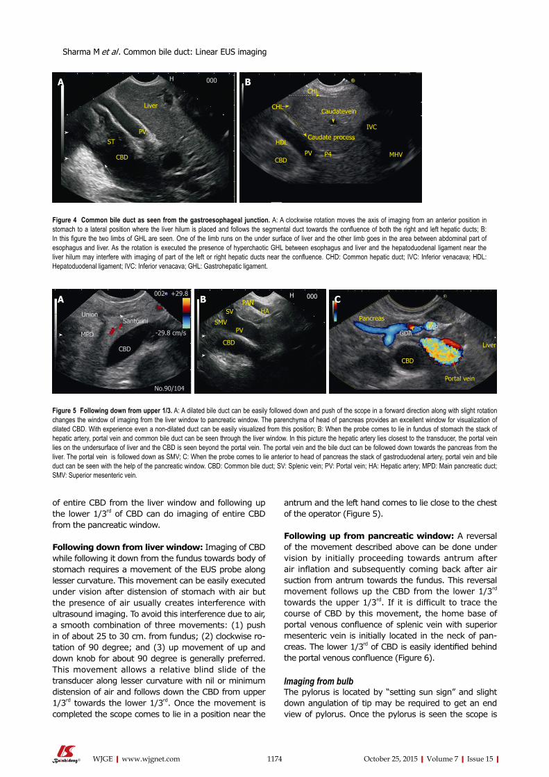
Figure 4 Common bile duct as seen from the gastroesophageal junction. A: A clockwise rotation moves the axis of imaging from an anterior position in stomach to a lateral position where the liver hilum is placed and follows the segmental duct towards the confluence of both the right and left hepatic ducts; B: In this figure the two limbs of GHL are seen. One of the limb runs on the under surface of liver and the other limb goes in the area between abdominal part of esophagus and liver. As the rotation is executed the presence of hyperchaotic GHL between esophagus and liver and the hepatoduodenal ligament near the liver hilum may interfere with imaging of part of the left or right hepatic ducts near the confluence. CHD: Common hepatic duct; IVC: Inferior venacava; HDL: Hepatoduodenal ligament; IVC: Inferior venacava; GHL: Gastrohepatic ligament.
Union
MPD
Santorini
CBD
002 +29.8
-29.8 cm/s
No.90/104
A PANSV HA
SMVPV
CBD
H 000B
Pancreas
CBDPV
Liver
Portal vein
GDAHAP
C
Figure 5 Following down from upper 1/3. A: A dilated bile duct can be easily followed down and push of the scope in a forward direction along with slight rotation changes the window of imaging from the liver window to pancreatic window. The parenchyma of head of pancreas provides an excellent window for visualization of dilated CBD. With experience even a non-dilated duct can be easily visualized from this position; B: When the probe comes to lie in fundus of stomach the stack of hepatic artery, portal vein and common bile duct can be seen through the liver window. In this picture the hepatic artery lies closest to the transducer, the portal vein lies on the undersurface of liver and the CBD is seen beyond the portal vein. The portal vein and the bile duct can be followed down towards the pancreas from the liver. The portal vein is followed down as SMV; C: When the probe comes to lie anterior to head of pancreas the stack of gastroduodenal artery, portal vein and bile duct can be seen with the help of the pancreatic window. CBD: Common bile duct; SV: Splenic vein; PV: Portal vein; HA: Hepatic artery; MPD: Main pancreatic duct; SMV: Superior mesenteric vein.
Sharma M et al . Common bile duct: Linear EUS imaging
1175WJGE|www.wjgnet.com October 25, 2015|Volume 7|Issue 15|
SMV
IVC
RRA
l
A
BOP
HOP CBD
PV
IVC
Right kidney
-7.46 cm/s
B
HA
HAPV
D2
RK
Pancreas
A1
CBD
C
CBDWall layers of CBD
Pancreas
D
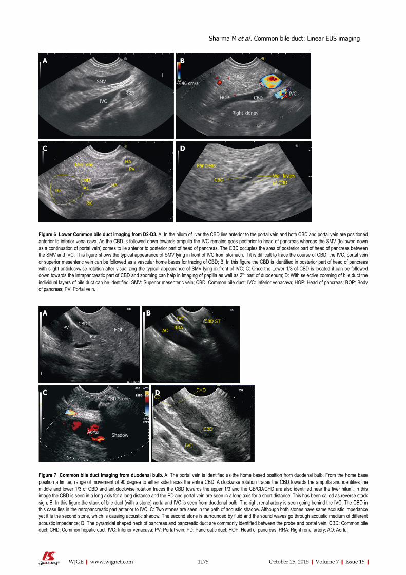
Figure 6 Lower Common bile duct imaging from D2-D3. A: In the hilum of liver the CBD lies anterior to the portal vein and both CBD and portal vein are positioned anterior to inferior vena cava. As the CBD is followed down towards ampulla the IVC remains goes posterior to head of pancreas whereas the SMV (followed down as a continuation of portal vein) comes to lie anterior to posterior part of head of pancreas. The CBD occupies the area of posterior part of head of pancreas between the SMV and IVC. This figure shows the typical appearance of SMV lying in front of IVC from stomach. If it is difficult to trace the course of CBD, the IVC, portal vein or superior mesenteric vein can be followed as a vascular home bases for tracing of CBD; B: In this figure the CBD is identified in posterior part of head of pancreas with slight anticlockwise rotation after visualizing the typical appearance of SMV lying in front of IVC; C: Once the Lower 1/3 of CBD is located it can be followed down towards the intrapancreatic part of CBD and zooming can help in imaging of papilla as well as 2nd part of duodenum; D: With selective zooming of bile duct the individual layers of bile duct can be identified. SMV: Superior mesenteric vein; CBD: Common bile duct; IVC: Inferior venacava; HOP: Head of pancreas; BOP: Body of pancreas; PV: Portal vein.
Figure 7 Common bile duct Imaging from duodenal bulb. A: The portal vein is identified as the home based position from duodenal bulb. From the home base position a limited range of movement of 90 degree to either side traces the entire CBD. A clockwise rotation traces the CBD towards the ampulla and identifies the middle and lower 1/3 of CBD and anticlockwise rotation traces the CBD towards the upper 1/3 and the GB/CD/CHD are also identified near the liver hilum. In this image the CBD is seen in a long axis for a long distance and the PD and portal vein are seen in a long axis for a short distance. This has been called as reverse stack sign; B: In this figure the stack of bile duct (with a stone) aorta and IVC is seen from duodenal bulb. The right renal artery is seen going behind the IVC. The CBD in this case lies in the retropancreatic part anterior to IVC; C: Two stones are seen in the path of acoustic shadow. Although both stones have same acoustic impedance yet it is the second stone, which is causing acoustic shadow. The second stone is surrounded by fluid and the sound waves go through acoustic medium of different acoustic impedance; D: The pyramidal shaped neck of pancreas and pancreatic duct are commonly identified between the probe and portal vein. CBD: Common bile duct; CHD: Common hepatic duct; IVC: Inferior venacava; PV: Portal vein; PD: Pancreatic duct; HOP: Head of pancreas; RRA: Right renal artery; AO: Aorta.
CBDHOP
PD
PV
A
AO
IVC
RRACBD ST
B
CBD Stone
ShadowAorta
IVC
C
CBD
CHDCD
IVC
D
Sharma M et al . Common bile duct: Linear EUS imaging
pushed into 1st part of duodenum with slight upwards angulation and imaging from bulb is started after
1176WJGE|www.wjgnet.com October 25, 2015|Volume 7|Issue 15|
CHD
RHD
LHD
Segment 4
A
CHD
LHD
3
2 4
B
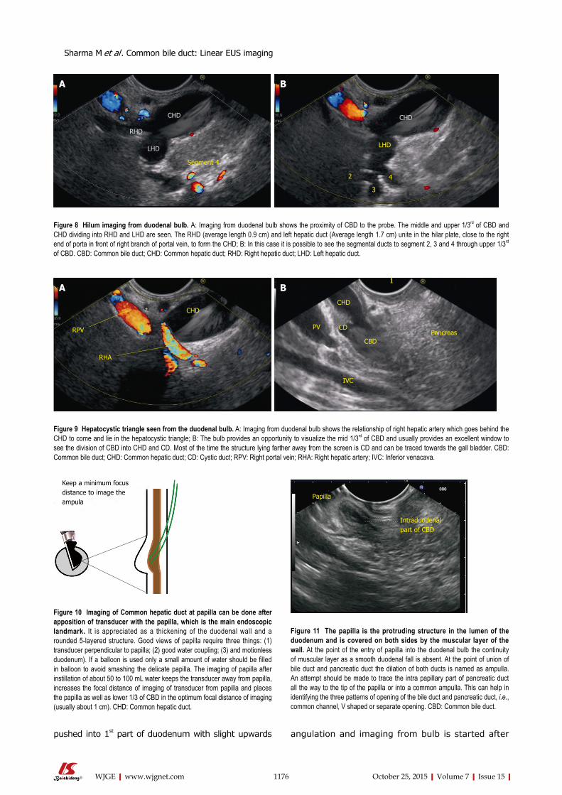
Figure 8 Hilum imaging from duodenal bulb. A: Imaging from duodenal bulb shows the proximity of CBD to the probe. The middle and upper 1/3rd of CBD and CHD dividing into RHD and LHD are seen. The RHD (average length 0.9 cm) and left hepatic duct (Average length 1.7 cm) unite in the hilar plate, close to the right end of porta in front of right branch of portal vein, to form the CHD; B: In this case it is possible to see the segmental ducts to segment 2, 3 and 4 through upper 1/3rd of CBD. CBD: Common bile duct; CHD: Common hepatic duct; RHD: Right hepatic duct; LHD: Left hepatic duct.
RPV
RHA
CHD
ACHD
PV CD
CBD
IVC
Pancreas
1B
Figure 9 Hepatocystic triangle seen from the duodenal bulb. A: Imaging from duodenal bulb shows the relationship of right hepatic artery which goes behind the CHD to come and lie in the hepatocystic triangle; B: The bulb provides an opportunity to visualize the mid 1/3rd of CBD and usually provides an excellent window to see the division of CBD into CHD and CD. Most of the time the structure lying farther away from the screen is CD and can be traced towards the gall bladder. CBD: Common bile duct; CHD: Common hepatic duct; CD: Cystic duct; RPV: Right portal vein; RHA: Right hepatic artery; IVC: Inferior venacava.
Papilla
Intraduodenalpart of CBD
Figure 11 The papilla is the protruding structure in the lumen of the duodenum and is covered on both sides by the muscular layer of the wall. At the point of the entry of papilla into the duodenal bulb the continuity of muscular layer as a smooth duodenal fall is absent. At the point of union of bile duct and pancreatic duct the dilation of both ducts is named as ampulla. An attempt should be made to trace the intra papillary part of pancreatic duct all the way to the tip of the papilla or into a common ampulla. This can help in identifying the three patterns of opening of the bile duct and pancreatic duct, i.e., common channel, V shaped or separate opening. CBD: Common bile duct.
Figure 10 Imaging of Common hepatic duct at papilla can be done after apposition of transducer with the papilla, which is the main endoscopic landmark. It is appreciated as a thickening of the duodenal wall and a rounded 5-layered structure. Good views of papilla require three things: (1) transducer perpendicular to papilla; (2) good water coupling; (3) and motionless duodenum). If a balloon is used only a small amount of water should be filled in balloon to avoid smashing the delicate papilla. The imaging of papilla after instillation of about 50 to 100 mL water keeps the transducer away from papilla, increases the focal distance of imaging of transducer from papilla and places the papilla as well as lower 1/3 of CBD in the optimum focal distance of imaging (usually about 1 cm). CHD: Common hepatic duct.
Keep a minimum focus distance to image the ampula
Sharma M et al . Common bile duct: Linear EUS imaging
establishing contact with posterior duodenal wall. The contact with wall is generally established by turning in an anticlockwise (ACW) direction with down angulation of up and down knobs. Sometimes in this imaging the ACW rotation of the scope may take the scope down and below the level of table in a straight scope position. With suitable rotation and minor adjustments of knobs a home base position is identified where the portal vein is seen on the far side of the screen going from 5 o’clock position to 11 o’clock position. In this home base
position the middle 1/3rd of CBD is commonly identified with slight adjustments of right and left knobs between the transducer and portal vein. Clockwise rotation from this position traces the lower 1/3rd of CBD and ACW rotation traces the upper 1/3rd of CBD as well as the cystic duct and gall bladder (Figures 79).
Imaging from duodenum Imaging from duodenum requires two key movements. The first is entry into 2nd part of duodenum and the
1177WJGE|www.wjgnet.com October 25, 2015|Volume 7|Issue 15|
CBD
D2
D1
++
+ + Calcul
1
+
3.0 mm1.9 mm
007A
CBD
PD
B
CBD
PDCalcul
Duodenum
C
CBD
D
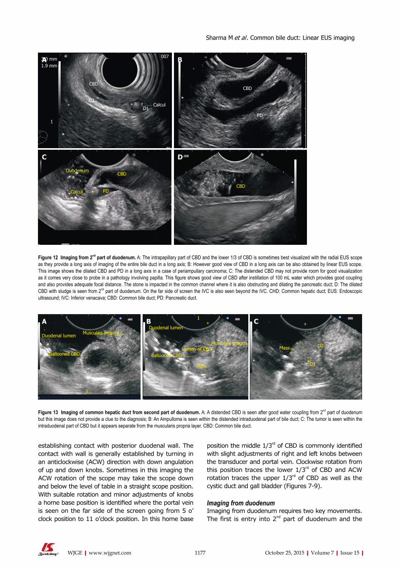
Figure 12 Imaging from 2nd part of duodenum. A: The intrapapillary part of CBD and the lower 1/3 of CBD is sometimes best visualized with the radial EUS scope as they provide a long axis of imaging of the entire bile duct in a long axis; B: However good view of CBD in a long axis can be also obtained by linear EUS scope. This image shows the dilated CBD and PD in a long axis in a case of periampullary carcinoma; C: The distended CBD may not provide room for good visualization as it comes very close to probe in a pathology involving papilla. This figure shows good view of CBD after instillation of 100 mL water which provides good coupling and also provides adequate focal distance. The stone is impacted in the common channel where it is also obstructing and dilating the pancreatic duct; D: The dilated CBD with sludge is seen from 2nd part of duodenum. On the far side of screen the IVC is also seen beyond the IVC. CHD: Common hepatic duct; EUS: Endoscopic ultrasound; IVC: Inferior venacava; CBD: Common bile duct; PD: Pancreatic duct.
Duodenal lumen Musculais propria.....
Ballooned CBD .....
1
ADuodenal lumen
.....Musculais propria
Ballooned CBD .....Lumen of CBD
Mass
+1B
Mass... D2
D1
+
+
+
+
++
+
C
Figure 13 Imaging of common hepatic duct from second part of duodenum. A: A distended CBD is seen after good water coupling from 2nd part of duodenum but this image does not provide a clue to the diagnosis; B: An Ampulloma is seen within the distended intraduodenal part of bile duct; C: The tumor is seen within the intraduodenal part of CBD but it appears separate from the muscularis propria layer. CBD: Common bile duct.
Sharma M et al . Common bile duct: Linear EUS imaging
second is deep intubation into 3rd part of duodenum.
Passage into D2: Entry into D2 is facilitated by engagement of the tip of the probe at D1/D2 junction (superior duodenal angle). Four movements of knob at the superior duodenal angle, i.e., “right turn of knob, up turn of knob, clockwise rotation of the scope and pulling back of the scope” help in passage of probe into second part of duodenum. These movements bring the scope in a short position and place the tip of scope near the papilla once the scope is shortened to about 55 cm. Slow pulling back for shortening can be done by
pulling the shaft of scope with the use of right hand or by the use of outward pressure on the shaft of scope by ulnar aspect of the left hand in an open right position. Endoscopic view should be always maintained during a combination of these movements while shortening to avoid a sudden jerk and entry of the transducer into 2nd part of duodenum.
Passage into D3: Once the second part of duodenum is entered two to three times pushing in and out is required to position the scope deeper into the third part
1178WJGE|www.wjgnet.com October 25, 2015|Volume 7|Issue 15|
GB CHD
CD
Liver
A
GB
CHD
Liver
MPVRPV
LPV
B
Liver
RPV
LPV
LHDRHD
C
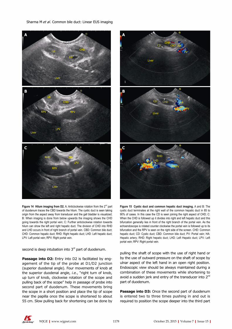
Figure 14 Hilum imaging from D2. A: Anticlockwise rotation from the 2nd part of duodenum traces the CBD towards the hilum. The cystic duct is seen taking origin from the aspect away from transducer and the gall bladder is visualized; B: When imaging is done from below upwards the imaging shows the CHD going towards the right portal vein; C: Further anticlockwise rotation towards hilum can show the left and right hepatic duct. The division of CHD into RHD and LHD occurs in front of right branch of portal vein. CBD: Common bile duct; CHD: Common hepatic duct; RHD: Right hepatic duct; LHD: Left hepatic duct; LPV: Left portal vein; RPV: Right portal vein.
CHDCD CBD HA
PV
Liver
1
A
CHDCD HA
Liver
PV
B
LHD
RHD
LPVRPV
C
Figure 15 Cystic duct and common hepatic duct imaging. A and B: The cystic duct terminates at the right wall of the common hepatic duct in 85 to 90% of cases. In this case the CD is seen joining the right aspect of CHD; C: When the CHD is followed up it divides into right and left hepatic duct and this bifurcation generally lies in front of the right branch of the portal vein. As the echoendoscope is rotated counter clockwise the portal vein is followed up to its bifurcation and the RPV is seen on the right side of the screen. CHD: Common hepatic duct; CD: Cystic duct; CBD: Common bile duct; PV: Portal vein; HA: Hepatic artery; RHD: Right hepatic duct; LHD: Left Hepatic duct; LPV: Left portal vein; RPV: Right portal vein.
Sharma M et al . Common bile duct: Linear EUS imaging
of duodenum.
Imaging from duodenum: From the third part of duodenum a combination of three movements, i.e., slow withdrawal up to the first part of duodenum, clockwise and ACW torque and upward movement of the up and down knobs is required for getting good views of lower 1/3rd of bile duct. This combined movement traces the CBD from the lower 1/3rd towards the upper 1/3rd but as the scope comes towards the first part of
duodenum it tends to slip back into stomach. Movement of the up knob in a fully up position and maintaining a clockwise stance during slow torque from the 2nd part of duodenum helps in preventing the scope from slipping back. Wedging the scope at D1/D2 junction with an inflated balloon is an alternative, which is preferred by some operators to prevent slipping back, but carries a small disadvantage of reverse intussusception of the 2nd part of duodenum into stomach.
In a small number of cases it may be difficult to trace a normal CBD during this combined movement as most of the lumen of CBD gets compressed due to the pressure of transducer. In such cases the combined movement should be done with a main thrust on ACW rotation till it visualizes the anechoic bile duct within the bean shaped hepatoduodenal ligament. A clockwise rotation with slight push and relaxation of the pressure on up and down knob (reverse of the combined move-ment) now traces the CBD from the liver hilum towards the papilla.
Imaging of CBD should be done from below the papilla from the third part of duodenum after instillation of water whenever pathology of papilla (stone or a periampullary tumor) causes distension of intraduodenal part of CBD. This technique provides adequate focal distance for imaging of papilla and good water coupling
1179WJGE|www.wjgnet.com October 25, 2015|Volume 7|Issue 15|
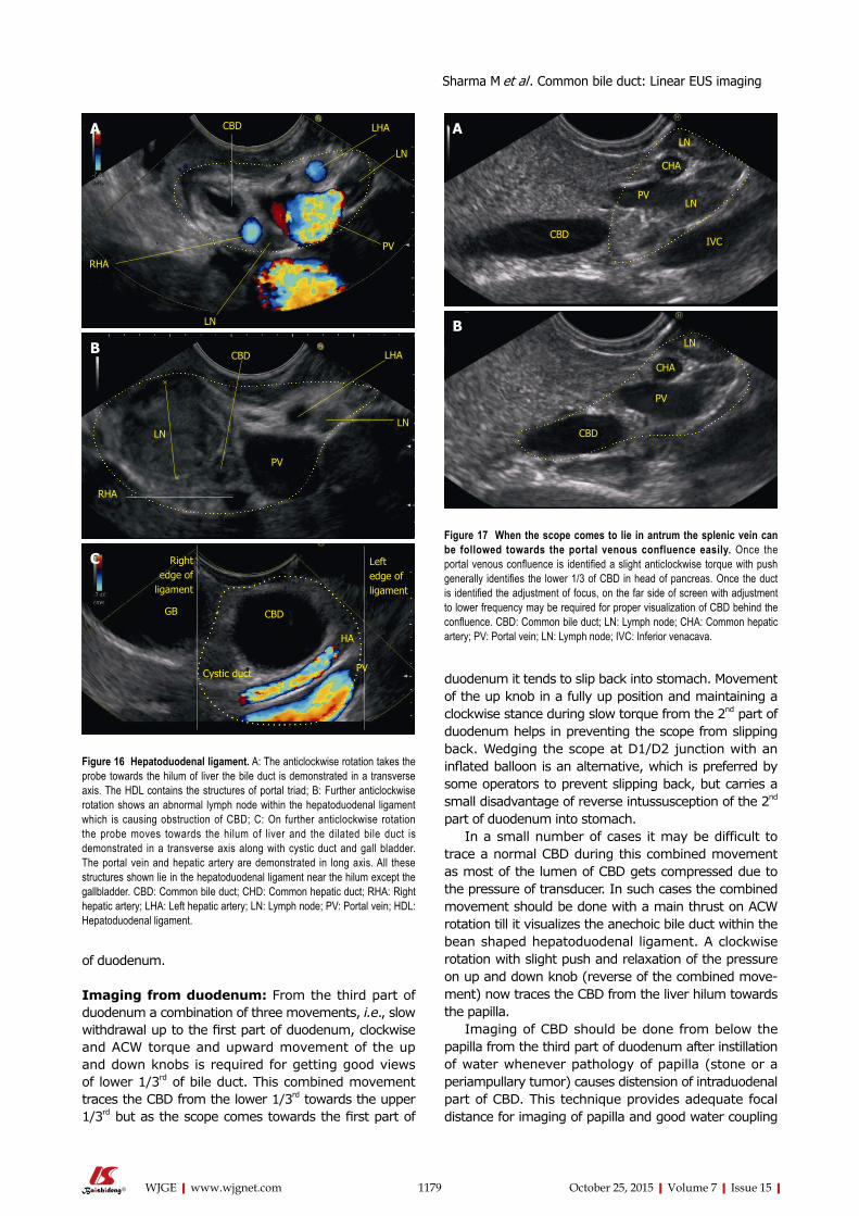
Figure 16 Hepatoduodenal ligament. A: The anticlockwise rotation takes the probe towards the hilum of liver the bile duct is demonstrated in a transverse axis. The HDL contains the structures of portal triad; B: Further anticlockwise rotation shows an abnormal lymph node within the hepatoduodenal ligament which is causing obstruction of CBD; C: On further anticlockwise rotation the probe moves towards the hilum of liver and the dilated bile duct is demonstrated in a transverse axis along with cystic duct and gall bladder. The portal vein and hepatic artery are demonstrated in long axis. All these structures shown lie in the hepatoduodenal ligament near the hilum except the gallbladder. CBD: Common bile duct; CHD: Common hepatic duct; RHA: Right hepatic artery; LHA: Left hepatic artery; LN: Lymph node; PV: Portal vein; HDL: Hepatoduodenal ligament.
CBD LHA
LN
PV
LN
RHA
B
CBDGB
HA
PV
Rightedge of
ligament
Leftedge ofligament
Cystic duct
C
LHA
RHA
LN
PV
LN
CBDALN
LN
CBDIVC
CHA
PV
A
LN
CBD
CHA
PV
B
Figure 17 When the scope comes to lie in antrum the splenic vein can be followed towards the portal venous confluence easily. Once the portal venous confluence is identified a slight anticlockwise torque with push generally identifies the lower 1/3 of CBD in head of pancreas. Once the duct is identified the adjustment of focus, on the far side of screen with adjustment to lower frequency may be required for proper visualization of CBD behind the confluence. CBD: Common bile duct; LN: Lymph node; CHA: Common hepatic artery; PV: Portal vein; LN: Lymph node; IVC: Inferior venacava.
Sharma M et al . Common bile duct: Linear EUS imaging
(Figures 1016).
Imaging from antrum This imaging is similar to imaging through the pancreatic window from stomach as already described above. It can be done if evaluation of CBD is considered necessary once the scope slips back from the 2nd part of duodenum or once the examination from duodenum is completed. As the scope comes to lie opposite the head of pancreas the pancreatic window provides optimum imaging of lower 1/3rd of CBD (Figure 17).
CONCLUSIONThe techniques described in the above section can be expected to reproduce the images as discussed in majority of cases and from most of the stations. The only station of CBD imaging which may not reproduce the images as described is from duodenal bulb. This difference in reproducing the images and a great variability of images comes mainly due to the variability of the position of scope (short loop, or J shaped position) and due to the use of balloon (nestled, wedged, withdrawn wedged, intussuscepted). The basic concept of imaging however remains simple: stomach shows mainly the upper 1/3rd of CBD, bulb shows mainly the middle 1/3rd of CBD and duodenum shows mainly the lower 1/3rd of CBD. The follow up imaging to trace entire CBD requires a clockwise rotation and push from upper 1/3rd of CBD. The follow up imaging to trace entire CBD requires an ACW rotation and pull from lower 1/3rd of CBD. The follow up imaging to trace entire CBD requires a clockwise rotation to trace the lower 1/3rd and an ACW rotation to trace the upper 1/3rd when imaging is started
from middle 1/3rd of CBD.
REFERENCES1 Dietrich CF. Endoscopic Ultrasound: An Introductory manual and
Atlas. New York: Thieme, 20062 Van Dam S, Sivak MV. Gastrointestinal Endosonography.
Philadelphia, Pennsylvania: Saunders, 19993 Rosch T, Will U, Chang KJ. Logitudianl Endosonography: Atlas and
Manual for Use in the Upper Gastrointestinal Tract. Germany, 20014 Gress FG. Ishan Bhattacharya. Endoscopic Ultrasonography.
Massachusetts: Wiley-Blackwell, 20015 Hawes RH. Paul Fockens. Endosonographyh. Philadelphia:
Saunders, 20066 Rameshbabu CS, Wani ZA, Rai P, Abdulqader A, Garg S, Sharma
M. Standard imaging techniques for assessment of portal venous system and its tributaries by linear endoscopic ultrasound: a pictorial essay. Endosc Ultrasound 2013; 2: 16-34 [PMID: 24949362 DOI: 10.4103/2303-9027.117724]
7 Sharma M, Rai P, Rameshbabu CS, Arya S. Techniques of imaging of pancreatic duct by linear endoscopic ultrasound. Endosc Ultrasound 2014; 3: 179-190 [PMID: 25184125 DOI: 10.4103/2303-9027.138793]
8 Sharma M, Pathak A, Rameshbabu CS, Rai P, Kirnake V, Shoukat A, Dietrich F C, Sharma SS, Imaging of Pancreas Divisum by Linear Array Endoscopic Ultrasonography. Endosc Ultrasound 2014; In press
9 Sharma M, Rai P, Rameshbabu CS, Senadhipan B. Imaging of peritoneal ligaments by endoscopic ultrasound (with videos). Endosc Ultrasound 2014; 4: 15-27 [PMID: 25789280 DOI: 10.4103/2303-9027.151317]
10 Sharma M, Rai P, Mehta V, Rameshbabu CS. Techniques of imaging of aorta and its 1st order branches by Endoscopic Ultra-sound. Endosc Ultrasound 2015; 4: 98-108 [DOI: 10.4103/2303-9027.156722]
11 Sharma M, Rameshbabu CS, Dietrich F C, Rai P, Bansal R, Pathak A. Endoscopic Ultrasound of the hepatoduodenal ligament and porta hepatis. Endosc Ultrasound 2014; In press
12 Sharma M, Pathak A, Shoukat A, Thomas S N, Mehta D. Seagulls of EUS. Endosc Ultrasound 2015; In Press
P- Reviewer: Kurtoglu E, Skok P S- Editor: Ji FF L- Editor: A E- Editor: Jiao XK
1180WJGE|www.wjgnet.com October 25, 2015|Volume 7|Issue 15|
Sharma M et al . Common bile duct: Linear EUS imaging

© 2015 Baishideng Publishing Group Inc. All rights reserved.
Published by Baishideng Publishing Group Inc8226 Regency Drive, Pleasanton, CA 94588, USA
Telephone: +1-925-223-8242Fax: +1-925-223-8243
E-mail: [email protected] Desk: http://www.wjgnet.com/esps/helpdesk.aspx
http://www.wjgnet.com