Embed Size (px)
Citation preview

The Magazine for Molecular Imaging Innovation
Imaging Life
Cover Story The Future of Molecular Imaging Has ArrivedPage 08
Outcomes Siemens‘ PETNET Solutions Partnership Delivers PET/CT Outcomes Page 24
SciencePET/CT in Radiation Therapy Planning for Cervical CarcinomaPage 42
Clinical CasexSPECT Bone Metastases in a Case of Myxoid LiposarcomaPage 50
Customer CareTips & Tricks: Setting Scan Range on the New Biograph mCT Flow Page 60
06
Issue Number 06SNMMI Edition | June 8-12, 2013

2 Imaging Life · June 2013 · www.siemens.com/imaginglife
“These innovative new platforms will change the face of molecular imaging by enabling more efficient and effective patient care.”Alexander R. Zimmermann, Vice President Marketing and Sales, Molecular Imaging, Siemens Healthcare
Editorial
Imaging Life · June 2013 · www.siemens.com/imaginglife 3
Editorial
Dear Reader,PET/CT and SPECT/CT exemplify how thewhole can be greater than the sum ofits parts. The combination of CT withthe two standard bearers of nuclearmedicine, PET and SPECT, added notonly a simple and reproducible meansof attenuation correction, but also the ability to put functional data into anatomical context.
However, while contributing enormously to patient management, neither has achieved its true potential. The stop-and-go movement of the patient table that characterizes PET/CT has constrained this modality to datacollection by bed positions, limitingthroughput, image resolution andquantification, even contributing toartifacts due to patient movement.
And, while SPECT/CT delivers the high sensitivity associated with SPECT, the addition of CT has not improvedspecificity, a shortcoming due to themechanical fusion of images. Thismechanical mismatch of data hasfurther limited SPECT/CT, as it has maderoutine quantification impossible.
As a recognized leader in medicalinnovation for more than 130 years,
Siemens is commited to pioneeringnew technologies that break throughthe limitations of molecular imaging to address the growing challenges intoday’s healthcare environment andprovide definitive and timely answersto physicians and their patients.
At this year’s annual Society of Nuclear Medicine and Molecular Imaging (SMNMI) annual meeting in Vancouver, BC, Canada, Siemens once again demonstrates its dedication to advancing the quality of patient care with the launch of two revolutionary new products engineered to break the bonds that have restrained PET/CT and SPECT/CT.
Our new Biograph mCT Flow™* replaces the stop-and-go motion of conventional PET/CT with FlowMotion™, which eases the patient steadily through the gantry, boosting throughput, providing accurate quantification, delivering the finest resolution** and increasing patient comfort.
Our new Symbia Intevo™*** integratesthe data obtained from SPECT and CT, combining the high sensitivity of SPECT with the high specificity of CT, and, for the first time, allowing routine
quantification. These clinical capabilities are so advanced that this new product represents a wholly new modality, which we call xSPECT.***
In this issue we describe the clinicalsignificance of FlowMotion and xSPECT,illustrating with the experience andexpectations of leaders in the molecularimaging community how theseinnovative new platforms will changethe face of molecular imaging byenabling more efficient and effectivepatient care.
Enjoy reading,
Alexander R. Zimmermann
* The Biograph mCT Flow and mCT Flow Edge are not commercially available in all countries. Due to Regula-tory reasons their future availability cannot be guar-anteed. Please contact your local Siemens organiza-tion for further details.
** Based on volumetric resolution available in competitiveliterature for systems greater than 70 cm bore siz. Data on file.
*** Symbia Intevo and xSPECT are not commercially available in all countries. Due to regulatory reasons their future availability cannot be guaranteed. Please contact your local Siemens organization for further details.
Alexander R. Zimmermann Vice President
Marketing and Sales Molecular Imaging
Siemens Healthcare

4 Imaging Life · June 2013 · www.siemens.com/imaginglife
Contents
Cover Story
Contents24 Siemens‘ PETNET Solutions Partnership Delivers PET/CT Outcomes
Cover Story08 The Future of Molecular
Imaging Has Arrived
News06 Explora One: One Integration,
Infinite Possibilities
07 Siemens Increases Patient Access to Amyloid Imaging in the United States
07 Siemens’ PETNET Solutions to Manufacture Amyloid Imaging PET Tracer in Europe
42 PET/CT in Radiation Therapy Planning for Cervical Carcinoma
08 The new Biograph mCT Flow™*
replaces the stop-and-go motion ofconventional PET/CT with FlowMotion™, which eases the patient smoothly through the gantry.Meanwhile, Symbia Intevo™**, theworld’s first xSPECT* system, integratesthe data obtained from SPECT andCT, combining the high sensitivity ofSPECT with the high specificity of CT,to produce high-resolution imagesand, for the first time, allow routine quantification. In this issue, read more about the clinical significance of these two revolutionary platforms and how leaders in the molecular imaging community expect this technology to change the face of molecular imaging.
* The Biograph mCT Flow and mCT Flow Edge are not commercially available in all countries. Due to Regula-tory reasons their future availability cannot be guar-anteed. Please contact your local Siemens organiza-tion for further details.
** Symbia Intevo and xSPECT are not commercially avail-able in all countries. Due to regulatory reasons their fu-ture availability cannot be guaranteed. Please contact your local Siemens organization for further details.

Imaging Life · June 2013 · www.siemens.com/imaginglife 5
Contents
60 Tips & Tricks: Setting Scan Range on the New Biograph mCT Flow
50 xSPECT Bone Metastases in a Case of Myxoid Liposarcoma
Outcomes24 Siemens’ PETNET Solutions
Partnership Delivers PET/CT Outcomes
26 Biograph mCT Paves the Road to Advances in PET/CT Quantification
30 IQ•SPECTStreamlinesCardiacImaging at Amphia Hospital
32 Biograph TruePoint 16 Meets Challenges at Fox Chase Cancer Center
34 xSPECT—A New Benchmark in Image Quality and Quantification
Science38 Village of Forgetfulness
42 PET/CT in Radiation Therapy Planning for Cervical Carcinoma
Clinical Case Studies46 FlowMotion 18FFDGPET•CT
Evaluation of Chemoradiation Response in a Case of Lung Carcinoma
48 xSPECT Imaging in a Patient with Diffuse Skeletal Metastases
50 xSPECT Bone Metastases in a Case of Myxoid Liposarcoma
52 AmyloidPET•CTinPatientswithSuspected Alzheimer’s Disease
54 18FFDGPET•CT-basedRadiationTherapy Planning in a Case of Cervical Carcinoma
56 Improved Detection of Hepatic Metastases from a Pancreatic Neuroendocrine Tumor with Simultaneous MR and 68Ga DOTATOC PET Acquisition
58 PET Myocardial Perfusion Combined with CT Angiography in Detection of Post- Revascularization Ischemia
Customer Care60 Tips & Tricks: Setting Scan Range
on the Biograph mCT Flow
62 Spanish Clinical University on Forefront of Research and Clinical Practice
64 2013 Image of the Year Competition
65 MI University 360: Connecting Physicians Around the Globe
66 Imprint
67 Subscription

6 Imaging Life · June 2013 · www.siemens.com/imaginglife
News
Explora One: One Integration, Infinite Possibilities Siemens unveiled its new Explora One chemistry box at the 20th International Symposium on Radiopharmaceutical Sciences (ISRS), on Jeju Island, Korea. Explora One is a fully integrated cassette-based solution for the production of commercially available and newly released 18F PET imaging biomarkers, as well as imaging biomarkers under development or in clinical trials.
One Easy, Integrated Workflow
Imaging biomarker production is dictated by the synthesis sequence, which may require specific steps, including synthesis, purification and reformulation. Traditionally, this meant that for users to produce a variety of tracers, they needed multiple separate hardware systems to perform the specific steps. Multiple individual pieces of hardware virtually eliminates any possibility of an integrated workflow between complex steps, increasing the risk of human error, which can lead to contamination or loss of a production run.Now, Siemens introduces Explora OneTM, a revolutionary system to address these requirements and provide an easy and integrated solution. A fully integrated hardware design aids compliance with a wide range of imaging biomarker production scenarios with the benefit of reduced lab space, which in turn reduces initial setup costs. It also virtually eliminates user error by providing an automated and integrated workflow for easy imaging biomarker production, improving production output efficiency and quality, while decreasing the risk of loss of revenue due to human error and contamination. Additionally, Explora One’s browser-based user interface makes it possible for users to perform tasks outside of the sterile environment, perform routine troubleshooting remotely and improve workflow efficiency. This feature also eliminates issues with computer compatibility or the need for additional
computer systems, providing another opportunity to reduce costs. Explora One simplifies the overall process to meet cGMP (current Good Manufacturing Practices) guidelines for the production of imaging biomarkers with features including tiered individual password protected user accounts, all in one tracer specific production reports for each initiated production sequence and the flexibility for users to choose from a wide variety of non-proprietary cGMP certified cassettes for production.
One System, Infinite Possibilities
PET clinical imaging is growing in demand and utilization with an increasing trend toward a more personalized, disease-specific approach to diagnosis, which requires disease process specific imaging biomarkers in order to better manage patient outcomes. To meet the demand of PET imaging growth and imaging biomarker variety, a reliable, efficient and flexible production system is vital. Explora One meets this need, providing protection for future PET imaging biomarker production, avenues to venture into newly released imaging biomarkers and the ability to expand with the creation of new synthesis sequences and partake in research.Explora One allows users the flexibility to purchase cassettes from multiple consumables vendor, thus allowing users the choice of a wider variety of non-proprietary cassettes for production compared to conventional solutions.
Explora One will be released with the functionality to produce more then eight imaging biomarkers, as well as with the capability to easily expand to newly released imaging biomarkers, including those imaging biomarkers held with intellectual property (IP) licensing. This flexibility enables users to adopt new tracers as they come to market. Furthermore, the system’s development mode facilitates the creation of new synthesis sequences either independently or as part of a structured trial.The solution is bundled with Siemens Remote Services (SRS), which provides a hotline for troubleshooting, arranging on site support and maintaining system logs to establish trending, reduce downtime and improve overall efficiency to help enable highly reliable production.Siemens Explora One‘s unique combination of features make this the ideal solution for 18F imaging biomarker production now and in the future.
The new Explora One chemistry box integrates all steps of the biomarker production process into one module.

Imaging Life · June 2013 · www.siemens.com/imaginglife 7
News
Siemens Increases Patient Access to Amyloid Imaging in the United StatesSiemens’ PETNET Solutions increased patient access to Eli Lilly and Company’s Amyvid™ (Florbetapir F 18 Injection) by expanding manufacturing and distribu-tion to a total of 17 metro areas in the United States. As of March 2013, Siemens’ PETNET Solutions is now com-mercially distributing Amyvid in Denver; Raleigh, NC; Boston; Minneapolis; Hackensack, NJ; Los Angeles, Fort Lauderdale, FL; Cleveland; Atlanta; Chicago; Dallas; Houston; Jacksonville, FL; Palo Alto, CA; Philadelphia; Seattle; and St. Louis. With the largest global network of posi-tron emission tomography (PET) radio-pharmaceutical production facilities, Siemens’ PETNET Solutions was the first company to sign a manufacturing and distribution agreement with Eli Lilly and Company in November 2011 for Amyvid.Siemens’ PETNET Solutions has made significant investments to standardize equipment and training processes across its entire production network to reduce the level of complexity and ensure consistency in its PET
radiopharmaceutical production. In addition, Siemens’ PETNET Solutions has invested in dedicated staff for the pro-duction of amyloid imaging biomarkers. As a result, Siemens’ PETNET Solutions is able to offer hospitals and imaging cen-ters a high level of delivery reliability, bolstering confidence in their ability to scan patients as scheduled. For instance, “Trident Medical Imaging Center has found Siemens’ PETNET Solutions to be extremely reliable in its production and distribution of Amyvid,” said Guy Messer, chief financial officer, Trident Imaging Center, Fayetteville, GA. Trident was the first imaging center in Georgia to administer Amyvid for a PET imaging exam.Since 1996, Siemens’ PETNET Solutions has leveraged its network of highly expe-rienced professionals and a recognized portfolio of programs designed to help customers establish and grow their PET imaging offerings and provide new ser-vices to their communities. This expertise and services continues to fuel the expan-sion of PET imaging in the United States.
“Siemens’ PETNET Solutions is proud to expand its production and distribution of Amyvid, increasing the availability of this radiopharmaceutical to patients in the USA,” said Dr. Christoph Zindel, CEO of PETNET Solutions. “The continual invest-ment in our network to standardize our equipment, as well as our integrated manufacturing and pharmacy facilities provides hospitals and imaging centers with broad coverage and extremely high reliability in proprietary radiopharmaceuti-cal production, helping them to offer this new PET/CT imaging exam to patients.”
Siemens’ PETNET Solutions to Manufacture Amyloid Imaging PET Tracer in EuropePETNET Solutions, Inc. has signed a European manufacturing services agreement with Eli Lilly and Company that grants Siemens’ PETNET Solutions the right to manufacture Lilly’s Amyvid™ (Florbetapir F 18 Injection) at its facilities located in the United Kingdom, France and Spain. First commercial doses of Amyvid produced by PETNET Solutions are expected to begin in the United Kingdom in June 2013, followed by Spain by the end of 2013, and France in 2014, pending regulatory approval of these sites.
Siemens’ PETNET Solutions will leverage its experience as the largest manufacturing and distributor of Amyvid in the USA to provide high delivery reliability in the European Union. Siemens’ PETNET Solutions has made significant investments throughout its entire global network to standardize operations, including new automated processes and redundant equipment , to serve clinical research demands, as well as immediate patient needs.“This agreement with Lilly is another proof point that Siemens is at the
forefront in providing leading PET imaging solutions to fight against the most challenging diseases, such as Alzheimer’s,” said Dr. Christoph Zindel, CEO of PETNET Solutions. “With our investments into our network over the last several years and our experience manufacturing Amyvid in the USA, we are confident that we will provide our customers in Europe the same level of high delivery reliability that our customers have come to expect from PETNET Solutions.”

8 Imaging Life · June 2013 · www.siemens.com/imaginglife
Cover Story
Data courtesy of University of Tennessee, Knoxville, TN, USA
Data courtesy of Ludwig Maximilian University,
Munich, Germany

Imaging Life · June 2013 · www.siemens.com/imaginglife 9
Cover Story
Nuclear medicine has long been a corner-stone of diagnostic imaging due to its ability to illustrate metabolic activity and provide physicians with the answers they need sooner than is possible with tradi-tional anatomical modalities. However, in today’s challenging healthcare environ-ment, where the growing demand for higher-quality, patient-centered care is matched only by the need for definitive and timely answers, the limitations of conventional PET/CT and SPECT/CT scan-ners are becoming more evident.Despite the high sensitivity of today’s SPECT/CT scanners that can aid physi-cans in early disease detection, the modality is restricted in its ability to provide definitive and timely answers. Its limited specificity, resulting from the use of mechanically fused images, often requires the need for follow-up proce-dures that delay patient care and poten-tially increase costs. Meanwhile, the stop-and-go technology of conventional PET/CT limits a physician’s ability to tailor the scanning to the needs of each patient and results in a one-size fits all approach to scanning that can lead to higher dose and unnecessary patient anxiety.Founded on the belief that the highest technical performance is only achieved when solutions deliver clinical outcome that can help improve patient health, Siemens has been a recognized leader in medical innovation for more than 130 years. From the first electromedical devices in 1896 to the latest molecular
The Future of Molecular Imaging Has Arrived Siemens has once again proved that the most distant technical horizons can be surpassed with consistent dedication to improving healthcare through the introduction of two new platforms that redefine molecular imaging. One transforms the one-size-fits-all PET/CT exam into a tailored suit; the other propels hybridized SPECT and CT beyond today’s state-of-the-art technology to qualify as a whole new modality.
By Greg Freiherr
imaging technologies, Siemens has a long history of pioneering technologi-cal achievements that help make the impossible, possible.In what may be the most significant advance in PET in more than a decade, Siemens’ new Biograph mCT Flow™* replaces the stop-and-go acquisition of conventional exams with FlowMotion™, easing the patient steadily through the gantry to boost throughput, improve uni-formity for better quantification and increase patient comfort. Taking PET/CT to a new level of performance, Biograph mCT Flow eliminates the need to collect data in overlapping bed positions by con-tinuously scanning the entire patient. The unavoidable necessity of overlaps in stop-and-go PET is eliminated with FlowMotion, as the patient moves smoothly through the Biograph mCT Flow gantry. These time savings can be used to produce higher resolution images, as the pace of the table’s con-tinuous motion slows to allow the detec-tor to gather more counts over demand organs and body areas. Biograph mCT Flow increases detector sensitivity edge-to-edge, boosting both the accuracy and reproducibility of quantitative values that characterize disease and health. The continuous motion helps assure patients that the scan is progressing, while efficiencies afforded by Biograph mCT Flow minimize radiation exposure.Siemens’ new Symbia IntevoTM** inte-grates data from SPECT and CT.
Leveraging this hybridized source of information, the new scanner delivers the high sensitivity commonly obtained with SPECT, the high specificity often sought in follow-up studies and, for the first time, quantification. Together these capabilities raise this technologi-cal advancement to the status of a new modality, which Siemens has dubbed xSPECT.**
In the revolutionary xSPECT exams per-formed with Symbia Intevo, image qual-ity reaches extraordinary resolution as data from the two systems are matched and processed together using algorithms that correct and localize SPECT informa-tion using CT information beyond the positional data. The exactness of this integration allows zoning and a corre-sponding image quality boost, as well as SPECT quantification, which is difficult to perform on conventional SPECT/CT. Fast and confident one-stop accurate results, a never-before-possible achievement that could possibly render tests, such as MR and biopsy unnecessary, potentially sav-ing time and reducing healthcare costs. In these unique ways, Biograph mCT Flow and Symbia Intevo could not only change the practice of molecular imaging, but also alter the foundation by which dis-ease is detected and characterized. They have the potential to replace uncertainty with certainty and streamline the pro-cesses underlying diagnosis and therapy monitoring, heralding a new age of molecular imaging and more efficient

10 Imaging Life · June 2013 · www.siemens.com/imaginglife
Cover Story
and effective patient management.Surgeons had removed the patient’s cancer and all of the thyroid tissue sur-rounding it. But there was a node of metabolic activity at the base of the neck, where metastatic disease would not be expected. In line with that, behind the jugular, was another, less intense node.If a traditional whole-body PET/CT had been performed, those cancerous nodes would likely have been missed. But Dr. Kirk A. Frey, MD, PhD, director of the PET Center at the University of Michigan Hospitals in Ann Arbor, MI, USA, was reading images acquired with a new generation of PET•CT, one that allows physicians to increase the resolution of selected body areas with-out requiring more time for the exam. “We wouldn’t have been confident about either node if not for the high-resolution scan,” Frey says. “It gave us the information we needed to make a confident diagnosis, and the surgeons a nice road map to remove the nodes.”The scan was acquired on Siemens’ new Biograph mCT Flow, arguably the most significant advancement in PET since it was hybridized more than a decade ago. Rather than bed posi-tions, defined by conventional stop-and-go movement of the patient table, scans with the new scanner, guided by FlowMotion, are focused according to body areas and organs as the patient moves continuously through the detector rings.In the case performed by Frey, the patient’s head and neck were scanned at a slow table speed. This allowed more counts to be gathered and high-resolution images to be produced. “The ability to do a single acquisition, while easily dwelling over areas you want to reconstruct at higher resolu-tion, is a big plus,” he says.The continuously moving patient table provides the foundation for software and hardware that together remedy the shortcomings of conventional, stop-and-go PET/CT. Images are higher
quality; quantification more sensitive and reproducible; dose and throughput optimized; and scans more comfortable for the patient.FlowMotion orchestrates image quality, doubling the resolution of conventional scanning for selected areas of the body and easily gating data acquisition to account for respiration as part of standard scanning protocol, making it routine. A new quantification paradigm improves noise uniformity in all dimensions, edge-to-edge, maximizing accuracy and boosting reproducibility. Throughput can be increased by scan-ning body areas at a steady, rapid pace. Alternatively, the table can be slowed to gather the necessary counts with a lower dose of radiopharmaceutical. Patient experience is addressed as well, as the continuous motion of the table reassures patients that the scan is progressing, free from the stop-and-go bed motion that characterizes con-ventional PET/CT.What makes FlowMotion unique clini-cally, however, is what matters most—the ability to detect, characterize and monitor disease with greater certainty than ever before.
Biograph Changes Patient Flow
Conventional PET/CT assigns rigidly sized bed positions, typically seven for an adult male. When performing a stop-and-go scan, the table moves into the first bed position and stops. It remains there, sta-tionary for a set time, while the ring detectors record the number of photon strikes resulting from coincidence events. The table then steps to the next position, where it stops again. This process repeats until the scan is done. Planning and scanning is limited to the fixed size of the detector and the bed positions. While technically possible in some scanners, the complexity of adjusting scan parameters in clinical stop-and-go protocols has limited its routine use. As a result, each bed or step acquisition typically lasts the same length of time and uses the same reconstruction matrix. Despite an increasingly competitive and rapidly changing healthcare environment that demands definitive and timely answers, hospitals and physicians have been con-fined to the limitations set by stop-and-go technology. This has resulted in
Siemens Unveils Continuous FlowMotion PET•CT
Continued on page 12
Biograph mCT Flow

Imaging Life · June 2013 · www.siemens.com/imaginglife 11
Cover Story
xSPECT: The Difference Between Seeing and KnowingWith the introduction of Symbia Intevo, Siemens is once again pioneering hybrid imaging. This new system weaves SPECT and CT data so tightly together that the system represents not just a new prod-uct, but a new modality.As the world’s first xSPECT** system, Symbia Intevo delivers not only the high sensitivity that the nuclear medicine community has come to expect from SPECT, but also extraordinary image quality. And best of all, for the first time, the SPECT data is fully quantitative.Peter Bartenstein, MD, chairman of the Department of Nuclear Medicine, Ludwig-Maximillians-University in Munich, Germany, describes xSPECT as providing excellent spatial resolution. “Because we can better locate the lesion and get a clearer idea of the extent of the lesion, xSPECT may result in more confident interpretation,” he says.“We are in the early stages of our evalua-tion of quantification with xSPECT,” adds Jerry Froelich, MD, director of Nuclear Medicine and Molecular Imaging at the University of Minnesota in Minneapolis, MN, USA. “I am excited about the possibilities.”Images generated by xSPECT, comprised
of SPECT and CT data, show excellent delineation between bone and soft tissue and the lesions present within, according to Zsolt Szabo, MD, PhD, professor of Radiology at Johns Hopkins Hospital. “It is much easier to localize lesions in the bone and do lesion characterization,” he says.
A Breed Apart
Despite the high sensitivity of today’s SPECT/CT scanners, which permit early disease detection, the modality is restricted by its ability to provide defini-tive and timely answers.While detecting virtually all cases of dis-ease, SPECT and SPECT/CT have always been labeled as “unclear medicine.” Physicians often have been forced to rely on other modalities, such as MRI, diagnos-tic CT, PET/CT or biopsy, to accurately char-acterize suspicious lesions, distinguishing inflammation, for example, from bone tumor. For this reason, SPECT/CT has been viewed as a modality that may generate as many questions as it does answers.Symbia Intevo (short for evolution of integration) overcomes the challenges that limit conventional SPECT/CT, by pro-viding the information that physicians need to find and differentiate disease. It
does so through a new, accurate align-ment method that completely integrates SPECT and CT. The resulting high-resolu-tion xSPECT images provide physicians with the potential to not only find dis-ease, but also to more confidently inter-pret images. Moreover, Symbia Intevo’s unique quantitative capabilities may pro-vide the ability to monitor and adjust treatments earlier by accurately measur-ing small differences. “The quantitative aspect is of paramount importance,” says Bartenstein. “Without it, you are really making an educated guess.”Symbia Intevo also has the potential to improve patient care and increase effi-ciency by cutting dose to the minimum needed to achieve high-quality diagnostic images—all while maximizing through-put. Its optimization of CT and SPECT data acquisition and processing affords the potential to cut dose in half and dou-ble the scan speed, thereby increasing patient well-being and throughput.As such, Symbia Intevo is a truly revolu-tionary product that represents not only a technological advance, but also an entirely new modality: xSPECT.
Building on Innovation
The Symbia Intevo xSPECT series is built upon the same roots as conventional SPECT and SPECT/CT. These roots extend back decades to the first gamma cam-eras, which produced planar images. These two-dimensional images indicated the presence and general location of sus-picious lesions, but little more. They gave way in the 1970s to images produced using single photon emission CT scan-ners. These images provided substan-tially higher resolution based on technol-ogy that evolved from CT scanners.The success of PET/CT led to the hybrid-ization of SPECT and CT a decade ago. But early SPECT/CT scanners did not meet expectations. Instead, they repre-sented mostly an advance in attenua-tion correction, as the CT components were incapable of delivering diagnostic information.
Continued on page 18Symbia Intevo

12 Imaging Life · June 2013 · www.siemens.com/imaginglife
Cover Story
best use of time. This flexibility over the different zones allows every scan to pro-duce optimal image quality.“In the past, we had to decide where to start gating, based on bed position, not anatomy,” Frey says. “I think the ability to use anatomically defined segmentation will drive the acquisition parameters in a sensible direction and raise the opportu-nity for individualized scanning protocols.” Gone is the need for time-consuming multiple scans in a conventional patient exam to accomplish higher contrast over a specific area. Gone, too, is the one-size-fits-all compromise that has belea-guered PET/CT since its inception.
FlowMotion Innovation
Underpinning the success of the new Biograph mCT Flow with FlowMotion is the precisely engineered table. Siemens began work on this technology with Dr. David W. Townsend PhD, a pioneer of PET/CT, more than 10 years ago, knowing this was the key to the future. The first step was the development of Siemens’ SMART patient handling system (PHS). A key component of the SMART PHS is the cantilevered bed with its hardened carbon fiber, which allows zero differen-tial deflection. This ensures that the patient remains correctly positioned from
higher dose, greater patient anxiety and lower efficiency.Siemens, a recognized leader in medical innovations for more than 130 years, has broken down these barriers with the introduction of Biograph mCT Flow with FlowMotion, the world’s first continu-ous motion PET/CT system. Instead of fitting the patient to bed positions defined by the old way of stop-and-go, Biograph mCT Flow fits the scan to each patient with a bed that moves in one continuous motion. Under the automatic control of Siemens’ FlowMotion, Biograph mCT Flow replaces this one-size-fits-all approach with protocols tailored for each organ. Up to four zones are assigned to a single scan. Each is customized to tailor fit organs and anatomic regions, for exam-ple, the high resolution scan of the head/neck or respiratory-gated scan of the lungs. The bed moves continuously, but its speed is adaptable throughout the scan based on the counts needed for each organ or zone. For example, in patients suspected of lung cancer, the new standard imaging protocol may set zone 1 (the head and neck) at a slower scan speed for high resolution to look for metastatic disease in the brain and lymph nodes. Zone 2 may cover the lungs at a slower scan speed and with gating. Zone 3 over the liver may maximize counts in case of liver metastasis. Zone 4 may be covered at increased speed in lower attenuating areas like the extremities to make the
Continued from page 10
Siemens Unveils Continuous FlowMotion PET•CT
Conventional Stop-and-Go
FlowMotion
Magnetically Driven Table
+ + + + + + + + + + + + + +

Imaging Life · June 2013 · www.siemens.com/imaginglife 13
Cover Story
the CT exam at the front of the gantry through the PET exam at the back, thereby preventing misregistration between the CT and PET images. The table can travel up to 200 mm per second when positioning the patient. The magnetic drive enables precise acquisi-tion speeds from 0.1 mm to 10 mm per second with submillimeter positioning accuracy and <0.1 percent velocity accu-racy, even carrying a patient weighing 227 kg/500 lbs. This precision cannot be achieved in the belt-driven CT bed design found on conventional PET/CT scanners. Further contributing to the engineering success of FlowMotion is the new Flow Advanced Computational System (ACS), which acquires imaging data from the scanner. Two solid-state drives, coupled to the detector array, help ensure a steady stream of data from the scanner to the ACS. These data are processed on the fly, thus resolving a shortcoming of stop-and-go scanning, which can only
record as many counts as the onboard storage device will allow.With Flow ACS, the data stream from the scanner for immediate processing with-out limiting signal collection. In conven-tional scanning, data accumulate for each bed position. FlowMotion has another advantage as each event is time and position “stamped” to indicate where and when the data were acquired so they can be rebinned for reconstruction and normalized based on their specific line of response. This new complexity, addressed by FlowMotion, propels PET•CT to the next level of performance, allowing every detector row in the scanner to see the body as it moves continuously through the scanner field of view (FOV).
Finest Detail in Every Scan
To provide an accurate diagnosis, physi-cians need an accurate image. However, conventional systems can lose defini-tion toward the edge of the FOV, miss small or low-grade lesions and are sub-ject to motion blurring, potentially com-promising the image and reducing diag-nostic confidence. With FlowMotion as the guiding princi-ple, Biograph mCT Flow provides unsur-passed image quality, which has always been the first priority at Siemens. Its OptisoHD (high-definition) detection system is designed to deliver the indus-try’s finest*** volumetric resolution of 95 mm3, with the industry’s best 400x400 reconstruction matrix.*** Siemens’ ultraHD•PET delivers increased image quality compared to conventional PET/CT technology by combining two important
1
1 FlowMotion supports the highest possible resolution for each organ with a high-resolution 400x400 matrix and HD•Chest for motion management. This helps achieve the finest volumetric resolution,*** which, for example, is critical for white and grey matter differentiation and early lesion detection. Data courtesy of University of Michigan, Ann Arbor, MI, USA
“There are parts of the whole-body image where reconstruction on a more resolute matrix offers real advantages...”Kirk Frey, MD, PhD Director of the PET Center University of Michigan Hospitals Ann Arbor, MI, USA
Solid-State Electronic Architecture Dynamic Data Processing

14 Imaging Life · June 2013 · www.siemens.com/imaginglife
Cover Story
innovations: HD•PET and Time of Flight (TOF). With the addition of z-Sharp™ technology that offers CT-isotropic resolution down to 0.28 mm at any position within the scan field, Biograph mCT Flow pushes the boundaries of spatial resolution. Additionally, by eliminating the need for individual bed positions, Biograph mCT Flow enables examination parameters, such as speed, image resolution and motion management to be adjusted to the precise dimensions of each organ.
At the University of Michigan Hospitals, Frey and colleagues are custom-design-ing FlowMotion protocols based on dis-ease populations to produce high-reso-
lution images of specific body areas, then making up time by increasing the table speed over areas of less concern.“There are parts of the whole-body image where reconstruction on a more resolute matrix offers real advantages,” Frey says. “Such areas might be the brain or head/neck region. Their interpretation
might benefit from reconstruction on a 400x400 matrix as opposed to the typi-cal 200 matrix for the body.”The increased resolution possible with FlowMotion proved useful when Frey performed a PET•CT to stage a lung can-cer patient. Initially diagnosed with a pulmonary mass, the patient com-plained of vision problems. Frey ordered a slow table time for the head. Reconstructed in a 400x400 pixel matrix, the images revealed a metaboli-cally active lesion in the occipital cortex.“This up-staged the patient to one with a distant metastatic deposit and further directed us in terms of the kind of lung cancer we were dealing with,” he says. “This was clearly an aggressive tumor type, raising the possibility that maybe it isn’t a typical squamous cell but rather a small cell cancer.”Identifying distant metastatic disease in the brain confirmed that the patient was stage IV, thus highlighting the likely change from non-small cell lung cancer (NSCLC) to small cell lung cancer (SCLC). This could have an important impact on
treatment strategy, because some drugs utilized for NSCLC are not effective against SCLC.These are just a few examples of what might be achieved with Biograph mCT Flow. The flexibility of the scanner and the ease by which protocols can be designed may lead to many others. “FlowMotion will stimulate the develop-ment of completely different protocols,” says Frank M. Bengel, MD, director of the Department of Nuclear Medicine at the Medizinische Hochschule, in Hannover, Germany. “That’s what I like about the technology.”
Unprecedented Quantification
In conventional stop-and-go scanning, where protocols are built around bed positions, data may be sampled anywhere in the FOV. Consequently, different values may be obtained depending on whether the sampling point was in the sweet spot of the FOV or near the edge of the detec-tor. Such differences could impact clinical decisions when using quantification to assess the effect of therapy. If the sam-pling points are not the same before and after the start of therapy, values may erro-neously indicate patient response or lack of it, directly influencing the manage-ment of the patient.Biograph mCT Flow promises to boost both the accuracy and reproducibility of quantification. Guided by FlowMotion, protocols can be built around organs rather than bed positions, helping to ensure that values are obtained when the point being quantified is in the sweet spot of the FOV. The continuous motion of the patient table means all points of interest pass through the sweet spot of the detector’s FOV at some time during the scan. This optimizes data collection, potentially increasing reproducibility, as the measurements are known to have been gathered in exactly the same way for each scan.Further, FlowMotion acquisition improves edge-to-edge noise uniformity compared with conventional stop-and-go. With Biograph mCT Flow, the acqui-sition is continuous, avoiding the loss of sensitivity that can occur when the
“FlowMotion will stimulate the development of completely different protocols. That’s what I like about the technology.”Frank M. Bengel, MD, Director, Dept. Nuclear Medicine Medizinische Hochschule, Hannover, Germany

Imaging Life · June 2013 · www.siemens.com/imaginglife 15
Cover Story
overlap between bed positions in con-ventional scanning is not sufficient. Noise uniformity is achieved throughout the FOV, all the way to the edge plane, thereby assuring the accuracy and repro-ducibility of standardized uptake values.Additionally, quality control algorithms built into the Biograph platform normal-ize data collection to help ensure the accuracy and reproducibility of acquired quantitative data, while Quanti•QC, an automatic quality check process, normal-izes and precisely calibrates the scanner nightly to the right specifications. Because the calibration is performed overnight, it does not negatively impact scheduling or reduce throughput.
Optimizing Dose and Throughput
FlowMotion further improves through-put by simplifying workflow with the means to easily integrate high-resolu-tion scans–and even respiratory gating–into a single scan. “From what I have seen, I don’t think it will be a great challenge to go from stop-and-go to FlowMotion,” says Jerry Froelich, MD, director of nuclear medicine and molecular imaging at the University of Minnesota in Minneapolis, MN, USA. “The interface makes it very straightforward, very easy to set up the protocols.” At a busy medical center performing rou-tine clinical studies, throughput may be a paramount concern. In such cases, Biograph mCT Flow can be set to deliver standard resolution, yet cover the whole-body scan in less time than if the scan had been performed using stop-and-go.While increased throughput is impor-tant, providing minimal radiation expo-sure to the patient is also a critical con-cern. The old way of scanning by bed position exposes the patient to more CT radiation than required. This is because the area covered by a bed position may be more than is needed to cover the organ or body area being targeted. Biograph mCT Flow solves this problem by scanning only where needed, elimi-nating the extra CT dose caused by over-scanning. Depending on the number of bed positions, this dose savings can rise
to 32 percent of the overall CT dose.“If you do the bed-by-bed acquisition, you need CT over your entire bed position, but you may not need to look at an image extending that far,” Bengel says. “Your area of interest might end right in the middle of the bed position, but you will need still to cover the whole position with CT. With FlowMotion, you can stop the CT scan where your area of interest ends.”Siemens’ CARE (Combined Applications to Reduce Exposure) and iterative recon-struction software, which minimize CT radiation exposure are enhanced by Biograph mCT Flow with FlowMotion and single source dual-energy CT. Similarly the company’s FAST (Fully Assisting Scanner Technologies) accelerates work-flow by helping the technologist plan, scan and process the data, as Biograph’s wide bore gantry measuring 78 cm in diameter bolsters patient comfort.Because FlowMotion is so efficient at counting coincidence events, radiation
dose from the radiopharmaceutical can be minimized as well. The operator can administer a lesser dose of radiopharma-ceutical and acquire data over the same length of time as if a conventional scan were being done. Radiation dose from the PET tracer might be reduced by half with True V, for example, from 12 to 6 millicuries, yet the same number of counts can be acquired and, therefore, image quality maintained. This flexibility in scanning is achieved with single-click simplicity through pro-tocols set by the operator and executed by algorithms built into Biograph mCT Flow. Despite its sophistication, the user interface is easy to learn, Frey says.“There was some anxiety among technol-ogists that this was going to further com-plicate their daily workflow, but I think that after they experienced it, they adopted the opposite opinion,” he says.In this way, FlowMotion and TrueV can boost throughput, reduce dose or find a
2 Accurate staging of lung cancer requires early detection of small lesions. Biograph mCT Flow allows for routine use of HD•Chest motion management techniques that enable delineation as well as quantification of small lesions. Data courtesy of University of Tennessee, Knoxville, TN, USA
2

16 Imaging Life · June 2013 · www.siemens.com/imaginglife
Cover Story
balance between the two that satisfies the clinical demands and the patient’s safety. This flexibility assures that the patient is exposed to radiation dose as low as rea-sonably achievable, the so-called ALARA principle, which is widely embraced by the imaging community and is becoming increasingly important to patients. “With FlowMotion, we also get a great marketing tool because it is what the physician needs and at the same time places the lowest possible radiation bur-den to the patient,” Bengel says.
Patient-Centric Imaging
Biograph mCT Flow supports not only higher quality imaging and reduced dose, but also patient comfort. Gone are the jarring steps that can unsettle the patient and potentially prompt involuntary patient motion which, in turn, can intro-duce motion artifacts into the images. Some technologists try to avoid such problems by alerting patients to upcom-ing steps. FlowMotion eliminates the need to do so, allowing them to
concentrate on other duties, as the con-tinuous motion of the table provides moment-to-moment feedback to the patient that the scan is progressing. “FlowMotion may be a relief for patients, because the patient will know the machine is performing,” says Koji Murakami, MD, PhD, head of the Division of Nuclear Medicine, Department of Radiology at Keio University School of Medicine in Keio, Japan. Otherwise, with stop-and-go, the patient may feel no activity for two minutes at a time, Froelich notes. “They are lying on the table; the table doesn’t move; they think nothing is happening,” he says. Likewise, when standard resolution is sufficient, table speed can be maximized over the length of the body, thereby boosting patient comfort.“There are some patients who cannot tol-erate standard scanning time and in those cases we have to decide the scan-ning time on what we expect the patient can manage,” Murakami says. “With FlowMotion, we can vary the whole-body scanning time according to the patient.”Extended time can be a big issue for patients undergoing conventional scan-ning. If, following a whole-body scan, higher resolution is needed, an additional, dedicated acquisition of a single-bed posi-tion must be done. The patient must be positioned in the detector rings so the region of interest is inside their FOV. And another scan must be done, adding sub-stantially to the overall exam time.
“FlowMotion may be a relief for patients, because the patient will know the machine is performing...”Koji Murakami, MD, PhD Head of the Division of Nuclear Medicine, Department of Radiology Keio University School of Medicine, Keio, Japan
Biograph mCT Flow improves patient comfort and offers the
lowest possible dose.

Imaging Life · June 2013 · www.siemens.com/imaginglife 17
Cover Story
Engineered as a true dual-modality scan-ner, Biograph mCT Flow further enhances the patient experience by meeting all diagnostic requirements in a single imag-ing session. Comprehensive diagnostic CT and PET imaging can now be offered with one room, one team and one integrated system. Such clinical flexibility saves pre-cious hospital space, cost and patient time while maximizing dual-modality uti-lization, patient experience and enabling business growth.“We rarely obtain the necessary and achievable resolution of the brain when it is part of a whole- body acquisition,” Frey says. “If we were to want to do a dedicated brain scan, it would require us to go back and re-image over that area. It would usually be a single-bed position and, if for some reason, the entire brain is not well centered, there could be diffi-culties with some of the anatomy being excluded (from the FOV).”If the point being sampled is on the edge of the FOV, quantitative accuracy
and image quality might be reduced with conventional technology. Not so with FlowMotion.“This revolutionary technology (FlowMotion) offers a much more flexi-ble approach to that kind of data collec-tion,” Froelich says.
The Future of PET•CT Has Arrived
By leveraging past advances in the Biograph mCT platform, Siemens blends the familiar with what is novel, trans-forming a long history of Siemens inno-vation in core PET•CT technologies into the foundation for this advanced and logical evolution of the modality, which is FlowMotion. The confluence of these varied technolo-gies and the engineering precision to take advantage of them has brought PET•CT solidly into the 21st century. It is a leap beyond stop-and-go, whose draw-backs were long accepted because there was no alternative. Now there is.
“This revolutionary technology (FlowMotion) offers a much more flexible approach...”Jerry Froelich, MD Director of Nuclear Medicine/Molecular Imaging University of Minnesota, Minneapolis, MN, USA
With Biograph mCT Flow, physicians are able to benefit from the finest*** image resolution in every patient situation and every organ. Furthering their ability to understand disease, FlowMotion enables accurate and reproducible quantification in every dimension. In addition, simple and precise range planning eliminates over-scanning and its associated radiation exposure, while at the same time stream-lining workflow. Biograph mCT Flow also incorporates proven solutions that support the usage of the lowest possible dose, all while scanning patients faster than ever before. Finally, FlowMotion’s sense of con-tinuous progress provides a more comfort-able exam experience for every patient. As a result, the new Biograph mCT Flow enables physicians to make unprece-dented progress in diagnosing and treat-ing the most challenging diseases, in effect, redefining the clinical decision- making process. Overcoming the limita-tions of conventional PET/CT, Biograph mCT Flow is the end of stop-and-go.
3 Biograph mCT Flow combines industry-leading volumetric PET resolution*** with advanced CT capabilities for increased diagnostic confidence. Data courtesy of Keio University, Tokyo, Japan
3

18 Imaging Life · June 2013 · www.siemens.com/imaginglife
Cover Story
Siemens broke from this trend with the introduction of its Symbia T series SPECT•CT systems, which featured advanced CT components capable of delivering diagnostic information. Yet, even these did not truly integrate the two modalities.
A Change of Perspective
SPECT/CT has always been based on the principle of separately reconstructing images that are then mechanically fused. While this method enables basic anatomi-cal localization of disease, the inherent misalignment of SPECT and CT prevents utilization of high-resolution CT proper-ties during SPECT reconstruction. As a result, the physician’s ability to character-ize and follow disease is limited.Precise data alignment is not possible with today’s SPECT/CT scanners as the low-fidelity SPECT is always used as the starting frame of reference, forcing the degradation of CT’s fine spatial resolu-tion. The computer cannot align images that are based upon different sets of coordinates. Therefore, when recon-structing two sets of data, algorithms need to rely on a common frame of ref-erence, defaulting to the lowest resolu-tion, a process called “down sampling.” As a result, high-resolution CT images are reduced from a 512x512 reconstruc-tion matrix size to the lowest common denominator, typically the 128x128 matrix size of the SPECT image. The engineering team from Siemens, understood that if SPECT and CT were to ever be truly integrated, a change of per-spective is required. For over a decade SPECT has been the foundation for the reconstruction of SPECT/CT data. Perhaps therein lies the problem. If CT is known for its fine volumetric resolution, doesn’t it follow that CT should be the foundation to align SPECT and CT? As the result of more than a decade of relentless engineering, Siemens is the world’s first molecular imaging company to break through this barrier, aligning SPECT and CT at the high-resolution
level of the xSPECT frame of reference— powered by exclusive innovations in image acquisition and reconstruction.
xSPECT Integration
With SPECT/CT, the data from each sys-tem is acquired separately and their images are reconstructed apart from each other. The two types—SPECT and CT images—are then mechanically fused, but the data themselves are never truly merged. Consequently, the images are often viewed independent from one another and are even displayed in sepa-
rate windows onscreen. “Right now, we have to switch among the images showing CT, SPECT and fusion,” Szabo says. “You really have to have a multidimensional mind because you are moving through slices of the human body and at the same time you have to think in terms of metabolic imaging.”Unlike conventional SPECT/CT, xSPECT fully integrates SPECT and CT data. Going beyond mechanical fusion, Symbia Intevo uses the high-precision CT as its common frame of reference. This enables both SPECT and CT to be
Continued from page 11
xSPECT: The Difference Between Seeing and Knowing
1 With Symbia Intevo, physicians are now able to have more diagnostic information to aid them in differentiating cancer from other forms of disease. Data courtesy of Friedrich Alexander University Erlangen, Nuernberg, Germany
Conventional SPECT/CT xSPECT1
Conventional SPECT/CTStep 1: Limited Alignment Step 2: Mechanical Fusion

Imaging Life · June 2013 · www.siemens.com/imaginglife 19
Cover Story
precisely and accurately aligned in a 256x256 high-resolution matrix size. The immediate benefit from this xSPECT alignment is a more complete and deep integration of SPECT functional informa-tion with CT’s anatomical precision. The resulting complete integration could set the standard for image quality in anatom-ical detail and functional clarity.“The CT and SPECT information are truly merged [in xSPECT] to provide new information that I haven’t had before,” says Froelich.
Innovative Technologies
Underlying this precise registration of SPECT and CT data are advanced detector technologies. New, slim detectors provide improved rotational uniformity and improved energy resolution that guard against deflection during gantry rotation that can degrade tomographic resolution. A newly developed rear bed support pre-vents defection of the patient table, yet allows a scan length of 202 cm, longer than any conventional SPECT/CT.*** This zero deflection table minimizes the use of correction models upon which
conventional SPECT/CT scanners depend, models whose inability to handle non-lin-ear deflections may result in artifacts and truncation that negatively impact image quality. Siemens’ proprietary reconstruction method, built on a conjugate-gradient iterative reconstruction algorithm, accounts for detector motion, gantry deflections, the sizes and shapes of colli-mator holes and the distance of the patient from the detectors. The data from SPECT and CT acquisitions are processed using a state-of-the-art, 64-bit computer architecture, which allows high-resolution image reconstruc-tion in a clinically acceptable time frame, thereby maintaining efficient workflow. Reconstructing the information acquired using the two modalities generates a sin-gle, high-resolution image. Details are sharp, thanks to the near perfect align-ment. This allows a resolution greater than would be technically possible with the SPECT detector alone. “Since you have a sharper image it may be easier and faster to find the lesion,” Szabo says.
See the Unseen
Conventional SPECT/CT systems may image disease, but they are poor at dif-ferentiating one disease from another or even healthy tissue from diseased. Their use may raise more questions than answers, leading physicians to order additional tests, such as MRI. This can delay treatment and increase cost. Symbia Intevo is different. Its underlying xSPECT alignment and fundamentally improved technology have the potential to differentiate cancer from other forms of disease, even in bone, which is espe-cially challenging for today’s conven-tional SPECT/CT systems. To help in their interpretations, physi-cians often consider SPECT/CT exams in the context of a patient’s age, recogniz-ing that abnormalities in the spine among older patients are typically degenerative, whereas those in the long bone are metastatic. But this is not always the case.“You look at the spine of an older person and you see lots of abnormalities that look like degenerative change. But you can have metastatic disease in a bed of degenerative change. These are the ones that come back to bite you,” Froelich says. “We can take some of these subjective interpretations away now and make an objective interpreta-tion because the xSPECT image contains supporting information needed to make the diagnosis.”
“The CT and SPECT information are truly merged [in xSPECT] to provide new information that I haven’t had before...” Jerry Froelich, MD Director of Nuclear Medicine and Molecular Imaging University of Minnesota, MN, USA
xSPECTStep 1: Accurate Alignment Step 2: Complete Integration

20 Imaging Life · June 2013 · www.siemens.com/imaginglife
Cover Story
The key is differentiating between bone and surrounding soft tissue. This is diffi-cult using conventional SPECT/CT, because of its poor resolution, but not Symbia Intevo, which uses high-resolu-tion CT to provide an accurate frame of reference to precisely align SPECT and CT data. Distinctions can be drawn using a unique application context-based reconstruction. The advanced algorithms underlying Symbia Intevo leverage attenuation coefficients to index each voxel into any of five classes: air, adipose, soft tissue, soft bone and cortical bone. These pro-vide the basis for a patient-specific linear attenuation map that can improve the SPECT resolution and enables physicians to confidently interpret and diagnose the most challenging diseases. Being able to see that the SPECT data originate from inside the bone rather than the surrounding soft tissue allows the diag-nostician to determine whether a malig-nancy is present. Clinical management differs significantly between the two diagnoses. And confidence in the con-clusion, based on the Symbia Intevo exam alone, means additional testing might be not necessary, which means an MRI study or bone biopsy can potentially be avoided along with the added time, cost and inconvenience, not to mention, in the case of biopsy, patient discomfort.
When evaluating bone scans with xSPECT, Froelich has found cases of can-cer that might have been missed if he had relied on conventional bone scans alone. “With the specificity and sensitivity from xSPECT, we may be able to charac-terize patients with one exam,” he says.In deciding on a course of treatment, physicians must determine whether a primary tumor, for example, in the pros-tate or breast has metastasized, which makes a huge difference in patient man-agement. “If there are no metastatic lesions, then the treatment is surgical; you remove the primary tumor and the patient is many times cured,” Szabo says. “In the case of metastatic disease, you have to consider radiation or che-motherapy. If the diagnosis is degenera-tive disease, then typically it is just pre-scribing pain medication if the patient suffers from pain.”Higher xSPECT image contrast and more precise lesion characterization provides physicians additional support in distin-guishing between degenerative disease and cancer. This facilitates physician decision making and potentially mini-mizes the need for costly CT, MRI or biopsy follow-ups. “In our practice, we want the patients to leave with an answer,” Froelich says. “With xSPECT the answers are in the images themselves, so I won’t have to do addi-tional studies outside of the department.”
Quantify the Difference
Once the physician is able to make a con-fident diagnosis, a treatment plan must be defined and monitored. Inherent limi-tations in conventional technology have prevented SPECT/CT from producing quantitative measurements, the
cornerstone of early and accurate evalua-tion of treatment response. Due to their inherent misalignment of data, conventional SPECT/CT systems lack the clinical information to reliably quantify the metabolic activity. Simply put, quantitative data cannot be reliably extracted and put into the context of the conventional SPECT/CT image.
“Since you have a sharper image it is expected to be easier and faster to find the lesion.”Zsolt Szabo, MD, PhD Professor of Radiology Director of Nuclear Medicine/Molecular Imaging Johns Hopkins Hospital, Baltimore, MD, USA
2 Symbia Intevo enables more precise lesion characterization than conventional PECT/CT bone imaging. Data courtesy of Johns Hopkins University, Baltimore, MD, USA
2

Imaging Life · June 2013 · www.siemens.com/imaginglife 21
Cover Story
“We want to be able to monitor therapy and track what is happening to the tumor,” Froelich says. “We can get the images, but the inability to quantify the data causes a lot of variability in our measurements.”Symbia Intevo provides accurate quanti-fication by integrating SPECT counts per voxel with CT’s volumetric tissue density. The precise xSPECT alignment of SPECT and CT data by Symbia Intevo makes tracer quantification possible. Accuracy and reproducibility is ensured through quality control using a precision 57Co source unique to Siemens, which pro-vides confidence that the quantitative measures are accurate and consistent over time. This combined with the most advanced reconstruction in nuclear medicine today, enables Symbia Intevo to deliver fully quantitative measure-ments of the region of interest. These measurements can be translated in units of Bq/ml, standard uptake values, counts per voxel and HU values. “We have the potential to be more accu-rate in assessing disease severity through quantification,” says Szabo. “And we will be able to quantify the response to therapy.”The improved image quality possible with xSPECT should provide very exact data sampling.
“I expect we will be able to place the cursor on the region very accurately, so we don’t cross boundaries between the bone and soft tissue when we are doing the analysis,” he says.Much of Bartenstein’s research has involved the quantitative assessment of radionuclide uptake as a means for plan-ning radiation therapy. Because SPECT/CT has been incapable of such quantita-tive measurements, he has used PET/CT. But this could change with xSPECT. “We think that xSPECT will go beyond the classic SPECT to allow this,” he says.As experience with xSPECT increases and the knowledge grows, the understand-ing of values representing normal and abnormal will naturally increase. In this way, CT may do for xSPECT what it has done for PET/CT by allowing absolute quantification of metabolic data.
Adapt the Lowest Dose
In molecular imaging, physcians strive to expose patients to the lowest possi-ble dose of radiation, while delivering diagnostic quality images. Symbia Intevo utilizes a wide range of technol-ogies to rein in dose.With CARE, Siemens has been highly successful integrating many innovations into its systems that significantly reduce
radiation dose compared to conven-tional SPECT/CT systems. CARE Dose 4D™ for instance uses a patient topo-gram to tailor radiation dose coming from the CT to fit the size and shape of the patient. The software varies the CT tube current according to the size of the patient and the density of body regions. For example, larger patients receive more dose than smaller patients, just as the shoulders get more dose than the thorax. Tube current is further refined in real time as the scanner plots moment-to-moment attenuation of the CT beam, adjusting current according to body regions, as well as different beam angles that occur as the tube rotates around the patient. Whereas conventional SPECT/CT systems deliver only one tube voltage, typically 120 kV, Symbia Intevo offers a range from 80 to 130 kV. The lower settings permit markedly lower patient radiation exposures. A tube voltage of 80 kV for cardiac attenuation correction, for exam-ple, reduces the dose as much as 74 per-cent compared with a conventional exposure at 120 kV.***
By offering automated dose modulation, flexible CT protocols and unique collima-tor design, Symbia Intevo enables up to 74 percent*** lower CT radiation and up to 26 percent*** reduction in injected dose to reduce long-term patient radia-tion risk.
Double the Throughput
Time affects all aspects of daily imaging from patient comfort to staff productivity.Conventional SPECT/CT systems rely on manual procedures to ensure their proper function. These procedures typi-cally run during the day, absorbing tech-nologists’ time and impeding workflow. Symbia Intevo has the potential to boost throughput through quality control pro-cedures that run automatically over-night, generating a report for technolo-gists to review the following morning.This Automatic Quality Control (AQC) saves up to an hour each day and
Symbia Intevo is the first system of its kind to allow easy, accurate and reproducible quantification.

22 Imaging Life · June 2013 · www.siemens.com/imaginglife
Cover Story
ensures that Symbia Intevo is always ready to scan. And because AQC does not involve the handling of the open radioactive sources, there is minimal risk of open-source spillage or exposure of technologists to radiation during quality control procedures they would otherwise have to perform on conven-tional SPECT/CTs.The Symbia Intevo also automates the exchange of collimators. A single click saves technologists up to five minutes per exchange. An auto-contouring fea-ture uses infrared sensors to optimize detector-to-patient distance during the scan, maximizing SPECT resolution, while sparing technologists the need to manually position detector heads. Scan speeds are optimized by using Siemens AUTOFORM collimators. Their proprietary design provides uniform septa wall thickness, which increases sensitivity by 26 percent, thereby accel-erating scan time.
Furthermore, cardiac scans can be per-formed in four minutes without sacrific-ing image quality through the use of IQ•SPECT Ultra-fast Cardiac. SMARTZOOM collimators magnify the heart, quadrupling sensitivity and accelerating the scan. Advanced detec-tor robotics onboard Symbia Intevo help ensure that the detectors precisely orbit the heart, just as the system’s
conjugate-gradient iterative reconstruc-tion algorithm correlates this orbital path to the geometry of the 48,000 col-limator holes to precisely map the counts in 3D space. By combining all of Symbia Intevo’s unique productivity features, institutions have the potential to realize up to 50 per-cent time savings and, subsequently, the potential to double patient throughput.
Symbia Intevo enables up to 74 percent*** lower CT radiation and up to 26 percent*** reduction in injected dose to reduce long-term patient radiation risk.
“Once we get the word out and people see what xSPECT can do, they are going to demand it as part of their care.”Jerry Froelich, MD Director of Nuclear Medicine and Molecular Imaging University of Minnesota, MN, USA

Imaging Life · June 2013 · www.siemens.com/imaginglife 23
Cover Story
The Difference Between Seeing and Knowing
Despite limitations of conventional scanners, patients—particularly the families of pediatric patients—increas-ingly have been coming to the nuclear medicine department at the University of Minnesota asking for SPECT/CT, according to Froelich.“They have done the research and know they can get better information with SPECT/CT,” he says. “What they don’t realize yet is that xSPECT will give them even more information.”Symbia Intevo and the new modality it represents could turn into a marketing
tool for the department, according to Froelich. “Once we get the word out and people see what xSPECT can do, they are going to demand it as part of their care,” he says.Now, more than ever, with the Symbia Intevo xSPECT series, healthcare practi-tioners have the potential to find the abnormalities early and, more impor-tantly, effectively characterize disease and monitor treatment response, thus setting a new standard in diagnostic imaging. Symbia Intevo, the world’s first xSPECT system makes the difference between seeing and knowing.
* The Biograph mCT Flow and mCT Flow Edge are not commercially available in all countries. Due to Regu-latory reasons their future availability cannot be guaranteed. Please contact your local Siemens orga-nization for further details.
** Symbia Intevo and xSPECT are not commercially available in all countries. Due to regulatory reasons their future availability cannot be guaranteed. Please contact your local Siemens organization for further details.
*** Based on volumetric resolution available in compe- titive literature for systems greater than 70 cm bore size. Data on file.
The statements by Siemens’ customers described herein are based on results that were achieved in the customer’s unique setting. Since there is no “typical” hospital and many variables exist (e.g., hospital size, case mix, level of IT adoption) there can be no guaran-tee that other customers will achieve the same results.
“Because we can much better locate the lesion and get a better idea of the extent of the lesion, xSPECT can improve diagnostic confidence.”Peter Bartenstein, MD Chairman, Department of Nuclear Medicine Ludwig-Maximillians University, Munich, Germany
Symbia Intevo may give physicians the ability to make progress in diagnosing and treating the most challenging diseases. Data courtesy of Johns Hopkins University, Baltimore, MD, USA
24 Imaging Life · June 2013 · www.siemens.com/imaginglife
Outcomes
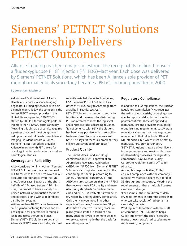
Siemens’ PETNET Solutions Partnership Delivers PET/CT OutcomesAlliance Imaging reached a major milestone–the receipt of its millionth dose of a fludeoxyglucose F 18* injection (18F FDG)–last year. Each dose was delivered by Siemens’ PETNET Solutions, which has been Alliance’s sole provider of PET radiopharmaceuticals since they became a PET/CT imaging provider in 2000.
By Jonathan Batchelor
A division of California-based Alliance Healthcare Services, Alliance Imaging began its PET imaging services with a sin-gle mobile unit. Today, the company is the largest PET/CT imaging provider in the United States, operating 130 PET/CTs staffed by 300 PET technologists perform-ing more than 140,000 exams annually. "Reaching this pinnacle of service required a partner that could meet our growing radiopharmaceutical needs," says Alliance Imaging President Richard A. Jones. Siemens’ PETNET Solutions provides Alliance Imaging with PET tracers for oncology imaging and staging, as well as neurological studies.
Coverage and Reliability
A key consideration in choosing Siemens’ PETNET Solutions as the sole source of PET tracers was the need “to cover all our accounts appropriately, even the rural ones,” Jones says. Because of the short half-life of 18F-based tracers, 110 min-utes, it is crucial to have a widely dis-persed network of production facilities and backups, along with a dependable distribution system.With more than 40 PET radiopharmaceuti-cal drug manufacturing facilities and dis-pensing nuclear pharmacies in multiple locations across the United States, Siemens’ PETNET Solutions serves all of Alliance's PET/CT assets, including its most
recently installed site in Anchorage, AK, USA. Siemens’ PETNET Solutions flies doses of 18F FDG daily to Anchorage from a facility in Seattle, WA, USA.PETNET Solutions has enough production facilities and the means for distributing PET radiotracers to meet the logistical challenges we face, according to Jones. “My experience with PETNET Solutions has been very positive with its reliability to deliver doses to us on a consistent basis,” he says. “If there is a problem, they still ensure coverage of our doses.”
Product Quality
The United States Food and Drug Administration (FDA) approval of an Abbreviated New Drug Application (ANDA) for 18F FDG from Siemens’ PETNET Solutions is an important element in the continuing partnership, according to Jones. Granted in February 2011, the ANDA ensures customers that the 18F FDG they receive meets FDA quality and man-ufacturing standards.“In nuclear medi-cine and PET/CT, it really starts with deliv-ery reliability and regulatory compliance. Only then can you move into other aspects of business," Jones notes. "If you don't have those two building blocks in place, you're limited in terms of how many customers you're going to be able to service. We've made that the basis for everything we do.”
Regulatory Compliance
In addition to FDA regulations, the Nuclear Regulatory Commission (NRC) regulates the radioactive materials, packaging, stor-age, transport and distribution of radio-pharmaceuticals. These are applied to manufacturers and providers through rig-orous licensing requirements. Lastly, state regulatory agencies may have regulatory requirements that fall outside FDA and NRC standards, but which must be met by manufacturers, providers or both.“PETNET Solutions is aware of our licens-ing requirements and works with us on implementing processes for regulatory compliance,” says Michael Culley, Corporate Radiation Safety Office for Alliance.Among his responsibilities, Culley ensures compliance with the company’s radioactive materials licenses, a total of 35 throughout the country. Meeting the requirements of these multiple licenses can be a challenge. “For example, there are different deliv-ery requirements in different states as to who can take receipt of radiopharma-ceuticals,” he notes. Working in collaboration with the staff at Siemens’ PETNET Solutions helps Culley implement the specific require-ments of each state's radioactive mate-rial licensing compliance.
Imaging Life · June 2013 · www.siemens.com/imaginglife 25
Outcomes
Strategic Partnership
"In addition to its work delivering radio-pharmaceuticals to existing Alliance sites, Siemens’ PETNET Solutions has provided invaluable service to us as we have started new facilities," Jones says.“Likewise, one of the main benefits to our relationship with PETNET Solutions has been its ability to scale with us as our business has grown."Culley notes that when Alliance takes on new accounts and new customers, they typically have to transition from other providers of PET radiopharmaceuticals. In these instances, Siemens’ PETNET Solutions ensures that the lines of com-munication with imaging centers are open and the information is flowing, he said. This is done through multiple con-ference calls to set up radiopharmaceuti-cal production schedules, backup plans in case a cyclotron goes offline, delivery dry
runs to scope out routes and timelines, and follow up to see that quality, reliabil-ity and expectations are met. “We work well together with PETNET Solutions to ensure our mutual custom-ers are well taken care of and that we're meeting the business objectives of our respective firms,” Culley stated.
Collaborating for Future Growth
A smooth transition for new accounts and customers is just one of the ways Siemens' PETNET Solutions is helping Alliance Imaging grow its business. Siemens' PETNET Solutions has also ini-tiated a pilot program for new PET radiopharmaceuticals to help Alliance expand its clinical PET service offerings beyond oncology PET imaging to the communities it serves. “Having access to new radiopharmaceuticals and being able to pilot new imaging programs is very important to us and our custom-ers,” Jones says."This exemplifies the evolving partner-ship between Alliance Imaging and Siemens’ PETNET Solutions," continues Jones, "But this collaboration extends well beyond supplying new tracers.“PETNET Solutions provides an ongoing resource for our account education, as well as keeping us up-to-date about reimbursement changes,” Jones says.“Based on the access they have to research and data, PETNET Solutions is in a good position to help us make the transition to a business consultative role with our clients,” he stated.As for the future, Alliance Imaging will continue the development of PET/CT assets as a major part of its business model, rey-ling on Siemens’ PETNET Solutions to assist in the planning, development and use of PET radiopharmaceuticals.“Our PET customers are looking to expand the services they're providing, while at the same time making sure their services are delivered reliably and effi-ciently,” Jones says.“Working hand-in-hand with PETNET Solutions enables us to provide quality services to our clients,” Jones says. “And that's huge.”
IndicationsFludeoxyglucose F 18 Injection is indi-cated for positron emission tomography (PET) imaging in the following settings:• Oncology: For assessment of abnormal
glucose metabolism to assist in the eval-uation of malignancy in patients with known or suspected abnormalities found by other testing modalities, or in patients with an existing diagnosis of cancer.
• Cardiology: For the identification of left ventricular myocardium with residual glucose metabolism and reversible loss of systolic function in patients with cor-onary artery disease and left ventricular dysfunction, when used together with myocardial perfusion imaging.
• Neurology: For the identification of regions of abnormal glucose metabo-lism associated with foci of epileptic seizures.
Important Safety Information
• Radiation Risks: Radiation-emitting products, including Fludeoxyglucose F 18 Injection, may increase the risk for cancer, especially in pediatric patients. Use the smallest dose neces-sary for imaging and ensure safe han-dling to protect the patient and health care worker.
• Blood Glucose Abnormalities: In the oncology and neurology setting, sub-optimal imaging may occur in patients with inadequately regulated blood glu-cose levels. In these patients, consider medical therapy and laboratory testing to assure at least two days of normo-glycemia prior to Fludeoxyglucose F 18 Injection administration.
• Adverse Reactions: Hypersensitivity reactions with pruritus, edema and rash have been reported; have emer-gency resuscitation equipment and personnel immediately available.
Fludeoxyglucose F 18 Injection for intravenous use, 0.74 to 7.40 GBq/mL (20 to 200 mCi/mL)
* The full prescribing information for the Fludeoxyglucose F 18 injection can be found on pages 68-70.
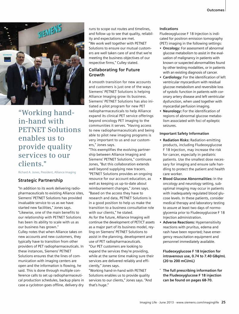
“Working hand- in-hand with PETNET Solutions enables us to provide quality services to our clients."Richard A. Jones, President, Alliance Imaging

26 Imaging Life · June 2013 · www.siemens.com/imaginglife
Outcomes
Biograph mCT Paves the Road to Advances in PET/CT QuantificationClinicians at the Clinical University of Navarra in Pamplona, Spain, are using SUV-uptake measurements to characterize cancers, identify patient response to therapy and detect cancer recurrence. Their work with Biograph
mCT 64 is laying the groundwork for wider use of these quantitative measures.
By Greg Freiherr
Clinical University of Navarra Pamplona, Spain

Imaging Life · June 2013 · www.siemens.com/imaginglife 27
Outcomes
Spain’s first state-of-the art academic research facility is breaking new ground in molecular imaging applications. Its most significant development, according to José Angel Richter, MD, director of nuclear medicine at the Clinical University of Navarra (Clínica Universidad de Navarra), has been the advancement of quantification as a supplement to the visualization capabilities of their Biograph™ mCT 64 scanner with TrueV and UltraHD•PET, the first such sophisticated PET•CT to be installed in Spain.Richter, working with other specialists in nuclear medicine there, Javiar Arbizu, MD, PhD and Elena Prieto, PhD, has per-formed phantom studies documenting the high level of quantitative accuracy that can be achieved with Biograph mCT. They have followed this work with clinical research indicating that quantification of lesions with Biograph mCT provides infor-mation critically important to making the best possible treatment decisions.A key measure is the standardized uptake value (SUV), which indicates the uptake of PET radiopharmaceuticals by body tissues. The change in SUV from one study to another during therapy, for example, quantitatively indicates how
the patient is responding. SUV measures have the potential to provide an objec-tive and reliable indicator. There are, however, different ways to measure SUV. One, called SUVmax, reflects the maximum uptake value. Another value used is the mean SUV within a group of voxels, usually defined around a lesion (SUVmean). A third method is to use the mean SUV in a group of voxels that have a value higher than 50 percent of the maximum value (SUV 50*).1
“The clinical implication of these results is important considering the potential shift from SUVmax to SUV 50,” Richter says.
Leading-Edge Technologies
The Biograph mCT with ultraHD•PET leverages time of flight (TOF) to pre-cisely measure the two photons created during a coincidence event, which occurs when a positron, emitted by the PET radiotracer, collides with an elec-tron—a matter/anti-matter collision that annihilates the positron and electron and, in the process, generates photons that travel in exactly opposite direc-tions. Biograph mCT’s OptisoHD detec-tor technology precisely measures in the trillionths of seconds the difference
between the times the two photons impact the detector ring. The most likely position of the positron annihilation is computed from this difference. This position, along with the point spread function (PSF), is algorithmically mod-eled into images that quantitatively describe the location of the radiotracer.Conventional PET/CT is much less exact. It relies on approximations of where the coincidence events occurred. Consequently, conventional PET/CT results provide a less distinct result.Benchmark testing by Siemens engineers has determined that Biograph mCT ultraHD•PET provides visual improve-ment in signal-to-noise** (SNR) of con-ventional PET/CT.2
Biograph mCT Advances Quantification
A decade of clinical use at facilities around the world has established PET/CT as a critically important tool in the diag-nosis and prognosis of patients with many types of cancer. But this hybrid still depends on expert interpretation of images. Accurate and reproducible quantification achieved with the Biograph mCT may provide greater
A 30-year-old obese male (185 kg) treated for primitive neuroectodermal tumor (PNET) by nephrectomy (November 2010). 18F FDG PET performed on June 2011 (left) using a conventional PET/CT shows peritoneal metastatic implants. Follow-up PET after 2 cycles of chemotherapy performed in September 2011 (right) using Biograph mCT 64 shows partial metabolic response with persistence of a peritoneal implant in the left flank. Please note the dramatically improved resolution of the Biograph mCT image (right) compared to that obtained from the previous generation system.

28 Imaging Life · June 2013 · www.siemens.com/imaginglife
Outcomes
confidence in SUV values, used to moni-tor therapeutic response, that accurately measure the radiopharmaceutical uptake associated with the disease state. Biograph mCT at Clínica Universidad de Navarra is helping to lay the foundation for the even wider use of quantification.In a phantom study, Richter and col-leagues compared the performance of Biograph mCT outfitted with ultraHD•PET against several varying parameters. Results were further com-pared against those obtained with con-ventional PET/CT. The effect of different reconstruction algorithms was also assessed, namely filtered back projec-tion—a long-time industry standard—against Siemens iterative reconstruction algorithm ordered-subset expectation maximization (OSEM).A NEMA body phantom with six spheres ranging in diameter from 10.1 to 37.6 millimeters were filled with varying sig-nal-to-background ratios using different background concentrations of radio-tracer.2
The phantom studies performed by the research team at the university docu-mented that SUVmax quantification, which measures peak standard uptake of 18F FDG*** in a targeted lesions, is overestimated for most reconstructions, except for OSEM, where attenuation and partial volume effect offset the overestimation. This work demonstrates that the SUV 50 may be the more con-sistent quantitative parameter. In clinical studies, presented at the European Association of Nuclear Medicine (EANM) Congress 2012 in Milan, Italy, the Spanish team found that the use of SUV 50 values improved spec-ificity over SUVmax quantitative analysis. This analysis of 18F FDG-PET/CT imaging produced a sensitivity and specificity for the identification of primary tumors of 98 percent and 55 percent respectively. The study examined the effect of image reconstruction using a number of tech-niques including PSF + TOF, OSEM, PSF alone and TOF alone.
Boosting Performance
Recent work has flourished with the upgrade of a conventional PET/CT scan-ner, which for much of the past decade was used to lay the foundation for the Spanish team’s recent developments. The Biograph mCT replacement boosted the team’s ability to detect lesions approximately 50 percent of the size of what was possible compared with the conventional PET/CT, a larger axial field of view, achieved with TrueV, reduced
scanning times and, as Richter pointed out, provides significantly higher quality information for diagnosis, therapy moni-toring and detection of recurrence.“The new generation of hybrid imaging scanners provides better PET image res-olution,” he says. “Besides, PET studies can be acquired at faster speeds. In gen-eral, advanced PET/CT protocols are much more reliable for detection and localization of tumoral disease.”Richter and his colleagues plan to use PET quantification to explore new
25-year-old male referred for PET•CT staging of Hodgkin's disease. 18F FDG PET performed in April 2012 (bottom row) shows gross mediastinal disease, as well as lesions in vertebrae, left ischium, left ileum and right acetabulum (stage IV). Interim PET performed in August 2012 (top row) after 2 cycles of BEACOP shows excellent metabolic response with minimum residual uptake in mediastinum of intensity below hepatic uptake.

Imaging Life · June 2013 · www.siemens.com/imaginglife 29
Outcomes
research avenues in lymph node stag-ing, therapeutic monitoring through metabolic criteria, called PET Response Criteria in Solid Tumors (PERCIST), and the characterization of tumors in the brain, liver and lungs. Accurate and pre-cise quantification may prove especially useful in lymphomas and for establish-ing the degree of differentiation and heterogeneity of brain tumors. While the use of PET/CT quantification to predict treatment response or staging is experimental at many institutions, the question of its clinical values is a foregone conclusion at Clinical University of Navarra.“In our case, quantification already is part of our routine clinical practice,” Richter says. The most successful use has been for patients being monitored for early ther-apeutic response in several types of tumors. Biograph mCT is particularly well-suited for these applications, he says, because the scanner “provides us with a high degree of confidence in the detection of very small residual disease. In addition, it helps us perform a more accurate cal-culation with SUVpeak through volumet-ric analysis.”
* The SUV 50 value is not automatically calculated on the Biograph mCT. The volume of interest (VOI) thresh-old has to be set by the customer.
** Conventionally reconstructed images compared to im-ages reconstructed with HD•PET.
*** Important safety information on Fludeoxyglucose F 18 injection can be found on page 25. The full prescrib-ing information can be found on pages 68-70.
The statements by Siemens’ customers described
herein are based on results that were achieved in
the customer’s unique setting. Since there is no
“typical” hospital and many variables exist (e.g., hos-
pital size, case mix, level of IT adoption) there can
be no guarantee that other customers will achieve
the same results.
References:
1 Jakoby, et al Phys Med Biol. 2011 Apr
21;56(8):2375-89
2 Prieto, Elena et. al (2012) Impact of time-of-flight
and point-spread-function reconstruction in stan-
dard uptake value quantification: A phantom
study. J Nucl Med, 53 (Supplement1), 160
“Biograph mCT provides us with a high degree of confidence in the detection of very small residual disease.”José Angel Richter, MD Director of Nuclear Medicine, Clinical University of Navarra Pamplona, Spain

30 Imaging Life · June 2013 · www.siemens.com/imaginglife
Outcomes
IQ•SPECT Streamlines Cardiac Imaging at Amphia HospitalThe nuclear medicine department at Amphia Hospital in Breda, The Netherlands, scans as many as 40 patients daily. In 2012, the hospital underwent major equipment changes. By introducing IQ•SPECT into their department, they more than made up for this change with a more dynamic workflow and happier patients.
By Shalmali Pal
The molecular imaging department at Amphia Hospital has taken the adage of “less is more” to heart. Last year, the team at the Molengracht campus, one of the hospital's multiple locations, wanted to replace a pair of SPECT cam-eras, but lacked the physical space to bring in two new SPECT•CTs. Instead, they removed an aging system and upgraded their existing Symbia™ T6 with IQ•SPECT technology, which reduces the acquisition time for myocar-dial perfusion SPECT, while maintaining or improving image quality. To further enhance their productivity, Amphia
Hospital also installed Symbia.net and syngo®.via software. Now, the Dutch hospital is acquiring molecular images in less time, experi-encing less patient movement and, con-sequently, fewer rescans, and imposing less patient discomfort during studies—all while increasing patient throughput.“We were surprised at the reduction in acquisition time from the industry stan-dard of 18 minutes [per patient] to 9 with IQ•SPECT—to the great relief of patients,” says Jan Akkermans, depart-ment manager. “[Complaints and move-ment from] patients during acquisition
were common before IQ•SPECT, causing us sometimes to stop before completing the acquisition.”IQ•SPECT added a collimator that increases the number of counts without compromising image resolution. Its software plots a cardio-centric orbit for the detectors, positioning the heart in the most sensitive part of the collima-tor. Proprietary algorithms reconstruct the molecular imaging data in three dimensions, while correcting for attenu-ation and scatter.
Amphia Hospital, Molengracht campus, Breda, The Netherlands

Imaging Life · June 2013 · www.siemens.com/imaginglife 31
Outcomes
Cutting Cardiac Imaging Time, Boosting Workflow
Since the upgrade in July 2012, the department has doubled the number of myocardial perfusion scans, performing up to 15 per morning. Key to their increased productivity has been effi-cient scheduling. Cardiac patients are scanned from 9 a.m. to 12:30 p.m., four days a week. In the pre-IQ•SPECT days, stress studies were done in the morn-ing, while the afternoon was dedicated to rest studies. “With IQ•SPECT, we are mixing stress and rest to achieve a constant flow of patients on the camera during the hours dedicated to cardiac imaging,” Akkermans says. The team has also worked out the staff-ing needed to manage patient flow and optimize procedure time. Two technolo-gists are allotted 15 minutes to get each patient on and off the Symbia T6 system. The department also uses two dressing rooms to keep the flow of patients com-ing. This helps the team accommodate the variety of patients, including those who are overweight or obese.“Our patients range in weight from 60 kg [132 lbs] to over 140 kg [308 lbs],” says nuclear medicine physician Jim Baas, MD. “Our current set-up allows us to accommodate all these patients with minimal disruption.” As for the scan-time itself, technologist Corne Snoijs says the nine-minute scan is as streamlined as they want to go. “We could shorten the SPECT part down to four or five minutes, but then we run the risk of missing one or two heartbeats," Snoijs says. “So in order to avoid exces-sive rescanning, we extend the scan time a few extra minutes.”After 12:30 p.m., the scanner is open for what Akkermans called “unexpected SPECT•CT” studies. These include screen-ing exams and tumor staging studies. “This means minimal collimator change to accommodate these other [non-cardiac] scans,” Snoijs says.Baas says the department wants to expand its imaging reach by setting up a
formal program to talk with referring physicians about the clinical value of the cardiac scans at Amphia Hospital, and how they can leverage the benefits of IQ•SPECT even more effectively.
Meeting Challenges
The increased throughput possible with IQ•SPECT has required the department to adjust its everyday routine. Applications specialists from Siemens visited Amphia Hospital to help the team streamline its workflow. For instance, prior to the upgrade, processing for one patient’s images was done in the roughly 20-minute period that the next patient was in the scanner. The quicker scans achieved using IQ•SPECT and the ability to mix stress and rest studies has shortened the time available to process images.The improved image quality possible with IQ•SPECT also has put a premium on registering the attenuation correction map correctly. Early in the use of the upgraded Symbia T6, the Siemens' sup-port staff recommended that the Dutch team make a two-day observational visit to another hospital using IQ•SPECT. Technologist training was a boon con-ferred by visiting a hospital experienced with IQ•SPECT. “It’s there that you can learn tips and tricks, what to look for," Snoijs points out.One such trick was the introduction of a 15-minute free period halfway through the day to absorb delays that may occur in the early morning scans. Additionally, the two technologists who took part in the off-site visits were responsible for on-the-job training of their colleagues. “I think it's an impor-tant point that we already had Symbia T6, so everyone was familiar with the machine,” Baas says. “We updated with
IQ•SPECT so our learning curve was dif-ferent than for a department that has to learn SPECT•CT and IQ•SPECT together. Even so, the Siemens Symbia T6 is an intuitive and easy-to-use scanner. I think that made a big difference.”
Looking Forward
Continuing to hone their skills with IQ•SPECT is high on the agenda for the Amphia team. At the moment, the staff use a very low mean dose of 500 MBq (dependent on patient height and weight), just as they are pushing the limits of low dose and productivity. “We are working at the lowest possible dose that we can,” Akkermans says, “and we allow just 15 minutes per patient.”If there are still gains to be made, they will come from a better understanding of the advantages of cardiac imaging with IQ•SPECT. This is an ongoing pro-cess, Akkermans says. In the meantime, the team hopes to broaden the use of IQ•SPECT through its non-traditional application. “I am curious to know if, in the near future, it will be possible to use IQ•SPECT to investigate small organs such as the thyroid*,” Baas says. “We can benefit from a technical improvement that would allow us to image these small organs. We are experimenting with this now. It’s a work in progress, but we think it will be very beneficial.”
“We were surprised at the reduction in acquisition time..."Jan Akkermans, Manager Nuclear Medicine Department, Amphia HospitalBreda, The Netherlands
* IQ•SPECT imaging is intended for cardiac imaging only. IQ•SPECT thyroid imaging is based on research only and is not commercially available.

32 Imaging Life · June 2013 · www.siemens.com/imaginglife
Outcomes
Biograph TruePoint 16 Meets Challenges at Fox Chase Cancer CenterAs one of fewer than 50 comprehensive cancer centers in the United States, the nonprofit Fox Chase Cancer Center in Philadelphia, Pennsylvania, relies on leading-edge Biograph PET•CT technology to support a highly collaborative research environment among some of the top cancer specialists. Researchers and faculty at the Center have won Nobel and Kyoto Prizes.
By Matthew Skoufalos
Four years ago, Fox Chase Cancer Center replaced its PET/CT with the Biograph™ TruePoint 16, which met the clinical and research needs of the Center while being economical. Its high-definition detector composed of lutetium oxyorthosilicate (LSO) scintillator crystal combined with HD•PET technology offers visual improvement in signal-to-noise* of non-HD•PET systems. The onboard 16-slice CT allows quick scans for attenuation correction, as it produces high-resolution CT images for anatomical reference. As a value-priced scanner, Biograph TruePoint was easy on the center’s bud-get, yet able to meet the challenges of a cancer center accustomed to pursuing advanced research initiatives and sup-porting a growing patient base. Fast data collection allows the capture of more counts over a given time, maximizing patient throughput and productivity, while boosting image quality to support advanced research protocols and diag-nostic confidence.Five to ten percent of all cases at Fox Chase involve research, says Jian Qin “Michael” Yu, MD, FRCPC, chief of nuclear medicine/PET at Fox Chase, whose scope of research spans imaging trials with cooperative groups like the Radiation Therapy Oncology Group (RTOG), Gynecologic Oncology Group
(GOG), Eastern Cooperative Oncology Group (ECOG), and American College of Radiology Imaging Network (ACRIN). “A lot of our physicians are on NCCN (National Comprehensive Cancer Network) guideline panels, and some of them are subcommittee chairs,” Yu says. “Because we’re a small institution in size, everybody really knows everybody. It makes the collaboration much easier. If you connect with the right clinical spe-cialist, you can do quite a lot.”And because of the prestige of Fox Chase as a founding memeber of NCCN, “a lot of pharmaceutical companies come here to try their ideas first,” he says.
Meeting New Demands
In 2005, Fox Chase was operating one of the first commercially available PET/CTs, scanning about 800 patients annu-ally. Four years later, the Center’s patient volume had doubled. Though notable in its day, the first-generation PET/CT unit at Fox Chase couldn’t keep up, according to Yu. “Throughput had become an issue,” he says. “Every year, our volume increased by about 20 percent. We needed some-thing faster.”In 2009, Fox Chase went shopping for a new PET/CT. Yu, his department and hos-pital colleagues considered the
Fox Chase Cancer Center Philadelphia, PA, USA

Imaging Life · June 2013 · www.siemens.com/imaginglife 33
Outcomes
multifaceted challenges the new scanner would have to face. After a year-long search, they installed the Siemens Biograph TruePoint 16, a “sturdy machine,” Yu says. “We know there’s definitely much more information available to us for data anal-ysis and processing with Biograph TruePoint 16,” Yu says. “It creates more possibilities.”For Yu, foremost among the features of the Biograph TruePoint 16 is the intuitive software application, syngo®.via,* for which Fox Chase was both an alpha and beta software test site. syngo.via enables Yu to transfer, configure and assign patient protocols throughout the facility network, saving time and allow-ing for greater ease of collaboration with his colleagues.“It (syngo.via) is really well organized,” Yu says. “If we have a study and I want to find out how it was acquired, I can open up the study on syngo.via and all of the acquisition parameters pop up.“I can also perform additional reconstruc-tions easily,” he continues. “That I found to be very convenient and very useful. I use that feature many, many times when I need to reconstruct PET images with dif-ferent parameters.”syngo.via puts information “at our finger-tips,” Yu adds. “That’s what makes the work easier and maybe faster.”Yu’s colleague at Fox Chase, medical physicist Mohan Doss, praised the dynamic image acquisition features of the Biograph TruePoint 16. The scanner offers Siemens’ SureView, which helps automate the parameters of various studies to produce high-quality images gathered at different times, filters and reconstructions, “which is very useful for a lot of research,” he says.
Optimizing Performance
In view of the present concerns in the radiological community regarding patient doses, advanced functions, like CARE Dose4D™, can help lower CT dose deliv-ered to patients by modulating the tube current in real time, while maintaining image quality. Performance is further
optimized by the TrueV wide field of view on the Biograph TruePoint 16 scanner, which is one-third larger than those of comparable systems.** The extra cover-age provides the opportunity to reduce radiopharmaceutical dose, while opening up scanning protocols to a greater degree of flexibility, including faster study times.“One of the main advantages of Biograph TruePoint 16 compared to our old system is its speed,” Doss says. “We do acquisi-tions of two to three minutes per bed position, as compared with five to six minutes on the old system. So, we’re able to do more patients than we could before.” Biograph TruePoint 16 can accommo-date as many as 15 patients per day in a regular shift, Yu says, but typically Fox Chase schedules 10 to 12 patients per day to maintain flexibility. “We always try to leave some room for the research patients,” Yu says, and the speed of Biograph TruePoint 16 “allows more room in the schedule” to accom-modate them.Donna Mosley, chief technologist at Fox Chase, agrees that the extra time has helped improve throughput at the facil-ity by a factor of at least two. That’s no easy feat, especially given that Biograph TruePoint 16 is the only PET/CT scanner her department operates.Mosley commended Siemens’ technical support staff for being proactive on scheduling preventive maintenance. The extra notice has “worked out quite well,” she says, giving her time to adjust her schedule to minimize the impact.“We have a good service staff with Siemens,” Mosley says. “We’ve had ser-vice done over the phone, which is very helpful to us. And we can call them and get them to reply very quickly.”
A Big Step Up
In addition to coming in at an affordable price, Doss says, Biograph TruePoint 16’s collection of advanced functionality pro-vides a variety of expanded options—so many, in fact, that he says it’s not even fair to compare this scanner to the one it replaced.Making a new equipment purchase ulti-mately meant changing vendors and
although she was comfortable when the final decisions were made, Mosley says she “was a little pessimistic because I didn’t know exactly what to expect, but I’m glad we made the switch.”Mosley believes the team has gotten value for its dollar. For Fox Chase, Yu says, the decision to purchase Biograph TruePoint 16 was reinforced by a combi-nation of attributes—price, research needs, clinical needs, support and ser-vice. “On all these criteria, Biograph TruePoint 16 has been a winner," he says.
*Conventionally reconstructed images compared to images reconstructed with HD•PET.
The statements by Siemens’ customers described herein are based on results that were achieved in the custom-er’s unique setting. Since there is no “typical” hospital and many variables exist (e.g., hospital size, case mix, level of IT adoption) there can be no guarantee that other customers will achieve the same results.

34 Imaging Life · June 2013 · www.siemens.com/imaginglife
Outcomes
xSPECT—A New Benchmark in Image Quality and QuantificationBy Partha Ghosh, MD and Patrick Sorger, CNMT, Molecular Imaging Business Unit, Siemens Healthcare
Introduction
The fusion of SPECT with diagnostic mul-tislice CT has been one of the key drivers in nuclear medicine. As 3D iterative reconstruction became clinically feasible, it improved SPECT resolution and con-trast.1 This, along with CT attenuation correction and lesion localization based on fusion of diagnostic CT and SPECT datasets, has improved accuracy and scope of SPECT imaging. One of the problems inherent to bone SPECT is the lower specificity, in spite of high sensitivity, due to the lower resolu-tion of SPECT. Diagnostic CT information about individual lesions, for example the degree of lysis and sclerosis in bone metastases, often helps lesion character-ization. However, several clinical ques-tions about the nature and extent of a lesion often remain unanswered due to the limited mechanical fusion of CT with a lower resolution SPECT. Another limitation of SPECT/CT has been the lack of accurate and reproduc-ible quantification, similar to what is available for PET. These limitations in conventional SPECT/CT makes quantita-tive evaluation of lesions using sequen-tial follow-up not possible in daily rou-tine. Currently, most decisions about lesion progression or therapy response in conventional SPECT/CT are made using visual assessments or lesion to background ratios.
xSPECT* Image Quality
The new modality xSPECT changes the frame of reference from SPECT to CT and allows precise alignment of the two
1 99mTc MDP bone SPECT reconstructed with 3D iterative reconstruction (top row) and xSPECT (bottom row) in a patient with a history of gastric carcinoma with normal distribution of tracer throughout the skeletal system. There is a visually significant improvement in image quality especially in the delineation of sharp margins of vertebral bodies, end plates and intervertebral discs, as well as vertebral canal, spinous and transverse processes. The vertebral canal, which is almost smeared over by spilled activity from the vertebral bodies in the conventional SPECT/CT, is clearly delineated on xSPECT. Conventional SPECT/CT shows complete absence of differentiation of ribs from transverse processes, while it is possible to clearly visualize and differentiate the vertebral end of the rib from transverse process on xSPECT images. Following injection of 370 MBq of 99mTc MDP, SPECT/CT imaging of the thoracic region was performed with a standard acquisition protocol of 64 frames with 20 seconds per frame. Diagnostic CT was performed as an integrated procedure prior to SPECT acquisition with 130 kV 50 mAs. The conventional SPECT/CT was reconstructed with 20 iterations and 4 subsets with a Gaussian filter. xSPECT reconstruction was also used with the same dataset using zone information obtained from the CT data. Data courtesy of University of Erlangen, Germany
1
Conventional SPECT/CT
xSPECT Bone

Imaging Life · June 2013 · www.siemens.com/imaginglife 35
Outcomes
modalities. This enables for instance the extraction and integration of medically relevant information from the CT into the SPECT creating a new xSPECT image. The extraction is based on delineation of skeletal tissue boundaries via a linear-attenuation coefficient-based segmenta-tion. This information is then used to reconstruct xSPECT Bone,* which achieves improvements in image quality and resolution.
Quantification* with xSPECT
Furthermore, xSPECT in conjuction with a new system calibration and advanced reconstruction enables the image to be reproducibly quantitative for activity concentration measured in units of Bq/ml for 99mTc labeled radiopharmaceuti-cals imaged using a LEHR collimator.2 Well counter-generated values of total injected dose and patient height and weight information, if provided, can yield standard uptake value (SUV) for the first time in SPECT. SUV generated by using xSPECT is not only reproduc-ible, but also no longer strongly system dependent, as the xSPECT is calibrated to a common standard. xSPECT enables SPECT SUV in a clinical setting and pre-pares the ground for future applications. The ability to generate such quantitative parameters reflects the potential of the true integration of SPECT and CTbeing pioneered by Siemens with the introduction of Symbia Intevo™,* the world's first xSPECT system.
xSPECT Bone in Skeletal Scintigraphy
Skeletal scintigraphy has been a main-stay of nuclear medicine for decades. It is the key modality for detection and characterization of skeletal metastases, as well as characterization of numerousnon-malignant lesions of the bone including fractures, infections, avascular necrosis, degenerative and osteoarthritic bone and joint lesions.Bone scintigraphy has evolved from pla-nar to SPECT with cross-sectional visual-ization. Diagnostic accuracy and confi-dence with bone SPECT was further improved by introduction of SPECT/CT.
xSPECT achieves sharper definition of bone dense margins and lesion boundar-ies. The resulting xSPECT images seem to better reflect the distribution of metaboli-cally active bone within the skeletal sys-tem with sharper differentiation between cortical and spongy bone in the verte-brae, as well as in flat bones like the pel-vis, scapula and skull. This also provides fine detail that helps differentiate between the cortex and marrow cavity in long bones and improved separation of bone from joint spaces—both for large joints such as knee and shoulder and small bones like carpals and tarsal bones.
xSPECT Quant*: Quantification in Bone Disease
Absolute quantification of skeletal metabolism by the measurement of bone concentration of tracer in Bq/ml has the potential to be of value in the assessment of bone graft function andretention. Droll et al4 performed bone SPECT in 104 hips in 93 patients with osteonecrosis of the femoral head treated with vascularized fibular graft-ing. The signal intensity of the graft was compared with the intensity of the ipsi-lateral proximal femoral diaphysis and
assigned a score of 1 if less than diaphy-sis, 2 if equal to diaphysis and 3 if greater than diaphysis. Graft failure was defined as conversion to total hip arthro-plasy. A total of 30 percent of the hips failed treatment with vascular fibular grafting and mean SPECT scores were significantly lower in the failed group compared to the group in which the bone graft was retained.This pattern of scoring of the intensity of tracer uptake and comparison with adja-cent normal bone can potentially be helped by the reproducible absolute quan-titative values of tracer uptake within bone that can be obtained by xSPECT.The intensity of lesional uptake visual-ized in bone SPECT has been used in osteoarthritic knee joints for comparison with arthroscopic findings in order to grade severity of involvement of menisci and femoral condyles. Siegel et al5 com-pared SPECT uptake intensity with arthroscopic findings in 41 patients and found significant correlation between uptake and severity of meniscal pathol-ogy. This suggested the possibility that quantification using xSPECT could potentially be used to help make surgical decisions in such patients.
2
2 xSPECT measurements in lumbar vertebrae of the patient in Figure 1 with normal tracer distribution in thoracic and lumbar vertebrae show tracer concentration of 69 kBq/ml and average SUV of 7.28 in the center of the T9 vertebral body. Injected dose: 574 MBq; patient weight: 64 kg/141 lb; patient height: 1.72m/ 5'8". xSPECT measurements yielded average absolute tracer concentration of 56.70 +/- 17.21 kBq/ml and average SUV of 7.02 +/- 1.67.3 Data courtesy of University of Erlangen, Germany
SUVmax 8.67SUVavg 7.28
69 kBq/ml
xSPECT Quant

36 Imaging Life · June 2013 · www.siemens.com/imaginglife
Outcomes
Clinical Impact of xSPECT Bone: Equivocal Bone Lesions
Equivocal bone lesions are a common concern in skeletal scintigraphy. xSPECT Bone images with improved lesion defi-nition, as well as absolute quantification of tracer uptake have the potential to improve the evaluation of such equivo-cal lesions. Such an equivocal lesion is highlighted in the example in Figure 5. A patient with renal cell carcinoma under-went 99mTc MDP bone SPECT/CT. The SPECT/CT image shows a solitary focal hypermetabolic lesion in the body of a cervical vertebrae. Conventional SPECT/CT, however, is not able to clearly define the extent of the lesion and involvement of pedicles. Since this was a solitary lesion, the possibility of a degenerative cervical lesion could not be ruled out. xSPECT defines the margins of the verte-bral body, pedicles, spine and transverse processes and clearly localizes the hypermetabolic foci to the left lateral part of the vertebral body without involvement of the pedicles or facet joints or lamina (Figure 6). Since the location of the lesion does not correlate with sites of degenerative changes, metastases is a possibility.
xSPECT Quantification: Infection Imaging
Quantitative evaluation of tracer concen-tration using xSPECT extends to all 99mTc labeled isotopes. One study where quan-tification will potentially add value is characterization of the intensity and severity of infection detected by 99mTc labeled leucocytes, as well as sequential evaluation of such infective process to detect impact of antibiotic therapy. SUV can potentially help improve evaluation of such infective foci by providing objec-tive values of uptake intensity although the clinical implications of SUV needs to be further ascertained. In cases of infec-tion in prosthetic joints, this can poten-tially help differentiate normal marrow hyperactivity from sites of infection, since intensity of uptake of labeled leu-cocytes is expected to be significantly higher in infection.6
Sequential imaging with 99mTc HMPAO-labeled leukocytes has been performed in ulcerative colitis to evaluate response to medical therapy.7 Twenty consecutive patients were imaged before and one week after initiation of medical therapy. The ratio of the maximum tracer uptake in the different colon segments to that of the lumbar vertebral bone marrow uptake was used as a semi-quantitative indicator of the intensity of colonic inflammation. All patients with clinical and endoscopic proof of response to medical therapy showed a positive response on sequential scintigraphy with significant decrease in the colonic seg-mental uptake ratios. The majority of the non-responders showed a >10% increase in the sum of segmental ratios.Most of these non-responders required colectomies. Since the ratio of the inflamed lesion to normal marrow is the key indicator in such cases, use of SUV based on quantitative xSPECT imaging could potentially be of major value in response assessment.
xSPECT Quantification: Lung Perfusion
99mTc MAA lung perfusion scintigraphy is used for the preoperative determination of functional lung volume in patients with lung carcinoma. Manual or semiau-tomatic determination of total perfused lung volume and regional lung function, as well as ratios of upper to lower lung zones, have been correlated with post-operative FEV1 and act as reliable pre-dictors of ventilatory function following lobectomy or pneumonectomy.8 Use of xSPECT based SUV may help further improve functional lung volume deter-mination.
xSPECT Quantification: Liver Tumor
Another potential application of abso-lute quantification with xSPECT is in the assessment of pulmonary shunting of intra-arterial hepatic infusion of 99mTc MAA, which is performed prior to intra-arterial radionuclide therapy for liver
3
3 Bone scintigraphy of a 66-year-old female patient with a history of adenocarcinoma of the lung. xSPECT Bone and conventional SPECT/CT in the pelvis and lumbar spine show higher uptake in the metastatic lesion in the sacroiliac joint with sharp lesion margins, as well as improved visualization of pelvic bones, sacral body, ala of sacrum and spinal canal. Focal hypermetabolic metastatic lesions in L1 and L4 vertebrae are well delineated on SPECT, with higher lesion intensity in xSPECT acquisitions. Small focal metastatic lesions in the acetabular margin and ischium are also better defined with higher lesion contrast. Data courtesy of Johns Hopkins University, Baltimore, MD, USA
xSPECT Bone
Conventional SPECT/CT
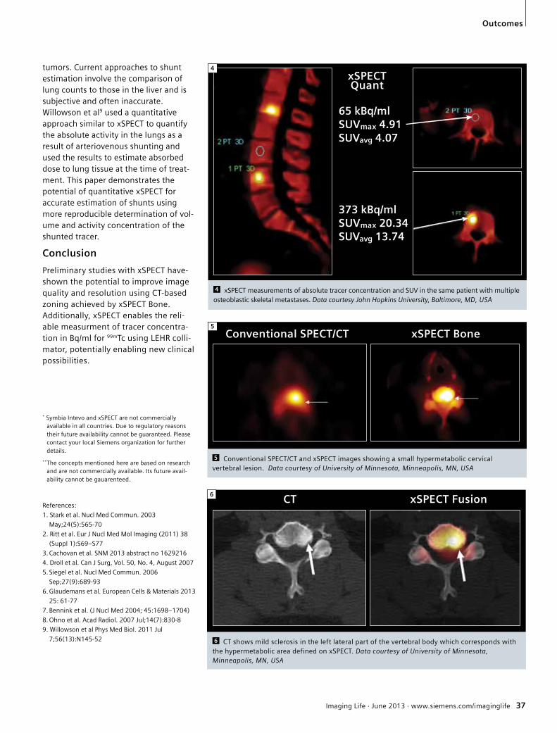
Imaging Life · June 2013 · www.siemens.com/imaginglife 37
Outcomes
tumors. Current approaches to shunt estimation involve the comparison of lung counts to those in the liver and is subjective and often inaccurate. Willowson et al9 used a quantitative approach similar to xSPECT to quantify the absolute activity in the lungs as a result of arteriovenous shunting and used the results to estimate absorbed dose to lung tissue at the time of treat-ment. This paper demonstrates the potential of quantitative xSPECT for accurate estimation of shunts using more reproducible determination of vol-ume and activity concentration of the shunted tracer.
Conclusion
Preliminary studies with xSPECT have-shown the potential to improve image quality and resolution using CT-based zoning achieved by xSPECT Bone.Additionally, xSPECT enables the reli-able measurment of tracer concentra-tion in Bq/ml for 99mTc using LEHR colli-mator, potentially enabling new clinical possibilities.
* Symbia Intevo and xSPECT are not commercially available in all countries. Due to regulatory reasons their future availability cannot be guaranteed. Please contact your local Siemens organization for further details.
**The concepts mentioned here are based on research and are not commercially available. Its future avail-ability cannot be gauarenteed.
References:
1. Stark et al. Nucl Med Commun. 2003
May;24(5):565-70
2. Ritt et al. Eur J Nucl Med Mol Imaging (2011) 38
(Suppl 1):S69–S77
3. Cachovan et al. SNM 2013 abstract no 1629216
4. Droll et al. Can J Surg, Vol. 50, No. 4, August 2007
5. Siegel et al. Nucl Med Commun. 2006
Sep;27(9):689-93
6. Glaudemans et al. European Cells & Materials 2013
25: 61-77
7. Bennink et al. (J Nucl Med 2004; 45:1698–1704)
8. Ohno et al. Acad Radiol. 2007 Jul;14(7):830-8
9. Willowson et al Phys Med Biol. 2011 Jul
7;56(13):N145-52
4
4 xSPECT measurements of absolute tracer concentration and SUV in the same patient with multiple osteoblastic skeletal metastases. Data courtesy John Hopkins University, Baltimore, MD, USA
65 kBq/ml SUVmax 4.91SUVavg 4.07
373 kBq/ml SUVmax 20.34SUVavg 13.74
xSPECT Quant
Conventional SPECT/CT xSPECT Bone5
5 Conventional SPECT/CT and xSPECT images showing a small hypermetabolic cervical vertebral lesion. Data courtesy of University of Minnesota, Minneapolis, MN, USA
6 CT xSPECT Fusion
6 CT shows mild sclerosis in the left lateral part of the vertebral body which corresponds with the hypermetabolic area defined on xSPECT. Data courtesy of University of Minnesota, Minneapolis, MN, USA

38 Imaging Life · June 2013 · www.siemens.com/imaginglife
Science
Dr. Andrés Villegas takes off his digital watch and wedding ring and carefully attaches the ring to the same clip that also holds his ID card. He slowly rolls up his shirtsleeves and puts on a blue smock that covers everything except his shirt collar and gray tie. Villegas now puts on a pair of latex gloves and — just to be on the safe side — a second pair over them. Only after he has done all this does he remove the cover from a white plastic tub in front of him and immerse his hand in a yellowish liquid. The tub contains half of a brain — and brains are Villegas’ specialty. Villegas is the Director of the biobank at Universidad de Antioquía in the northwestern Colombian city of Medellín. It has a col-lection of about 200 human brains. Villegas has personally dissected and
Village of Forgetfulness A large number of unusually young people in the mountainous region of Antioquía in Colombia suffer from a hereditary form of Alzheimer’s disease. An international team of researchers is testing a special medication to be administered long before any symptoms appear. If this approach proves effective, it would be a major breakthrough in the treatment of Alzheimer’s. A pilot study in Medellín is drawing on imaging technology from Siemens.
By Andreas Kleinschmidt Individuals afflicted with this form of Alzheimer’s reach the final stage of the disease at the age of 47 on average. By contrast, the more common form of Alzheimer’s sets in at the age of 65 or older. Early-onset Alzheimer’s is caused by a simple genetic defect on Chromosome 14. Over a period of
prepared around three quarters of them for further study. He now carefully places the dripping half brain onto a stainless steel tray. “This brain has shrunk consid-erably,” he says. “Look here and you can see some conspicuously deep grooves that look like the wrinkles in a dried-out grape.” He pauses and then gives the diagnosis. “This patient was only 56,” he says, “and what you see here is the result of early-onset Alzheimer’s disease.” Nowhere else in the world are as many people afflicted by this special form of dementia as here in Colombia, in the department of Antioquía.Although early-onset Alzheimer’s is very similar to the typical form of the disease, there is one major difference — namely, that in some cases the first symptoms occur before the victim turns 40. It begins with forgetfulness and progresses to disorientation and delusional ideas.

Imaging Life · June 2013 · www.siemens.com/imaginglife 39
Science
around 300 years, this defective gene has spread throughout a widely branched family that now has around 5,000 members. Experts refer to it as the “Paisa mutation” because the people living in the region around Medellín are known in Colombia as Paisas.Villegas gets to know many of the patients while they’re still alive. After they die, he removes their brains and studies them. “A brain can tell you a lot, but it can’t tell you everything,” he says as he places the half brain back into the formalin solution. “The ideal research case is when you can follow the course of the disease while the patient is still alive.” The other half of the brain he’s studying is stored along with hundreds of others in a large freezer. Villegas does the same thing with all the brains: He puts one half in a solution and the other in ice at -78 degrees Celsius.Dr. Adam Fleisher from the Banner Alzheimer’s Institute in Arizona has traveled from Phoenix to Medellín specifically to contribute to the launch of an ambitious prevention trial, which is part of a program known as the Alzheimer’s Prevention Initiative. “We want to develop a preclinical treatment for Alzheimer’s,” Fleisher explains. “In other words, we’re aiming to come up with a treatment that can prevent the disease or delay its outbreak, or at least slow its progress.” Most drugs for treating Alzheimer’s that have been tested to date have proved to be more or less insufficiently effective for fighting this terrible disease. Fleisher and his research colleagues believe this may be
due to the fact that the medications were administered after it was too late for the patients. “You may have to start the treatments before the onset of symptoms,” Fleisher explains. “We’ve been able to show that Alzheimer’s causes changes to the brain long before patients display the first signs of dementia — in some cases, 20 years before the first symptoms occur. For example, researchers have discovered that the onset of Alzheimer’s is preceded by the accumulation of plaques of beta amyloid proteins. These plaques form a type of crust in brain cells, which then slowly die as a result. By the time patients become forgetful, their brains may already be irreparably damaged.” Indeed, in many cases the patients’ brains have already significantly shrunk by the time they and those around them notice the first signs of forgetfulness. “We think it’s likely that we have the right weapon for fighting Alzheimer’s already — the problem may be that we are not using it until it’s too late,” says Fleisher. The weapon being tested in the study in Medellín is called crenezumab. This medi-cation is designed to attach itself to the amyloid and allow the patient’s immune system to render the protein harmless before it begins forming plaques.
Perfect Population. How can researchers know whether their hypothesis is correct? Should they give crenezumab to healthy people, for example? If they did, they would have to wait decades to find out whether the test subjects displayed fewer cases of
Alzheimer’s than a control group that would not have been given any medication. Such a plan would therefore be impractical at best. A population predisposed to developing Alzheimer’s, on the other hand, would be ideal — which is where the Antioquía patients come in. That’s because all it takes is a genetic test to reliably determine if these individuals will eventually be afflicted with the disease. Such patients therefore constitute a perfect population for clinical studies of the medication.Although patients with early-onset Alzheimer’s are also to be found in other parts of the world, the large number of cases around Medellín helps to ensure a high level of reliability for the results of the clinical study. That’s why the study in Antioquía will soon be under way with 300 participants, all of whom are between the ages of 30 and 60 and have not yet shown any symptoms. Excellent imaging technology is an essential part of the research program. The Banner Alzheimer’s Institute has made use of Siemens’ latest generation of PET-CT devices (a combination of a computer tomograph and a positron-emission tomograph) to make amyloid plaques and their continuing growth in Alzheimer’s patients visible in detail for the first time (see Pictures of the Future, Fall 2012, p. 92). Fleisher and his col-leagues are now hoping that such plaques will never form to begin with in the patients in Antioquía who will be treated with crenezumab at an early stage. “We have divided participants into two groups,” says Fleisher.“One group will be given the drug, the other group will receive a placebo. Repeated MRI, PET-CT scans, spinal fluid assessments, and cognitive testing in Medellín will help us to draw conclusions over the next two to five years regarding the effectiveness of the medication.” The tragedies caused by the frequency of Alzheimer’s in the region can be seen in towns such as Belmira, Angostura,
Near Medellín, cases of dementia abound. Andrés Villegas dissected dozens of brains searching for causes. Church records helped to identify the answer: hereditary Alzheimer’s disease.

40 Imaging Life · June 2013 · www.siemens.com/imaginglife
Science
and Yarumal, all of which are about a two hour drive north of Medellín. Most of the people afflicted with early-onset Alzheimer’s live here, and this degenerative brain disease has cut down many in the best years of their lives. There are no adequate facilities to care for them in these areas. They are usually taken care of by family members. Maria is one such caregiver.
Tied to a chair. Maria is 83 years old. Despite the fact that she herself doesn’t carry the Paisa mutation, her life has been shaped by Alzheimer’s. Her husband died of the disease over 20 years ago, and four of her 16 children suffer from it. Her son Alejandro died three years ago from Alzheimer’s at the age of 56. It was so difficult to care for him that he was sometimes tied to a chair to keep him from wandering around aimlessly. “This is not an isolated case,” says Claudia Madrigal, a psychologist at the hospital in Yarumal. It has barred windows, a few horses are tied at the hospital’s entrance, and a poster in the reception area advertises for Alcoholics Anonymous. “Thirty years ago, nobody knew what was going on here,” Madri gal says. “People got used to their middleaged relatives becoming forgetful, then aggressive, and finally demented and wasting away. At the time, many people believed a supernatural force was destroying people’s souls, so the sick individuals were locked away and their meals were given to them under the door. Only rarely did families allow an autopsy after the patients died. However, attitudes have changed over the last 20 years.” This change was mainly due to Dr. Francisco Lopera.
Bisected Brains.Lopera works in the Department of Neurosciences at Universidad de Antioquía. When he was a child, he used to live in Yarumal, where he began noticing the strange frequency of occurrences of dementia during the early 1980s. “It was a puzzle that I
wanted to solve,” he explains. Lopera and Lucia Madrigal, Claudia’s aunt and a nurse at the time, went from house to house asking for blood samples from afflicted patients. He also visited wakes and asked family members for permission to examine their loved ones’ brains. Amazingly, he did all this during a time when violent drug gangs controlled the region. Lopera was the first to discover that the sick people of Yarumal were suffering from a
quite expensive. According to estimates, the total cost of dementia illnesses worldwide in 2010 was more than $600 billion, which corresponds to around one percent of the gross world product. Madelyn Gutierrez knows figures like that by heart. She sits in her windowless office in Medellín located not too far from the Biobank and its halved brains. The air conditioner rattles lightly as patients assemble for a study that will begin shortly. Gutierrez is the young
Doctors, including Francisco Lopera and Lucía Madrigal of Universidad de Antioquía (above); Adam Fleisher and Eric Reiman of Banner Alzheimer’s Institute are providing new hope at PTU (top right) and in the village of Yarumal; and Héctor Zuluaga (bottom right) of the the Pablo Tobon Uribe Hospitál (PTU).
hereditary form of Alzheimer’s.It’s not just Colombians who urgently hope for progress to be made in the treatment of Alzheimer’s. The number of people afflicted with the disease worldwide will increase dramatically over the next few decades, largely as a result of higher life expectancy. That’s because more than 95 percent of Alzheimer’s cases around the world correspond to the typical form of the disease rather than early-onset Alzheimer’s, and the likelihood of a given individual becoming afflicted with Alzheimer’s doubles every five years from the age of 65. “If we could delay the average age when Alzheimer’s sets in by five years, we could reduce the absolute number of cases by 50 percent,” says Lopera. This would be a huge help not only to many Alzheimer’s patients and their families but also to healthcare systems, because proper care can be
psychologist who is coordinating the study and ensuring that it adheres to clinical trial standards. Among other things, the 300 participants will undergo tests to determine their cognitive capa-bilities and will also be sent on a regular basis over the next few years to the nearby Pablo Tobón Uribe Hospital for brain scans carried out with PET-CT devices from Siemens.Gutierrez will make sure the patients come to Medellín from their villages and show up on time for the examinations. “We need to comply with the highest standards for clinical studies, including ethical standards,” she explains. “I’m the one in charge of reminding everybody to be meticulous with their documenta-tion, because everything we do must be transparent.” Even minor methodologi-cal errors could jeopardize the results of this sophisticated and costly study.

Imaging Life · June 2013 · www.siemens.com/imaginglife 41
Science
“The big question is whether the beta amyloid in the brains of Alzheimer’s patients actually causes the disease, or if it’s merely an additional symptom,” Gutierrez says. “If it’s the cause, then drugs that inhibit its accumulation should help to prevent the disease, but if the plaques are a secondary sign, you would expect that the drugs would be ineffective even if they are administered at a very early stage. So far this has been a matter of discussion, but through this study we want to settle the issue once and for all.”Some scientists doubt that amyloid holds the key to understanding Alzheimer’s, wondering whether the key may rather be a protein called tau that makes up neurofibrillary tangles. Changes in other biomarkers, such as the tau proteins, have in fact been observed in Alzheimer’s patients but they need not contradict the amyloid hypothesis.“One of the goals of our trial is to provide a better test of the amyloid hypothesis than the trials that have been conducted in clinically affected patients, when the treatment might be too little too late,” says Dr. Eric Reiman, Executive Director of the Banner Alzheimer’s Institute. Institute researchers did in fact conduct an initial small-scale study that involved flying a few dozen patients from Colombia to the institute in Phoenix, in the United States, where among other things they were examined with Siemens PET-CTs for signs of amyloid plaques.The patients had to come to Arizona because the complex examinations couldn’t be carried out in Medellín at that time.
International Attention. Today, no one has to be flown to the U.S. for the more extensive current study, because the complex imaging technology is now available at Pablo Tobón Uribe Hospital in Medellín. The hospital is considered one of the best in Colombia and is well prepared for the study’s numerous participants. Dr. Héctor Zuluaga offers a look at the Siemens magnetic resonance tomography unit, which can reveal brain shrinkage in dementia patients, as well as the recently procured Siemens PET-CT, which makes amyloid plaques visible. The radioactive isotopes needed to operate the units are still being flown in from Colombia’s capital, Bogotá, but a Siemens cyclotron will soon go into operation in Medellín to supply the hospital with isotopes. “We often ask our patients to rate the hospital’s technical equipment, and we have an average grade of 4.99 — from a maximum score of five,” Zuluaga explains.For decades, Lopera has been laboriously processing data, some of which he collected in life-threatening situations in the mountains of Colombia, and he has done all this largely unnoticed by the international research community. His first research project related to early onset Alzheimer’s had a budget of $500. Then, a few years ago, the floodgates
opened as Alzheimer’s researchers from around the globe suddenly became interested in Lopera’s patients. The clinical study that is now beginning has a budget of more than $100 million.Lopera’s hair has by now turned white, but he’s full of energy and optimism. “We believe in the amyloid hypothesis,” he says, “and if it turns out to be correct, the study would be a huge success, because it could bring us closer to an effective treatment for Alzheimer’s.” And if the hypothesis cannot be confirmed? “The study would still be a success,” Lopera explains, “because we would at least know that we have to start all over again in Alzheimer’s research.” In the best case, there might be a drug in a few years that could slow down the progress of Alzheimer’s. “When I was a kid, I was very interested in UFOs and I wanted to be an astronomer or an astronaut,” Lopera recalls. “Then somebody told me UFOs exist only in one’s brain. So I became a doctor — a doctor who looks into people’s brains.” Villegas is now cleaning up his lab. Hygiene is extremely important — the tissue samples might, for example, be contaminated with highly infectious prions. Villegas closes the top of the tub containing the half brain disfigured by Alzheimer’s. “Some people look at a brain and see only a wrinkled mass,” Villegas says. “I look through the microscope and see complex structures and entire landscapes.” After a brief pause, he adds, “Brains are marvelously beautiful.”
“When somebody told me that UFOs exist only in people’s brains, I became a doctor – so that I could look into people’s brains.”
Names of patients have been alteredArticle reprinted from the Spring 2013 issue of Pictures of the Future. The full issue is available online at www.siemens.com/pof

42 Imaging Life | June 2013 | www.siemens.com/imaginglife
Science
PET/CT in Radiation Therapy Planning for Cervical CarcinomaBy Partha Ghosh, MD, Molecular Imaging Business Unit, Siemens Healthcare
With a worldwide annual incidence over 500,000 and more than 275,000 deaths, cervical cancer is the third most common gynecological cancer in the world and the second most common cancer in women.1 Approximately 43% of patients with newly diagnosed cervical cancer present with advanced disease.2
Treatment options vary according to the tumor stage and nodal status.
Accurate pre-treatment staging and assessment of prognostic factors is vital for tailoring the correct therapeutic regime. Early-stage disease is treated with surgery alone. Locally advanced disease or lymph node involvement is treated with chemo-
radiotherapy, typically external beam radiotherapy with concurrent administration of intravenous cisplatin chemotherapy, often followed by intracavitary brachytherapy. The 5-year overall survival rates with this therapy approach range from 67% to 80%.2 Tumor recurrence is common, occurring in approximately one-third of treated patients. Most recurrences occur within the first 2 years after therapy.3
18F FDG PET/CT in Staging ofCervical Carcinoma
Fludeoxyglucose F 18 (18F FDG)* PET/CT has been demonstrated to be highly effective for staging of cervical carcinoma, particularly in the
evaluation of pelvic, inguinal and para-aortic lymph node metastases. In a study involving 24 patients, PET/CT showed a sensitivity and specificity of 83% and 92% respectively for detection of pelvic lymph nodal metastases, while MRI showed a sensitivity of only 50%.4 Accurate delineation of internal iliac and pre-sacral nodal metastases by PET/CT has a significant impact on radiation fields. Nodal staging is also one of the key prognostic factors. 18F FDG PET/CT can detect disease in nodes that may not meet the size criteria of MRI or CT, both within the pelvis and in the para-aortic region and beyond. In a recent meta data analysis, 5 PET or PET/CT showed a pooled sensitivity of 82% and specificity of 95%, while CT showed 50% and 92%; and MRI, 56% and 91%, respectively.
Chou et al6 compared 18F FDG PET and MRI for preoperative staging in 83 patients with stage IB/IIB cervical adenocarcinoma. Pelvic and para-aortic nodal metastases were detected by surgical sampling and histopathology in 32.5% and 8.4% of patients, respectively. For detection of para-aortic nodal metastases, PET showed a sensitivity of 66.7%, while that for MRI was only 25%. MRI and PET had similar sensitivities for detection of pelvic nodal metastases. SUV
max >5.3 in the
primary cervical lesion was correlated with presence of lymph nodal metastases and deep stromal invasion. All of these factors were associated with poor prognosis.
Presence of 18F FDG-PET-positive pelvic and para-aortic lymph nodes is of major prognostic significance. In a recent study, PET/CT-based nodal metastases detection led to modifications in the extent of the radiotherapy field in 34% of patients and to major alterations in treatment plans in 23% of patients with
1 18F FDG PET•CT in a patient with cervical carcinoma for primary staging. Large glucose avid pelvic mass with associated necrotic center indenting the posterior wall of bladder reflects the primary tumor. Hypermetabolic lymph node metastases are visualized in the presacral and internal Iliac nodal groups. No distant metastases are visualized. Study was performed on BiographTM mCT with ultraHD•PET (time of flight combined with point spread function (PSF)) with sharp delineation of primary tumor margins and high contrast in the pelvic nodal metastases. Data courtesy of Institute Villas Boas, Brasilia, Brazil.
1

Imaging Life | June 2013 | www.siemens.com/imaginglife 43
Science
widespread disease.7 In 54% of patients with locally advanced cervical carcinoma, PET/CT detected para-aortic and pelvic lymph nodes (LN) that were not found on MRI.
GTV Definition Using PET/CT
Incorporation of PET/CT in radiation therapy planning of cervical carcinoma primarily aims to define the gross tumor volume (GTV) and clinical target volume (CTV) more accurately by incorporating viable tumor and involved nodes. Understanding tumor metabolism helps dose escalation to regions of viable tumor, and reduces dose to surrounding normal tissue, resulting in lower toxicity and long-term sequelae, such as fibrosis.
Improvements in PET/CT technology incorporated into Biograph mCT with the finest volumetric resolution** and increased lesion contrast with time of flight (TOF) have been instrumental in sharp delineation of tumor margins for accurate GTV definition.
Tsai et al8 performed a prospective, randomized clinical trial to determine the impact of PET on the detection of extrapelvic metastases and radiation field design. The trial included previously untreated stage I-IVA cervical cancer patients with pelvic lymph node metastases detected on MRI, but without para-aortic lymph node metastases. A total of 129 such patients were randomized to receive either pre-treatment PET/CT or conventional imaging workup without PET/CT. MRI and PET/CT images were used to determine the need for extended- versus standard-radiation fields in the control and study groups, respectively. All primary cervical tumors were 18F FDG-avid. Of the 66 patients, 48 (73%) demonstrated pelvic lymph node involvement by PET. In 7 patients (11%), use of PET for RT planning led to modification of radiation fields, which included extension of radiation fields to cover the para-aortic region or to include an omental tumor deposit detected by PET.
Optimum SUVmax
threshold for PET/CT-based tumor volume delineation for determination of GTV is under active study. Upasani et al9 found a 30%
SUVmax
to have the best correlation with MRI-based tumor volume in primary cervical carcinoma.
PET/CT-based GTV delineation for intensity-modulated radiation therapy (IMRT)-based dose boost has been shown to be effective for local control compared to conventional irradiation. Kidd et al10 compared 135 patients of newly diagnosed cervical carcinoma, who were treated by PET/CT-guided IMRT to 317 patients who received conventional pelvic irradiation. GTV was delineated out of the 18F FDG-avid cervical lesion, using a threshold of 40% of SUV
max. PET/CT was also used to
define the upper borders of the para-aortic fields. Patients in both treatment groups had follow-up with PET/CT 3 months after the completion of therapy. After a mean follow-up of 52 months, the IMRT group demonstrated improved overall survival and lower recurrence rates. Furthermore, bowel and bladder toxicity was significantly lower in patients treated with IMRT.
Similar results were obtained by Narayan et al,11 who compared PET/CT- and MRI-based radiation plans in patients with locally advanced cervical cancer. PET/CT-based planning led to the decision of pelvic boost in 14 of 27 patients compared to only 6 when the decision was based solely on MRI. PET findings also necessitated 4 patients to receive additional extended-field radiation therapy (EFRT).
PET/CT-based Dose Escalation
Dose escalation to PET-positive pelvic and para-aortic nodes has been demonstrated to achieve good local control. Grigsby et al12 achieved a dose of 69.4 Gy to PET-positive pelvic nodes 2-3 cm in size, and 74.1 Gy to nodes >3 cm. Nodal failure rate was <2%. None of the PET-negative nodes showed progression. This supports the approach of dose escalation to PET-positive nodes. while optimizing dose to PET-negative nodes. This achieved higher radiation dose to viable tumor without exceeding toxicity constraints.
Several studies have taken a similar approach to dose escalation. Chao et al13 assessed the impact of PET/CT on the subsequent management of 47 cervical cancer patients with suspected
para-aortic, inguinal or supraclavicular nodal metastases on CT and MRI images. Patients with involved pelvic lymph nodes received 45 Gy. In cases of para-aortic nodal involvement, the radiotherapy field was extended to the T12-L1 intervertebral space. Inguinal nodal metastases were given a boost to a total dose of 60-65 Gy delivered via conventional fractionation. Involved supraclavicular nodes were treated synchronously with pelvic radiotherapy via parallel-opposed radiation fields to a total dose of 30-60 Gy (conventional fractionation). PET/CT had a positive clinical impact in 21 (44.7%) of the 47 patients. The treatment field was modified in 8 patients with extension of the radiation field due to additional para-aortic nodal metastases or change to palliative therapy due to discovery of widespread metastases (3 patients). Six patients were downstaged because MRI-positive lymph nodes were negative on PET/CT with negative subsequent biopsies.
Esthappan et al14 reported PET/CT-based radiation plans of 10 cervical cancer patients with involved para-aortic nodal metastases, who were treated with IMRT. Gross tumor, as well as para-aortic and pelvic nodal disease were contoured using a threshold of 40% SUV
max. A dose of 60 Gy was
prescribed to nodal GTV, while nodal planning target volume (PTV) received 50 Gy. Approximately 50% of the kidney received at least 16 Gy; 50% of each vertebral body received at least 44 Gy; and less than 15% of the volume of the bowels received at least 45 Gy. These dose-volume histograms (DVH) reflect achievable optimization targets in patients treated with dose-escalated EFRT.
Mutic et al15 performed PET-guided IMRT in 4 cervical cancer patients with para-aortic nodal involvement. Technical and dosimetric feasibility of dose escalation was assessed based on PET images fused with CT simulation data. A 2-field, mono-isocentric radiotherapy plan was proposed. The whole pelvis up to the L4-L5 interspace level was treated using conventional treatment methods delivering a total dose of 50.4 Gy. At the same time, the para-aortic nodes were treated using IMRT, allowing PET-positive nodal GTV

44 Imaging Life | June 2013 | www.siemens.com/imaginglife
Science
and CTV to receive dose escalation from conventional 45 Gy to 59.4 and 50.4 Gy, respectively, in 33 fractions.Dose escalation to the nodal bed was possible while maintaining acceptable doses to surrounding critical structures.
All these studies demonstrate the feasibility of dose escalation to pelvic and para-aortic nodes based on PET/CT positivity with acceptable doses to bladder and bowel walls.
The following clinical example illustrates the impact of PET/CT-based radiation planning. The initial staging 18F FDG PET/CT and MRI of a patient with bulky cervical tumor was proven on biopsy to be differentiated squamous cell carcinoma (Figure 2). MRI revealed a bulky cervical mass with bilateral parametrial involvement. PET/CT showed increased metabolic activity in the cervix with SUV
max of 17
along with multiple hypermetabolic pelvic, inguinal and para-aortic nodal metastases.
Radiotherapy combined with concurrent chemotherapy (CCRT, weekly 40mg/ m2 cisplatin) was planned. The planned targets, including the whole pelvis, bilateral inguinal regions and para-aortic region (Figure 3), were treated with up to 50.4 Gy over 28 fractions by RapidArc technique with image-guided setup. Since the longitudinal field size was 40 cm, two overlapping RapidArc plans were combined to cover the full length of the treatment field without hot
spots (Figure 3). The primary cervical tumor was boosted by Ir-192 brachytherapy for 20.9 Gy/5 fractions.
There was clinical regression of primary tumor following CCRT. A follow-up 18F FDG PET/CT study was performed after 3 months, which showed absence of significant tracer uptake in the primary cervical tumor, as well as pelvic and para-aortic nodal lesions. However, there was a new hypermetabolic left supraclavicular nodal metastases (Figure 4), which was subsequently treated with radiation (Figure 5).
Therapy Response Assessment 18F FDG PET/CT performed treatment has been used to assess response to radiation therapy, as well as adaption of the external beam radiation plan based on change in tumor size and metabolic activity. Bjurberg et al16 performed sequential 18F FDG PET/CT in 32 patients with locally advanced cervical carcinoma before and during the third week of radiation therapy. Subsequent follow-up was performed at 3 months post RT. Six patients
with no previously known nodal metastases demonstrated lymph node metastases only on pre-therapy PET. In these patients, the RT plan was modified and the boost volume increased. Seven of the 32 patients showed complete response after 3 weeks of RT, after a mean irradiation dose of 23 Gy (range, 16-27 Gy). None of these patients relapsed. Eleven of the 25 patients with persistent hypermetabolic tumor at 3 weeks of RT relapsed as was evident on the follow-up 18F FDG PET/CT. In spite of radiation related inflammatory changes, the subset of patients who showed early response following radiation therapy as evident on mid-therapy PET/CT had excellent prognosis. These findings support the use of mid-therapy PET/CT as an indicator, not only for therapy response, but also as a prognostic indicator. Patients with inadequate response on mid-therapy PET/CT may benefit from therapy modification and dose boost.
2 Staging PET•CT with Biograph mCT in a case of cervical carcinoma with nodal metastases, along with pelvic MRI sagittal section. Data courtesy of Chang Gung Memorial Hospital, Linkou, Taiwan.
2
3 RapidArc therapy plan for primary cervical tumor with pelvic and para-aortic nodal irradiation based on Biograph mCT PET•CT scan. Data courtesy of Chang Gung Memorial Hospital, Linkou, Taiwan.
3
4 Post-radiation follow-up Biograph mCT PET•CT scan shows regression of primary cervical tumor and para-aortic nodal metastases. However, a new supra-clavicular metastatic lesion is visible. Data courtesy of Chang Gung Memorial Hospital, Linkou, Taiwan.
4

Imaging Life | June 2013 | www.siemens.com/imaginglife 45
Science
A similar study evaluated the impact of PET/CT performed during mid-radiation therapy on metastatic lymph nodes.17 Twenty-two patients with cervical cancer with lymph node metastases underwent pre-RT and inter-RT PET/CT (after median 63 Gy to involved nodes). The radiation plan was adapted based on inter-RT PET/CT study. SUV
max
of involved nodes decreased from a pre-therapy mean of 5.2 to 1.1 (inter-RT PET/CT). Approximately 79% of patients had a complete metabolic response. Metabolic response was closely correlated with a decrease in lymph nodal volume seen on CT. For 27% of patients, the radiation plan was modified based on inter-RT PET/CT. This study concluded that interim response assessment by mid-therapy PET/CT can help physicians identify patients with suboptimal response, who may require more aggressive therapy.
Grigsby et al18 performed a retrospective analysis of 152 patients with cervical carcinoma who underwent pre-therapy, as well as post-therapy PET performed at a mean interval of 3 months following external irradiation, intracavitary brachytherapy and cisplatin chemotherapy. Patients with complete metabolic response with resolution of tumor-18F FDG uptake in post-therapy PET had a 92% overall survival rate over 5 years. However, patients with persistent abnormal 18F FDG uptake in cervical tumor or pelvic lymph nodes had a poor 5-year survival rate of only 46%. None of the patients who developed new 18F FDG-
avid lesions in regions not previously irradiated survived 5 years.
PET/CT Detection of Tumor Recurrence
Recurrence in the site of primary tumor or in pelvic, para-aortic or supraclavicular nodes is commonly associated with advanced cervical carcinoma. Tumor recurrence rate was 6.8% at 5 years in a large series of 2,997 patients with stage I-II squamous cell carcinoma of cervix.19 However, 5-year-recurrence rates can be 30% for stage III/IV cancer. PET has demonstrated high sensitivity for detection of recurrent cervical tumor.20 18F FDG PET has been shown to be effective in detection of tumor recurrence in both symptomatic and asymptomatic women.21 In a group of 44 women previously treated for cervical carcinoma, PET detected recurrence in 66.7% of the symptomatic group compared to 30.8% of asymptomatic women.The sensitivity of PET for recurrent disease in asymptomatic women was 80%, while that in symptomatic women was 100%. High lesion contrast using combined TOF and ultraHD•PET on Biograph mCT vs. conventional reconstruction methods has the potential for improving early detection of recurrence.
Conclusion
Use of 18F FDG PET/CT, especially state-of-the-art high-resolution Biograph scanners, for planning of external-beam therapy or brachytherapy for cervical carcinoma positively impacts therapy outcomes by potentially improving target delineation to help detect early nodal metastases, as well as tumor recurrence. Dose escalation based on PET/CT-guided IMRT may also help improve local control and reduce toxicity. Sequential follow-up PET/CT has been demonstrated to be an effective method of therapy response evaluation showing early changes in tumor metabolism at a time when morphological changes are limited. Intra-therapy PET/CT can help differentiate responders from non-responders and help guide adaptive radiation therapy.
References:1.
2.
3. 4.
5. 6. 7.
8.
9. 10.
11. 12.
WHO Annual Report- Human Papillomavirus and related cancers : Summary Report Update. No-vember 15,2010 National Cancer Institute. Surveillance, Epidemiol-ogy and End Results (SEER) cancer statistics re-view, 1975-2002JClin Oncol. 2004;22:872–880 Narayan et al Int J Gynecol Cancer. 2001 Jul-Aug;11(4):263-71Choi et al Cancer Sci. 2010 Jun;101(6):1471-9 EJNM (2010) 37:728–735 Akkas et al Nucl Med Commun. 2012 Oct;33(10):1081-8 Int J Radiat Oncol Biol Phys. 2010 Feb 1;76(2):477-84Int J Gynecol Cancer. 2012 Jul;22(6):1031-6 Int J Radiat Oncol Biol Phys. 2010 Jul 15;77(4):1085-91Int J Gynecol Cancer. 2001 Jul-Aug;11(4):263-71 Int J Radiat Oncol Biol Phys. 2004 Jul 1;59(3):706-
13. 14.
15.
12Gynecol Oncol. 2008 Aug;110(2):172-8 Int J Radiat Oncol Biol Phys. 2008 Nov 15;72(4):1134-9 Int J Radiat Oncol Biol Phys. 2003 Jan 1;55(1):28-35
16. Int J Gynecol Cancer. 2009 Dec;19(9):1600-517. Yoon et al Int J Gynecol Cancer. 2011
May;21(4):699-70518. J Clin Oncol 22:2167-217119. Eifel et al Int J Gynecol Cancer. 2006
May-Jun;16(3):1106-1120. Park et al Korean J Radiol 2000;1:51-5521. Unger et al Gynecol Oncol. 2004 Jul;94(1):212-6
* Important safety information on Fludeoxyglu-cose F 18 injection can be found on pages 25.
The full prescribing information can be found on pages 68-70.
** Based on volumetric resolution available in
competitive literature for systems greater than
70 cm bore size. Data on file.
5 IMRT plan of supraclavicular nodal metastases. The left supraclavicular metastases were treated with IMRT. Using PET/CT-based radiation plan, 45 Gy was delivered over 15 fractions. The neck lesion was well controlled by CCRT. Patient remains disease free 2 years following IMRT.
5

46 Imaging Life · June 2013 · www.siemens.com/imaginglife
Clinical Case Studies
HISTORYA 74-year-old man with progressive cough and radiographic evidence of a space occupying lesion in the lung under-went Fludeoxyglucose F18* (18F FDG) PET•CT. The study for intial staging was performed on a Biograph™ mCT 64 PET•CT 1 hour following injection of 10 mCi of 18F FDG. A stop-and-go sequential bed acquisition was performed over 8 bed positions with a scan time of 1.5 min per bed position. The PET•CT study demonstrated a large, hypermetabolic, right lower lobe tumor located in the upper posterior part with central necrotic zone as seen in the PET image (Figure 1). No well defined nodal metastases were visualized. The hilar uptake was interpreted as inflam-matory changes.
Case 1FlowMotion 18F FDG PET•CT Evaluation of Chemoradiation Response in a Case of Lung Carcinoma By Partha Ghosh, MD and Patrick Sorger, CNMT, Molecular Imaging Business Unit, Siemens Healthcare
Case study data provided by University of Tennessee Medical Center, Knoxville, TN, USA
DIaGNOSISThe patient was subjected to combined chemotherapy and radiation and, subsequently, underwent a follow-up 18F FDG PET•CT performed on Biograph mCT Flow (Figures 2-4). Immediately fol-lowing the stop-and-go acquisition, a second PET acquisition using continuous FlowMotion™ scanning was performed.The table flow speed was 2 mm/sec in the head and neck area. The region of the thorax and upper abdomen was acquired with a slower 1.0 mm/sec acquisition speed since the lung was the primary area of interest. The rest of the body was acquired with a faster acquisi-tion speed of 1.5 mm/sec. Maximum intensity projection (MIP) images (Figure 2) show absence of sig-nificant tracer uptake in the right lung
1 Pre-therapy 18F FDG PET.
1 Pre-Therapy
lesion, suggesting complete response to therapy. There are inflammatory changes in the right hilum. The generalized increase in uptake in the vertebral mar-row and ribs suggest post-chemotherapy flare response. The increased noise at the edges of the image in stop-and-go acqui-sition (Figure 2 - black arrows) are absent in images acquired with FlowMotion technology, reflecting edge-to-edge sen-sitivity and end-plane image quality.
COMMENTSIn view of the complete absence of resid-ual tracer uptake in the lung lesion, the patient was labeled as tumor free and clinical follow-up was recommended. The FlowMotion acquisition demon-strated the flexibility offered by this technology through freely selectable scan ranges with variable scan parame-ters in order to acquire higher count statistics in the organs of interest and to accelerate the scan in regions with little or no probability of clinical pathology. In this patient, the lung was scanned at a slower speed in order to generate higher counts in the region of interest, while the lower abdomen and extremi-ties were scanned faster in order to optimize acquisition time. The freely selectable scan range avoids the limita-tions of the fixed-scan ranges of individ-ual bed positions and requisite overlaps, allowing the appropriate scan speed for exactly determinable acquisition ranges as in this clinical example.

Imaging Life · June 2013 · www.siemens.com/imaginglife 47
Clinical Case Studies
* Important safety information on Fludeoxyglucose F 18 injection can be found on page 25. The full prescribing information can be found on pages 68-70.
ExaMINaTION PROTOCOlScanner Biograph mCT Flow 64
Dose 10 mCi (370 MBq) 18F FDG
Scan Delay 60 minutes post injection
Parameters 1.5 min/bed Stop-and-Go2 mm/sec; 1 mm/sec and 1.5 mm/sec FlowMotion
CT 120 kV, 45 eff mAs, 5 mm slice thickness
2 Comparison of post-therapy 18F FDG PET MIP images acquired with stop-and-go and FlowMotion. The FlowMotion image shows uniform edge-to-edge noise (arrows).
Post-Therapy2
Stop-and-Go acquisition8 beds 40% overlap
200x200 matrix reconFlowMotion
200x200 matrix recon
1.5min/ bed
1.5min/ bed
1.5min/ bed
1.5min/ bed
1.5min/ bed
2.0mm/ sec
1.0mm/sec
1.5 mm/sec
3 Comparison of coronal thin MIP images of post-therapy 18F FDG PET performed with stop-and-go and FlowMotion acquisition. Coronal images show generalized increase in tracer uptake in the vertebral marrow with small focal cold areas (arrows).
Post-Therapy3
Stop-and-Go acquisition8 beds 40% overlap
200x200 matrix reconFlowMotion
200x200 matrix recon
1.5min/ bed
1.5min/ bed
1.5min/ bed
1.5min/ bed
1.5min/ bed
2.0mm/ sec
1.0mm/sec
1.5 mm/sec
4 Comparison of sagittal images of stop-and-go and FlowMotion acquisition.
Post-Therapy4
Stop-and-Go acquisition8 beds 40% overlap
200x200 matrix reconFlowMotion
200x200 matrix recon
1.5min/ bed
1.5min/ bed
1.5min/ bed
1.5min/ bed
1.5min/ bed
2.0mm/ sec
1.0mm/sec
1.5 mm/sec
5 CT and fused PET•CT images show focal cold areas in the vertebrae. CT images through T7 and T11 vertebrae show well circumscribed focal areas of lower density (white arrows) of vertebral spongy bone, which corresponds to the cold vertebral focal areas seen on PET and the fused PET•CT images. The pattern of hypodensity of CT and the cold areas within vertebral marrow seen on PET suggest that these lesions are vertebral body hemangiomas.
Post-Therapy5
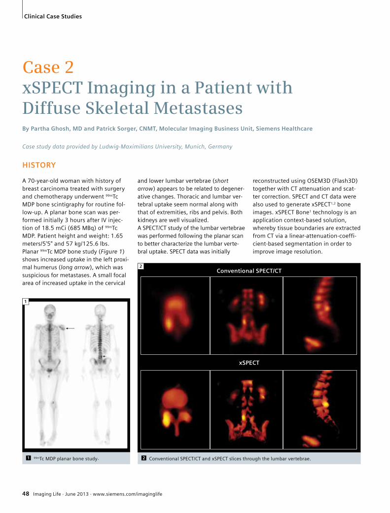
48 Imaging Life · June 2013 · www.siemens.com/imaginglife
Clinical Case Studies
HISTORY
A 70-year-old woman with history of breast carcinoma treated with surgery and chemotherapy underwent 99mTc MDP bone scintigraphy for routine fol-low-up. A planar bone scan was per-formed initially 3 hours after IV injec-tion of 18.5 mCi (685 MBq) of 99mTc MDP. Patient height and weight: 1.65 meters/5'5" and 57 kg/125.6 lbs. Planar 99mTc MDP bone study (Figure 1) shows increased uptake in the left proxi-mal humerus (long arrow), which was suspicious for metastases. A small focal area of increased uptake in the cervical
Case 2xSPECT Imaging in a Patient with Diffuse Skeletal Metastases By Partha Ghosh, MD and Patrick Sorger, CNMT, Molecular Imaging Business Unit, Siemens Healthcare
Case study data provided by Ludwig-Maximilians University, Munich, Germany
reconstructed using OSEM3D (Flash3D) together with CT attenuation and scat-ter correction. SPECT and CT data were also used to generate xSPECT1,2 bone images. xSPECT Bone1 technology is an application context-based solution, whereby tissue boundaries are extracted from CT via a linear-attenuation-coeffi-cient-based segmentation in order to improve image resolution.
and lower lumbar vertebrae (short arrow) appears to be related to degener-ative changes. Thoracic and lumbar ver-tebral uptake seem normal along with that of extremities, ribs and pelvis. Both kidneys are well visualized.A SPECT/CT study of the lumbar vertebrae was performed following the planar scan to better characterize the lumbar verte-bral uptake. SPECT data was initially
1 99mTc MDP planar bone study.
1
2 Conventional SPECT/CT and xSPECT slices through the lumbar vertebrae.
2Conventional SPECT/CT
xSPECT

Imaging Life · June 2013 · www.siemens.com/imaginglife 49
Clinical Case Studies
aNalYSIS
xSPECT shows higher intensity of uptake secondary to facet arthropathy in the right facet joint in L5 vertebrae com-pared to conventional SPECT/CT (Figure 2), as well as clear and sharp delineation of lamina, spinous process and spinal canal. Uptake of tracer in the lumbar vertebrae appears uniform. Visual evaluation of the CT and fused images (Figure 3), however, show a dif-ferent clinical picture. CT shows diffuse sclerosis involving all the lumbar verte-brae, including the body of the sacrum and the lamina and spinous processes. Such diffuse sclerosis could potentially reflect diffuse osseous metastases. Visually the 99mTc MDP uptake in the scle-rotic vertebrae appear uniform without any focal increase. Visualization of kid-neys and bladder activity exclude a "superscan" appearance.Using xSPECT-based quantification1,2 of SPECT tracer concentration in Bq/ml and using injected dose and patient height and weight information, the standard uptake value (SUV) of individual voxels, as well as volumes, could be calculated (Figure 4).
COMMENTS
Absolute quantification of tracer con-centration in the lumbar vertebrae with the new modality, xSPECT, shows 157 kBq/ml of 99mTc MDP in the center of the body of L3 vertebrae (arrow) with an SUV average (SUVavg) of 14.95, which is
approximately 2 times higher than that of normal (SUV average of 7.02).2,3 This high SUV within the lumbar vertebrae along with a diffuse sclerosis on CT is reflective of diffuse osseous metasta-ses. The planar bone study did not show diffuse vertebral hypermetabo-lism and both kidneys were visualized, excluding a superscan appearance. This can be explained by response of the dif-fuse vertebral metastases to chemo-therapy, but with persistent higher level of vertebral metabolism, due to the increased bone turnover within the sclerotic component, as evident in the increased tracer concentration and SUV
within the vertebrae. Measurements using xSPECT data in lumbar vertebrae of 8 normal female patients2,3 (average 64 years of age; injected dose 537 +/- 82 MBq) yielded average bone tracer activity concentra-tion (AC) of 56.70 +/- 17.21 kBq/ml and average SUV of 7.02 +/- 1.67.2
3 CT and xSPECT images of lumbar vertebrae.
3 CT
xSPECT
1. Symbia Intevo and xSPECT are not commercially available in all countries. Due to regulatory reasons their future availability cannot be guaranteed. Please contact your local Siemens organization for further details.
2. These are preliminary quantitative values defined on prototype system with prototype calibration, and are based on research. There can be no guarantee that customers will achieve the same results. Final results may vary.
3. Cachovan, Michal; Vija, A. Hans.; Hornegger, Joachim; Kuwert, Torsten1. "Quantitative bone SPECT with a novel multimodal reconstruction", J. Nucl.
Med. Meeting Abstracts, 2013; Vol.TBD
4 Quantitative evaluation of 99mTc MDP absolute tracer concentration within the lumbar vertebral body in kBq/ml and calculated vertebral SUV2 enabled by xSPECT technology.
4
ExaMINaTION PROTOCOlScanner SymbiaTM with xSPECT
Technology
Dose 18.5 mCi (685 MBq) 99mTc MDP
Scan Delay 3 hours post injection
Parameters 32 frames, 25 sec/frame
CT 130 kVp, 90 eff mAs, 3 mm slice

50 Imaging Life · June 2013 · www.siemens.com/imaginglife
Clinical Case Studies
HISTORYA 45-year-old man with history of biopsy proven myxoid liposarcoma in the left thigh, which was treated with surgery presented for follow-up. A 99mTc MDP bone scan was performed to eval-uate for skeletal metastases. A bone scan was performed on the new modal-ity, xSPECT*, 3 hours after an injection of 23.5 mCi of 99mTc MDP. Initial planar whole-body images (Figure 1) were fol-lowed by an xSPECT prototype study of the pelvis.
Case 3xSPECT Bone Metastases in a Case of Myxoid Liposarcoma By Jerry Froelich, MD
Case study data provided by the University of Minnesota, MN, USA
1 99mTc MDP whole-body planar bone scan. Whole-body planar images show area of increased uptake in the left acetabulum. No other abnormal focal area of increased uptake was visualized. To further evaluate the left acetabular lesion, the SPECT/CT study was performed immediately following the planar study.
aNalYSISUsing list mode SPECT data and CT- based zone maps, xSPECT was per-formed for the pelvis. As seen on Figure 2, conventional SPECT/CT Flash 3D shows mild increase of uptake in the anterior left acetabu-lum, extending to the superior left pubic ramus. Prelimary xSPECT reconstructions show increased intensity of tracer uptake in the acetabular and pubic lesions, along with small hypodense areas within the anterior acetabular
lesion (arrow). The variegated uptake pattern and the lower uptake intensity of the lesion visualized on the SPECT/CT and xSPECT compared to intensely hypermetabolic tumors like osteosarcoma could reflect the myxoid pathology of the primary tumor. CT shows focal lytic area in the anterior acetabulum and superior pubic ramus with surrounding mild sclerosis, which corresponds to the regions of hypointen-sity and increased uptake of 99mTc MDP
1
2 Conventional SPECT/CT and xSPECT of the pelvis and bilateral hip joints. SPECT was reconstructed using iterative reconstruction (Flash3D) with 4 iterations and 21 subsets. CT-based attenuation correction was also performed.
2Conventional SPECT/CT
xSPECT
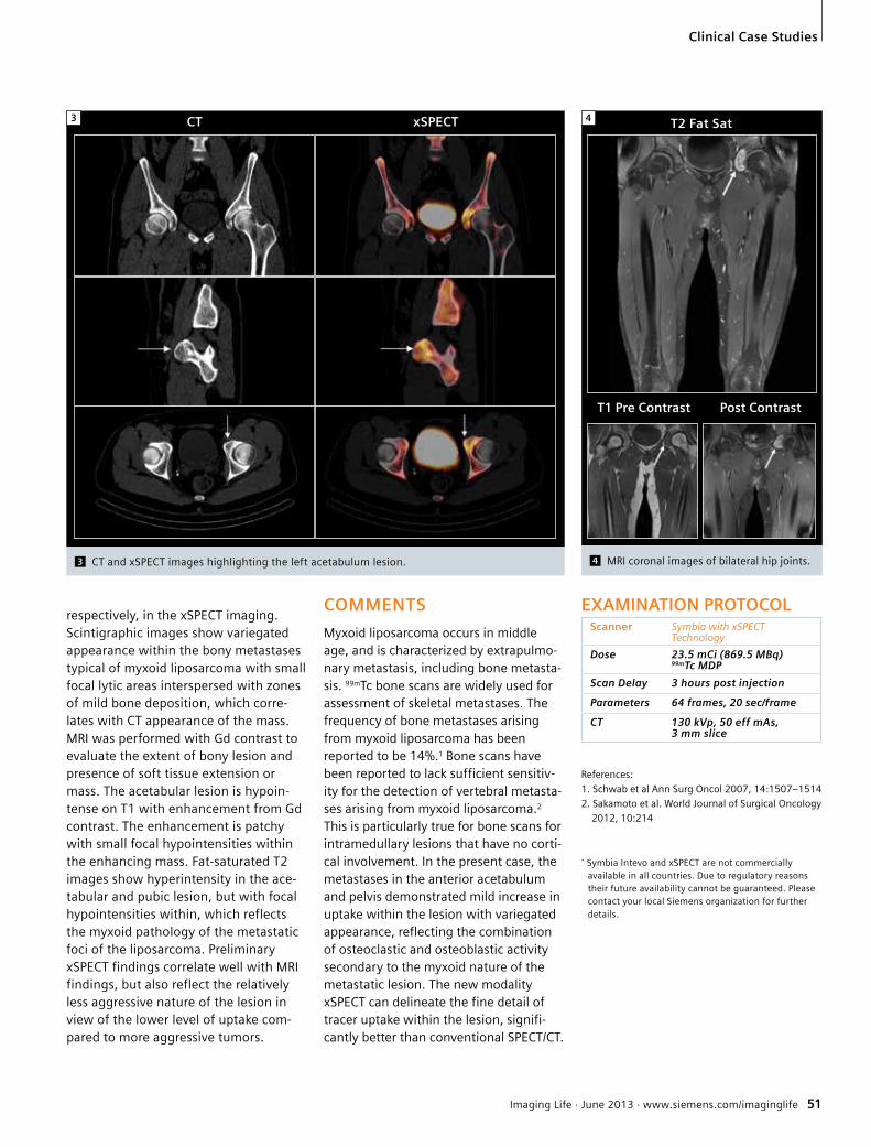
Imaging Life · June 2013 · www.siemens.com/imaginglife 51
Clinical Case Studies
respectively, in the xSPECT imaging. Scintigraphic images show variegated appearance within the bony metastases typical of myxoid liposarcoma with small focal lytic areas interspersed with zones of mild bone deposition, which corre-lates with CT appearance of the mass. MRI was performed with Gd contrast to evaluate the extent of bony lesion and presence of soft tissue extension or mass. The acetabular lesion is hypoin-tense on T1 with enhancement from Gd contrast. The enhancement is patchy with small focal hypointensities within the enhancing mass. Fat-saturated T2 images show hyperintensity in the ace-tabular and pubic lesion, but with focal hypointensities within, which reflects the myxoid pathology of the metastatic foci of the liposarcoma. Preliminary xSPECT findings correlate well with MRI findings, but also reflect the relatively less aggressive nature of the lesion in view of the lower level of uptake com-pared to more aggressive tumors.
COMMENTS
Myxoid liposarcoma occurs in middle age, and is characterized by extrapulmo-nary metastasis, including bone metasta-sis. 99mTc bone scans are widely used for assessment of skeletal metastases. The frequency of bone metastases arising from myxoid liposarcoma has been reported to be 14%.1 Bone scans have been reported to lack sufficient sensitiv-ity for the detection of vertebral metasta-ses arising from myxoid liposarcoma.2 This is particularly true for bone scans for intramedullary lesions that have no corti-cal involvement. In the present case, the metastases in the anterior acetabulum and pelvis demonstrated mild increase in uptake within the lesion with variegated appearance, reflecting the combination of osteoclastic and osteoblastic activity secondary to the myxoid nature of the metastatic lesion. The new modality xSPECT can delineate the fine detail of tracer uptake within the lesion, signifi-cantly better than conventional SPECT/CT.
ExaMINaTION PROTOCOlScanner Symbia with xSPECT
Technology
Dose 23.5 mCi (869.5 MBq) 99mTc MDP
Scan Delay 3 hours post injection
Parameters 64 frames, 20 sec/frame
CT 130 kVp, 50 eff mAs, 3 mm slice
3
3 CT and xSPECT images highlighting the left acetabulum lesion.
CT xSPECT
References:
1. Schwab et al Ann Surg Oncol 2007, 14:1507–1514
2. Sakamoto et al. World Journal of Surgical Oncology
2012, 10:214
* Symbia Intevo and xSPECT are not commercially available in all countries. Due to regulatory reasons their future availability cannot be guaranteed. Please contact your local Siemens organization for further details.
4 MRI coronal images of bilateral hip joints.
4 T2 Fat Sat
T1 Pre Contrast Post Contrast
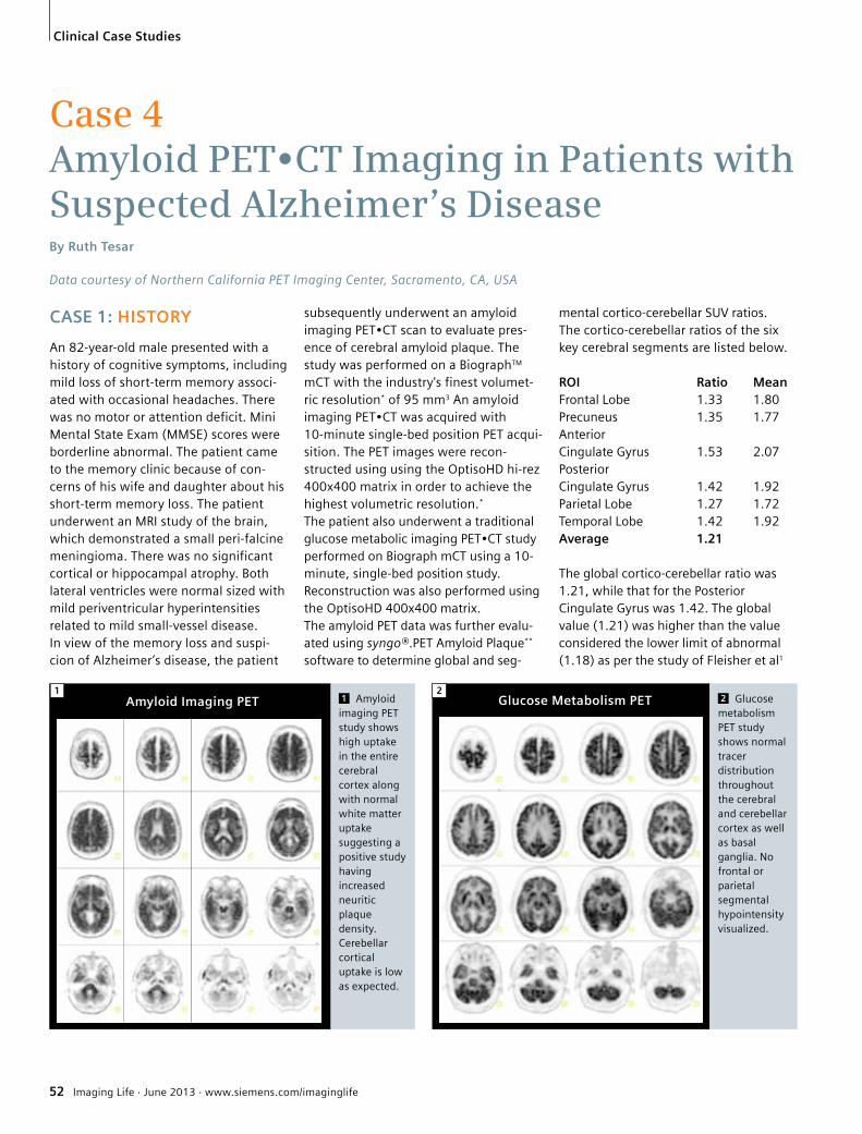
52 Imaging Life · June 2013 · www.siemens.com/imaginglife
Clinical Case Studies
CaSE 1: HISTORY
An 82-year-old male presented with a history of cognitive symptoms, including mild loss of short-term memory associ-ated with occasional headaches. There was no motor or attention deficit. Mini Mental State Exam (MMSE) scores were borderline abnormal. The patient came to the memory clinic because of con-cerns of his wife and daughter about his short-term memory loss. The patient underwent an MRI study of the brain, which demonstrated a small peri-falcine meningioma. There was no significant cortical or hippocampal atrophy. Both lateral ventricles were normal sized with mild periventricular hyperintensities related to mild small-vessel disease.In view of the memory loss and suspi-cion of Alzheimer’s disease, the patient
Case 4Amyloid PET•CT Imaging in Patients with Suspected Alzheimer’s DiseaseBy Ruth Tesar
Data courtesy of Northern California PET Imaging Center, Sacramento, CA, USA
subsequently underwent an amyloid imaging PET•CT scan to evaluate pres-ence of cerebral amyloid plaque. The study was performed on a BiographTM mCT with the industry's finest volumet-ric resolution* of 95 mm3 An amyloid imaging PET•CT was acquired with 10-minute single-bed position PET acqui-sition. The PET images were recon-structed using using the OptisoHD hi-rez 400x400 matrix in order to achieve the highest volumetric resolution.*
The patient also underwent a traditional glucose metabolic imaging PET•CT study performed on Biograph mCT using a 10- minute, single-bed position study. Reconstruction was also performed using the OptisoHD 400x400 matrix. The amyloid PET data was further evalu-ated using syngo®.PET Amyloid Plaque** software to determine global and seg-
mental cortico-cerebellar SUV ratios. The cortico-cerebellar ratios of the six key cerebral segments are listed below.
ROI Ratio MeanFrontal Lobe 1.33 1.80Precuneus 1.35 1.77Anterior Cingulate Gyrus 1.53 2.07Posterior Cingulate Gyrus 1.42 1.92Parietal Lobe 1.27 1.72Temporal Lobe 1.42 1.92average 1.21
The global cortico-cerebellar ratio was 1.21, while that for the Posterior Cingulate Gyrus was 1.42. The global value (1.21) was higher than the value considered the lower limit of abnormal (1.18) as per the study of Fleisher et al1
1 Amyloid imaging PET study shows high uptake in the entire cerebral cortex along with normal white matter uptake suggesting a positive study having increased neuritic plaque density. Cerebellar cortical uptake is low as expected.
amyloid Imaging PET1
2 Glucose metabolism PET study shows normal tracer distribution throughout the cerebral and cerebellar cortex as well as basal ganglia. No frontal or parietal segmental hypointensity visualized.
Glucose Metabolism PET2

Imaging Life · June 2013 · www.siemens.com/imaginglife 53
Clinical Case Studies
*Based on volumetric resolution available in competitive literature for systems greater than 70 cm bore size. Data on file.
**syngo.PET Amyloid Plaque is intended for use only with approved amyloid radiopharmaceuticals in the country of use. Users should review the drug labeling for approved uses.
References:
1. Arch sy. 2011 Nov;68(11):1404-11
derived from post mortem analysis of brains of patients with Alzheimer’s dis-ease. Elevated global and segmental ratios also support the impression of increased cortical amyloid plaque depos-its in this patient. Cortico-cerebellar ratio analysis using syngo.PET Amyloid Plaque aims to sup-port visual evaluation of amyloid PET imaging and provide additional diagnos-tic information.
aNalYSIS In view of the amyloid PET findings of high cortical amyloid plaque burden in a patient with cognitive symptoms includ-ing short term memory loss, the pres-ence of Alzheimer’s disease could not be ruled out. In view of the absence of cor-tical and hippocampal atrophy, normal cortical glucose metabolism and high
cortical amyloid levels, early Alzheimer’s disease was deemed a possibility.
CaSE 2: HISTORY A 71-year-old female patient presented with progressive memory dysfunction to her neurologist. She underwent brain MRI, which demonstrated subtle hippo-campal atrophy along with mild diffuse cortical atrophy. Lateral ventricles were within normal limits with small periven-tricular hyperdensities suggestive of mild subcortical small vessel disease.In view of the suspicion of Alzheimer’s disease, the patient underwent an amy-loid imaging PET•CT study for evaluation of the extent of cerebral amyloid plaque in order to rule out the disease. The study was performed on a Biograph mCT with a 10-minute, single-bed position PET study following CT. The PET images
were reconstructed at 400x400 matrix. The high volumetric resolution* achieved with Biograph mCT when combined with 400x400 matrix reconstruction is able to improve the physicians ability to differenti-ate between tracer uptake in the white matter, considered normal, and uptake in the grey matter, which can signal the pres-ence of amyloid plaques. The patient further underwent traditional glucose metabolism PET•CT, which was performed on Biograph mCT with similar acquisition and reconstruction parameters.
aNalYSIS Presence of a high amount of cortical amyloid plaque in this patient with neu-rologically documented progressive memory dysfunction was suggestive of the possibility of Alzheimer’s disease. Mild hippocampal atrophy on MRI and bilateral parietal hypometabolism seen on glucose metabolism PET provided reinforcement to the possibility of Alzheimer’s disease.
3 Amyloid and glucose metabolism PET images at the same levels show increased cortical amyloid uptake with normal cortical glucose uptake. The normal level of retention of amyloid tracer in the white matter is clearly defined as is the increased glucose metabolism uptake in the basal ganglia. CT slices at the same level shows normal ventricular size and absence of significant cortical atrophy.
amyloid Imaging PET Gluclose Metabolism PET CT
3
4 Amyloid PET images show diffuse increase in cortical uptake of tracer suggesting high cortical amyloid density. Cerebellar uptake is low as expected. This study appears positive for cerebral amyloid.
amyloid Imaging PET4
5 Glucose metabolism PET study shows bilateral parietal hypometabolism typically associated with Alzheimer’s disease. The frontal and occipital cortex, basal ganglia and cerebellum show normal uptake.
Glucose Metabolism PET5

54 Imaging Life · June 2013 · www.siemens.com/imaginglife
Clinical Case StudiesClinical Case Studies
HISTORY
A 63-year-old woman without signifi-cant past medical histories presented with post-menopausal vaginal bleeding for 2 years. Gynecological examination revealed a bulky cervical mass without direct pelvic side wall invasion. A cervi-cal biopsy showed very poorly differenti-ated squamous cell carcinoma with extensive necrosis.
DIaGNOSIS
The patient underwent Fludeoxyglucose F18* (18F FDG) PET•CT, as well as MRI for primary staging.The study was performed on Biograph™ mCT 64 with the industry’s finest** volumetric resolution, which improves lesion margin delineation for more accurate Gross Tumor Volume (GTV) determination. High-lesion contrast achieved using ultraHD•PET (Time of Flight and Point Spread Function (PSF) reconstruction combined) improves lesion delineation, especially for small primary tumors and recurrences, as well as for small lymph node metastases. PET showed increased metabolic activity in the cervix with a maximum standard-ized uptake value (SUVmax) of 27 (Figure 1). No nodal or distant metastasis was found. MRI revealed a 74 mm x 71 mm x 63 mm mass within the cervix . There was no enlargement of the pelvic nodes, para-aortic nodes and inguinal nodes. Final clinical staging was cT2b2N0M0 by 2009 American Joint Committee on Cancer staging system (FIGO stage IIb2).
Case 518F FDG PET•CT-based Radiation Therapy Planning in a Case of Cervical CarcinomaBy Partha Ghosh, MD, Molecular Imaging Business Unit, Siemens Healthcare
Case study data courtesy of Jack Wang, MD and Tsu Chen Yen, MD, Chang Gung Memorial Hospital, Linkou, Taiwan
A definitive concurrent chemo-radiother-apy (CCRT) with weekly cisplatin (40 mg/m2) was arranged. Radiation was deliv-ered by conventional four-field box tech-nique. Doses of large-field radiation to the whole pelvis were 45 Gy/25 fractions (Figure 2A), followed by a true pelvis boost, including the whole uterus, to a total dose of 54 Gy/30 fractions (Figure 2B). Brachytherapy was administered as 5 fractions with 4 Gy/fraction to point A.Overall, the patient tolerated combined treatment well and did not require any unplanned treatment breaks. Acute grade II diarrhea was noted during CCRT. At the end of treatment, the surface of the cervix showed complete tumor regression with little granulation change by clinical pelvic examination. MRI and PET follow-up scans were done during
2 Isodose curves of the radiotherapy planning.
2A
2
2B
1 Pre-therapy PET•CT of patient with large cervical mass.
1
1

Imaging Life · June 2013 · www.siemens.com/imaginglife 55
Clinical Case Studies
the fourth week of treatment and 2 months after completing treatment. The images on the fourth week illus-trated a partial response of the primary tumor (Figure 3B). However, PET•CT findings of post-CCRT study suggested residual tumor over the fundus of uterus (Figure 3C). High-lesion contrast achieved on Biograph mCT was instru-mental in the detection of a small resid-ual tumor in post-chemoradiation study. Based on the PET•CT findings and the post-CCRT status of this patient, a sal-vage radical hysterectomy was per-formed one month later. The final pathology revealed a 1.5 x 1 cm tumor mass over the endometrium with inva-sion to 10% of the myometrial wall thickness. The other sites, including cer-vix and parametrium, were all cancer-free. This patient is still alive without disease recurrence.This case illustrates the value of 18F FDG PET•CT for radiation therapy planning and therapy response evaluation in cer-vical carcinoma. PET•CT demonstrated high sensitivity for detection of small residual tumor after CCRT and helped guide the salvage therapy.
ExaMINaTION PROTOCOlScanner Biograph mCT
Scan dose 10 mCi (370 MBq) 18F FDG injection
Scan protocol 150 minutes post-injection delay, 4-min/bed PET acquisition
3 Pre- and post-therapy PET•CT images. Images were acquired before treatment (3A), 4th week during CCRT (3B), and 2 months after CCRT (3C).
3A 3B 3C
Indications
Fludeoxyglucose F 18 Injection is indi-cated for positron emission tomography (PET) imaging in the following settings:• Oncology: For assessment of abnormal
glucose metabolism to assist in the eval-uation of malignancy in patients withknown or suspected abnormalities foundby other testing modalities, or in patients with an existing diagnosis of cancer.
• Cardiology: For the identification ofleft ventricular myocardium with resid-ual glucose metabolism and reversibleloss of systolic function in patients withcoronary artery disease and left ventric-ular dysfunction, when used togetherwith myocardial perfusion imaging.
• Neurology: For the identification ofregions of abnormal glucose metabo-lism associated with foci of epilepticseizures.
Important Safety Information
• Radiation Risks: Radiation-emittingproducts, including FludeoxyglucoseF 18 Injection, may increase the riskfor cancer, especially in pediatricpatients. Use the smallest dose neces-sary for imaging and ensure safe han-dling to protect the patient and healthcare worker.
• Blood Glucose abnormalities: In theoncology and neurology setting, sub-optimal imaging may occur in patientswith inadequately regulated blood glu-cose levels. In these patients, considermedical therapy and laboratory testingto assure at least two days of normo-glycemia prior to Fludeoxyglucose F 18Injection administration.
• adverse Reactions: Hypersensitivityreactions with pruritus, edema andrash have been reported; have emer-gency resuscitation equipment andpersonnel immediately available.
* The full prescribing information for the Fludeoxyglu-cose F 18 injection can be found on pages 68-70.
**Based on volumetric resolution available in compe- titive literature for systems greater than 70 cm bore size. Data on file.
3

56 Imaging Life · June 2013 · www.siemens.com/imaginglife
Clinical Case Studies
HISTORYA 66-year-old man presented with a his-tory of abdominal pain, flushes and diar-rhea. Initial investigation detected a pancreatic mass, which was confirmed to be a pancreatic neuroendocrine tumor (carcinoid). High serum 5-HIAA
and presence of carcinoid-syndrome-like features like diarrhea suggested a high possibility of the presence of extra-pan-creatic metastases. A 68Ga DOTATOC* MR and PET study was performed on the Biograph mMR™. Simultaneous
acquisition of 68Ga DOTATOC* PET with breath-hold, fat-suppressed, MR sequences of the abdomen, as well as Gadolinium (Gd) contrast-enhanced, abdominal MR sequences were per-formed on the Biograph mMR system.
Case 6Improved Detection of Hepatic Metastases from a Pancreatic Neuroendocrine Tumor with Simultaneous MR and 68Ga DOTATOC PET AcquisitionBy Hilmar Kuehl, MD
Case study data provided by University of Essen, Essen, Germany
1 Biograph mMR acquisitions show small, tracer-avid, liver metastases.
Simultaneous MR and PET Gd MRarterial phase
1

Imaging Life · June 2013 · www.siemens.com/imaginglife 57
Clinical Case Studies
DIaGNOSIS
Simultaneously-acquired MR and PET images show 3 hypermetabolic liver metastases (arrows) with corresponding contrast enhancements in all the 3 met-astatic lesions in the fat-suppressed, simultaneously-acquired MR images. The exact localization of the liver lesion due to simultaneous MR and PET acquisi-tion can be demonstrated by the increased PET tracer uptake in the metastases adjacent to the inferior vena cava, which also shows high signal on fat suppression MR. The MR and PET sig-nals match exactly on the simultaneous MR and PET images.
2 Post Gd-contrast, fat-suppression MR images show multiple focal-liver lesions.
2
MR images also show multiple small-enhancing liver lesions apart from the 3 metastatic lesions previously defined on simultaneous MR and PET images. Positive PET uptake in the liver lesions confirms that these lesions are function-ing neuroendocrine tumor metastases due to uptake of 68Ga DOTATOC*, thereby giving improved specificity to the study. The simultaneous MR and PET study, therefore, delivers high sensitivity and specificity in combination.
* 68Ga DOTATOC is not currently recognized by the U.S. Food and Drug Administration (FDA) or other regulatory agencies as being safe and effective, and Siemens does not make any claims regarding its use.
References:
1. Beiderwellen et al Invest Radiol. 2013 Mar 13.
ExaMINaTION PROTOCOlScanner Biograph mMR
Dose 150 MBq 68Ga DOTATOC
MR T1 Flash, T2 FatSat, TIRM,Gd enhanced VIBE
PET 4 min/bed simultaneousacquisition
COMMENTS
This case example is from a series of 8 patients with neuroendocrine tumors, who were evaluated with 68Ga DOTATOC* PET/CT, as well as on simultaneous MR and PET.1 Of the 8 patients, 5 were con-firmed to have malignant neuroendo-crine tumors based on histopathology and follow-up. All malignant lesions detected on PET/CT were also identified on MR and PET. One liver lesion, which was labeled as indeterminate on PET/CT, was identified as a metastasis on MR and PET because of a diffusion restriction on diffusion-weighted imaging. PET/CT, however, was more sensitive for detec-tion of metastases in the lung and lymph nodes, as well as a sclerotic bone lesion. The SUVmax measured by PET/CT and by simultaneous MR and PET showed a strong correlation.This clinical case example demonstrates the advantage of simultaneous MR and PET for detection of liver metastases due to the combination of a high signal on fat-suppression MR and 68Ga DOTATOC* uptake on PET. The simultaneous acqui-sition on Biograph mMR ensures precise registration between PET and MR even for small lesions.

58 Imaging Life · June 2013 · www.siemens.com/imaginglife
Clinical Case Studies
HISTORYA 74-year-old man with history of coro-nary artery disease treated with bypass grafting (CABG) 4 years ago, presented to the hospital with worsening exertional dyspnea and occasional chest pain. His resting ECG showed sinus rhythm with no acute stress changes. He was referred for a stress-rest Rubidium-82 (Rb-82) PET•CT myocardial perfusion study. Dynamic Rb-82 stress-rest myocardial perfusion study was performed on a
Siemens BiographTM mCT 40. Dosage of Rb infusion was based on patient’s body weight at 15 MBq/kg. A rest study was first obtained. Imaging acquisition was over 8 minutes starting from time of Rb-82 infusion. This was followed by dipyridamole (persantin) infusion at 0.14 mg/kg/min over 4 minutes. Stress imaging was then acquired at peak stress (8 minutes after start of persan-tin infusion).
Case 7PET•CT Myocardial Perfusion Combined with CT Angiography in Detection of Post-Revascularization IschemiaBy Seng Chuan Ong, MD, FRCR, Hee Kit lai , MD, MRCP and Robert Kwok, MD, FRCR
Study was performed at Mount Elizabeth Hospital Singapore (Parkway Group), Singapore
A CT coronary angiography (CTA) was performed at the end of the PETperfusion study. Stress and rest PET perfusion studies were evaluated with 4DMSPECT, and the CT angiography was evaluated using syngo® Circulation. Biograph mCT with LSO crystals and ultraFast electronics provides high count rate capability for excellent image quality in dynamic Rb-82
1 Rb-82 Myocardial Perfusion PET.
1
STRESS
REST
STRESS
REST
STRESS
REST

Imaging Life · June 2013 · www.siemens.com/imaginglife 59
Clinical Case Studies
myocardial perfusion imaging, espe-cially in obese patients. The unique detector design of Biograph mCT avoids detector saturation at higher doses of Rubidium and thus is able to perform dynamic PET studies for myocardial blood flow estimations at the higher Rubidium doses required for obese patients. syngo Circulation is a Siemens software that evaluates CT angiography from all ven-dors and is able to fuse PET data and CT angiography with volume rendering in order to improve diagnostic confidence.
DIaGNOSIS
Comparison of static short-axis, horizon-tal-axis and vertical-long-axis slices of stress and rest Rb 82 myocardial perfu-sion PET•CT studies show a large perfu-sion defect in the lateral wall in the stress images, which corresponds to ter-ritory perfused by the left circumflex artery, with complete reversibility as demonstrated by normal uptake at rest. There is mild to moderate left ventricu-lar dilatation during stress, which reflects the severity of lateral wall isch-emia. The rest of the left ventricle (LV) myocardium shows normal perfusion.
COMMENTS
In view of the stress-induced perfusion defect, the CT angiography was per-formed to evaluate arterial and venous graft patency in this patient with history of CABG. CT angiography showed patent left internal mammary artery (LIMA) graft to mid left anterior descending artery (LAD), as well as patent venous graft to distal right coronary artery (RCA). The venous graft to first obtuse marginal (OM1) was patent at origin, as well as the entire length of the graft. However, the graft insertion at the level of proxi-mal OM1 appeared irregular with absence of contrast runoff into OM1 distal to the graft insertion. This sug-gested the possibility of an obstruction to contrast flow at the insertion level, which may explain the stress-induced
ischemia in the lateral wall exactly cor-responding to the OM1 territory. Native vessels including LAD, left cir-cumflex were heavily calcified and nar-row throughout their entire extent, which reflects shunting of blood to the patent grafts. Fusion of stress and rest PET perfusion and coronary tree derived from CTA using syngo Circulation show the extent of stress-induced ischemia with com-plete normalization of perfusion at rest (thin arrow). The ischemic zone corre-sponds to the myocardium supplied by OM1 with irregularities at the insertion of the venous graft to OM1 (thick arrow). The LIMA graft to LAD and venous graft to RCA are distinctly patent with no corresponding ischemic zones. In view of the patency of all other grafts and absence of ischemia in all of LV myocardium except the lateral wall (OM1) distribution, as well as the irreg-ularities visualized on the CTA at the insertion of venous graft to OM1 and lack of opacification of the mid and
distal OM1 on CTA, there appeared to be a strong possibility of a graft block at the OM1 insertion which, if confirmed by catheter angiography, is amenable to an angioplasty. Combined imaging Biograph mCT 40 of Rb-82, stress-rest myocardial perfusion and CT coronary angiography along with hybrid display of coronary tree with volume-rendered, quantitatively accurate PET perfusion data using syngo Circulation was key to identification of the culprit lesion at the graft insertion.
2 Fusion and volume rendering of PET perfusion and angiography using syngo Circulation.
2
STRESS
REST
ExaMINaTION PROTOCOlScanner Biograph mCT 40
Dose 40 mCi (15 MBq/kg) Rb-82 injection
Acquisition Dynamic List mode
CT Low dose for CTAC
CT Coronary angiography
Dose 80 ml contrast injection
CT 100 kV, 300 eff mAs, 1 mm slice
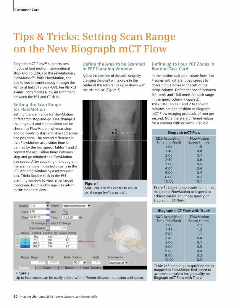
60 Imaging Life · June 2013 · www.siemens.com/imaginglife
Customer Care
Figure 1Small circle in the center to adjust axial range (yellow arrow).
Tips & Tricks: Setting Scan Range on the New Biograph mCT Flow Biograph mCT Flow™ supports two modes of bed motion, conventional stop-and-go (S&G) or the revolutionary FlowMotionTM. With FlowMotion, the bed is moved continuously through the PET axial field of view (FOV). For PET•CT exams, both modes allow an alignment between the PET and CT data.
Setting the Scan Range for FlowMotion Setting the scan range for FlowMotion differs from stop-and-go. One change is that any start and stop position can be chosen by FlowMotion, whereas stop-and-go needs to start and stop at discrete bed positions. The second difference is that FlowMotion acquisition time is defined by the bed speed. Tables 1 and 2 convert the acquisition times between stop-and-go min/bed and FlowMotion bed speed. After acquiring the topogram, the scan range is indicated visually in the PET Planning window by a rectangular box. Trick: Double click in the PET planning window to view an enlarged topogram. Double click again to return to the standard view.
Define the Area to be Scanned in PET Planning Window
Adjust the position of the axial range by dragging the small white circle in the center of the scan range up or down with the left mouse (Figure 1).
Figure 2Up to four zones can be easily added with different distance, duration and speed.
Biograph mCT Flow with TrueV
S&G Acquisition FlowMotion Time (min/bed) Speed (mm/s) 1:00 2.1 1:48 1.2 2:00 1.1 2:48 0.8 3:00 0.7 4:00 0.5 5:00 0.4 8:00 0.3 10:00 0.2
Table 2: Stop-and-go acquisition times mapped to FlowMotion bed speed to achieve equivalent image quality on Biograph mCT Flow with TrueV.
Biograph mCT Flow
S&G Acquisition FlowMotion Time (min/bed) Speed (mm/s) 1:00 1.5 1:48 0.8 2:00 0.7 2:30 0.6 3:00 0.5 4:00 0.4 5:00 0.3 6:00 0.2 10:00 0.1
Table 1: Stop-and-go acquisition times mapped to FlowMotion bed speed to achieve equivalent image quality on Biograph mCT Flow.
Define up to Four PET Zones in Routine Task Card
In the routine task card, create from 1 to 4 zones with different bed speeds by checking the boxes to the left of the range column. Define the speed between 0.1 mm/s and 10.0 mm/s for each range in the speed column (Figure 2).Trick: Use Tables 1 and 2 to convert minutes per bed position to Biograph mCT Flow imaging protocols of mm per second. Note there are different values for a scanner with or without TrueV.

Imaging Life · June 2013 · www.siemens.com/imaginglife 61
Customer Care
Redefine Scan Range in PET Planning Window
Scan range is typically defined by the protocol, but can be redefined by dragging the scan range reference lines in the PET Planning window (Figure 3). Changing the length of a range in the PET planning window changes the distance value on the routine task card (Figure 2).Tip: Scan ranges are indicated visually by color coded bars on the scan reference bar on the left side of the PET Planning window (Figure 3) corresponding to the colored zone numbers in the range column of the routine task card (Figure 2).Trick: The length of ranges can also be changed easily by dragging the dotted lines in the PET Planning window. Changing the length of a range in the PET Planning window changes the distance value on the routine task card.
Figure 4 Scan Range - Match CT Range.
Figure 3 Scan range reference bar (yellow arrow).Scan range reference lines (red arrows).
Figure 5 Overview: Setting Scan Range in FlowMotion Planning Window
1) Adjust the position of the axial range by dragging the small white circle
2) Drag the scan range reference lines to further define scan range
3) Click the box to add a new zone
4) Define the speed for each zone
5) Color-coded PET FlowMotion regions correspond to the color coded zones on the routine subtask card
Match CT Match CT Behavior of PET•CT PlanningRange Slice Location
Checked Checked CT range snaps automatically to PET scan range. The PET image slice location and thickness is then matched to the CT.
Unchecked Checked The CT and PET scan ranges are independently defined. The PET image slice location and thickness will be matched to the CT for the planned PET. scan range.
Table 3: Match CT Range option to behavior of PET•CT planning.
Trick: For FlowMotion protocols, on the routine task card, if you check Scan Range - Match CT Range (Figure 4), syngo® automatically adjusts the CT field of view to match the PET field of view. Correspondingly, if the CT range is changed, the PET range is automatically changed to match. If Match CT Range is left unchecked, the CT and PET ranges will not automatically match (Table 3).
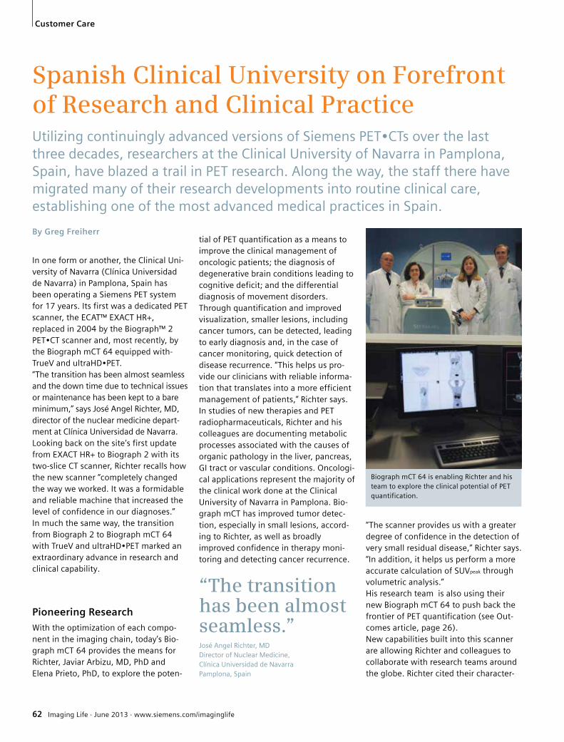
62 Imaging Life · June 2013 · www.siemens.com/imaginglife
Customer Care
Spanish Clinical University on Forefront of Research and Clinical PracticeUtilizing continuingly advanced versions of Siemens PET•CTs over the last three decades, researchers at the Clinical University of Navarra in Pamplona, Spain, have blazed a trail in PET research. Along the way, the staff there have migrated many of their research developments into routine clinical care, establishing one of the most advanced medical practices in Spain.
Biograph mCT 64 is enabling Richter and his team to explore the clinical potential of PET quantification.
By Greg Freiherr
In one form or another, the Clinical Uni-versity of Navarra (Clínica Universidad de Navarra) in Pamplona, Spain has been operating a Siemens PET system for 17 years. Its first was a dedicated PET scanner, the ECAT™ EXACT HR+, replaced in 2004 by the Biograph™ 2 PET•CT scanner and, most recently, by the Biograph mCT 64 equipped with-TrueV and ultraHD•PET.“The transition has been almost seamless and the down time due to technical issues or maintenance has been kept to a bare minimum,” says José Angel Richter, MD, director of the nuclear medicine depart-ment at Clínica Universidad de Navarra.Looking back on the site’s first update from EXACT HR+ to Biograph 2 with its two-slice CT scanner, Richter recalls how the new scanner “completely changed the way we worked. It was a formidable and reliable machine that increased the level of confidence in our diagnoses.” In much the same way, the transition from Biograph 2 to Biograph mCT 64 with TrueV and ultraHD•PET marked an extraordinary advance in research and clinical capability.
Pioneering Research
With the optimization of each compo-nent in the imaging chain, today’s Bio-graph mCT 64 provides the means for Richter, Javiar Arbizu, MD, PhD and Elena Prieto, PhD, to explore the poten-
tial of PET quantification as a means to improve the clinical management of oncologic patients; the diagnosis of degenerative brain conditions leading to cognitive deficit; and the differential diagnosis of movement disorders. Through quantification and improved visualization, smaller lesions, including cancer tumors, can be detected, leading to early diagnosis and, in the case of cancer monitoring, quick detection of disease recurrence. “This helps us pro-vide our clinicians with reliable informa-tion that translates into a more efficient management of patients,” Richter says. In studies of new therapies and PET radiopharmaceuticals, Richter and his colleagues are documenting metabolic processes associated with the causes of organic pathology in the liver, pancreas, GI tract or vascular conditions. Oncologi-cal applications represent the majority of the clinical work done at the Clinical University of Navarra in Pamplona. Bio-graph mCT has improved tumor detec-tion, especially in small lesions, accord-ing to Richter, as well as broadly improved confidence in therapy moni-toring and detecting cancer recurrence.
“The scanner provides us with a greater degree of confidence in the detection of very small residual disease,” Richter says. “In addition, it helps us perform a more accurate calculation of SUVpeak through volumetric analysis.”His research team is also using their new Biograph mCT 64 to push back the frontier of PET quantification (see Out-comes article, page 26). New capabilities built into this scanner are allowing Richter and colleagues to collaborate with research teams around the globe. Richter cited their character-
“The transition has been almost seamless.”José Angel Richter, MDDirector of Nuclear Medicine, Clínica Universidad de Navarra Pamplona, Spain

Imaging Life · June 2013 · www.siemens.com/imaginglife 63
Customer Care
ization of lung nodules through the International Early Lung Cancer Program (I-ELCAP) program and participation in the assessment of PET/CT colonography in high-risk patients as an example of the results of this collaboration. The Spanish researchers are also leveraging Biograph mCT to move into areas out-side oncology.
Clinical Translation
While much of Clinical University of Navarra’s efforts have focused on research, their latest scanner has been and continues to be used extensively in everyday clinical studies. The main objective behind migrating to this most recent scanner, in fact, was to support the clinical utilization of PET.“The new equipment has made it possi-ble to expand the number of clinical applications, which in turn has enabled our nuclear medicine service to see patients with a wider variety of condi-tions,” Richter says.The Biograph mCT 64 has also improved productivity. Scan speed is up dramati-cally, he says.“The speed with which we can perform more accurate studies—be it for diagnos-tic purposes, for monitoring response to therapy or finding disease recurrence—helps us provide our clinicians with reli-able information that translates into a
more efficient management of patients,” Richter says. “And, generally speaking, this helps increase the number and types of cases we can see in a given period of time.”Siemens’ TrueV has extended the axial field of view by 33 percent, increasing count rate by more than 70 percent, thereby providing the option of lowering radiation dose rates or reducing scan times up to 50 percent.
Patient Centric
Biograph mCT 64 has also improved patient safety. Its Adaptive Dose Shield uses collimators to block unnecessary radiation, just as CARE Dose4D modu-lates X-ray tube current to precisely shape the radiation pattern to the patient’s body, cutting dose by as much 66 per-cent. Quanti•QC calibrates and tunes the system after hours when the system is not scanning patients, ensuring consis-tent and optimal quantitative perfor-mance. Siemens Molecular and Anatomi-cal Registration Technologies (SMART)
attack the traditional problems of inher-ent scanner drift and inaccurate attenua-tion correction that can occur due to mis-registration of CT and PET data.Not surprisingly, given the extensive use of Siemens PET•CT in oncology research over the past three decades, the Biograph mCT is proving especially helpful in rou-tine clinical use for ruling out malignan-cies, improving the guidance of biopsy of positive lesions that were difficult to characterize, and in the early detection of cancer recurrence, Richter says.He describes the decision to migrate to Biograph mCT 64 and its many advanced features as being a natural evolution from the first Siemens PET equipment installed in the mid-1990s.“We felt comfortable and knowledgeable about the equipment, so at one point we felt we were ready for an update to the next generation of scanners,” Richter says. “We are extremely satisfied with the performance of our new equipment.”
“We are extremely satisfied with the performance of our new equipment.”José Angel Richter, MDDirector of Nuclear Medicine, Clínica Universidad de Navarra Pamplona, Spain
Evolution of PET at the Clinical University of Navarra
1996
ECAT EXACT HR+
2004
Biograph 2
2011
Biograph mCT 64 with TrueV Ultra•HD
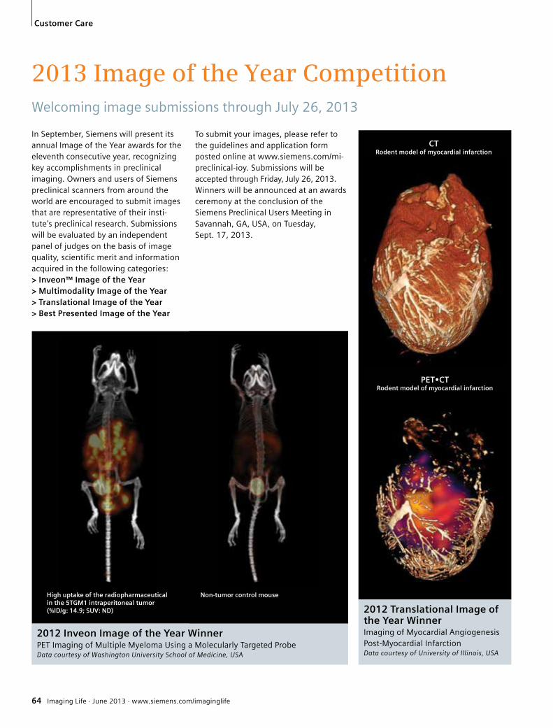
64 Imaging Life · June 2013 · www.siemens.com/imaginglife
Customer Care
2013 Image of the Year Competition Welcoming image submissions through July 26, 2013
In September, Siemens will present its annual Image of the Year awards for the eleventh consecutive year, recognizing key accomplishments in preclinical imaging. Owners and users of Siemens preclinical scanners from around the world are encouraged to submit images that are representative of their insti-tute’s preclinical research. Submissions will be evaluated by an independent panel of judges on the basis of image quality, scientific merit and information acquired in the following categories:> Inveon™ Image of the Year > Multimodality Image of the Year > Translational Image of the Year > Best Presented Image of the Year
To submit your images, please refer to the guidelines and application form posted online at www.siemens.com/mi-preclinical-ioy. Submissions will be accepted through Friday, July 26, 2013. Winners will be announced at an awards ceremony at the conclusion of the Siemens Preclinical Users Meeting in Savannah, GA, USA, on Tuesday, Sept. 17, 2013.
2012 Inveon Image of the Year WinnerPET Imaging of Multiple Myeloma Using a Molecularly Targeted ProbeData courtesy of Washington University School of Medicine, USA
2012 Translational Image of the Year WinnerImaging of Myocardial Angiogenesis Post-Myocardial InfarctionData courtesy of University of Illinois, USA
High uptake of the radiopharmaceutical in the 5TGM1 intraperitoneal tumor (%ID/g: 14.9; SUV: ND)
Non-tumor control mouse
PET•CT Rodent model of myocardial infarction
CT Rodent model of myocardial infarction

Imaging Life · June 2013 · www.siemens.com/imaginglife 65
Customer Care
MI University 360: Connecting Physicians Around the GlobeAs a worldwide innovation leader in medical imaging, Siemens has accrued extensive knowledge and technical expertise in the field of molecular imaging. This experience has been applied to providing excellent systems, an extensive portfolio of educational programs and innovative, flexible and comprehensive service solutions that enable you to concentrate on what is most important: patient care.
“I included my conceptual imaging protocols in MIU 360 to educate other physicians in the molecular imagingcommunity on how Iintend to achieve theclearest images [on thenew Biograph mCTFlow™] with FlowMotion.”Kirk A. Frey, MD, PhD University of Michigan, Ann Arbor, MI, USA
Expanding Practice Expertise
As the field of molecular imaging contin-ues its rapid growth, the industry is con-stantly changing. MIU 360 enables imag-ing physicians to keep an eye on newprotocols from industry leaders and tobring new techniques into their dailyworkflow. Despite the dramatic successes of molecular imaging technology over the last decade, challenges still persist, and the need for standardizing best prac-tices remains a hot topic among physi-cians. MIU 360 offers you the ability toaccess case studies, whitepapers andprotocols to help quickly find guidanceon practice expertise.
Driving Referrals
Clinical knowledge and practice expertiseare the first steps, but physicians also face competitive and financial pressures to grow their patient base, enhance theircommunity image and use resourcesmore efficiently. Through MIU 360, youcan find tips on promoting the benefits ofmolecular imaging and access tools thatcan help you drive referrals and strengthen a successful imaging practice.To experience this one-stop resource,which helps you maintain and increaseyour clinical and operational expertiseand assists you in getting the most out ofyour investment, visit:www.siemens.com/MIU360.
With MI University 360 (MIU 360), ournew customer portal, Siemens hasexpanded its educational resources tohelp you gain additional clinical knowl-edge, expand practice expertise anddrive referrals to grow your practice.
Gaining Clinical Knowledge
MIU 360 connects physicians across theglobe, providing a platform to addresstopics such as how to optimize dose,increase accuracy in radiation therapy orimprove detection of coronary artery dis-ease. You can find answers to these challenges, learn how to maximize the benefits that can be achieved with Sie-mens advanced technology and deepen your clinical expertise.

66 Imaging Life · June 2013 · www.siemens.com/imaginglife
Imprint
Imaging Life © 2013 by Siemens Medical Solutions USA, Inc. All rights reserved.
Publisher: Siemens Medical Solutions USA, Inc. Healthcare Sector Molecular Imaging 2501 N. Barrington Road Hoffman Estates, IL 60192 USA Telephone: + 1 847 304-7700 www.siemens.com/mi
Editor: Catherine Eby [email protected]
Editorial Consulting: The Freiherr Group Wisconsin, USA
Design Consulting: 360 Media, Inc., Colorado, USA
Printer: Tewell Warren Printing, Colorado, USA
Photo Credit: Charles Brooks photography Tennessee, USA
Clinical Images: Cover Story: P131_28; P166_34; P158_32; S122-23, S135_23; S154_21; xSPECT Outcomes Article: A115_21; S118_22; S131_20; S116_22; A115_21; Clinical Case Studies: Case #1- P186_34; Case #2 - S160_23; Case #3 - S125_20;
Note in accordance with § 33 Para.1 of the German Federal Data Protection Law: Dispatch is made using an address file which is maintained with the aid of an automated data processing system.
Siemens Molecular Imaging reserves the right to modify the design and specifications contained herein without prior notice. Trademarks and service marks used in this material are property and service names may be trademarks or registered trademarks of their respective holders.
We remind our readers that when printed, X-ray films never disclose all the information content of the original. Artifacts in CT, MR, SPECT, SPECT/CT, PET, PET/CT and PET/MR images are recognizable by their typical features and are generally distinguishable from existing pathology. As referenced below, healthcare practioners are expected to utilize their own learning, training and expertise in evaluating images.
Please contact your local Siemens sales representative for the most current information.
Note: Original images always lose a certain amount of detail when reproduced. All comparative claims derived from competitive data at the time of printing. Data on file.
The consent of the authors and publisher are required for the reprint or reuse of an article. Please contact Siemens for further information. Suggestions, proposals and information are always welcome; they are carefully examined and submitted to the editorial board for attention. Imaging Life is not responsible for loss, damage or any other injury to unsolicited manuscripts or materials.
We welcome your questions and comments about the editorial content of Imaging Life. Please contact us at [email protected].
Imaging Life is available on the internet: www.siemens.com/imaginglife
Imaging Life also has a free iPad and iPhone App available. From your iPad or iPhone, go to the App Store and search “Imaging Life,” then download.
Some of the imaging biomarkers in this publication are not currently recognized by the U.S. Food and Drug Administration (FDA) or other regulatory agencies as being safe and effective, and Siemens does not make any claims regarding their use.
DISCLAIMERS: Imaging Life: “The information presented in this magazine is for illustration only and is not intended to be relied upon by the reader for instruction as to the practice of medicine. Healthcare practitioners reading this information are reminded that they must use their own learning, training and expertise in dealing with their individual patients. This material does not substitute for that duty and is not intend-ed by Siemens Healthcare to be used for any purpose in that regard.” Contrast Agents: “The drugs and doses mentioned herein are consistent with the approved labeling for uses and/or indications of the drug. The treating physician bears the sole responsibility for the diagnosis and treatment of patients, including drugs and doses prescribed in connection with such use. The Operating Instructions must always be strictly followed when operating your Siemens system. The source for the technical data is the corresponding data sheets.” Trademarks: “All trade-marks mentioned in this document are property of their respective owners.” Results: “The outcomes achieved by the Siemens customers de-scribed herein were achieved in the customer’s unique setting. Since there is no “typical” hospital and many variables exist (e.g., hospital size, case mix, level of IT adoption), there can be no guarantee that others will achieve the same results.”

Imaging Life · June 2013 · www.siemens.com/imaginglife 67
Subscription
Imaging LifeEverything from the world of molecular imaging innovations. This semi-annual magazine presents clinical case reports and applications, customer experiences and product news and is primarily designed for physicians, hospital management and researchers.
AXIOM InnovationsEverything from the worlds of interventional radiology, cardiology, fluoroscopy and radiography. This semi-annual magazine is primar-ily designed for physicians, physicists, researchers and medical technical personnel.
MAGNETOM FlashEverything from the world of magnetic resonance imaging. The magazine presents case reports, technology, product news and how-to articles. It is primarily designed for physicians, physicists and medical technical personnel.
SOMATOM SessionsEverything from the world of computed tomography. With its innovations, clinical applications and visions, this semiannual magazine is primarily designed for physicians, physicists, researchers and medical technical personnel.
Medical SolutionsInnovations and trends in healthcare. The magazine is designed especially for members of hospital man-agement, administration personnel and heads of medical departments.
There‘s an Imaging Life App!Visit Apple‘s App Store and search ‘Imaging Life‘ to download your free app today.
Siemens Healthcare – Customer MagazinesOur customer magazine family offers the latest information and background for every healthcare field. From the hospital director to the radiological assistant – here, you can quickly find information relevant to your needs.
For current and past issues and to order the magazines, please visit www.siemens.com/healthcare-magazine.
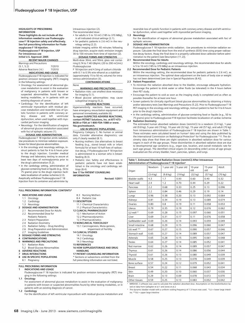
68 Imaging Life · June 2013 · www.siemens.com/imaginglife
Fludeoxyglucose F 18 Injection, USP
HIGHLIGHTS OF PRESCRIBING INFORMATIONThese highlights do not include all the information needed to use Fludeoxyglu-cose F 18 Injection safely and effectively. See full prescribing information for Flude-oxyglucose F 18 Injection.Fludeoxyglucose F 18 Injection, USPFor intravenous use Initial U.S. Approval: 2005
RECENT MAJOR CHANGESWarnings and Precautions (5.1, 5.2) 7/2010Adverse Reactions ( 6 ) 7/2010
INDICATIONS AND USAGEFludeoxyglucose F18 Injection is indicated for positron emission tomography (PET) imaging in the following settings: • Oncology: For assessment of abnormal glu-
cose metabolism to assist in the evaluation of malignancy in patients with known or suspected abnormalities found by other testing modalities, or in patients with an existing diagnosis of cancer.
• Cardiology: For the identification of left ventricular myocardium with residual glu-cose metabolism and reversible loss of sys-tolic function in patients with coronary ar-tery disease and left ventricular dysfunction, when used together with myo-cardial perfusion imaging.
• Neurology: For the identification of regions of abnormal glucose metabolism associated with foci of epileptic seizures (1).
DOSAGE AND ADMINISTRATIONFludeoxyglucose F 18 Injection emits radiation. Use procedures to minimize radiation exposure. Screen for blood glucose abnormalities. • In the oncology and neurology settings, in-
struct patients to fast for 4 to 6 hours prior to the drug’s injection. Consider medical therapy and laboratory testing to assure at least two days of normoglycemia prior to the drug’s administration (5.2).
• In the cardiology setting, administration of glucose-containing food or liquids (e.g., 50 to 75 grams) prior to the drug’s injection facili-tates localization of cardiac ischemia (2.3).
Aseptically withdraw Fludeoxyglucose F 18 Injection from its container and administer by
intravenous injection (2). The recommended dose: • for adults is 5 to 10 mCi (185 to 370 MBq),
in all indicated clinical settings (2.1). • for pediatric patients is 2.6 mCi in the neu-
rology setting (2.2).Initiate imaging within 40 minutes following drug injection; acquire static emission images 30 to 100 minutes from time of injection (2).
DOSAGE FORMS AND STRENGTHSMulti-dose 30mL and 50mL glass vial contai-ning 0.74 to 7.40 GBq/mL (20 to 200 mCi/mL) FludeoxyglucoseF 18 Injection and 4.5mg of sodium chloride with 0.1 to 0.5% w/w ethanol as a stabilizer (approximately 15 to 50 mL volume) for intra-venous administration (3).
CONTRAINDICATIONSNone
WARNINGS AND PRECAUTIONS• Radiation risks: use smallest dose necessary
for imaging (5.1). • Blood glucose adnormalities: may cause
suboptimal imaging (5.2).ADVERSE REACTIONS
Hypersensitivity reactions have occurred; have emergency resuscitation equipment and personnel immediately available (6). To report SUSPECTED ADVERSE REACTIONS, contact PETNET Solutions, Inc. at 877-473-8638 or FDA at 1-800-FDA-1088 or www.fda.gov/medwatch.
USE IN SPECIFIC POPULATIONSPregnancy Category C: No human or animal data. Consider alternative diagnostics; use only if clearly needed (8.1). • Nursing mothers: Use alternatives to breast
feeding (e.g., stored breast milk or infant formula) for at least 10 half-lives of radioac-tive decay, if Fludeoxyglucose F 18 Injection is administered to a woman who is breast-feeding (8.3).
• Pediatric Use: Safety and effectiveness in pediatric patients have not been estab-lished in the oncology and cardiology set-tings (8.4).
See 17 for PATIENT COUNSELING INFORMATION Revised: 1/2011
FULL PRESCRIBING INFORMATION: CONTENTS*
reversible loss of systolic function in patients with coronary artery disease and left ventricu-lar dysfunction, when used together with myocardial perfusion imaging.
1.3 Neurology For the identification of regions of abnormal glucose metabolism associated with foci of epileptic seizures.
2 DOSAGE AND ADMINISTRATION Fludeoxyglucose F 18 Injection emits radiation. Use procedures to minimize radiation ex-posure. Calculate the final dose from the end of synthesis (EOS) time using proper radioac-tive decay factors. Assay the final dose in a properly calibrated dose calibrator before admi-nistration to the patient [see Description (11.2)].
2.1 Recommended Dose for Adults Within the oncology, cardiology and neurology settings, the recommended dose for adults is 5 to 10 mCi (185 to 370 MBq) as an intravenous injection.
2.2 Recommended Dose for Pediatric PatientsWithin the neurology setting, the recommended dose for pediatric patients is 2.6 mCi, as an intravenous injection. The optimal dose adjustment on the basis of body size or weight has not been determined [see Use in Special Populations (8.4)].
2.3 Patient Preparation• To minimize the radiation absorbed dose to the bladder, encourage adequate hydration.
Encourage the patient to drink water or other fluids (as tolerated) in the 4 hours before their PET study.
• Encourage the patient to void as soon as the imaging study is completed and as often as possible thereafter for at least one hour.
• Screen patients for clinically significant blood glucose abnormalities by obtaining a history and/or laboratory tests [see Warnings and Precautions (5.2)]. Prior to Fludeoxyglucose F 18 PET imaging in the oncology and neurology settings, instruct patient to fast for 4 to 6 hours prior to the drug’s injection.
• In the cardiology setting, administration of glucose-containing food or liquids (e.g., 50 to 75 grams) prior to Fludeoxyglucose F18 Injection facilitates localization of cardiac ischemia
2.4 Radiation DosimetryThe estimated human absorbed radiation doses (rem/mCi) to a newborn (3.4 kg), 1-year old (9.8 kg), 5-year old (19 kg), 10-year old (32 kg), 15-year old (57 kg), and adult (70 kg) from intravenous administration of Fludeoxyglucose F 18 Injection are shown in Table 1. These estimates were calculated based on human2 data and using the data published by the International Commission on Radiological Protection4 for Fludeoxyglucose 18 F. The do-simetry data show that there are slight variations in absorbed radiation dose for various organs in each of the age groups. These dissimilarities in absorbed radiation dose are due to developmental age variations (e.g., organ size, location, and overall metabolic rate for each age group). The identified critical organs (in descending order) across all age groups evaluated are the urinary bladder, heart, pancreas, spleen, and lungs.
Table 1. Estimated Absorbed Radiation Doses (rem/mCi) After Intravenous Administration of Fludeoxyglucose F-18 Injectiona
Organ
Newborn 1-year old 5-year old 10-year old
15-year old
Adult
(3.4 kg) (9.8 kg) (19 kg) (32 kg) (57 kg) (70 kg)
Bladder wallb 4.3 1.7 0.93 0.60 0.40 0.32
Heart wall 2.4 1.2 0.70 0.44 0.29 0.22
Pancreas 2.2 0.68 0.33 0.25 0.13 0.096
Spleen 2.2 0.84 0.46 0.29 0.19 0.14
Lungs 0.96 0.38 0.20 0.13 0.092 0.064
Kidneys 0.81 0.34 0.19 0.13 0.089 0.074
Ovaries 0.80 0.8 0.19 0.11 0.058 0.053
Uterus 0.79 0.35 0.19 0.12 0.076 0.062
LLI wall * 0.69 0.28 0.15 0.097 0.060 0.051
Liver 0.69 0.31 0.17 0.11 0.076 0.058
Gallbladder wall 0.69 0.26 0.14 0.093 0.059 0.049
Small intestine 0.68 0.29 0.15 0.096 0.060 0.047
ULI wall ** 0.67 0.27 0.15 0.090 0.057 0.046
Stomach wall 0.65 0.27 0.14 0.089 0.057 0.047
Adrenals 0.65 0.28 0.15 0.095 0.061 0.048
Testes 0.64 0.27 0.14 0.085 0.052 0.041
Red marrow 0.62 0.26 0.14 0.089 0.057 0.047
Thymus 0.61 0.26 0.14 0.086 0.056 0.044
Thyroid 0.61 0.26 0.13 0.080 0.049 0.039
Muscle 0.58 0.25 0.13 0.078 0.049 0.039
Bone surface 0.57 0.24 0.12 0.079 0.052 0.041
Breast 0.54 0.22 0.11 0.068 0.043 0.034
Skin 0.49 0.20 0.10 0.060 0.037 0.030
Brain 0.29 0.13 0.09 0.078 0.072 0.070
Other tissues 0.59 0.25 0.13 0.083 0.052 0.042a MIRDOSE 2 software was used to calculate the radiation absorbed dose. Assumptions on the biodistribution ba-
sed on data from Gallagher et al.1 and Jones et al.2 b The dynamic bladder model with a uniform voiding frequency of 1.5 hours was used. *LLI = lower large intesti-
ne; **ULI = upper large intestine
1 INDICATIONS AND USAGE1.1 Oncology1.2 Cardiology1.3 Neurology
2 DOSAGE AND ADMINISTRATION2.1 Recommended Dose for Adults2.2 Recommended Dose for Pediatric Patients2.3 Patient Preparation2.4 Radiation Dosimetry2.5 Radiation Safety – Drug Handling2.6 Drug Preparation and Administration2.7 Imaging Guidelines
3 DOSAGE FORMS AND STRENGTHS4 CONTRAINDICATIONS5 WARNINGS AND PRECAUTIONS
5.1 Radiation Risks5.2 Blood Glucose Abnormalities
6 ADVERSE REACTIONS7 DRUG INTERACTIONS8 USE IN SPECIFIC POPULATIONS
8.1 Pregnancy
8.3 Nursing Mothers8.4 Pediatric Use
11 DESCRIPTION11.1 Chemical Characteristics11.2 Physical Characteristics
12 CLINICAL PHARMACOLOGY12.1 Mechanism of Action12.2 Pharmacodynamics12.3 Pharmacokinetics
13 NONCLINICAL TOXICOLOGY13.1 Carcinogenesis, Muta-genesis, Impairment of Fertility
14 CLINICAL STUDIES14.1 Oncology14.2 Cardiology14.3 Neurology
15 REFERENCES16 HOW SUPPLIED/STORAGE AND DRUG
HANDLING17 PATIENT COUNSELING INFORMATION* Sections or subsections omitted from the full prescribing information are not listed.
FULL PRESCRIBING INFORMATION1 INDICATIONS AND USAGE
Fludeoxyglucose F 18 Injection is indicated for positron emission tomography (PET) ima-ging in the following settings:
1.1 Oncology For assessment of abnormal glucose metabolism to assist in the evaluation of malignancy in patients with known or suspected abnormalities found by other testing modalities, or in patients with an existing diagnosis of cancer.
1.2 Cardiology For the identification of left ventricular myocardium with residual glucose metabolism and

Imaging Life · June 2013 · www.siemens.com/imaginglife 69
Fludeoxyglucose F 18 Injection, USP
2.5 Radiation Safety – Drug Handling• Use waterproof gloves, effective radiation shielding, and appropriate safety measures
when handling Fludeoxyglucose F 18 Injection to avoid unnecessary radiation exposure to the patient, occupational workers, clinical personnel and other persons.
• Radiopharmaceuticals should be used by or under the control of physicians who are quali-fied by specific training and experience in the safe use and handling of radionuclides, and whose experience and training have been approved by the appropriate governmental agency authorized to license the use of radionuclides.
• Calculate the final dose from the end of synthesis (EOS) time using proper radioactive de-cay factors. Assay the final dose in a properly calibrated dose calibrator before administra-tion to the patient [see Description (11.2)].
• The dose of Fludeoxyglucose F 18 used in a given patient should be minimized consistent with the objectives of the procedure, and the nature of the radiation detection devices em-ployed.
2.6 Drug Preparation and Administration• Calculate the necessary volume to administer based on calibration time and dose. • Aseptically withdraw Fludeoxyglucose F 18 Injection from its container. • Inspect Fludeoxyglucose F 18 Injection visually for particulate matter and discoloration be-
fore administration, whenever solution and container permit. • Do not administer the drug if it contains particulate matter or discoloration; dispose of
these unacceptable or unused preparations in a safe manner, in compliance with applicable regulations.
• Use Fludeoxyglucose F 18 Injection within 12 hours from the EOS. 2.7 Imaging Guidelines
• Initiate imaging within 40 minutes following Fludeoxyglucose F 18 Injection administration. • Acquire static emission images 30 to 100 minutes from the time of injection.
3 DOSAGE FORMS AND STRENGTHS Multiple-dose 30 mL and 50 mL glass vial containing 0.74 to 7.40 GBq/mL (20 to 200 mCi/mL) of Fludeoxyglucose F 18 Injection and 4.5 mg of sodium chloride with 0.1 to 0.5% w/w ethanol as a stabilizer (approximately 15 to 50 mL volume) for intravenous administration.
4 CONTRAINDICATIONS None
5 WARNINGS AND PRECAUTIONS 5.1 Radiation Risks
Radiation-emitting products, including Fludeoxyglucose F 18 Injection, may increase the risk for cancer, especially in pediatric patients. Use the smallest dose necessary for imaging and ensure safe handling to protect the patient and health care worker [see Dosage and Administration (2.5)].
5.2 Blood Glucose Abnormalities In the oncology and neurology setting, suboptimal imaging may occur in patients with ina-dequately regulated blood glucose levels. In these patients, consider medical therapy and laboratory testing to assure at least two days of normoglycemia prior to Fludeoxyglucose F 18 Injection administration.
6 ADVERSE REACTIONS Hypersensitivity reactions with pruritus, edema and rash have been reported in the post-marketing setting. Have emergency resuscitation equipment and personnel immediately available.
7 DRUG INTERACTIONS The possibility of interactions of Fludeoxyglucose F 18 Injection with other drugs taken by patients undergoing PET imaging has not been studied.
8 USE IN SPECIFIC POPULATIONS8.1 Pregnancy
Pregnancy Category C Animal reproduction studies have not been conducted with Fludeoxyglucose F 18 Injec-tion. It is also not known whether Fludeoxyglucose F 18 Injection can cause fetal harm when administered to a pregnant woman or can affect reproduction capacity. Consider al-ternative diagnostic tests in a pregnant woman; administer Fludeoxyglucose F 18 Injection only if clearly needed.
8.3 Nursing MothersIt is not known whether Fludeoxyglucose F 18 Injection is excreted in human milk. Consider alternative diagnostic tests in women who are breast-feeding. Use alternatives to breast feeding (e.g., stored breast milk or infant formula) for at least 10 half-lives of radioactive decay, if Fludeoxyglucose F 18 Injection is administered to a woman who is breast-feeding.
8.4 Pediatric Use The safety and effectiveness of Fludeoxyglucose F 18 Injection in pediatric patients with epilepsy is established on the basis of studies in adult and pediatric patients. In pediatric patients with epilepsy, the recommended dose is 2.6 mCi. The optimal dose adjustment on the basis of body size or weight has not been determined. In the oncology or cardiology settings, the safety and effectiveness of Fludeoxyglucose F 18 Injection have not been esta-blished in pediatric patients.
11 DESCRIPTION 11.1 Chemical Characteristics
Fludeoxyglucose F 18 Injection is a positron emitting radiopharmaceutical that is used for diagnostic purposes in conjunction with positron emission tomography (PET) imaging. The active ingredient 2-deoxy-2-[18F]fluoro-D-glucose has the molecular formula of C6H1118FO5 with a molecular weight of 181.26, and has the following chemical structure:
Fludeoxyglucose F 18 Injection is provided as a ready to use sterile, pyrogen free, clear, co-lorless solution. Each mL contains between 0.740 to 7.40GBq (20.0 to 200 mCi) of 2-deo-
xy-2-[18F]fluoro-D-glucose at the EOS, 4.5 mg of sodium chloride and 0.1 to 0.5% w/w ethanol as a stabilizer. The pH of the solution is between 4.5 and 7.5. The solution is packa-ged in a multiple-dose glass vial and does not contain any preservative.
11.2 Physical Characteristics Fluorine F 18 decays by emitting positron to Oxygen O 16 (stable) and has a physical half-life of 109.7 minutes. The principal photons useful for imaging are the dual 511 keV gamma photons, that are produced and emitted simultaneously in opposite direction when the po-sitron interacts with an electron (Table 2).
Table 2. Pricipal Radiation Emission Data for Fluorine F18
Radiation/Emission % Per Disintegration Mean Energy
Positron (b+) 96.73 249.8 keV
Gamma (±)* 193.46 511.0 keV
*Produced by positron annihilation From: Kocher, D.C. Radioactive Decay Tables DOE/TIC-I 1026, 89 (1981)The specific gamma ray constant (point source air kerma coefficient) for fluorine F 18 is 5.7 R/hr/mCi (1.35 x 10⁻⁶ Gy/hr/kBq) at 1 cm. The half-value layer (HVL) for the 511 keV photons is 4 mm lead (Pb). The range of attenuation coefficients for this radionuclide as a function of lead shield thickness is shown in Table 3. For example, the interposition of an 8 mm thickness of Pb, with a coefficient of attenuation of 0.25, will decrease the external radiation by 75%.
Table 3. Radiation Attenuation of 511 keV Photons by lead (Pb) shielding
Shield thickness (Pb) mm Coefficient of attenuation
0 0.00
4 0.50
8 0.25
13 0.10
26 0.01
39 0.001
52 0.0001
For use in correcting for physical decay of this radionuclide, the fractions remaining at selected intervals after calibration are shown in Table 4.
Table 4. Physical Decay Chart for Fluorine F18
Minutes Fraction Remaining
0* 1.000
15 0.909
30 0.826
60 0.683
110 0.500
220 0.250
*calibration time
12 CLINICAL PHARMACOLOGY 12.1 Mechanism of Action
Fludeoxyglucose F 18 is a glucose analog that concentrates in cells that rely upon glucose as an energy source, or in cells whose dependence on glucose increases under pathophy-siological conditions. Fludeoxyglucose F 18 is transported through the cell membrane by facilitative glucose transporter proteins and is phosphorylated within the cell to [18F] FDG-6-phosphate by the enzyme hexokinase. Once phosphorylated it cannot exit until it is de-phosphorylated by glucose-6-phosphatase. Therefore, within a given tissue or pathophy-siological process, the retention and clearance of Fludeoxyglucose F 18 reflect a balance involving glucose transporter, hexokinase and glucose-6-phosphatase activities. When allo-wance is made for the kinetic differences between glucose and Fludeoxyglucose F 18 trans-port and phosphorylation (expressed as the ‚‘lumped constant‘‘ ratio), Fludeoxyglucose F 18 is used to assess glucose metabolism. In comparison to background activity of the specific organ or tissue type, regions of decrea-sed or absent uptake of Fludeoxyglucose F 18 reflect the decrease or absence of glucose metabolism. Regions of increased uptake of Fludeoxyglucose F 18 reflect greater than nor-mal rates of glucose metabolism.
12.2 Pharmacodynamics Fludeoxyglucose F 18 Injection is rapidly distributed to all organs of the body after intrave-nous administration. After background clearance of Fludeoxyglucose F 18 Injection, opti-mal PET imaging is generally achieved between 30 to 40 minutes after administration. In cancer, the cells are generally characterized by enhanced glucose metabolism partially due to (1) an increase in activity of glucose transporters, (2) an increased rate of phospho-rylation activity, (3) a reduction of phosphatase activity or, (4) a dynamic alteration in the balance among all these processes. However, glucose metabolism of cancer as reflected by Fludeoxyglucose F 18 accumulation shows considerable variability. Depending on tumor type, stage, and location, Fludeoxyglucose F 18 accumulation may be increased, normal, or decreased. Also, inflammatory cells can have the same variability of uptake of Fludeoxy-glucose F 18.In the heart, under normal aerobic conditions, the myocardium meets the bulk of its ener-gy requirements by oxidizing free fatty acids. Most of the exogenous glucose taken up by the myocyte is converted into glycogen. However, under ischemic conditions, the oxidation of free fatty acids decreases, exogenous glucose becomes the preferred myocardial sub-
23
56
91011
1314
Hashmark guide for AMERISOURCEAMERISOURCE.
1. Bars 1-3 are always white2. Start bar is bar #43. Code is numbered item followed by letter of alphabet. Ex: 01N4. Use Hashmark Bar Code Guide to create bar
Code 93R - McKESSON
LIGHT DARK1 1 12 1 23 1 34 2 15 2 26 2 37 3 18 3 29 3 30 1 4
A=01B=12C=23D=34E=45F=56G=67H=78I=89J=00K=11L=22M=33
N=44O=55P=66Q=77R=88S=99T=10U=21V=32W=43X=54Y=65Z=76
HASHMARK BAR CODE GUIDE

70 Imaging Life · June 2013 · www.siemens.com/imaginglife
Fludeoxyglucose F 18 Injection, USP
strate, glycolysis is stimulated, and glucose taken up by the myocyte is metabolized imme-diately instead of being converted into glycogen. Under these conditions, phosphorylated Fludeoxyglucose F 18 accumulates in the myocyte and can be detected with PET imaging. In the brain, cells normally rely on aerobic metabolism. In epilepsy, the glucose metabolism varies. Generally, during a seizure, glucose metabolism increases. Interictally, the seizure focus tends to be hypometabolic.
12.3 Pharmacokinetics Distribution: In four healthy male volunteers, receiving an intravenous administration of 30 seconds in duration, the arterial blood level profile for Fludeoxyglucose F 18 decayed triex-ponentially. The effective half-life ranges of the three phases were 0.2 to 0.3 minutes, 10 to 13 minutes with a mean and standard deviation (STD) of 11.6 (±) 1.1 min, and 80 to 95 minutes with a mean and STD of 88 (±) 4 min. Plasma protein binding of Fludeoxyglucose F 18 has not been studied. Metabolism: Fludeoxyglucose F 18 is transported into cells and phosphorylated to [18F]-FDG-6- phosphate at a rate proportional to the rate of glucose utilization within that tissue. [F18]-FDG-6-phosphate presumably is metabolized to 2-deoxy-2-[F18]fluoro-6-phospho-D-mannose([F 18]FDM-6-phosphate). Fludeoxyglucose F 18 Injection may contain several impurities (e.g., 2-deoxy-2-chloro-D-glucose (ClDG)). Biodistribution and metabolism of ClDG are presumed to be similar to Flu-deoxyglucose F 18 and would be expected to result in intracellular formation of 2-deoxy-2-chloro-6-phospho-D-glucose (ClDG-6-phosphate) and 2-deoxy-2-chloro-6-phospho-D-mannose (ClDM-6-phosphate). The phosphorylated deoxy-glucose compounds are dephosphorylated and the resulting compounds (FDG, FDM, ClDG, and ClDM) presumably leave cells by passive diffusion. Fludeoxyglucose F 18 and related compounds are cleared from non-cardiac tissues within 3 to 24 hours after administration. Clearance from the cardiac tissue may require more than 96 hours. Fludeoxyglucose F 18 that is not involved in glucose metabolism in any tissue is then excreted in the urine. Elimination: Fludeoxyglucose F 18 is cleared from most tissues within 24 hours and can be eliminated from the body unchanged in the urine. Three elimination phases have been identified in the reviewed literature. Within 33 minutes, a mean of 3.9% of the administra-ted radioactive dose was measured in the urine. The amount of radiation exposure of the urinary bladder at two hours post-administration suggests that 20.6% (mean) of the radio-active dose was present in the bladder. Special Populations: The pharmacokinetics of Fludeoxyglucose F 18 Injection have not been studied in renally-impaired, hepatically impaired or pediatric patients. Fludeoxyglucose F 18 is eliminated through the renal system. Avoid excessive radiation exposure to this organ system and adjacent tissues. The effects of fasting, varying blood sugar levels, conditions of glucose intolerance, and diabetes mellitus on Fludeoxyglucose F 18 distribution in humans have not been ascer-tained [see Warnings and Precautions (5.2)].
13 NONCLINICAL TOXICOLOGY 13.1 Carcinogenesis, Mutagenesis, Impairment of Fertility
Animal studies have not been performed to evaluate the Fludeoxyglucose F 18 Injection carcinogenic potential, mutagenic potential or effects on fertility.
14 CLINICAL STUDIES 14.1 Oncology
The efficacy of Fludeoxyglucose F 18 Injection in positron emission tomography cancer imaging was demonstrated in 16 independent studies. These studies prospectively evalua-ted the use of Fludeoxyglucose F 18 in patients with suspected or known malignancies, in-cluding non-small cell lung cancer, colo-rectal, pancreatic, breast, thyroid, melanoma, Hodgkin‘s and non-Hodgkin‘s lymphoma, and various types of metastatic cancers to lung, liver, bone, and axillary nodes. All these studies had at least 50 patients and used patholo-gy as a standard of truth. The Fludeoxyglucose F 18 Injection doses in the studies ranged from 200 MBq to 740 MBq with a median and mean dose of 370 MBq. In the studies, the diagnostic performance of Fludeoxyglucose F 18 Injection varied with the type of cancer, size of cancer, and other clinical conditions. False negative and false positive scans were observed. Negative Fludeoxyglucose F 18 Injection PET scans do not exclude the diagnosis of cancer. Positive Fludeoxyglucose F 18 Injection PET scans can not replace pathology to establish a diagnosis of cancer. Non-malignant conditions such as fungal infections, inflammatory processes and benign tumors have patterns of increased glucose metabolism that may give rise to false-positive scans. The efficacy of Fludeoxyglu-cose F 18 Injection PET imaging in cancer screening was not studied.
14.2 Cardiology The efficacy of Fludeoxyglucose F 18 Injection for cardiac use was demonstrated in ten in-dependent, prospective studies of patients with coronary artery disease and chronic left ventricular systolic dysfunction who were scheduled to undergo coronary revasculariza-tion. Before revascularization, patients underwent PET imaging with Fludeoxyglucose F 18 Injection (74 to 370 MBq, 2 to 10 mCi) and perfusion imaging with other diagnostic radio-pharmaceuticals. Doses of Fludeoxyglucose F 18 Injection ranged from 74 to 370 MBq (2 to 10 mCi). Segmental, left ventricular, wall-motion assessments of asynergic areas made before revascularization were compared in a blinded manner to assessments made after successful revascularization to identify myocardial segments with functional recovery. Left ventricular myocardial segments were predicted to have reversible loss of systolic func-tion if they showed Fludeoxyglucose F 18 accumulation and reduced perfusion (i.e., flow-metabolism mismatch). Conversely, myocardial segments were predicted to have irreversi-ble loss of systolic function if they showed reductions in both Fludeoxyglucose F 18 accumulation and perfusion (i.e., matched defects). Findings of flow-metabolism mismatch in a myocardial segment may suggest that success-ful revascularization will restore myocardial function in that segment. However, false-posi-tive tests occur regularly, and the decision to have a patient undergo revascularization should not be based on PET findings alone. Similarly, findings of a matched defect in a myocardial segment may suggest that myocardial function will not recover in that seg-ment, even if it is successfully revascularized. However, false-negative tests occur regular-ly, and the decision to recommend against coronary revascularization, or to recommend a cardiac transplant, should not be based on PET findings alone. The reversibility of segmen-
tal dysfunction as predicted with Fludeoxyglucose F 18 PET imaging depends on successful coronary revascularization. Therefore, in patients with a low likelihood of successful revas-cularization, the diagnostic usefulness of PET imaging with Fludeoxyglucose F 18 Injection is more limited.
14.3 Neurology In a prospective, open label trial, Fludeoxyglucose F 18 Injection was evaluated in 86 pati-ents with epilepsy. Each patient received a dose of Fludeoxyglucose F 18 Injection in the range of 185 to 370 MBq (5 to 10 mCi). The mean age was 16.4 years (range: 4 months to 58 years; of these, 42 patients were less than 12 years and 16 patients were less than 2 years old). Patients had a known diagnosis of complex partial epilepsy and were under evaluation for surgical treatment of their seizure disorder. Seizure foci had been previously identified on ictal EEGs and sphenoidal EEGs. Fludeoxyglucose F 18 Injection PET imaging confirmed previous diagnostic findings in 16% (14/87) of the patients; in 34% (30/87) of the patients, Fludeoxyglucose F 18 Injection PET images provided new findings. In 32% (27/87), imaging with Fludeoxyglucose F 18 Injection was inconclusive. The impact of the-se imaging findings on clinical outcomes is not known. Several other studies comparing imaging with Fludeoxyglucose F 18 Injection results to subsphenoidal EEG, MRI and/or surgical findings supported the concept that the degree of hypometabolism corresponds to areas of confirmed epileptogenic foci. The safety and ef-fectiveness of Fludeoxyglucose F 18 Injection to distinguish idiopathic epileptogenic foci from tumors or other brain lesions that may cause seizures have not been established.
15 REFERENCES1. Gallagher B.M., Ansari A., Atkins H., Casella V., Christman D.R., Fowler J.S., Ido T., Mac-
Gregor R.R., Som P., Wan C.N., Wolf A.P., Kuhl D.E., and Reivich M. “Radiopharmaceuticals XXVII. 18F-labeled 2-deoxy-2-fluoro-d-glucose as a radiopharmaceutical for measuring re-gional myocardial glucose metabolism in vivo: tissue distribution and imaging studies in animals,” J Nucl Med, 1977; 18, 990-6.
2. Jones S.C., Alavi, A., Christman D., Montanez, I., Wolf, A.P., and Reivich M. “The radiation dosimetry of 2 [F-18] fluoro-2-deoxy-D-glucose in man,” J Nucl Med, 1982; 23, 613-617.
3. Kocher, D.C. “Radioactive Decay Tables: A handbook of decay data for application to radia-tion dosimetry and radiological assessments,” 1981, DOE/TIC-I 1026, 89.
4. ICRP Publication 53, Volume 18, No. l-4,1987, pages 75-76. 16 HOW SUPPLIED/STORAGE AND DRUG HANDLING
Fludeoxyglucose F 18 Injection is supplied in a multi-dose, capped 30 mL and 50 mL glass vial containing between 0.740 to 7.40 GBq/mL (20 to 200 mCi/mL), of no carrier added 2-deoxy-2-[F 18] fluoro-D-glucose, at end of synthesis, in approximately 15 to 50 mL. The contents of each vial are sterile, pyrogen-free and preservative-free. NDC 40028-511-30; 40028-511-50 Receipt, transfer, handling, possession, or use of this product is subject to the radioactive material regulations and licensing requirements of the U.S. Nuclear Regulatory Commissi-on, Agreement States or Licensing States as appropriate.Store the Fludeoxyglucose F 18 Injection vial upright in a lead shielded container at 25°C (77°F); excursions permitted to 15-30°C (59-86°F). Store and dispose of Fludeoxyglucose F 18 Injection in accordance with the regulations and a general license, or its equivalent, of an Agreement State or a Licensing State. The expiration date and time are provided on the container label. Use Fludeoxyglucose F 18 Injection within 12 hours from the EOS time.
17 PATIENT COUNSELING INFORMATIONInstruct patients in procedures that increase renal clearance of radioactivity. Encourage patients to:
• drink water or other fluids (as tolerated) in the 4 hours before their PET study. • void as soon as the imaging study is completed and as often as possible thereafter for at
least one hour.
Manufactured by: PETNET Solutions Inc. 810 Innovation Drive Knoxville, TN 37932Distributed by: PETNET Solutions Inc. 810 Innovation Drive Knoxville, TN 37932
PN0002262 Rev. AMarch 1, 2011


On account of certain regional limitations of sales rights and service availability, we cannot guarantee that all products included in this brochure are available through the Siemens sales organization worldwide. Availability and packaging may vary by country and is subject to change without prior notice. Some/All of the features and products described herein may not be available in the United States.
The information in this document contains general technical descriptions of specifications and options as well as standard and optional features which do not always have to be present in individual cases.
Siemens reserves the right to modify the design, packaging, specifications and options described herein without prior notice.
Please contact your local Siemens sales representative for the most current information.
Note: Any technical data contained in this document may vary within defined tolerances. Original images always lose a certain amount of detail when reproduced.
www.siemens.com/healthcare-magazine
Global Business Unit
Siemens Medical Solutions USA, Inc.Molecular Imaging2501 N. Barrington RoadHoffman Estates, IL 60192USATelephone: + 1 847 304-7700www.siemens.com/mi
Local Contact Information
Asia/Pacific:Siemens Medical SolutionsAsia Pacific HeadquartersThe Siemens Center60 MacPherson RoadSingapore 348615Phone: +65 9622 - 2026 www.siemens.com/healthcare
Canada:Siemens Canada LimitedMedical Solutions2185 Derry Road WestMississauga ON L5N 7A6CanadaPhone: +1 905 819 - 5800 www.siemens.com/healthcare
Europe/Africa/Middle East:Siemens AGMedical SolutionsHenkestraße 127D-91052 ErlangenGermanyPhone: +49 9131 84 - 0 www.siemens.com/healthcare
Latin America:Siemens S.A.Medical SolutionsAvenida de Pte. Julio A. Roca No 516, Piso 7C1067ABN Buenos Aires ArgentinaPhone: +54 11 4340 - 8400 www.siemens.com/healthcare
USA:Siemens Medical Solutions USA, Inc.51 Valley Stream ParkwayMalvern, PA 19355-1406USAPhone: +1-888-826 - 9702 www.siemens.com/healthcare
Global SiemensHealthcare Headquarters
Siemens AGHealthcare SectorHenkestraße 12791052 ErlangenGermanyPhone: +49 9131 84 - 0www.siemens.com/healthcare
Global Siemens Headquarters
Siemens AGWittelsbacherplatz 280333 MuenchenGermany
MI-1868.RM.360.PDF Only | © 03.2015, Siemens AG