Embed Size (px)
Citation preview
IMAGING
INDICATIONGUIDELINES
Your partner in outpatientradiology
2 | IMAGING INDICATION GUIDELINES
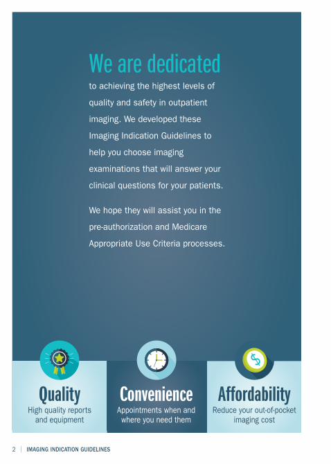
We are dedicatedto achieving the highest levels of
quality and safety in outpatient
imaging. We developed these
Imaging Indication Guidelines to
help you choose imaging
examinations that will answer your
clinical questions for your patients.
We hope they will assist you in the
pre-authorization and Medicare
Appropriate Use Criteria processes.
QualityHigh quality reportsand equipment
ConvenienceAppointments when andwhere you need them
AffordabilityReduce your out-of-pocket
imaging cost

IMAGING INDICATION GUIDELINES | 3
Notes

4 | IMAGING INDICATION GUIDELINES
Notes
IMAGING INDICATION GUIDELINES | 5
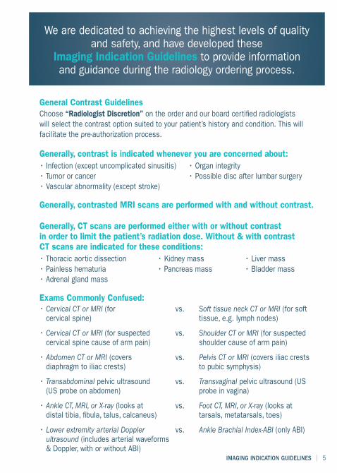
General Contrast GuidelinesChoose “Radiologist Discretion” on the order and our board certified radiologistswill select the contrast option suited to your patient’s history and condition. This willfacilitate the pre-authorization process.
Generally, contrast is indicated whenever you are concerned about:• Infection (except uncomplicated sinusitis) • Organ integrity• Tumor or cancer • Possible disc after lumbar surgery• Vascular abnormality (except stroke)
Generally, contrasted MRI scans are performed with and without contrast.
Generally, CT scans are performed either with or without contrastin order to limit the patient’s radiation dose. Without & with contrastCT scans are indicated for these conditions:• Thoracic aortic dissection • Kidney mass • Liver mass• Painless hematuria • Pancreas mass • Bladder mass• Adrenal gland mass
Exams Commonly Confused:• Cervical CT or MRI (forcervical spine)
• Cervical CT or MRI (for suspectedcervical spine cause of arm pain)
• Abdomen CT or MRI (coversdiaphragm to iliac crests)
• Transabdominal pelvic ultrasound(US probe on abdomen)
• Ankle CT, MRI, or X-ray (looks atdistal tibia, fibula, talus, calcaneus)
• Lower extremity arterial Dopplerultrasound (includes arterial waveforms& Doppler, with or without ABI)
vs. Soft tissue neck CT or MRI (for softtissue, e.g. lymph nodes)
vs. Shoulder CT or MRI (for suspectedshoulder cause of arm pain)
vs. Pelvis CT or MRI (covers iliac creststo pubic symphysis)
vs. Transvaginal pelvic ultrasound (USprobe in vagina)
vs. Foot CT, MRI, or X-ray (looks attarsals, metatarsals, toes)
vs. Ankle Brachial Index-ABI (only ABI)
We are dedicated to achieving the highest levels of qualityand safety, and have developed these
Imaging Indication Guidelines to provide informationand guidance during the radiology ordering process.
6 | IMAGING INDICATION GUIDELINES
The information provided in this guide is not intended to be a substitute for a licensed radiologist’s recommendation. The material provided is strictlyan informative guideline for the most probable scan ordered. Specific questions should be directed to the radiologist or the imaging technologist. Ourradiologists reserve the right to recommend an alternative exam based on the patient clinical history and diagnosis provided by the ordering provider.
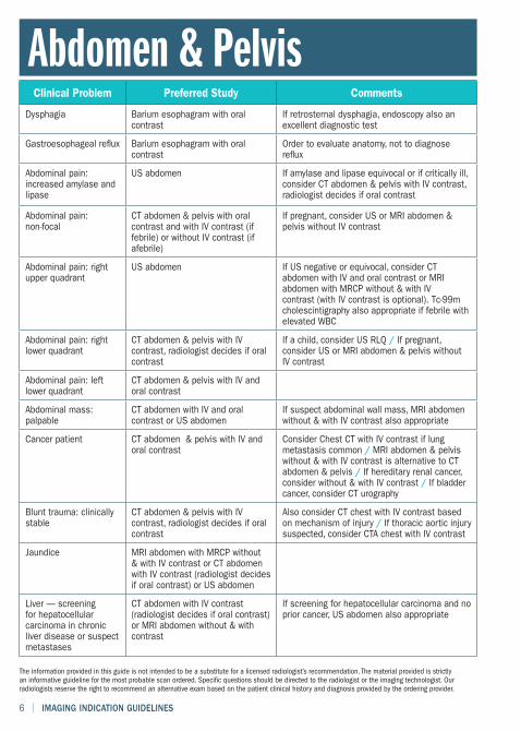
Clinical Problem Preferred Study Comments
Dysphagia Barium esophagram with oralcontrast
If retrosternal dysphagia, endoscopy also anexcellent diagnostic test
Gastroesophageal reflux Barium esophagram with oralcontrast
Order to evaluate anatomy, not to diagnosereflux
Abdominal pain:increased amylase andlipase
US abdomen If amylase and lipase equivocal or if critically ill,consider CT abdomen & pelvis with IV contrast,radiologist decides if oral contrast
Abdominal pain:non-focal
CT abdomen & pelvis with oralcontrast and with IV contrast (iffebrile) or without IV contrast (ifafebrile)
If pregnant, consider US or MRI abdomen &pelvis without IV contrast
Abdominal pain: rightupper quadrant
US abdomen If US negative or equivocal, consider CTabdomen with IV and oral contrast or MRIabdomen with MRCP without & with IVcontrast (with IV contrast is optional). Tc-99mcholescintigraphy also appropriate if febrile withelevated WBC
Abdominal pain: rightlower quadrant
CT abdomen & pelvis with IVcontrast, radiologist decides if oralcontrast
If a child, consider US RLQ / If pregnant,consider US or MRI abdomen & pelvis withoutIV contrast
Abdominal pain: leftlower quadrant
CT abdomen & pelvis with IV andoral contrast
Abdominal mass:palpable
CT abdomen with IV and oralcontrast or US abdomen
If suspect abdominal wall mass, MRI abdomenwithout & with IV contrast also appropriate
Cancer patient CT abdomen & pelvis with IV andoral contrast
Consider Chest CT with IV contrast if lungmetastasis common / MRI abdomen & pelviswithout & with IV contrast is alternative to CTabdomen & pelvis / If hereditary renal cancer,consider without & with IV contrast / If bladdercancer, consider CT urography
Blunt trauma: clinicallystable
CT abdomen & pelvis with IVcontrast, radiologist decides if oralcontrast
Also consider CT chest with IV contrast basedon mechanism of injury / If thoracic aortic injurysuspected, consider CTA chest with IV contrast
Jaundice MRI abdomen with MRCP without& with IV contrast or CT abdomenwith IV contrast (radiologist decidesif oral contrast) or US abdomen
Liver — screeningfor hepatocellularcarcinoma in chronicliver disease or suspectmetastases
CT abdomen with IV contrast(radiologist decides if oral contrast)or MRI abdomen without & withcontrast
If screening for hepatocellular carcinoma and noprior cancer, US abdomen also appropriate
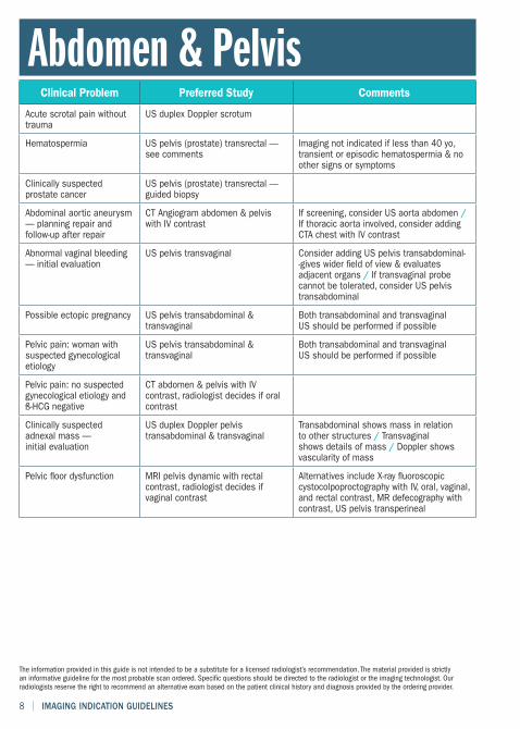
Abdomen & Pelvis
IMAGING INDICATION GUIDELINES | 7
The information provided in this guide is not intended to be a substitute for a licensed radiologist’s recommendation. The material provided is strictlyan informative guideline for the most probable scan ordered. Specific questions should be directed to the radiologist or the imaging technologist. Ourradiologists reserve the right to recommend an alternative exam based on the patient clinical history and diagnosis provided by the ordering provider.
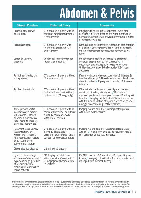
Abdomen & PelvisClinical Problem Preferred Study Comments
Suspect small bowelobstruction
CT abdomen & pelvis with IVcontrast, radiologist decidesif oral contrast
If high-grade obstruction suspected, avoid oralcontrast / If intermittent or low-grade obstructionsuspected, consider CT or MRI enteroclysis (neutralcontrast by NG tube)
Crohn’s disease CT abdomen & pelvis withIV and oral contrast or CTenterography
Consider MRI enterography if nonacute presentationor a child / Enterography uses neutral contrast bymouth (enteroclysis uses neutral contrast by NGtube)
Upper or Lower GIbleeding
Endoscopy is recommendedrather than imaging
If endoscopy negative or cannot be performed,consider angiography (CT or catheter) / Ifendoscopy and angiography negative for lowerGI bleeding, consider 99mTc-labeled RBC scanabdomen
Painful hematuria, r/okidney stone
CT abdomen & pelvis withoutIV or oral contrast
If recurrent stone disease, consider US kidneys &bladder with X-ray KUB to decrease overall radiationdose to patient / If pregnant, consider US kidneys& bladder
Painless hematuria CT abdomen & pelvis withoutand with IV contrast, withoutoral contrast (CT urography)
If hematuria due to renal parenchymal disease,consider US kidneys & bladder / If child andmacroscopic hematuria or proteinuria, US kidneys &bladder / Imaging not indicated if hematuria clearswith therapy, cessation of vigorous exercise or afterurologic procedure (e.g. catheterization)
Acute pyelonephritisin complicated patient(eg, diabetes, stones,prior renal surgery, notresponding to therapy,immunocompromised)
CT abdomen & pelvis with IVcontrast (preferred) or without& with IV contrast—bothwithout oral contrast
Imaging not indicated for uncomplicated patientwith acute pyelonephritis
Recurrent lower urinarytract infections inwomen with frequentreinfections, risk factorsor no response toconventional therapy
CT abdomen & pelvis without& with IV contrast (CTUrogram), oral contrast only ifsuspect enterovesical fistula
Imaging not indicated for uncomplicated patientwith UTI / If child with atypical or recurrent febrileUTI, consider US kidneys & bladder
Chronic kidney disease US kidneys & bladder
Hypertension — highsuspicion of renovascularhypertension (e.g. failureof medical therapy,progressive renal failure,young patient)
MR Angiogram abdomenwithout & with IV contrast orCT Angiogram abdomen withIV contrast
If eGFR less than 30, consider US duplex Dopplerkidney / Imaging not indicated for hypertension wellmanaged with medical therapy
8 | IMAGING INDICATION GUIDELINES
The information provided in this guide is not intended to be a substitute for a licensed radiologist’s recommendation. The material provided is strictlyan informative guideline for the most probable scan ordered. Specific questions should be directed to the radiologist or the imaging technologist. Ourradiologists reserve the right to recommend an alternative exam based on the patient clinical history and diagnosis provided by the ordering provider.
Clinical Problem Preferred Study Comments
Acute scrotal pain withouttrauma
US duplex Doppler scrotum
Hematospermia US pelvis (prostate) transrectal —see comments
Imaging not indicated if less than 40 yo,transient or episodic hematospermia & noother signs or symptoms
Clinically suspectedprostate cancer
US pelvis (prostate) transrectal —guided biopsy
Abdominal aortic aneurysm— planning repair andfollow-up after repair
CT Angiogram abdomen & pelviswith IV contrast
If screening, consider US aorta abdomen /If thoracic aorta involved, consider addingCTA chest with IV contrast
Abnormal vaginal bleeding— initial evaluation
US pelvis transvaginal Consider adding US pelvis transabdominal--gives wider field of view & evaluatesadjacent organs / If transvaginal probecannot be tolerated, consider US pelvistransabdominal
Possible ectopic pregnancy US pelvis transabdominal &transvaginal
Both transabdominal and transvaginalUS should be performed if possible
Pelvic pain: woman withsuspected gynecologicaletiology
US pelvis transabdominal &transvaginal
Both transabdominal and transvaginalUS should be performed if possible
Pelvic pain: no suspectedgynecological etiology andß-HCG negative
CT abdomen & pelvis with IVcontrast, radiologist decides if oralcontrast
Clinically suspectedadnexal mass —initial evaluation
US duplex Doppler pelvistransabdominal & transvaginal
Transabdominal shows mass in relationto other structures / Transvaginalshows details of mass / Doppler showsvascularity of mass
Pelvic floor dysfunction MRI pelvis dynamic with rectalcontrast, radiologist decides ifvaginal contrast
Alternatives include X-ray fluoroscopiccystocolpoproctography with IV, oral, vaginal,and rectal contrast, MR defecography withcontrast, US pelvis transperineal
Abdomen & Pelvis
IMAGING INDICATION GUIDELINES | 9
The information provided in this guide is not intended to be a substitute for a licensed radiologist’s recommendation. The material provided is strictlyan informative guideline for the most probable scan ordered. Specific questions should be directed to the radiologist or the imaging technologist. Ourradiologists reserve the right to recommend an alternative exam based on the patient clinical history and diagnosis provided by the ordering provider.
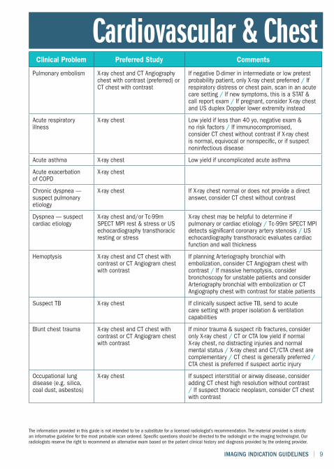
Clinical Problem Preferred Study Comments
Pulmonary embolism X-ray chest and CT Angiographychest with contrast (preferred) orCT chest with contrast
If negative D-dimer in intermediate or low pretestprobability patient, only X-ray chest preferred / Ifrespiratory distress or chest pain, scan in an acutecare setting / If new symptoms, this is a STAT &call report exam / If pregnant, consider X-ray chestand US duplex Doppler lower extremity instead
Acute respiratoryillness
X-ray chest Low yield if less than 40 yo, negative exam &no risk factors / If immunocompromised,consider CT chest without contrast if X-ray chestis normal, equivocal or nonspecific, or if suspectnoninfectious disease
Acute asthma X-ray chest Low yield if uncomplicated acute asthma
Acute exacerbationof COPD
X-ray chest
Chronic dyspnea —suspect pulmonaryetiology
X-ray chest If X-ray chest normal or does not provide a directanswer, consider CT chest without contrast
Dyspnea — suspectcardiac etiology
X-ray chest and/or Tc-99mSPECT MPI rest & stress or USechocardiography transthoracicresting or stress
X-ray chest may be helpful to determine ifpulmonary or cardiac etiology / Tc-99m SPECT MPIdetects significant coronary artery stenosis / USechocardiography transthoracic evaluates cardiacfunction and wall thickness
Hemoptysis X-ray chest and CT chest withcontrast or CT Angiogram chestwith contrast
If planning Arteriography bronchial withembolization, consider CT Angiogram chest withcontrast / If massive hemoptysis, considerbronchoscopy for unstable patients and considerArteriography bronchial with embolization or CTAngiography chest with contrast for stable patients
Suspect TB X-ray chest If clinically suspect active TB, send to acutecare setting with proper isolation & ventilationcapabilities
Blunt chest trauma X-ray chest and CT chest withcontrast or CT Angiogram chestwith contrast
If minor trauma & suspect rib fractures, consideronly X-ray chest / CT or CTA low yield if normalX-ray chest, no distracting injuries and normalmental status / X-ray chest and CT/CTA chest arecomplementary / CT chest is generally preferred /CTA chest is preferred if suspect aortic injury
Occupational lungdisease (e.g. silica,coal dust, asbestos)
X-ray chest If suspect interstitial or airway disease, consideradding CT chest high resolution without contrast/ If suspect thoracic neoplasm, consider CT chestwith contrast
Cardiovascular & Chest
10 | IMAGING INDICATION GUIDELINES
The information provided in this guide is not intended to be a substitute for a licensed radiologist’s recommendation. The material provided is strictlyan informative guideline for the most probable scan ordered. Specific questions should be directed to the radiologist or the imaging technologist. Ourradiologists reserve the right to recommend an alternative exam based on the patient clinical history and diagnosis provided by the ordering provider.
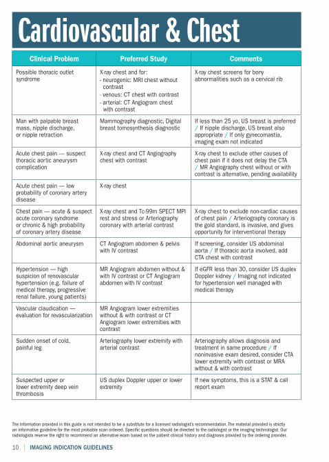
Clinical Problem Preferred Study Comments
Possible thoracic outletsyndrome
X-ray chest and for:- neurogenic: MRI chest withoutcontrast- venous: CT chest with contrast- arterial: CT Angiogram chestwith contrast
X-ray chest screens for bonyabnormalities such as a cervical rib
Man with palpable breastmass, nipple discharge,or nipple retraction
Mammography diagnostic, Digitalbreast tomosynthesis diagnostic
If less than 25 yo, US breast is preferred/ If nipple discharge, US breast alsoappropriate / If only gynecomastia,imaging exam not indicated
Acute chest pain — suspectthoracic aortic aneurysmcomplication
X-ray chest and CT Angiographychest with contrast
X-ray chest to exclude other causes ofchest pain if it does not delay the CTA/ MR Angiography chest without or withcontrast is alternative, pending availability
Acute chest pain — lowprobability of coronary arterydisease
X-ray chest
Chest pain — acute & suspectacute coronary syndromeor chronic & high probabilityof coronary artery disease
X-ray chest and Tc-99m SPECT MPIrest and stress or Arteriographycoronary with arterial contrast
X-ray chest to exclude non-cardiac causesof chest pain / Arteriography coronary isthe gold standard, is invasive, and givesopportunity for interventional therapy
Abdominal aortic aneurysm CT Angiogram abdomen & pelviswith IV contrast
If screening, consider US abdominalaorta / If thoracic aorta involved, addCTA chest with contrast
Hypertension — highsuspicion of renovascularhypertension (e.g. failure ofmedical therapy, progressiverenal failure, young patients)
MR Angiogram abdomen without &with IV contrast or CT Angiogramabdomen with IV contrast
If eGFR less than 30, consider US duplexDoppler kidney / Imaging not indicatedfor hypertension well managed withmedical therapy
Vascular claudication —evaluation for revascularization
MR Angiogram lower extremitieswithout & with contrast or CTAngiogram lower extremities withcontrast
Sudden onset of cold,painful leg
Arteriography lower extremity witharterial contrast
Arteriography allows diagnosis andtreatment in same procedure / Ifnoninvasive exam desired, consider CTAlower extremity with contrast or MRAwithout & with contrast
Suspected upper orlower extremity deep veinthrombosis
US duplex Doppler upper or lowerextremity
If new symptoms, this is a STAT & callreport exam
Cardiovascular & Chest
IMAGING INDICATION GUIDELINES | 11
The information provided in this guide is not intended to be a substitute for a licensed radiologist’s recommendation. The material provided is strictlyan informative guideline for the most probable scan ordered. Specific questions should be directed to the radiologist or the imaging technologist. Ourradiologists reserve the right to recommend an alternative exam based on the patient clinical history and diagnosis provided by the ordering provider.
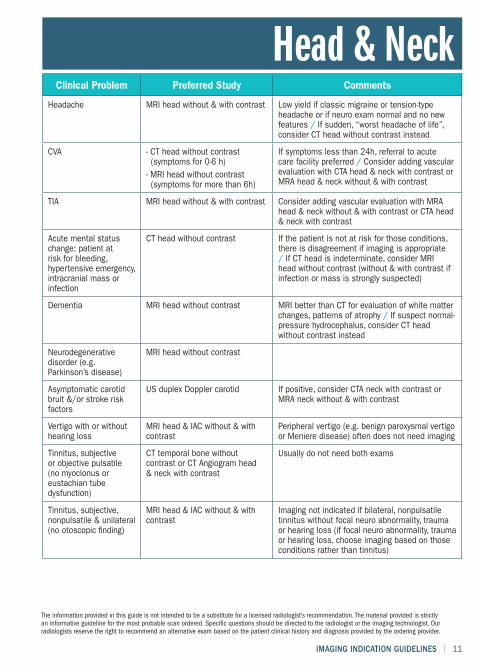
Clinical Problem Preferred Study Comments
Headache MRI head without & with contrast Low yield if classic migraine or tension-typeheadache or if neuro exam normal and no newfeatures / If sudden, “worst headache of life”,consider CT head without contrast instead
CVA - CT head without contrast(symptoms for 0-6 h)- MRI head without contrast(symptoms for more than 6h)
If symptoms less than 24h, referral to acutecare facility preferred / Consider adding vascularevaluation with CTA head & neck with contrast orMRA head & neck without & with contrast
TIA MRI head without & with contrast Consider adding vascular evaluation with MRAhead & neck without & with contrast or CTA head& neck with contrast
Acute mental statuschange: patient atrisk for bleeding,hypertensive emergency,intracranial mass orinfection
CT head without contrast If the patient is not at risk for those conditions,there is disagreement if imaging is appropriate/ If CT head is indeterminate, consider MRIhead without contrast (without & with contrast ifinfection or mass is strongly suspected)
Dementia MRI head without contrast MRI better than CT for evaluation of white matterchanges, patterns of atrophy / If suspect normal-pressure hydrocephalus, consider CT headwithout contrast instead
Neurodegenerativedisorder (e.g.Parkinson’s disease)
MRI head without contrast
Asymptomatic carotidbruit &/or stroke riskfactors
US duplex Doppler carotid If positive, consider CTA neck with contrast orMRA neck without & with contrast
Vertigo with or withouthearing loss
MRI head & IAC without & withcontrast
Peripheral vertigo (e.g. benign paroxysmal vertigoor Meniere disease) often does not need imaging
Tinnitus, subjectiveor objective pulsatile(no myoclonus oreustachian tubedysfunction)
CT temporal bone withoutcontrast or CT Angiogram head& neck with contrast
Usually do not need both exams
Tinnitus, subjective,nonpulsatile & unilateral(no otoscopic finding)
MRI head & IAC without & withcontrast
Imaging not indicated if bilateral, nonpulsatiletinnitus without focal neuro abnormality, traumaor hearing loss (if focal neuro abnormality, traumaor hearing loss, choose imaging based on thoseconditions rather than tinnitus)
Head & Neck
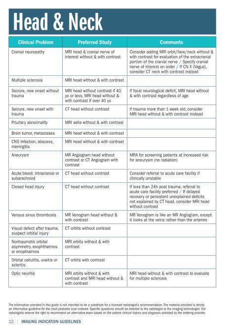
12 | IMAGING INDICATION GUIDELINES
The information provided in this guide is not intended to be a substitute for a licensed radiologist’s recommendation. The material provided is strictlyan informative guideline for the most probable scan ordered. Specific questions should be directed to the radiologist or the imaging technologist. Ourradiologists reserve the right to recommend an alternative exam based on the patient clinical history and diagnosis provided by the ordering provider.
Clinical Problem Preferred Study Comments
Cranial neuropathy MRI head & cranial nerve ofinterest without & with contrast
Consider adding MRI orbit/face/neck without &with contrast for evaluation of the extracranialportion of the cranial nerve / Specify cranialnerve of interest on order / If CN X (Vagus),consider CT neck with contrast instead
Multiple sclerosis MRI head without & with contrast
Seizure, new onset withouttrauma
MRI head without contrast if 40yo or less, MRI head without &with contrast if over 40 yo
If focal neurological deficit, MRI head without& with contrast regardless of age
Seizure, new onset withtrauma
CT head without contrast If trauma more than 1 week old, considerMRI head without & with contrast instead
Pituitary abnormality MRI sella without & with contrast
Brain tumor, metastases MRI head without & with contrast
CNS infection, abscess,meningitis
MRI head without & with contrast
Aneurysm MR Angiogram head withoutcontrast or CT Angiogram withcontrast
MRA for screening patients at increased riskfor aneurysm (no radiation)
Acute bleed: intracranial orsubarachnoid
CT head without contrast Consider referral to acute care facility ifclinically unstable
Closed head injury CT head without contrast If less than 24h post trauma, referral toacute care facility preferred / If delayedrecovery or persistent unexplained deficitsnot explained by CT head, consider MRI headwithout contrast
Venous sinus thrombosis MR Venogram head without &with contrast
MR Venogram is like an MR Angiogram, exceptit looks at the veins rather than the arteries
Visual defect after trauma,suspect orbital injury
CT orbits without contrast
Nontraumatic orbitalasymmetry, exophthalmosor enopthalmos
MRI orbits without & withcontrast
Orbital cellulitis, uveitis orscleritis
CT orbits with contrast
Optic neuritis MRI orbits without & withcontrast and MRI head without &with contrast
MRI head without & with contrast to evaluatefor multiple sclerosis
Head & Neck
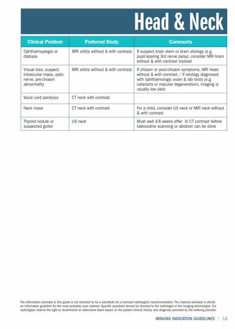
IMAGING INDICATION GUIDELINES | 13
The information provided in this guide is not intended to be a substitute for a licensed radiologist’s recommendation. The material provided is strictlyan informative guideline for the most probable scan ordered. Specific questions should be directed to the radiologist or the imaging technologist. Ourradiologists reserve the right to recommend an alternative exam based on the patient clinical history and diagnosis provided by the ordering provider.
Clinical Problem Preferred Study Comments
Ophthalmoplegia ordiplopia
MRI orbits without & with contrast If suspect brain stem or brain etiology (e.g.pupil-sparing 3rd nerve palsy), consider MRI brainwithout & with contrast instead
Visual loss, suspectintraocular mass, opticnerve, pre-chiasmabnormality
MRI orbits without & with contrast If chiasm or post-chiasm symptoms, MRI headwithout & with contrast / If etiology diagnosedwith ophthalmologic exam & lab tests (e.g.cataracts or macular degeneration), imaging isusually low yield
Vocal cord paralysis CT neck with contrast
Neck mass CT neck with contrast For a child, consider US neck or MRI neck without& with contrast
Thyroid nodule orsuspected goiter
US neck Must wait 4-8 weeks after IV CT contrast beforeradioiodine scanning or ablation can be done
Head & Neck
14 | IMAGING INDICATION GUIDELINES
The information provided in this guide is not intended to be a substitute for a licensed radiologist’s recommendation. The material provided is strictlyan informative guideline for the most probable scan ordered. Specific questions should be directed to the radiologist or the imaging technologist. Ourradiologists reserve the right to recommend an alternative exam based on the patient clinical history and diagnosis provided by the ordering provider.
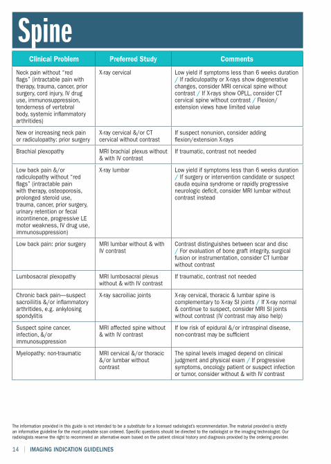
Clinical Problem Preferred Study Comments
Neck pain without “redflags” (intractable pain withtherapy, trauma, cancer, priorsurgery, cord injury, IV druguse, immunosuppression,tenderness of vertebralbody, systemic inflammatoryarthritides)
X-ray cervical Low yield if symptoms less than 6 weeks duration/ If radiculopathy or X-rays show degenerativechanges, consider MRI cervical spine withoutcontrast / If X-rays show OPLL, consider CTcervical spine without contrast / Flexion/extension views have limited value
New or increasing neck painor radiculopathy: prior surgery
X-ray cervical &/or CTcervical without contrast
If suspect nonunion, consider addingflexion/extension X-rays
Brachial plexopathy MRI brachial plexus without& with IV contrast
If traumatic, contrast not needed
Low back pain &/orradiculopathy without “redflags” (intractable painwith therapy, osteoporosis,prolonged steroid use,trauma, cancer, prior surgery,urinary retention or fecalincontinence, progressive LEmotor weakness, IV drug use,immunosuppression)
X-ray lumbar Low yield if symptoms less than 6 weeks duration/ If surgery or intervention candidate or suspectcauda equina syndrome or rapidly progressiveneurologic deficit, consider MRI lumbar withoutcontrast instead
Low back pain: prior surgery MRI lumbar without & withIV contrast
Contrast distinguishes between scar and disc/ For evaluation of bone graft integrity, surgicalfusion or instrumentation, consider CT lumbarwithout contrast
Lumbosacral plexopathy MRI lumbosacral plexuswithout & with IV contrast
If traumatic, contrast not needed
Chronic back pain—suspectsacroiliitis &/or inflammatoryarthritides, e.g. ankylosingspondylitis
X-ray sacroiliac joints X-ray cervical, thoracic & lumbar spine iscomplementary to X-ray SI joints / If X-ray normal& continue to suspect, consider MRI SI jointswithout contrast (IV contrast may also help)
Suspect spine cancer,infection, &/orimmunosuppression
MRI affected spine without& with IV contrast
If low risk of epidural &/or intraspinal disease,non-contrast may be sufficient
Myelopathy: non-traumatic MRI cervical &/or thoracic&/or lumbar withoutcontrast
The spinal levels imaged depend on clinicaljudgment and physical exam / If progressivesymptoms, oncology patient or suspect infectionor tumor, consider without & with IV contrast
Spine
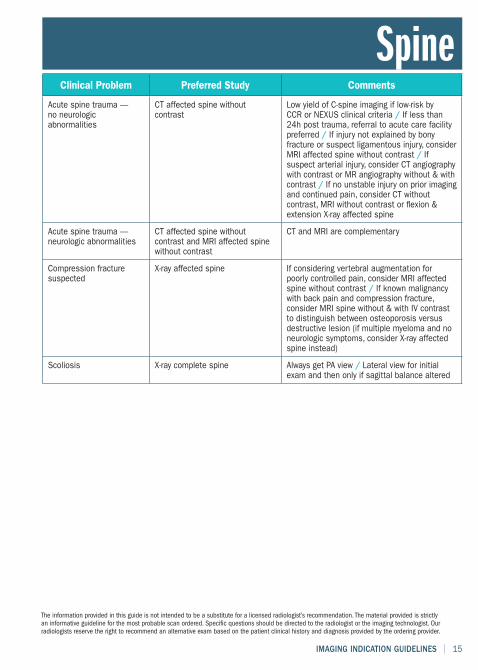
IMAGING INDICATION GUIDELINES | 15
The information provided in this guide is not intended to be a substitute for a licensed radiologist’s recommendation. The material provided is strictlyan informative guideline for the most probable scan ordered. Specific questions should be directed to the radiologist or the imaging technologist. Ourradiologists reserve the right to recommend an alternative exam based on the patient clinical history and diagnosis provided by the ordering provider.
Clinical Problem Preferred Study Comments
Acute spine trauma —no neurologicabnormalities
CT affected spine withoutcontrast
Low yield of C-spine imaging if low-risk byCCR or NEXUS clinical criteria / If less than24h post trauma, referral to acute care facilitypreferred / If injury not explained by bonyfracture or suspect ligamentous injury, considerMRI affected spine without contrast / Ifsuspect arterial injury, consider CT angiographywith contrast or MR angiography without & withcontrast / If no unstable injury on prior imagingand continued pain, consider CT withoutcontrast, MRI without contrast or flexion &extension X-ray affected spine
Acute spine trauma —neurologic abnormalities
CT affected spine withoutcontrast and MRI affected spinewithout contrast
CT and MRI are complementary
Compression fracturesuspected
X-ray affected spine If considering vertebral augmentation forpoorly controlled pain, consider MRI affectedspine without contrast / If known malignancywith back pain and compression fracture,consider MRI spine without & with IV contrastto distinguish between osteoporosis versusdestructive lesion (if multiple myeloma and noneurologic symptoms, consider X-ray affectedspine instead)
Scoliosis X-ray complete spine Always get PA view / Lateral view for initialexam and then only if sagittal balance altered
Spine
16 | IMAGING INDICATION GUIDELINES
The information provided in this guide is not intended to be a substitute for a licensed radiologist’s recommendation. The material provided is strictlyan informative guideline for the most probable scan ordered. Specific questions should be directed to the radiologist or the imaging technologist. Ourradiologists reserve the right to recommend an alternative exam based on the patient clinical history and diagnosis provided by the ordering provider.
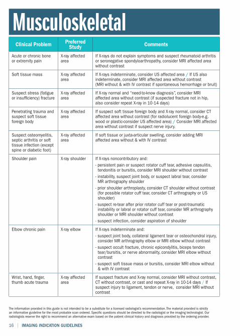
Clinical Problem PreferredStudy Comments
Acute or chronic boneor extremity pain
X-ray affectedarea
If X-rays do not explain symptoms and suspect rheumatoid arthritisor seronegative spondyloarthropathy, consider MRI affected areawithout contrast
Soft tissue mass X-ray affectedarea
If X-rays indeterminate, consider US affected area / If US alsoindeterminate, consider MRI affected area without contrast(MRI without & with IV contrast if spontaneous hemorrhage or bruit)
Suspect stress (fatigueor insufficiency) fracture
X-ray affectedarea
If X-ray normal and “need-to-know diagnosis”, consider MRIaffected area without contrast (if suspected fracture not in hip,also consider repeat X-ray in 10-14 days)
Penetrating trauma andsuspect soft tissueforeign body
X-ray affectedarea
If suspect soft tissue foreign body and X-ray normal, consider CTaffected area without contrast (for radiolucent foreign body-e.g.wood or plastic-consider US affected area) / Consider MRI affectedarea without contrast if suspect nerve injury.
Suspect osteomyelitis,septic arthritis or softtissue infection (exceptspine or diabetic foot)
X-ray affectedarea
If soft tissue or juxta-articular swelling, consider adding MRIaffected area without & with IV contrast
Shoulder pain X-ray shoulder If X-rays noncontributory and:- persistent pain or suspect rotator cuff tear, adhesive capsulitis,tendonitis or bursitis, consider MRI shoulder without contrast- instability, suspect joint body, or suspect labral tear, considerMR arthrography shoulder- prior shoulder arthroplasty, consider CT shoulder without contrast(for possible rotator cuff tear, consider CT arthrography or USshoulder)- suspect re-tear after prior rotator cuff tear or post-traumaticinstability or labral or rotator cuff tear, consider MR arthrographyshoulder or MRI shoulder without contrast- suspect infection, consider aspiration of shoulder
Elbow chronic pain X-ray elbow If X-rays indeterminate and:- suspect joint body, collateral ligament tear or osteochondral injury,consider MR arthrography elbow or MRI elbow without contrast- suspect occult fracture, chronic epicondylitis, biceps tendontear/bursitis, or nerve abnormality, consider MRI elbow withoutcontrast- suspect soft tissue mass or bursitis, consider MRI elbow without& with IV contrast
Wrist, hand, finger,thumb acute trauma
X-ray affectedarea
If suspect fracture and X-ray normal, consider MRI without contrast,CT without contrast, or cast and repeat X-ray in 10-14 days / Ifsuspect injury to ligament, tendon or nerve, consider MRI withoutcontrast
Musculoskeletal
IMAGING INDICATION GUIDELINES | 17
The information provided in this guide is not intended to be a substitute for a licensed radiologist’s recommendation. The material provided is strictlyan informative guideline for the most probable scan ordered. Specific questions should be directed to the radiologist or the imaging technologist. Ourradiologists reserve the right to recommend an alternative exam based on the patient clinical history and diagnosis provided by the ordering provider.
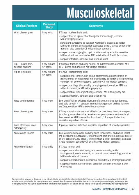
Clinical Problem PreferredStudy Comments
Wrist chronic pain X-ray wrist If X-rays indeterminate and:- suspect tear of ligament or triangular fibrocartilage, considerMR arthrography wrist- persistent symptoms or suspect Kienböck’s disease, considerMRI wrist without contrast (for suspected occult, stress or nonunionfracture, also consider CT wrist without contrast)- suspect mass or ganglion cyst or inflammatory arthritis, considerMRI wrist without contrast or MRI wrist without & with IV contrast- suspect infection, consider aspiration of wrist
Hip — acute pain,suspect fracture
X-ray hip andAP pelvis
If suspect fracture and X-ray normal or indeterminate, consider MRIor CT pelvis and affected hip without contrast
Hip chronic pain X-ray hip andAP pelvis
If X-rays indeterminate and:
- suspect bone, tendon, soft tissue abnormality, osteonecrosis orpainful metal-on-metal total hip arthroplasty, consider MRI hip withoutcontrast (for osteoid osteoma, consider CT hip without contrast)- suspect cartilage abnormality or impingement, consider MRI hipwithout contrast or MR arthrography hip- suspect labral tear or joint body, consider MR arthrography hip- suspect infection, consider aspiration of hip
Knee acute trauma X-ray knee Low yield if fall or twisting injury, no effusion, no focal tenderness,and able to walk / If suspect internal derangement and no fractureon X-ray, consider MRI knee without contrast
Knee chronic pain X-ray knee If X-ray normal or shows joint effusion or prior bone injury, or suspectjoint body, osteochondritis dessicans or post-op recurrent meniscaltear, consider MRI knee without contrast / If suspect infection,consider aspiration of knee
Knee after total kneearthroplasty
X-ray knee If pain or suspect infection, consider aspiration of knee by specialist
Ankle acute trauma X-ray ankle Low yield if able to walk, no bony point tenderness, and neuro intact(no peripheral neuropathy) / If persistent pain and no X-rays at time ofinjury, consider X-ray ankle / If more than 1 week persistent pain andX-rays negative, consider CT or MRI ankle without contrast
Ankle chronic pain X-ray ankle If X-rays normal and:- suspect osteochondral injury, tendon abnormality, ankleimpingement, ankle instability or pain of uncertain etiology, considerMRI ankle without contrast- suspect osteochondritis dessicans, consider MR arthrography ankle- suspect inflammatory arthritis, consider MRI ankle without & withIV contrast
Musculoskeletal
18 | IMAGING INDICATION GUIDELINES
The information provided in this guide is not intended to be a substitute for a licensed radiologist’s recommendation. The material provided is strictlyan informative guideline for the most probable scan ordered. Specific questions should be directed to the radiologist or the imaging technologist. Ourradiologists reserve the right to recommend an alternative exam based on the patient clinical history and diagnosis provided by the ordering provider.
Clinical Problem Preferred Study Comments
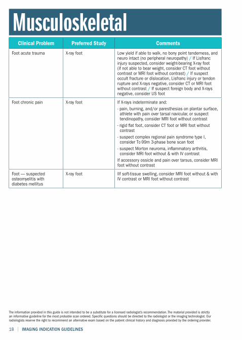
Foot acute trauma X-ray foot Low yield if able to walk, no bony point tenderness, andneuro intact (no peripheral neuropathy) / If Lisfrancinjury suspected, consider weight-bearing X-ray foot(if not able to bear weight, consider CT foot withoutcontrast or MRI foot without contrast) / If suspectoccult fracture or dislocation, Lisfranc injury or tendonrupture and X-rays negative, consider CT or MRI footwithout contrast / If suspect foreign body and X-raysnegative, consider US foot
Foot chronic pain X-ray foot If X-rays indeterminate and:- pain, burning, and/or paresthesias on plantar surface,athlete with pain over tarsal navicular, or suspecttendinopathy, consider MRI foot without contrast- rigid flat foot, consider CT foot or MRI foot withoutcontrast- suspect complex regional pain syndrome type I,consider Tc-99m 3-phase bone scan foot- suspect Morton neuroma, inflammatory arthritis,consider MRI foot without & with IV contrastIf accessory ossicle and pain over tarsus, consider MRIfoot without contrast
Foot — suspectedosteomyelitis withdiabetes mellitus
X-ray foot IIf soft-tissue swelling, consider MRI foot without & withIV contrast or MRI foot without contrast
Musculoskeletal
IMAGING INDICATION GUIDELINES | 19
The information provided in this guide is not intended to be a substitute for a licensed radiologist’s recommendation. The material provided is strictlyan informative guideline for the most probable scan ordered. Specific questions should be directed to the radiologist or the imaging technologist. Ourradiologists reserve the right to recommend an alternative exam based on the patient clinical history and diagnosis provided by the ordering provider.
Clinical Problem Preferred Study Comments
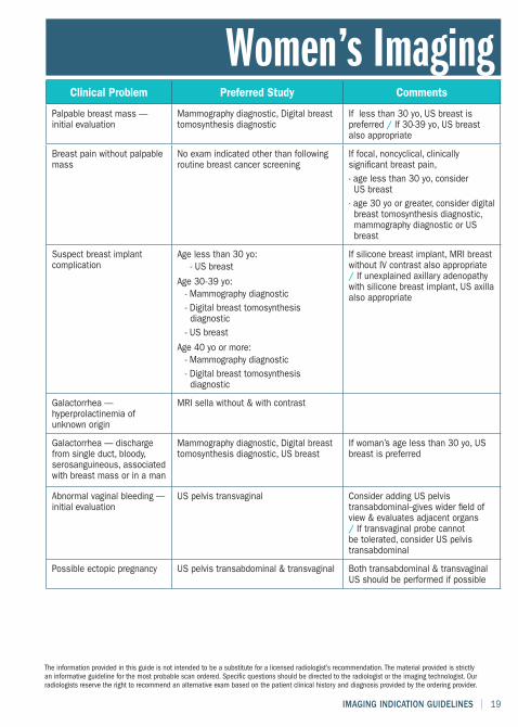
Palpable breast mass —initial evaluation
Mammography diagnostic, Digital breasttomosynthesis diagnostic
If less than 30 yo, US breast ispreferred / If 30-39 yo, US breastalso appropriate
Breast pain without palpablemass
No exam indicated other than followingroutine breast cancer screening
If focal, noncyclical, clinicallysignificant breast pain,- age less than 30 yo, considerUS breast- age 30 yo or greater, consider digitalbreast tomosynthesis diagnostic,mammography diagnostic or USbreast
Suspect breast implantcomplication
Age less than 30 yo:- US breast
Age 30-39 yo:- Mammography diagnostic- Digital breast tomosynthesisdiagnostic- US breast
Age 40 yo or more:- Mammography diagnostic- Digital breast tomosynthesisdiagnostic
If silicone breast implant, MRI breastwithout IV contrast also appropriate/ If unexplained axillary adenopathywith silicone breast implant, US axillaalso appropriate
Galactorrhea —hyperprolactinemia ofunknown origin
MRI sella without & with contrast
Galactorrhea — dischargefrom single duct, bloody,serosanguineous, associatedwith breast mass or in a man
Mammography diagnostic, Digital breasttomosynthesis diagnostic, US breast
If woman’s age less than 30 yo, USbreast is preferred
Abnormal vaginal bleeding —initial evaluation
US pelvis transvaginal Consider adding US pelvistransabdominal--gives wider field ofview & evaluates adjacent organs/ If transvaginal probe cannotbe tolerated, consider US pelvistransabdominal
Possible ectopic pregnancy US pelvis transabdominal & transvaginal Both transabdominal & transvaginalUS should be performed if possible
Women’s Imaging
20 | IMAGING INDICATION GUIDELINES
The information provided in this guide is not intended to be a substitute for a licensed radiologist’s recommendation. The material provided is strictlyan informative guideline for the most probable scan ordered. Specific questions should be directed to the radiologist or the imaging technologist. Ourradiologists reserve the right to recommend an alternative exam based on the patient clinical history and diagnosis provided by the ordering provider.
Clinical Problem Preferred Study Comments
Pelvic pain: woman withsuspected gynecologicaletiology
US pelvis transabdominal& transvaginal
Both transabdominal & transvaginalUS should be performed if possible
Pelvic pain: no suspectedgynecological etiology andß-HCG negative
CT abdomen & pelviswith IV contrast,radiologist decides if oralcontrast
Clinically suspectedadnexal mass — initialevaluation
US Doppler pelvistransabdominal &transvaginal
Transabdominal shows mass in relation to otherstructures / Transvaginal shows details of mass /Doppler shows vascularity of mass
Pelvic floor dysfunction MRI pelvis dynamicwith rectal contrast,radiologist decides ifvaginal contrast
Alternatives include X-ray fluoroscopiccystocolpoproctography with IV, oral, vaginal, and rectalcontrast, MRI defecography with contrast, US pelvistransperineal
Women’s Imaging
IMAGING INDICATION GUIDELINES | 21
The information provided in this guide is not intended to be a substitute for a licensed radiologist’s recommendation. The material provided is strictlyan informative guideline for the most probable scan ordered. Specific questions should be directed to the radiologist or the imaging technologist. Ourradiologists reserve the right to recommend an alternative exam based on the patient clinical history and diagnosis provided by the ordering provider.
Screening PreferredStudy
Timing and Indicationsfor Screening Comments
Breast cancerscreening
Mammographyscreening,Digital breasttomosynthesisscreening
Every year, starting at:- Low risk: 40 yo- BRCA carrier/ relative: age 30 yo- 1° relative with premenopausalbreast cancer: later of age 30 yoor 10 years less than relative’s ageat diagnosis- Mantle radiation age 10-30 yo: 8 yafter radiation but older than 25 yo- Diagnosis of lobular neoplasia,atypical ductal hyperplasia (ADH),breast carcinoma:
Digital breast tomosynthesisscreening may be helpful if lessthan 50 yo or if dense breasts/ If high risk (BRCA carrier/relative, mantle radiationage 10-30 yo or with 20% orgreater lifetime risk), consideradding MRI breast without &with contrast / If pregnant orlactating, recommendations arethe same
Lung cancerscreening
CT chestscreening withoutcontrast
Every year for patients:- Age 55-80 yo- Without lung cancer symptoms- 30 or more pack-year smokinghistory- Current smoker or stopped withinpast 15 years- Counselled on smoking cessation
CT done with very low radiationdose
Coronaryartery diseasescreening
CT coronaryartery calciumscoring or UScarotid intimamedial thickness
Asymptomatic with intermediate risk(10%-20% 10-year risk) for coronaryartery disease
Useful for reclassifyingintermediate risk patients tolow or high risk / If low riskpatient with family history ofpremature coronary arterydisease, may be helpful
Abdominalaortic aneurysmscreening
US aortaabdomen
Men over 65 yo, especially withhypertension, smoking, coronaryartery disease, 1st degree malerelative with AAA
Peripheralvascular diseasescreening
US duplexDoppler lowerextremities withankle-brachialindex
Older than 50 yo with history ofdiabetes or smoking
Colon cancerscreening
CT colonography Every 5 years after negative screen,starting at 50 yo or more
Recommend if incompletecolonoscopy or refusal ofoptical colonoscopy / Ifhigh risk for colon cancer,recommend optical colonoscopy/ Need colon prep
Screening
22 | IMAGING INDICATION GUIDELINES
The information provided in this guide is not intended to be a substitute for a licensed radiologist’s recommendation. The material provided is strictlyan informative guideline for the most probable scan ordered. Specific questions should be directed to the radiologist or the imaging technologist. Ourradiologists reserve the right to recommend an alternative exam based on the patient clinical history and diagnosis provided by the ordering provider.
Screening PreferredStudy
Timing and Indicationsfor Screening Comments
Osteoporosisscreening
DXA lumbar spineand hip(s)
Every 2 years until bone mineraldensity stabilizes unless risk factors*or treatment changes- All women age 65 yo & older and menage 70 yo & older- Women younger than 65 yo and menyounger than 70 yo with risk factors*- 50 yo & older with wrist, hip, spineor proximal humerus fracture withminimal or no trauma- any age with insufficiency fracture,osteopenia on imaging, conditionsthat could alter bone mineral density
*Risk factors include:- current smoker- loss of height, thoracic kyphosis- estrogen deficiency- maternal hip fracture after 50 yo- body weight less than 127 lb/57.6 kg- amenorrhea for more than 1 yearbefore 42 yo
Fracture risk based on T- andZ-score / Follow bone mineraldensity (BMD), not the T- orZ-score / No fracture risk data forpremenopausal women or menunder 50 yo / If BMI over 35kg/m2 , very large or very smallbody height, or advanced spinedegenerative disease, considerQuantitative CT lumbar spine &hips without contrast
Ovariancancerscreening
None No screening recommended foraverage-risk / No proven benefitto screening with CA-125 &/orUS for high-risk
Screening
IMAGING INDICATION GUIDELINES | 23
The information provided in this guide is not intended to be a substitute for a licensed radiologist’s recommendation. The material provided is strictlyan informative guideline for the most probable scan ordered. Specific questions should be directed to the radiologist or the imaging technologist. Ourradiologists reserve the right to recommend an alternative exam based on the patient clinical history and diagnosis provided by the ordering provider.
IV ContrastWe Screen For:
Decreased Renal Function• If eGFR below 30 mL/min/1.73 m2, the radiologist may give less or no contrast
Allergies• Mild allergies (e.g., rash, hives, swelling of eyes &/or face)
– Recommend steroid premedication in non-emergent situations• Moderate or severe allergies (e.g., inability to breathe, becoming unconscious)
– Recommend giving IV contrast in an acute care setting aftersteroid premedication
• Arthrograms and myelograms usually not affected
Pregnancy• IV contrast can cross the placenta• CT/X-ray contrast
– No available data for harm to fetus• MRI contrast
– May accumulate in amniotic fluid– Uncertain if leads to harm &/or NSF in fetus or mother– Recommend no MRI contrast unless:
1) information cannot be acquired without contrast,2) the information would affect the care of the patient and fetusduring the pregnancy, or
3) it is not prudent to wait to obtain this information until thepatient is no longer pregnant
Breastfeeding and MRI/CT/X-ray Contrast• Safe to continue breast-feeding after receiving contrast according to available data• If concerned, stop breast-feeding for 24 hours with active expression and discardingof breast milk from both breasts during that period
Metformin and CT/X-ray Contrast• If eGFR below 30 mL/min/1.73 m2
– Stop metformin for 48 hours after contrast– Recommend renal function test after 48 hours before resuming metformin
Thyroid Abnormalities and CT or X-ray Contrast• If a radioactive iodine thyroid scan or radioactive iodine uptake is planned,do before the patient has iodinated CT or X-ray contrast
Our Commitment to Your Patients and Their Safety
24 | IMAGING INDICATION GUIDELINES
The information provided in this guide is not intended to be a substitute for a licensed radiologist’s recommendation. The material provided is strictlyan informative guideline for the most probable scan ordered. Specific questions should be directed to the radiologist or the imaging technologist. Ourradiologists reserve the right to recommend an alternative exam based on the patient clinical history and diagnosis provided by the ordering provider.
MRI SafetyWe screen for implants and any metal in or on patients before they go in the MRI scanner.
Radiation SafetyWe strive to limit radiation exposure while producing quality CT and X-ray examinations.
Suggested Premedication Protocols for Contrast Allergy
Protocol 1• #16 of Methylprednisolone (Medrol) 4mgLabel: Take eight (8) by mouth 12 hours and 2 hours before the examis scheduled.
• And consider adding #2 of Diphenhydramine (Benadryl) 25mgLabel: Take one or two (1 or 2) by mouth 1 hour before the exam is scheduled.Do not drive or operate heavy machinery for 4-6 hours after taking.
OR
Protocol 2• #3 of Prednisone 50mgLabel: Take one (1) by mouth 13 hours, 7 hours and 1 hour before the examis scheduled.
• And #2 of Diphenhydramine (Benadryl) 25mgLabel: Take one or two (1 or 2) by mouth 1 hour before the exam is scheduled.Do not drive or operate heavy machinery for 4-6 hours after taking.
Our Commitment to Your Patients and Their Safety
IMAGING INDICATION GUIDELINES | 25
The information provided in this guide is not intended to be a substitute for a licensed radiologist’s recommendation. The material provided is strictlyan informative guideline for the most probable scan ordered. Specific questions should be directed to the radiologist or the imaging technologist. Ourradiologists reserve the right to recommend an alternative exam based on the patient clinical history and diagnosis provided by the ordering provider.
Recommendations for Claustrophobic Patients
Patients with pain, anxiety, restlessness or claustrophobia• For optimal image quality, it is important for patients to lie still duringtheir CT and MRI exams.
Patients, consider:• Taking your prescribed pain medication about ½ hour before the examis to start
• Bringing a friend or family member to sit in the scanner room to reada book aloud or gently talk to you
• Bringing a favorite CD for listening during the exam
• Do not drive if taking an anxiolytic or if the pain medication instructionsadvise against it
Doctors, consider:• Prescribing a single dose of an oral anxiolytic to take about ½ hour beforethe exam is to start. These should not be given to women who are pregnantor breastfeeding. Oral anxiolytics include, but are not limited to:
– diazepam (Valium) (2-10 mg)
– lorazepam (Ativan) (0.5-1 mg only for those over 12 yo)
– alprazolam (Xanax) (0.25-0.5 mg for those over 18 yo)
– oxazepam (Serax) (10-15 mg only for those over 12 yo)
2020
ALABAMAMobileOpen MRI & Imaging6576 Airport BoulevardMobile, Alabama 36608T: 251.460.4112F: 251.460.4590Toll-Free: 888.833.4674Tax ID #: 62-1664765NPI: 1548228323
FLORIDAPanama CityOpen MRI & Imaging2525 Highway 77 (MLK Blvd.)Panama City, FL 32405T: 850.873.6900F: 850.873.6902Tax ID #: 59-3551727NPI: 1225096936
Pensacola Open MRI4511 North Davis Hwy.,Suite 1BPensacola, FL 32503T: 850.484.8454F: 850.484.7754Tax ID #: 59-3551727NPI: 1235197930
Results availablein 24 hours• Faxed results• Secure online access toimages, reports and scheduling
• Preliminary andSTAT reports availableupon request
• EMR interface capabilities