Embed Size (px)
Citation preview

brain swelling and mass effect which may require
medical or neurosurgical intervention.
The imaging modalities used in acute stroke
patients are CT, MRI and Doppler studies.
Role of CT in Stroke Imaging
CT is the quickest, cheapest and most easily
accessible imaging modality. It is the first imaging
done for these patients. It has the highest sensitivity
for detecting intracranial hemorrhage. With helical
and multislice CT, even slight movement of the
patient does not seriously compromise detail on the
scan as repeat imaging of specific areas can be
done without delay.
Hemorrhage is most readily picked up by a CT
when compared to MRI. A hematoma is seen as an
area of hyperdensity (bright) within the brain pa-
renchyma. An infarct on the other hand is usually
seen as an area of hypodensity (dark). Fig 1 shows
a hematoma in the putaminal region on the right
side surrounded by oedema.
TOPIC IN FOCUS—Imaging in stroke
Imaging in Stroke
Dr. Sunithi Elizabeth Mani, Assoc. Professor, Department of Radiology, CMC, Vellore.
The window period for thrombolysis for stroke
is very short and hence early, definitive diagnosis is
an essential component of management. Radiologi-
cal imaging for detection of stroke and description
of the arterial territory involved plays a vital role in
this regard both for diagnosis and therapy.
Aims of imaging in acute stroke:
1. Exclusion of hematoma
Exclusion of hematoma is important as this is a
contraindication for thrombolytic therapy.
2. Differentiate between infarcted tissue
and penumbra
Identification of the penumbra (area of risk for
infarction, which is not already infracted) is neces-
sary. This area is the target for reperfusion therapy.
3. Identify stenosis or occlusion of major
extra- and intracranial arteries.
The detection of stenosis or occlusion of the
artery helps in deciding whether endovascular in-
tervention is feasible or not.
Apart from these, imaging helps in identifying
Fig. 1: Right putaminal
hemorrhage
CMI 13:2 April 2015 15
Fig. 2: Right middle cerebral artery territory infarct with brain
swelling
Fig. 3: Yellow arrow points to nor-mal lentiform nucleus visualization, This is obscured on the other side.

grey-white interface of the insular cortex is known
as the ‘Insular ribbon’ sign. The insula is supplied
by the distal branches of the MCA and is suscepti-
ble to ischemia.
CT ANGIOGRAPHY AND CT PERFU-
SION STUDIES
CT perfusion study can be done to assess the
blood flow patterns in the brain which helps in
early detection of ischemia, areas of poor perfu-
sion and whether there is occlusion or stenosis of a
vessel . Contrast is injected intravenously during
the CT and a series of images are taken over a pe-
riod of time. Coloured images indicate regions
ofpoor perfusion which is the penumbra and pa-
SUBTLE SIGNS ON CT
CT may be appear to be normal in about 30%
of patients with stroke but careful examination
will prove otherwise. Some of the subtle signs of
an infarct which can be missed are:
1. Obscured lentiform nucleus sign: The
lentiform nucleus and the adjacent internal capsule
are usually well demarcated. In an infarct involv-
ing this region, the entire region appears uni-
formly dark or grey. (Fig.3)
2. Loss of sulcation: The sulci on the cerebral
surface are less evident on the side of an infarct
due to cerebral oedema. (Fig. 4)
3. Hyperdense vessel sign: A thrombus in a
vessel may be seen as a bright line in the CT
(Fig.5). This indicates that the thrombus has oc-
curred in the last 90 minutes. This sign has a high
specificity (100%) but only moderate sensitivity
(30%) in diagnosing a stroke. Loss of grey-white
differentiation along with a hyperdense vessel in-
creases the sensitivity. Hyperdense vessel sign is
indicative of a large vessel occlusion and therefore
predicts a poor stroke outcome (positive predictive
value of 91%).
4. Insular ribbon sign: The loss of the normal
Fig. 4: shows an infarct on the left side—note the loss of grey-white differentiation and loss of sulci sug-gestive of oedema in the left cere-bral hemisphere.
Fig. 5: White arrow points to hyperdensity in the left middle cerebral ar-
tery (patient had right sided weakness) indicating thrombus within. This
Advantages of CT
1. It is cheap and quick.
2. Less vulnerable to motion artefacts.
3. Very sensitive in detecting hemorrhage.
Disadvantages of CT
1. Not very sensitive in detection of very early or
hyperacute infarcts.
2. Small infarcts may be missed.
3. Exposure to ionizing radiation
TOPIC IN FOCUS—Imaging in stroke
CMI 13:2 April 2015 16

and dark in ADC) indicates non salvageable tissue.
On MR Perfusion, the tissue having poor perfusion
is identified and this indicates tissue at risk of in-
farction. Hence, the difference between these areas
shows the area that is potentially salvageable by
thrombolysis therapy. (Fig.7)
tients with these findings will benefit from throm-
bolysis.
Role of MRI T1 and T2 weighted imaging have a relatively
poor sensitivity in detecting ischemia. The introduc-
tion of Diffusion Weighted Imaging (DWI) how-
ever has revolutionized the imaging of acute stroke
patients both for diagnosis and management.
DWI images have a much higher sensitivity
and specificity than CT in detection of an acute
infarct and also an infarct can be detected within
minutes after it has occurred. (Fig.6)
DWI images indicate completely infracted tis-
sue or tissue that cannot be salvaged. Infarcted tis-
sue is bright on the DWI and dark on the ADC.
Concept of diffusion perfusion mismatch
The area of diffusion restriction (bright in DWI
Fig. 5: CT series showing progression of right middle cerebellar stroke over time: A: 3 hours, B: 16
hours, C: 24 hours, D: After decompressive craniectomy
Fig 6.A- FLAIR images showing slow flow in right MCA branches
in the sylvian fissure (arrow). Fig 6.B. DWI imaging showing
wedge shaped infarct and Fig 6.C: ADC image showing hypoin-
tensity in the same region.
Fig.7: DWI image showing infarct and perfu-
sion deficit on MR Perfusion image. There is
a mismatch between the two and hence
there is potentially salvageable tissue.
Restricted diffusion (grey) << perfusion
deficit (black) – good prognosis
Restricted diffusion = perfusion deficit
– poor prognosis
Restricted diffusion >> perfusion deficit
– subacute infarct/ reperfusion
A B C
TOPIC IN FOCUS—Imaging in stroke
CMI 13:2 April 2015 17
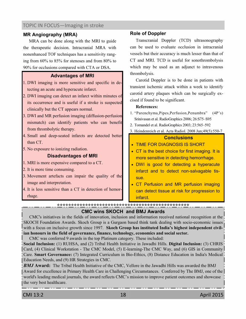
Role of Doppler
Transcranial Doppler (TCD) ultrasonography
can be used to evaluate occlusion in intracranial
vessels but their accuracy is much lesser than that of
CT and MRI. TCD is useful for sonothrombolysis
which may be used as an adjunct to intravenous
thrombolysis.
Carotid Doppler is to be done in patients with
transient ischemic attack within a week to identify
carotid artery plaques which can be surgically ex-
cised if found to be significant.
References:
1. “Parenchyma,Pipes,Perfusion,Penumbra” (4P’s)
Srinivasan et al. RadioGraphics 2006; 26:S75–S95
2. Tomandel et al. RadioGraphics 2003; 23:565–592
3. Heindenreich et al. Acta Radiol. 2008 Jun;49(5):550-7
MR Angiography (MRA)
MRA can be done along with the MRI to guide
the therapeutic decision. Intracranial MRA with
nonenhanced TOF techniques has a sensitivity rang-
ing from 60% to 85% for stenoses and from 80% to
90% for occlusions compared with CTA or DSA.
Conclusions
TIME FOR DIAGNOSIS IS SHORT
CT is the best choice for first imaging. It is
more sensitive in detecting hemorrhage.
DWI is good for detecting a hyperacute
infarct and to detect non-salvagable tis-
sue.
CT Perfusion and MR perfusion imaging
can detect tissue at risk for progression to
infarct.
Advantages of MRI
1. DWI imaging is more sensitive and specific in de-
tecting an acute and hyperacute infarct.
2. DWI imaging can detect an infarct within minutes of
its occurrence and is useful if a stroke is suspected
clinically but the CT appears normal.
3. DWI and MR perfusion imaging (diffusion-perfusion
mismatch) can identify patients who can benefit
from thrombolytic therapy.
4. Small and deep-seated infarcts are detected better
than CT.
5. No exposure to ionizing radiation.
Disadvantages of MRI
1. MRI is more expensive compared to a CT.
2. It is more time consuming.
3. Movement artefacts can impair the quality of the
image and interpretation.
4. It is less sensitive than a CT in detection of hemor-
rhage.
TOPIC IN FOCUS—Imaging in stroke
CMI 13:2 April 2015 18
CMC wins SKOCH and BMJ Awards CMC's initiatives in the fields of innovation, inclusion and information received national recognition at the
SKOCH Foundation Awards. Skoch Group is a Gurgaon based think tank dealing with socio-economic issues
with a focus on inclusive growth since 1997. Skoch Group has instituted India’s highest independent civil-
ian honours in the field of governance, finance, technology, economics and social sector.
CMC was conferred 9 awards in the top Platinum category. These included:
Social Inclusion: (1) RUHSA, and (2) Tribal Health Initiative in Jawadhi Hills. Digital Inclusion: (3) CHRIS
Card, (4) Clinical Workstation - The CMC Model, (5) E-learning-The CMC Way, and (6) GIS in Community
Care. Smart Governance: (7) Integrated Curriculum in Bio-Ethics, (8) Distance Education in India's Medical
Education Needs, and (9) HR Strategies in CMC.
BMJ Award: The Tribal Health Initiative of the CMC, Vellore in the Jawadhi Hills was awarded the BMJ
Award for excellence in Primary Health Care in Challenging Circumstances. Conferred by The BMJ, one of the
world's leading medical journals, the award reflects CMC’s mission to improve patient outcomes and showcase
the very best healthcare.
***********************************************


![Magnetic Resonance Imaging in Experimental Stroke and ... et al 2015 [STROKE] MRI.pdf · Magnetic Resonance Imaging (MRI) is an invaluable and ver - satile imaging tool used in both](https://img.pdfslide.us/doc/110x75/5e640f9afb16267f7a1e1295/magnetic-resonance-imaging-in-experimental-stroke-and-et-al-2015-stroke-mripdf.jpg)