Embed Size (px)
Citation preview
64 IMAGING ESSENTIALS
Ultrasonography of Peritoneal
and Retroperitoneal Spaces
and Abdominal Lymph NodesClifford R. Berry, DVM, DACVR; Elizabeth Huyhn, DVM; and Danielle Mauragis, CVT
University of Florida
IMAGING ESSENTIALS
The peritoneal and retroperitoneal spaces are assessed
throughout the ultrasound examination of the entire
abdomen. The abdominal lymph nodes (lymphocenters),
which are located throughout the abdomen, drain specific
regional organs and areas. This article reviews the normal
ultrasonographic appearance of these structures in dogs
and cats as well as commonly encountered abnormalities.
PERITONEUM AND
RETROPERITONEUM
Normal Findings
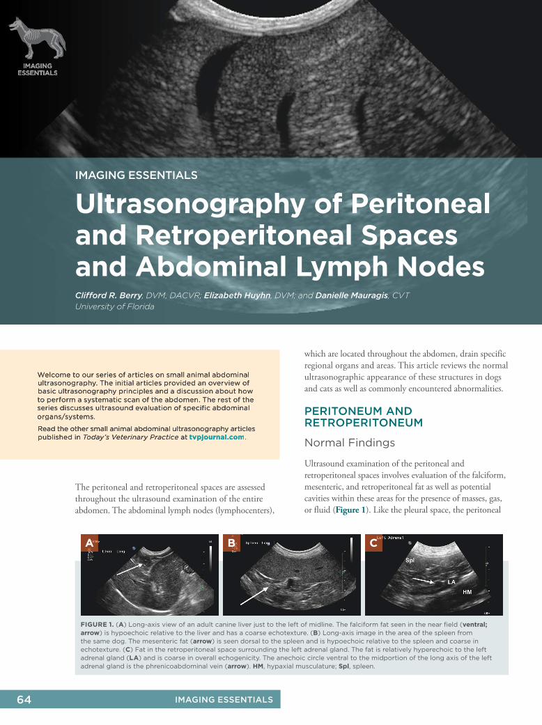
Ultrasound examination of the peritoneal and
retroperitoneal spaces involves evaluation of the falciform,
mesenteric, and retroperitoneal fat as well as potential
cavities within these areas for the presence of masses, gas,
or fluid (Figure 1). Like the pleural space, the peritoneal
Welcome to our series of articles on small animal abdominal ultrasonography. The initial articles provided an overview of basic ultrasonography principles and a discussion about how to perform a systematic scan of the abdomen. The rest of the series discusses ultrasound evaluation of specific abdominal organs/systems.
Read the other small animal abdominal ultrasonography articles published in Today’s Veterinary Practice at tvpjournal.com.
IMAGING
ESSENTIALS
FIGURE 1. (A) Long-axis view of an adult canine liver just to the left of midline. The falciform fat seen in the near field (ventral;
arrow) is hypoechoic relative to the liver and has a coarse echotexture. (B) Long-axis image in the area of the spleen from
the same dog. The mesenteric fat (arrow) is seen dorsal to the spleen and is hypoechoic relative to the spleen and coarse in
echotexture. (C) Fat in the retroperitoneal space surrounding the left adrenal gland. The fat is relatively hyperechoic to the left
adrenal gland (LA) and is coarse in overall echogenicity. The anechoic circle ventral to the midportion of the long axis of the left
adrenal gland is the phrenicoabdominal vein (arrow). HM, hypaxial musculature; Spl, spleen.
A B C
65JULY/AUGUST 2017 ■ TVPJOURNAL.COM
IMAGING ESSENTIALS
space is a closed cavity with a serous mesothelial lining.1
A scant amount of physiologic peritoneal effusion,
typically not seen on ultrasonography, is normal; this
effusion serves as a lubricant for the peritoneal organs.
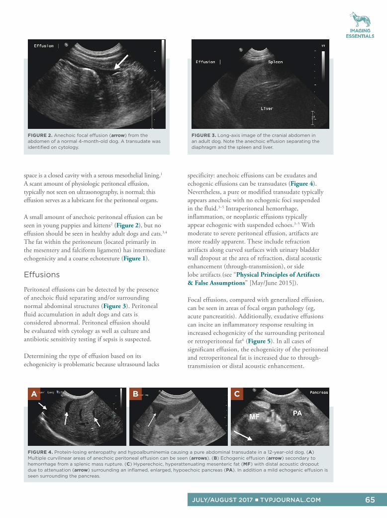
A small amount of anechoic peritoneal effusion can be
seen in young puppies and kittens2 (Figure 2), but no
effusion should be seen in healthy adult dogs and cats.3,4
The fat within the peritoneum (located primarily in
the mesentery and falciform ligament) has intermediate
echogenicity and a coarse echotexture (Figure 1).
Effusions
Peritoneal effusions can be detected by the presence
of anechoic fluid separating and/or surrounding
normal abdominal structures (Figure 3). Peritoneal
fluid accumulation in adult dogs and cats is
considered abnormal. Peritoneal effusion should
be evaluated with cytology as well as culture and
antibiotic sensitivity testing if sepsis is suspected.
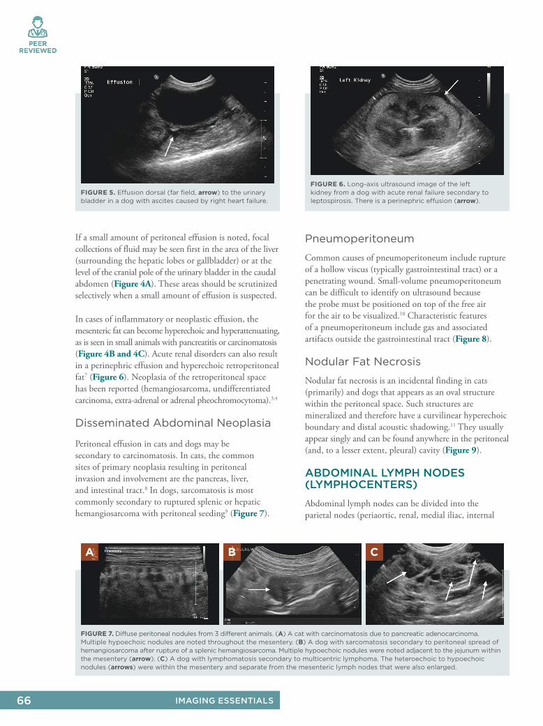
Determining the type of effusion based on its
echogenicity is problematic because ultrasound lacks
specificity: anechoic effusions can be exudates and
echogenic effusions can be transudates (Figure 4).
Nevertheless, a pure or modified transudate typically
appears anechoic with no echogenic foci suspended
in the fluid.3–5 Intraperitoneal hemorrhage,
inflammation, or neoplastic effusions typically
appear echogenic with suspended echoes.3–5 With
moderate to severe peritoneal effusion, artifacts are
more readily apparent. These include refraction
artifacts along curved surfaces with urinary bladder
wall dropout at the area of refraction, distal acoustic
enhancement (through-transmission), or side
lobe artifacts (see “Physical Principles of Artifacts
& False Assumptions” [May/June 2015]).
Focal effusions, compared with generalized effusion,
can be seen in areas of focal organ pathology (eg,
acute pancreatitis). Additionally, exudative effusions
can incite an inflammatory response resulting in
increased echogenicity of the surrounding peritoneal
or retroperitoneal fat6 (Figure 5). In all cases of
significant effusion, the echogenicity of the peritoneal
and retroperitoneal fat is increased due to through-
transmission or distal acoustic enhancement.
FIGURE 3. Long-axis image of the cranial abdomen in
an adult dog. Note the anechoic effusion separating the
diaphragm and the spleen and liver.
FIGURE 2. Anechoic focal effusion (arrow) from the
abdomen of a normal 4-month-old dog. A transudate was
identified on cytology.
FIGURE 4. Protein-losing enteropathy and hypoalbuminemia causing a pure abdominal transudate in a 12-year-old dog. (A)
Multiple curvilinear areas of anechoic peritoneal effusion can be seen (arrows). (B) Echogenic effusion (arrow) secondary to
hemorrhage from a splenic mass rupture. (C) Hyperechoic, hyperattenuating mesenteric fat (MF) with distal acoustic dropout
due to attenuation (arrow) surrounding an inflamed, enlarged, hypoechoic pancreas (PA). In addition a mild echogenic effusion is
seen surrounding the pancreas.
A B C
66 IMAGING ESSENTIALS
PEER
REVIEWED
If a small amount of peritoneal effusion is noted, focal
collections of fluid may be seen first in the area of the liver
(surrounding the hepatic lobes or gallbladder) or at the
level of the cranial pole of the urinary bladder in the caudal
abdomen (Figure 4A). These areas should be scrutinized
selectively when a small amount of effusion is suspected.
In cases of inflammatory or neoplastic effusion, the
mesenteric fat can become hyperechoic and hyperattenuating,
as is seen in small animals with pancreatitis or carcinomatosis
(Figure 4B and 4C). Acute renal disorders can also result
in a perinephric effusion and hyperechoic retroperitoneal
fat7 (Figure 6). Neoplasia of the retroperitoneal space
has been reported (hemangiosarcoma, undifferentiated
carcinoma, extra-adrenal or adrenal pheochromocytoma).3,4
Disseminated Abdominal Neoplasia
Peritoneal effusion in cats and dogs may be
secondary to carcinomatosis. In cats, the common
sites of primary neoplasia resulting in peritoneal
invasion and involvement are the pancreas, liver,
and intestinal tract.8 In dogs, sarcomatosis is most
commonly secondary to ruptured splenic or hepatic
hemangiosarcoma with peritoneal seeding9 (Figure 7).
Pneumoperitoneum
Common causes of pneumoperitoneum include rupture
of a hollow viscus (typically gastrointestinal tract) or a
penetrating wound. Small-volume pneumoperitoneum
can be difficult to identify on ultrasound because
the probe must be positioned on top of the free air
for the air to be visualized.10 Characteristic features
of a pneumoperitoneum include gas and associated
artifacts outside the gastrointestinal tract (Figure 8).
Nodular Fat Necrosis
Nodular fat necrosis is an incidental finding in cats
(primarily) and dogs that appears as an oval structure
within the peritoneal space. Such structures are
mineralized and therefore have a curvilinear hyperechoic
boundary and distal acoustic shadowing.11 They usually
appear singly and can be found anywhere in the peritoneal
(and, to a lesser extent, pleural) cavity (Figure 9).
ABDOMINAL LYMPH NODES (LYMPHOCENTERS)
Abdominal lymph nodes can be divided into the
parietal nodes (periaortic, renal, medial iliac, internal
FIGURE 5. Effusion dorsal (far field, arrow) to the urinary
bladder in a dog with ascites caused by right heart failure.
FIGURE 6. Long-axis ultrasound image of the left
kidney from a dog with acute renal failure secondary to
leptospirosis. There is a perinephric effusion (arrow).
FIGURE 7. Diffuse peritoneal nodules from 3 different animals. (A) A cat with carcinomatosis due to pancreatic adenocarcinoma.
Multiple hypoechoic nodules are noted throughout the mesentery. (B) A dog with sarcomatosis secondary to peritoneal spread of
hemangiosarcoma after rupture of a splenic hemangiosarcoma. Multiple hypoechoic nodules were noted adjacent to the jejunum within
the mesentery (arrow). (C) A dog with lymphomatosis secondary to multicentric lymphoma. The heteroechoic to hypoechoic
nodules (arrows) were within the mesentery and separate from the mesenteric lymph nodes that were also enlarged.
A B C
67JULY/AUGUST 2017 ■ TVPJOURNAL.COM
IMAGING ESSENTIALS
iliac, and sacral) and visceral nodes (hepatic, splenic,
gastric, pancreaticoduodenal, jejunal, ileocolic,
and colic).3,4 A number of abdominal lymph nodes
are not routinely seen on ultrasonography.
Abdominal lymph nodes vary dramatically in size and shape
depending on the age of the animal and the location of the
node.12 Assessment of abdominal lymph nodes requires the
sonographer to understand the normal anatomic location
of the individual nodes as well as the regional anatomy,
particularly the vascular anatomy, because lymph nodes
are found surrounding specific abdominal vessels.6,13–16
Parietal Nodes
The medial iliac lymph nodes are found caudal to the deep
circumflex arteries along the lateral margins of the origins
of the left and right external iliac arteries from the aorta
(aortic trifurcation). These nodes are located immediately
cranial to, at, or just caudal to the trifurcation of the
caudal abdominal aorta.16 They can be dorsolateral, lateral,
or ventrolateral to the caudal abdominal vasculature.
To identify medial iliac lymph nodes, therefore, it is
necessary to sweep the transducer in a dorsoventral
direction while imaging the vessels in long axis.
Translation motion in the transverse plane while imaging
the caudal aorta at the level of the trifurcation is also
very useful to identify these nodes, often using a
paralumbar acoustic window.
In larger dogs, the medial iliac lymph nodes are typically 2
to 4 cm in length. They can be seen as fusiform to oval in
shape and are isoechoic to slightly hypoechoic (relative to
the surrounding fat) with a faint outer hyperechoic capsule.
These nodes can be evaluated in long-axis (sagittal) or short-
axis (transverse) view (Figure 10) and are usually 3 to 5 mm
in thickness in the adult dog.2 The medial iliac lymph nodes
receive afferent lymphatics that drain the caudal abdomen,
pelvis, tail, and pelvic limbs. Features of malignancy
that have been described include enlarged, round,
hypoechoic to anechoic internal echogenicity with little
echotexture.17 In addition, focal effusion or hyperechoic
fat may surround the abnormal lymph node in dogs.17
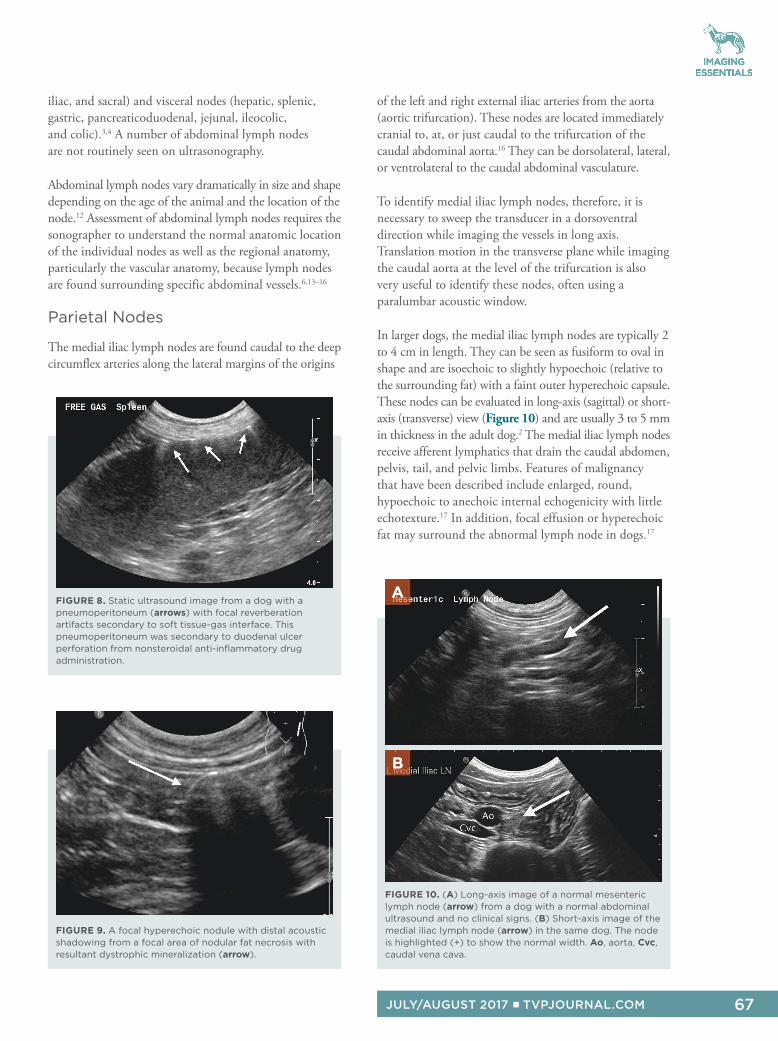
FIGURE 8. Static ultrasound image from a dog with a
pneumoperitoneum (arrows) with focal reverberation
artifacts secondary to soft tissue-gas interface. This
pneumoperitoneum was secondary to duodenal ulcer
perforation from nonsteroidal anti-inflammatory drug
administration.
FIGURE 9. A focal hyperechoic nodule with distal acoustic
shadowing from a focal area of nodular fat necrosis with
resultant dystrophic mineralization (arrow).
FIGURE 10. (A) Long-axis image of a normal mesenteric
lymph node (arrow) from a dog with a normal abdominal
ultrasound and no clinical signs. (B) Short-axis image of the
medial iliac lymph node (arrow) in the same dog. The node
is highlighted (+) to show the normal width. Ao, aorta, Cvc,
caudal vena cava.
A
B
68 IMAGING ESSENTIALS
PEER
REVIEWED
The internal iliac (formerly “hypogastric”) and sacral
lymph nodes are found between the origin of the
external and internal iliac arteries and alongside the
median sacral artery, respectively. These lymph nodes
receive afferent lymphatics from the rectum, pelvic canal,
anal glands, and perineal region. Although these nodes
are not routinely identified, this area should be evaluated
as metastases from tumors in these regions do occur
with enlargement of these specific lymph nodes, and the
internal iliac lymph nodes may then be appreciated.18,19
Unless severely enlarged, the sacral lymph nodes
are typically not visible ultrasonographically due
to their position in the pelvic canal. They are
obscured by the shadow from the pubic bones.
Visceral Nodes
The jejunal or mesenteric lymph nodes are the largest
lymph nodes in the abdomen. They are located
around the cranial mesenteric artery and vein in the
right cranial to middle abdomen just to the right of
the umbilicus (Figure 11). These lymph nodes are
vermiform, cylindrical, and elongated and measure
up to 0.5 cm thick and up to 3 to 4 cm long.20
Contrast ultrasonography has been described to better
characterize lymph node enlargement patterns.21
The jejunal lymph nodes are usually reactive and
enlarged in young dogs and cats up to 1 year of age
(Figure 11).3,4,13 They can be heteroechoic with
multiple peripheral hypoechoic nodules. The jejunal
or mesenteric lymph nodes are commonly enlarged in
inflammatory (eg, secondary to inflammatory bowel
disease), infectious (eg, pythiosis), and neoplastic (eg,
metastatic disease from adenocarcinoma or involvement
in multicentric round cell neoplasia; Figures 12 and 13)
disorders of the gastrointestinal tract.
The appearance of the cisterni chyli has been
reported as an anechoic tubular structure, without
detectable flow, at the right dorsolateral aspect
of the aorta at the level of the cranial mesenteric
artery. The shape and size of the cisterna chyli in an
individual dog can vary during the same ultrasound
examination and between different examinations.22
SUMMARY
As in all cases of abdominal disease, increases or
decreases in overall echogenicity are subjective, and
sonographers must be familiar with how the peritoneal
and retroperitoneal spaces and abdominal lymph nodes
appear in normal dogs and cats when scanned with their
machines. Severe enlargement of abdominal lymph
nodes is usually an indicator of neoplasia (multicentric or
metastatic); however, mild to moderate enlargement can
indicate either neoplasia or reactive lymphadenopathy
secondary to inflammation or infection.
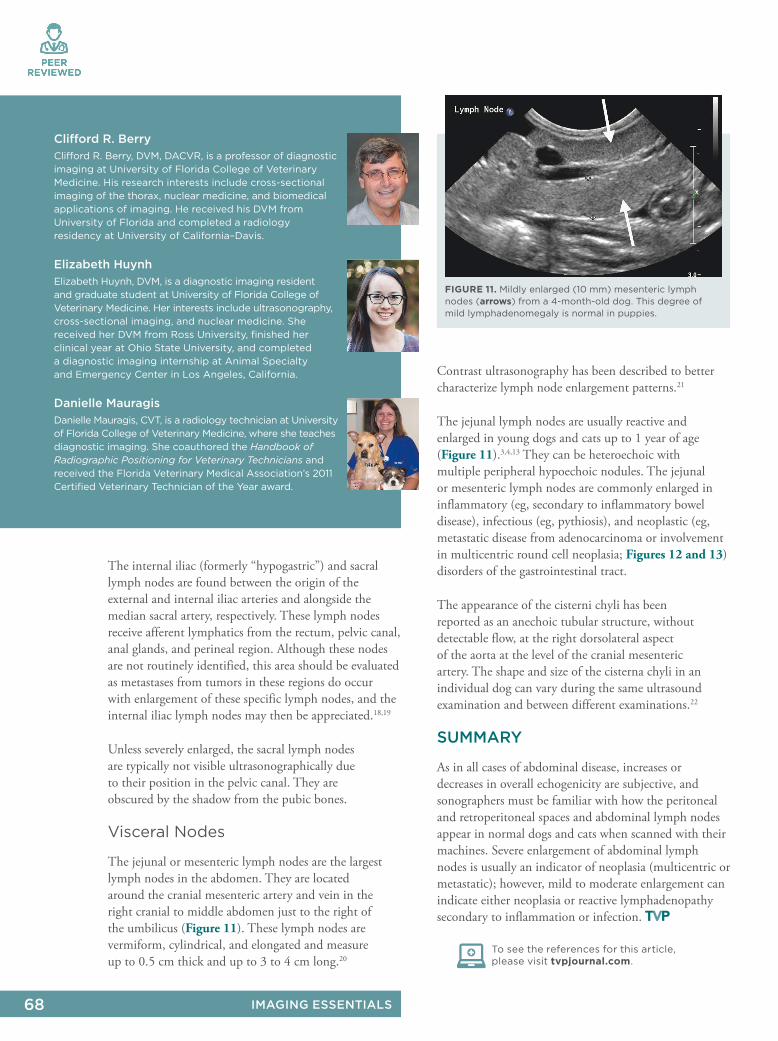
FIGURE 11. Mildly enlarged (10 mm) mesenteric lymph
nodes (arrows) from a 4-month-old dog. This degree of
mild lymphadenomegaly is normal in puppies.
Clifford R. Berry
Clifford R. Berry, DVM, DACVR, is a professor of diagnostic
imaging at University of Florida College of Veterinary
Medicine. His research interests include cross-sectional
imaging of the thorax, nuclear medicine, and biomedical
applications of imaging. He received his DVM from
University of Florida and completed a radiology
residency at University of California–Davis.
Elizabeth Huynh
Elizabeth Huynh, DVM, is a diagnostic imaging resident
and graduate student at University of Florida College of
Veterinary Medicine. Her interests include ultrasonography,
cross-sectional imaging, and nuclear medicine. She
received her DVM from Ross University, finished her
clinical year at Ohio State University, and completed
a diagnostic imaging internship at Animal Specialty
and Emergency Center in Los Angeles, California.
Danielle Mauragis
Danielle Mauragis, CVT, is a radiology technician at University
of Florida College of Veterinary Medicine, where she teaches
diagnostic imaging. She coauthored the Handbook of
Radiographic Positioning for Veterinary Technicians and
received the Florida Veterinary Medical Association’s 2011
Certified Veterinary Technician of the Year award.
To see the references for this article,
please visit tvpjournal.com.
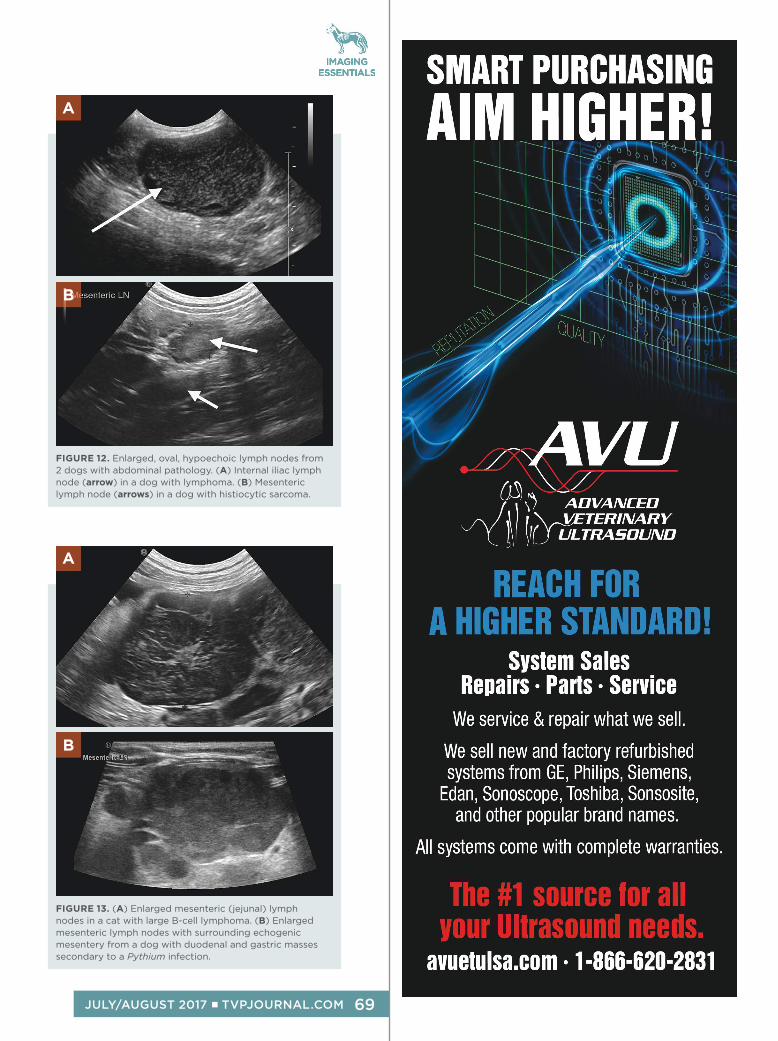
FIGURE 12. Enlarged, oval, hypoechoic lymph nodes from
2 dogs with abdominal pathology. (A) Internal iliac lymph
node (arrow) in a dog with lymphoma. (B) Mesenteric
lymph node (arrows) in a dog with histiocytic sarcoma.
A
B
FIGURE 13. (A) Enlarged mesenteric (jejunal) lymph
nodes in a cat with large B-cell lymphoma. (B) Enlarged
mesenteric lymph nodes with surrounding echogenic
mesentery from a dog with duodenal and gastric masses
secondary to a Pythium infection.
A
B
IMAGING ESSENTIALS
69JULY/AUGUST 2017 ■ TVPJOURNAL.COM