Embed Size (px)
Citation preview
Imaging and Phase I Study of111In- and 90Y-labeled Anti-LewisY
Monoclonal Antibody B3
Lee H. Pai-Scherf, Jorge A. Carrasquillo,Chang Paik, Otto Gansow, Millie Whatley,Deb Pearson, Keith Webber, Michael Hamilton,Carmen Allegra, Martin Brechbiel,Mark C. Willingham, and Ira Pastan 1
Laboratory of Molecular Biology [L. H. P-S., I. P.], Department ofNuclear Medicine, Warren G. Magnuson Clinical Cancer Center[J. A. C., C. P., M. W.], Medicine Branch [D. P., M. H., C. A.],Chemistry Section, ROB [O. G., M. B.], National Cancer Institute,NIH, Bethesda, Maryland 20892; Food and Drug Administration,Bethesda, Maryland 20892 [K. W.]; and Department of Pathology,Wake Forest University, Winston-Salem, North Carolina 27157[M. C. W.]
ABSTRACTB3 is a murine monoclonal antibody (mAb) that recog-
nizes a LewisY carbohydrate antigen present on the surfaceof many carcinomas. An imaging and Phase I trial wasperformed to study the ability of 111In-mAb B3 to imageknown metastasis and determine the maximum tolerateddose (MTD), dose-limiting toxicity (DLT), kinetics, and bio-distribution of 90Y-mAb B3. Patients (n5 26) with advancedepithelial tumors that express the LewisY antigen were en-tered. All patients received 5 mCi of 111In-mAb B3 forimaging. 90Y-mAb B3 doses were escalated from 5 to 25 mCiin 5-mCi increments. 111In-mAb B3 and 90Y-mAb B3 werecoadministered over a 1-h infusion. Definite tumor imagingwas observed in 20 of 26 patients. Sites imaged includedlung, liver, bone, and soft tissues. The MTD of90Y-mAb B3was determined to be 20 mCi. The DLTs were neutropeniaand thrombocytopenia. Tumor doses ranged from 7.7 to 65.1rad/mCi. 111In- and 90Y-mAb B3 serum pharmacokinetics(n 5 23) were found to be similar. The amount of B3administered (5, 10, and 50 mg) did not alter the pharma-cokinetics. Bone marrow biopsies (n 5 23) showed 0.003860.0016% of injected dose/gram for111In-mAb B3 comparedto 0.00466 0.0017% of injected dose/gram for90Y-mAb B3(P 5 0.009). When given to patients with carcinomas thatexpress the LewisY antigen, 111In-mAb B3 demonstratedgood tumor localization. The MTD of 90Y-mAb B3 is 20mCi, with myelosuppression as the DLT. Higher doses ofradioactivity need to be delivered to achieve an antitumor
effect. Humanized mAb B3 is being developed for evaluationin radioimmunotherapy. A clinical trial to explore the use ofhigher doses of90Y-mAb B3 with autologous stem cell sup-port is planned.
INTRODUCTIONThe use of radiolabeled mAbs2 for radioimmunotherapy
has been evaluated extensively in hematological malignanciesand in some epithelial cancers (1, 2). Whereas therapeuticresponses have been observed in patients with lymphoma andleukemia (3–6), results in patients with epithelial tumors havebeen disappointing (7–9). These poor responses are generallyattributed to limitations in antibody delivery and unfavorabletumor dosimetry. Tumor dose is affected by the delivery ofradiolabeled antibody, which is in turn dependent on physicalbarriers to antibody delivery (10, 11), antigen density, andphysical characteristics of the radionuclide (12).
In this study, we determined whether mAb B3, a murineIgG1k that reacts with the LewisY carbohydrate epitope (B3antigen; Ref. 13), would serve as a target for radioimmuno-therapy. This epitope is present on a large number of glycopro-teins and is abundantly and uniformly expressed by most car-cinomas, including.95% of colorectal cancer, 80% of breastcancer, and 60% of non-small cell lung cancer as well asesophageal, gastric, pancreatic, ovarian, and bladder carcino-mas. In contrast, mAb B3 has limited reactivity with normaltissues. mAb B3 has been chemically linked to a truncated formof Pseudomonasexotoxin to form immunotoxin B3-LysPE38(LMB-1). In a Phase I clinical trial, LMB-1 was given to 38cancer patients with tumors that react with mAb B3. In thatstudy, five objective responses (1 complete remission, 1 partialremission, and 3 minor responses) were observed (14), indicat-ing that the B3 antigen can be used as target for cancer therapy.
To explore the usefulness of B3 antigen as a target forradioimmunotherapy, preclinical experiments were conducted.When injected into immunodeficient mice bearing a humanepidermoid carcinoma that expresses the B3 antigen,111In-B3showed selective and progressive accumulation at the tumor site(15). These results and preclinical biodistribution studies using88Y-mAb B3 (16, 17) indicated that radiolabeled B3 warrantsfurther clinical evaluation.
Whereas the largest number of radioimmunotherapy stud-ies have focused on using131I-labeled antibodies, several lim-itations have been identified, including rapid dehalogenation
Received 6/8/99; revised 2/7/00; accepted 2/16/00.The costs of publication of this article were defrayed in part by thepayment of page charges. This article must therefore be hereby markedadvertisementin accordance with 18 U.S.C. Section 1734 solely toindicate this fact.1 To whom requests for reprints should be addressed, at Laboratory ofMolecular Biology, National Cancer Institute, Building 37, Room 4E16,37 Convent Drive MSC 4255, Bethesda, MD 20892-4255. Phone: (301)496-4797; Fax: (301) 402-1344.
2 The abbreviations used are: mAb, monoclonal antibody; HSA, humanserum albumin; % ID, percentage of injected dose; HAMA, humanantimouse antibody; AUC, area under the curve; MTD, maximumtolerated dose; SPECT, single-photon emission computed tomography;ANC, absolute neutrophil count; CT, computed tomography; DLT,dose-limiting toxicity.
1720Vol. 6, 1720–1730, May 2000 Clinical Cancer Research
Research. on May 26, 2020. © 2000 American Association for Cancerclincancerres.aacrjournals.org Downloaded from
(18, 19) and emission of high-energyg-rays, which imposescertain radiation safety constraints.90Y has been evaluated as analternative to131I for radioimmunotherapy because of its readyavailability from a90Sr/90Y generator (20) and its physical andbiological characteristics (21–24). Whereas90Y has favorablecharacteristics for therapy [t1/2 5 64 h; pure b-emission(Emax 5 2.28 MeV)], the lack ofg-ray emission makes itsuboptimal for imaging and assessing biodistribution (12). Totrace the biodistribution of90Y, 111In has been used as a surro-gate marker because it has similar coordination chemistry (25,26) and metabolic handling (19, 22). In this study, we carefullycompared the differences in biodistribution between111In- and90Y-labeled B3. Prior studies using weaker chelates have shownsignificant differences between111In and 90Y (23, 24). Newerchelates have greaterin vitro and in vivo stability (16, 27, 28).Nevertheless, even with improved chelates, some differencesbetween111In and 90Y have been observed (17, 24). Using adifferent antibody labeled with111In and 90Y via the 1B4Mchelate (also known as Mx-diethylenetriamine pentaacetic acid;Ref. 27) in patients with adult T-cell leukemia (3), we havepreviously shown that there are differences in biodistributionbetween these two radiolabels, although these differences weresmall (29). Other investigators have also used the same chelateconjugate to label other antibodies (30–32); nevertheless, this isthe first detailed pharmacokinetic comparison of these twoisotopes using this chelate in epithelial tumors.
This study presents the results of a clinical trial in patientswith advanced carcinomas that express the B3 antigen. Westudied the ability of111In-1B4M-mAb B3 to image knownmetastasis and performed a Phase I trial to determine the tox-icities, pharmacokinetics, and the MTD of90Y-1B4M-mAb B3.
MATERIALS AND METHODSB3 mAb. mAb B3 is a murine IgG1 developed as de-
scribed previously (13). The mAb B3 used for this clinical trialwas purified by Verax Co. (Lebanon, NH) from low serumculture medium using ion-exchange chromatography. It wasover 95% pure as established by SDS-PAGE.
Conjugation and Labeling. The B3 mAb was conju-gated to 2-(4-isothiocyantobenzyl)-6-methyl-diethylenetriaminepentaacetic acid (1B4M-diethylenetriamine pentaacetic acid;Ref. 27). Radiolabeling was performed with pharmaceuticalgrade111In (DuPont New England Nuclear, Wilmington, DE)for imaging and/or pharmaceutical grade90Y for therapy (Du-Pont New England Nuclear). In brief, 1.0–1.2 mg of conjugatedmAb B3 was put into a polypropylene vial that served as thereaction vessel. For111In, 10.2–23.2 mCi were added to thereaction vessel and allowed to react for 1 h. For90Y labeling, thestarting amount of radioactivity and antibody dose depended onthe dose level to be used. Typically, 8.9–53.3 mCi of90Y wereincubated with 1.08–4.31 mg of the conjugate for 15 min. Afterthe initial eight patients’90Y labeling, the method was modifiedto add ascorbate (11 mg/0.05 ml) as a radioprotectant during theincubation with90Y. Excess DTPA (1024
M) was then added tothe incubation mixtures to form complexes with unreacted ionicisotope. The mAb B3-bound fraction was separated by prepar-ative size-exclusion high-performance liquid chromatography(3). Purification resulted in a final product with.99% antibody-
bound111In or 90Y. The90Y fraction was mixed with 25% HSAto yield a 2.5% HSA solution. Purity was determined by instantthin-layer chromatography using silica gel-impregnated glassfiber sheets (2;2;1, 10% ammonium formate in water/metha-nol/0.2 M citric acid) and paper chromatography using salinesolvent and Whatmann #1 paper pretreated with 5% HSA. Thefinal product was filtered using a sterile 0.22-mm low-protein-binding filter (Millex-GV; Millipore, Inc., Bedford, MA). Thespecific activities of the111In-mAb B3 doses (n5 26) rangedfrom 4.3–13.0 mCi/mg (7.36 1.8 mCi/mg). The total111Inactivity injected ranged from 3.5–5 mCi. The specific activitiesof the 90Y-mAb B3 doses (n5 23) ranged from 5.3–12.9mCi/mg (8.46 1.7 mCi/mg), with individual doses of 5–25 mCiof 90Y-mAb B3. All products passed sterility and pyrogentesting. The111In-labeled products were injected within 72 h ofpreparation. Twenty-two of 2390Y-mAb B3 doses were injectedthe day of labeling, whereas one product was injected the nextday (24 h). The dose injected the day after labeling was retestedbefore injection and showed similar protein-bound radioactivity.
The immunoreactivity of the radiolabeled products wastested using a modification of the cell-binding assay describedby Lindmo et al. (33). In brief, an increasing number of A431cells were incubated in 6-well plates in cell numbers rangingfrom 4 3 105 to 1 3 106. A small fixed amount of theradiolabeled B3 was added to the wells and incubated for 2 h.After incubation, the wells were washed, and the percentage ofactivity bound to cells was determined. Overall, the cell bindingassay for the111In and 90Y preparations was not significantlydifferent, with a mean6 SD of 696 10% and 696 17% for111In- and 90Y-mAb B3, respectively. When the immunoreac-tivity from 90Y preparations labeled in the absence of ascorbicacid were compared with those labeled in the presence ofascorbic acid, a significant difference was observed (586 10%and 746 18%, respectively).
Patient Selection. Adult patients with metastatic gastro-intestinal tract, breast, non-small cell lung, bladder, and ovariancarcinoma who had failed standard therapy were eligible for thisstudy. Tumors expressed B3 antigen on$30% of the tumorcells as determined by immunohistochemistry. Tumor histologywas confirmed by a NIH pathologist. Other eligibility criteriaincluded: (a) advanced unresectable disease; (b) failed conven-tional chemotherapy; (c) Eastern Cooperative Oncology Groupperformance status of#2; (d) a minimum life expectancy of 3months; (e) serum creatinine, 1.6 mg/dl; (f) serum bilirubin,1.5 mg/dl; (g) absolute neutrophil count (ANC). 2,000/mm3;and (h) platelets. 100,000/mm3. Patients with clinically sig-nificant cardiac disease (New York Heart Association grade 3 or4) were excluded, as were those with infectious disease thatrequired antibiotic therapy, brain metastasis, prior exposure tomurine antibodies, pregnancy, or lactation. Patients who hadreceived bone marrow transplant therapy, more than 3 chemo-therapy regimens, pelvic radiation, or local radiation to morethan one site were excluded. The clinical protocol and theconsent form were approved by the Institutional Review Boardof the National Cancer Institute. Informed consent was obtainedfrom all patients before participation in this study.
Study Design. An outline of the protocol design and thenumber of patients entered in each group are shown in Table 1.In the initial portion of this study, three patients received 5 mCi
1721Clinical Cancer Research
Research. on May 26, 2020. © 2000 American Association for Cancerclincancerres.aacrjournals.org Downloaded from
of 111In-mAb B3. The objective was to determine whether therewould be any gross unexpected sites of localization of theantibody or acute toxicities that would prompt us not to proceedwith the therapy portion of the study. To provide potentialtherapeutic benefit, these initial patients were offered therapywith 5 mCi of 90Y-mAb B3, if they had tumor imaging, noHAMAs, and no toxic side effects or unexpected, undesirabletissue accumulation as compared with other111In-labeled anti-bodies. In the second portion of the study, we evaluated whetherthe amount of antibody resulted in dose-dependent changes inbiodistribution. Three groups of three patients each received111In (5 mCi)- and90Y-mAb B3 (5 mCi) mixed together with 5,10, or 50 mg of unlabeled mAb B3. In the third and main partof the protocol, the MTD of90Y was determined. Groupscontaining three patients each received escalating doses of90Y-mAb B3 for therapy coinfused with 5 mCi of111In and a totalof 10 mg (based on a lack of dose-dependent changes at thehigher amounts of mAb B3). The90Y doses were escalated in5-mCi intervals. Patients with hematological toxicity, grade 3were eligible for retreatment with the same dose of90Y if theyhad no evidence of disease progression and remained HAMAnegative.
111In-mAb B3 and90Y-mAb B3 were coadministered i.v.over a 1-h infusion in an outpatient setting.
Pharmacokinetics. Intravascular kinetics were deter-mined by counting111In or 90Y radioactivity in blood andplasma aliquots obtained at the following times after the end ofinfusion: 5 min, 30 min, 1 h, 2 h, 6 h, 1 day, and daily for up to7 days after the end of the infusion. The % ID/ml was obtainedby comparing the counts to a standard of the injected dose. Theplasma and blood volumes were estimated at each time oftreatment using a nomogram based on body surface area (34).Using the latter estimated volumes and the % ID/ml, the total %ID in the blood and plasma volume was calculated. Because theinfusion time was short compared with the disposition half-life(t1/2), the intravascular data were treated similar to an i.v. bolus.The % ID/ml of blood or plasma was fitted to a biexponentialcurve to obtain both thea andb phaset1/2 using a least-squaresfit algorithm. Conventional pharmacokinetic parameters werethen derived (35). The AUCs for the blood or plasma curveswere calculated in two steps. First, the AUC from the end ofantibody infusion (To) to 168 h was obtained by trapezoidalintegration of the decay-corrected blood and plasma data, andthen the terminal AUC was estimated using the terminal clear-ance rate to extrapolate from the activity retained at the last
measured time point. Using this data, we then estimated addi-tional pharmacokinetic parameters, including volume of distri-bution, clearance, and half-life (35). Serial 24-h urine collec-tions were obtained for up to 96 h so that we could compare theurinary excretion of the two tracers. Whole body clearance of111In was determined from the imaging data (see below).
Imaging. Scintillation camera images were first recordedup to six times with a large field of view dual-headed gammacamera starting within;2 h of the end of the infusion and dailyfor up to 6 or 7 days. Analogue and digital images of anteriorand posterior whole body as well as spot views (5–10 min/image) were obtained. For quantitative imaging, a 20% windowcentered over the 247 keV photopeak of111In was obtained. Ascatter correction method was utilized. The images were cor-rected for attenuation using a99mTc flood source and allowingfor the energy differences and the sensitivity of the gammacamera. A geometric mean image was then generated (in unitsof mCi/pixel). For visual interpretation, individual anterior andposterior images were reviewed. SPECT of the chest, abdomen,and pelvis was recorded using a medium energy collimator;SPECT was typically performed 4 to 5 days after injection. The111In SPECT images were obtained for visual assessment uti-lizing a 20% window centered over the 174 and 247 keVphotopeak of111In; the images were reconstructed using aHamming filter with a high cutoff frequency of 0.75 cycle/cm.All images were interpreted by one experienced nuclear medi-cine physician.
Two quantitative image regions of interest were drawnover the liver, spleen, and L4 vertebral body on the geometricmean images. The integrated radioactivity in the organs (AUC)was then determined by trapezoidal integration up to the lasttime point imaged (typically 7 days), and the remaining AUCwas determined by extrapolation using the terminalt1/2 ofclearance. The whole bodyt1/2 was obtained by fitting thegeometric mean concentration of111In activity of the anteriorand posterior whole body scans. Using the residence timesobtained from the data above and the individual organ size, asdetermined from CT, the medical internal radiation dose methodwas used to calculate organ dosimetry (36). In brief, the integralof radioactivity in a given organ, blood, or tissue was divided byits weight (mCizh/g) and multiplied by the mean energy emittedper nuclear transition of theb particle from90Y (1.99 gzRad/mCi/h), assuming an absorbed dose fraction of 1. The activity inL4 was determined from the region of interest analysis andintegrated over time. This activity was normalized by the gramsof bone marrow in L4 as estimated in standard man (37). Thedose to the bone marrow was then estimated as described above.
Counting Methods. Dual isotope counting of111In and90Y was performed on the patient samples. The111In g-raypeaks were counted in a gamma counter using a 100–500 keVenergy setting. Because90Y is counted with,4% efficiency ina gamma counter, Cerenkov counting in a beta counter was alsoutilized. Cerenkov counting was performed using an energyrange of 0–200 keV (A4530D Packard, Downes Grove, IL) in abeta counter. Because Cerenkov counting is sensitive to quenchand geometry, all samples were processed in a similar andreproducible manner as described previously (29), which in-cluded solubilizing the samples with SDS and bleaching themwith 30% hydrogen peroxide to minimize quench. The counts in
Table 1 Protocol outline
Imaging(111In-B3)
(mCi)
Therapy(90Y-B3)
(mCi)Total B3
(mg) n
5 5 (optional) 5 35 5 5 35 5 10 35 5 50 35 10 10 35 15 10 35 20 10 65 25 10 2
1722Differences between111In and90Y-labeled mAb B3
Research. on May 26, 2020. © 2000 American Association for Cancerclincancerres.aacrjournals.org Downloaded from
the samples were referred back to a standard of the injected dosethat had been prepared in a similar fashion using the patients’plasma or blood to mimic the quench. The counts obtained in thegamma and beta counters were corrected for cross-talk anddecay.
Bone Marrow Biopsies. Twenty-five patients underwentbone marrow biopsy of the posterior iliac spine within 5–7 daysafter mAb injection. Thirteen biopsies were performed 6 daysafter therapy, eight were performed at 5 days after therapy, andfour were performed at 7 days after initial therapy. The biopsycores were weighed on an analytical balance and put in a conicaltube with 10 ml of PBS for 1 h and analyzed as describedpreviously (29). The core was broken with a jagged-edged glassrod. This was centrifuged for 10 min at 6403 g, and thesupernatant was removed and counted (saline fraction). Thepelleted core was broken with a jagged-edged glass rod andmixed with 0.5 ml of 10% SDS. The core was heated to 56°C for30 min in an attempt to remove any cell-bound activity. Afterthe sample cooled, 0.4 ml of 30% hydrogen peroxide was addedas bleach, and the mixture was incubated at 56°C for 1 h tobleach the sample. Ten ml of distilled water were added, and thesample was again centrifuged for 10 min. The supernatant wasseparated for counting (SDS fraction). Perchloric acid (0.2 ml)was then added to the remaining bone chips, and the mixturewas incubated at 56°C until the bone was dissolved (bonefraction). This sample was again treated with hydrogen peroxideas described above. After cooling, the sample was transferred toa counting vial with 10 ml of distilled water. All samples werethen counted in the gamma and beta counters with the appro-priate decay and cross-talk corrections.
To attempt to find parameters that predict bone marrowtoxicity, we correlated the dose to bone marrow based onimaging, bone marrow biopsy, and blood retention of90Y. Thedose to the blood was calculated as described above, and thedose to the marrow was then determined by multiplying timesthe red marrow;blood ratio as described by Sgouroset al. (38).The dose from marrow biopsies was determined by calculatingthe activity concentrated in the marrow by gamma counting ofa precisely weighted specimen. We assumed that there was nobiological clearance from the marrow, based on gamma cameraimaging. The dose was then calculated as described above, withno corrections for cortical bone. In addition, we correlatedtoxicity to the bone marrow with administered activity or ad-ministered activity corrected for body weight.
HAMA Assays. HAMA assay was performed as de-scribed previously using a high-performance liquid chromatog-raphy method (39). More than 10% complex formation using a125I isotype matched nonspecific mAb (BL-3) and125I-labeledB3 was considered positive.
Statistics. To compare independent data,111In and 90Ypatient data obtained from the initial dual-injection study wereused. Pairedt test or Wilcoxon rank signed test (when data werenot normally distributed) was performed to assess the differ-ences in biodistribution between the two radiolabels. Pearson’scorrelation coefficient was used to evaluate the relationshipbetween nominal data, and Spearman correlation coefficient wasused with ordinal data.
RESULTSPatient Characteristics and Clinical Observation.
Twenty-six patients were entered into this study. Their clinicalcharacteristics are shown in Table 2. There were 12 men and 14women (age range, 39–73 years; mean age, 58 years). Twentypatients had colorectal cancer, 2 patients had esophageal cancer,1 patient had gastric carcinoma, 1 patient had carcinoma of theampulla of Vater, 1 patient had breast cancer, and 1 patient hadbronchioalveolar cancer. Prior therapy and sites of metastasisare shown in Table 2. Immunohistochemical staining of B3 wasperformed on paraffin-embedded tumor blocks from the primarysurgery in all cases. Homogeneous B3 expression was found in23 of 26 patients. In 3 of 26 cases, specimens were poorlypreserved (tightly fixed). However in all three cases, sectionsindicated that in the better preserved areas, more than 30% ofthe tumor cells were clearly positive for B3 antigen.
Infusion of unlabeled and radiolabeled antibody was welltolerated, accompanied by minimal or no side effects. Nonhe-matological toxicities were mild and transient (grade 1 and 2),with low-grade fever observed in four patients within 24 h afterdosing. Rigor and chills were reported in one patient. Grade 1arthralgia (one patient), myalgia (two patients), and fatigue (onepatient) were observed at 2–4 weeks after dosing. Patients weretreated with acetaminophen or ibuprofen as needed. It is un-likely that these symptoms were related to the therapy. Asexpected, the DLT was myelosuppression (see below). Therewere no significant changes in the chemistry profiles related to
Table 2 Patient characteristics
n 5 26Sex: 12 male, 14 femaleAge: 39–73 yrs (mean5 58)Tumor histology
20 colon carcinomas2 esophageal cancers1 gastric cancer1 breast cancer1 bronchoalveolar cancer1 cancer of the ampulla of Vater
Prior therapyChemotherapy
1 regimen 11 (42%)2 regimens 10 (38%)3 regimens 4 (15%)
Hormonal 1 (3%)Biological (INF)a 6 (26%)Radiation (1 site) 3 (11%)
No. of metastatic sites1 site 10 (38%)2–3 sites 14 (54%)$4 sites 2 (7%)
Sites of metastasis, number of patientsLiver, 17Lung, 13Soft tissue, 8 (perit, 4; pleura, 1; abdominal wall, 3)Lymph nodes, 8 (chest 2, abdomen 6)Bone, 3Stomach, 2Effusion, 6 (pleura, 2; ascites, 4)a INF, interferon.
1723Clinical Cancer Research
Research. on May 26, 2020. © 2000 American Association for Cancerclincancerres.aacrjournals.org Downloaded from
the mAb treatment. The disease status remained stable for 6weeks in four patients. There were no clinical responses to theinfusion of 90Y-mAb B3.
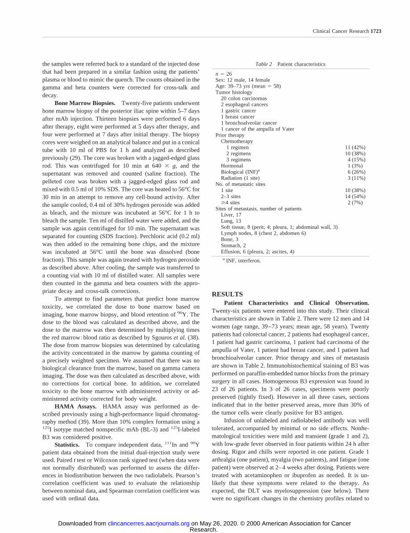
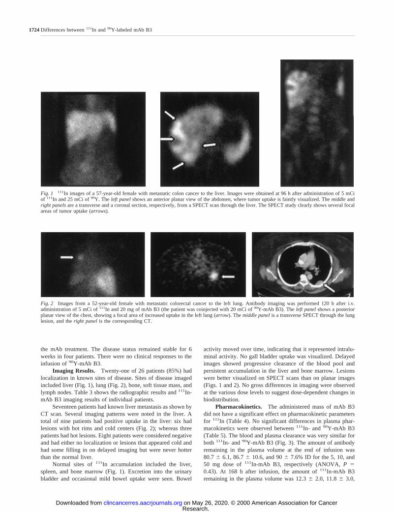
Imaging Results. Twenty-one of 26 patients (85%) hadlocalization in known sites of disease. Sites of disease imagedincluded liver (Fig. 1), lung (Fig. 2), bone, soft tissue mass, andlymph nodes. Table 3 shows the radiographic results and111In-mAb B3 imaging results of individual patients.
Seventeen patients had known liver metastasis as shown byCT scan. Several imaging patterns were noted in the liver. Atotal of nine patients had positive uptake in the liver: six hadlesions with hot rims and cold centers (Fig. 2); whereas threepatients had hot lesions. Eight patients were considered negativeand had either no localization or lesions that appeared cold andhad some filling in on delayed imaging but were never hotterthan the normal liver.
Normal sites of111In accumulation included the liver,spleen, and bone marrow (Fig. 1). Excretion into the urinarybladder and occasional mild bowel uptake were seen. Bowel
activity moved over time, indicating that it represented intralu-minal activity. No gall bladder uptake was visualized. Delayedimages showed progressive clearance of the blood pool andpersistent accumulation in the liver and bone marrow. Lesionswere better visualized on SPECT scans than on planar images(Figs. 1 and 2). No gross differences in imaging were observedat the various dose levels to suggest dose-dependent changes inbiodistribution.
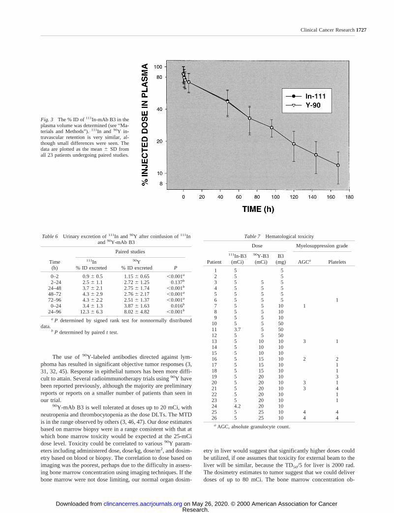
Pharmacokinetics. The administered mass of mAb B3did not have a significant effect on pharmacokinetic parametersfor 111In (Table 4). No significant differences in plasma phar-macokinetics were observed between111In- and 90Y-mAb B3(Table 5). The blood and plasma clearance was very similar forboth 111In- and90Y-mAb B3 (Fig. 3). The amount of antibodyremaining in the plasma volume at the end of infusion was80.76 6.1, 86.76 10.6, and 906 7.6% ID for the 5, 10, and50 mg dose of111In-mAb B3, respectively (ANOVA,P 50.43). At 168 h after infusion, the amount of111In-mAb B3remaining in the plasma volume was 12.36 2.0, 11.86 3.0,
Fig. 1 111In images of a 57-year-old female with metastatic colon cancer to the liver. Images were obtained at 96 h after administration of 5 mCiof 111In and 25 mCi of90Y. The left panelshows an anterior planar view of the abdomen, where tumor uptake is faintly visualized. Themiddleandright panelsare a transverse and a coronal section, respectively, from a SPECT scan through the liver. The SPECT study clearly shows several focalareas of tumor uptake (arrows).
Fig. 2 Images from a 52-year-old female with metastatic colorectal cancer to the left lung. Antibody imaging was performed 120 h after i.v.administration of 5 mCi of111In and 20 mg of mAb B3 (the patient was coinjected with 20 mCi of90Y-mAb B3). Theleft panelshows a posteriorplanar view of the chest, showing a focal area of increased uptake in the left lung (arrow). Themiddle panelis a transverse SPECT through the lunglesion, and theright panel is the corresponding CT.
1724Differences between111In and90Y-labeled mAb B3
Research. on May 26, 2020. © 2000 American Association for Cancerclincancerres.aacrjournals.org Downloaded from
and 126 1.6% ID, for the 5, 10, and 50 mg dose, respectively(ANOVA, P 5 0.96).
Antibody dose did not result in significant differences inthe 0–96-h urinary excretion of111In or 90Y. Patients receiving
5 (n 5 3), 10 (n5 17), or 50 mg (n5 3) of mAb B3 showeda median urinary excretion (0–96 h) of 11.9%, 15.1%, and12.2%, respectively (Kruskal-Wallis one-way ANOVA,P 50.26). Similarly, the median urinary excretion of90Y at thecorresponding time and doses was 9.7%, 11.1%, and 8.3%,respectively (P5 0.06).
The amount of90Y injected did not affect the urinaryexcretion (0–96 h) of90Y. Patients receiving;5 (n 5 3), 10(n 5 3), 15 (n5 3), or 20 mCi (n5 6) showed no significantdifferences in urinary excretion of90Y with 8.5 6 2.2, 11.161.0, 18.9 6 15.1, and 13.36 3.7% ID excreted in 96 h,respectively (ANOVA,P 5 0.35).
The urinary excretion of111In and 90Y in the intervalcollected is shown in Table 6. The urinary excretion of90Y inthe first 24 h was greater than that of111In [3.9 6 1.6%compared with 3.46 1.3% ID, respectively (P5 0.016)].However, at 48–96 h, the urine excretion of90Y was less thanthat of 111In [8.0 6 4.8% ID excreted compared with 12.366.3% ID, respectively (P5 0.016)].
Hematological Toxicity. The dose and the hematologi-cal toxicity in individual patients are shown in Table 7. Hema-tologic DLT for this study was defined as an ANC, 500/ml for.5 days or platelets, 25,000/ml for.5 days. At doses of 5 and10 mCi of 90Y-mAb B3, minimal to no hematological toxicitywas observed. Grade 2 and grade 3 thrombocytopenia wereobserved at 15 and 20 mCi of90Y-mAb B3. One instance ofDLT occurred in one patient at 20 mCi of90Y (patient 21); as aresult, a total of six patients was treated at this dose level.Dose-limiting neutropenia and thrombocytopenia occurred intwo patients who received 25 mCi of90Y (patients 25 and 26).Grade 4 neutropenia (absolute granulocyte count, 500) wasobserved at day 28 after dosing and lasted 5 and 18 days. Onepatient required hospital admission and i.v. antibiotics for fever.Both patients received granulocyte colony-stimulating factor.Grade 4 thrombocytopenia (platelets5 25,000/ml) requiringplatelet transfusion was observed at 18 and 25 days after dosing,lasting 42 days in one patient and 11 days in another patient.
In an attempt to obtain parameters that would predicttoxicity, correlation of various indices of administered activityor bone marrow dose was correlated to either a percentage dropin WBC, ANC, or platelets or to toxicity grade in these param-eters (Table 8). With the exception of estimates of bone marrowdose from imaging, the majority of the parameters showed agood correlation with changes in peripheral blood counts. Thecorrelation of drop in blood counts or bone marrow toxicitybased on the dose (mCi) administered correlated in the samerange as the other parameters that corrected the dose (mCi)based on weight. The estimates of bone marrow dose based onthe blood radioactivity or the90Y accumulation in the bonemarrow biopsy were in the same range and were not betterpredictors of toxicity than the administered activity.
Bone Marrow. The dose of antibody (mg) did not alterthe localization of radioactivity in the bone marrow. The meanconcentration of111In in the bone marrow was not significantlydifferent for the 5, 10, or 50 mg doses and had a mean of 0.0043,0.00391, and 0.00554% ID/g (ANOVA,P 5 0.516). Similarly,no significant difference in the concentration of90Y was seen inthe bone marrow.
The concentration of radioactivity in the bone marrow did
Table 3 Radiographic and111In-mAb B3 imaging results
Patient Radiographic results 111In imaging results
1 Lung Lung1Liver Liver 2 (fill in only)
Abdomen false1 (bowel)2 Lung Lung1
Liver Liver 2 (cold center)3 Lung Lung1
Bone Bone1Mediastinum/hilar LNa Mediastinum1/ hilar 1
4 Liver Liver 2 (cold center)Periaortic LN Periaortic1
5 Pancreatic mass Pancreatic mass26 Liver Liver 1 (hot rim)7 Bone (rib) Bone1
Lung (0.5 cm) Lung nodule2Presaccral area false1
8 Pleura (RLL) Pleura1Liver Liver 2Retroperitoneal LN Retroperitoneal LN2EG junction mass EG junction mass2
9 Liver Liver 2 (fill in)10 Liver Liver 1 (hot rim)
Periportal LN Periportal LN2Presaccral mass Presaccral mass1
11 Abdominal wall Abdominal wall112 Liver Liver 2 (fill in)13 Lung Lung1
Hilar LN Hilar LN 214 Liver Liver 2 (fill in)15 Lung Lung1
Liver16 Lung Lung117 Liver Liver 1 (hot rim)
Lung Lung1Retroperitoneal LN Retroperitoneal LN2Ascites Ascites1
18 Liver Liver 1 (hot rim)Pelvic mass Pelvic mass1Perihepatic mass Perihepatic mass1Ascites Ascites1
19 Abdominal wall mass Abdominal wall mass1Liver Liver 2 (cold lesions)Lung Lung2Retroperitoneal LN Retroperitoneal LN2Ascites Ascites1
20 Stomach (linitis plastica) Stomach2Ascites Ascites1
21 Liver Liver 1 (hot rim, some cold)Peritoneal mass Peritoneal mass1
22 Liver Liver 1 (hot rim)Retroperitoneal LN Retroperitoneal LN2
23 Liver Liver 1 (hot lesions)Lung Lung1Pleura Pleura1
24 Liver Liver 1 (hot lesions)Bone Bone1Pleural effusion Pleural effusion2
25 Liver Liver 1 (hot rim)Mesentery mass Mesenteric mass1
26 Lung Lung1a LN, lymph node; RLL, right lower lobe; EG, esophageal-gastric.
1725Clinical Cancer Research
Research. on May 26, 2020. © 2000 American Association for Cancerclincancerres.aacrjournals.org Downloaded from
not vary depending on the dose of90Y administered. The meanactivities of111In in the bone marrow for the 5, 10, 15, 20, and25 mCi doses were not significantly different with a mean of0.0039, 0.0029, 0.004, 0.0027, and 0.0044% ID/g, respectively(ANOVA, P 5 0.123). Similarly the % ID/g of90Y in the bonemarrow was not affected by the administered dose (ANOVA,P 5 0.56).
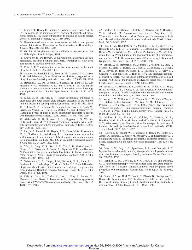
The concentration of111In in the bone marrow was lowerthan that for90Y in the 17 patients receiving 10 mg of antibodywith escalating doses of90Y. The concentration of111In was0.00346 0.0015% ID/g, whereas that for90Y was 0.004760.0015% ID/g. The distribution of the111In and 90Y in thecompartments of the bone measured (saline, SDS, and bone)were different for111In and90Y (Fig. 4).
On processing of the bone marrow, only a small proportionof the activity was lost due to processing. In the case of111In,we recovered a mean of 956 15% of the nonprocessed biopsy.In contrast, because of improved efficiency of bremmstrahlunggeneration after processing, the90Y apparent recovery (efficien-cy) was 1346 29% of the original activity.
Dosimetry. The doses to liver, spleen, bone marrow, andtumor were estimated based on the111In biodistribution. Themean dose to the liver and spleen was 19.46 4.5 and 22.26 8.6rad/mCi, respectively. The maximum total dose to the liver inthis study was 576 rad, and the maximum total dose to thespleen was 716 rad. The dose to the bone marrow based on theAUC of blood was 4.86 1.6 rad/mCi, and the marrow doseestimated from the biopsy was 8.36 3.1 rad/mCi. Many of thetumors visualized were very extensive and overlapped withother tumor or normal structures; in some cases, their size couldnot be determined. The doses to tumors were variable andranged from 7.7–65.1 rad/mCi. The mean dose to tumor was25.16 18.3 rad/mCi.
Tumor Response. No tumor responses were observed inthis study. Stable disease was seen in 6 of 24 patients. No patientcould be retreated because all patients developed HAMA re-sponse within 5–6 weeks after dosing.
DISCUSSIONDuring the past decade, several tumor-specific carbohy-
drate antigens have been identified (40). Because of their abun-dant expression on the surface of many epithelial cancers andtheir relative tissue specificity, surface carbohydrates such asLewisY are ideal for targeted therapy. Clinical trials in cancerpatients have been conducted using anti-LewisY antibodieslinked to toxins (14) or chemotherapy agents (41). In this study,we examined the imaging, toxicities, and pharmacokinetics ofan anti-LewisY antibody termed B3 labeled with radionuclides111In and90Y.
We have shown here that111In-mAb B3 has good sensi-tivity for imaging metastatic epithelial carcinomas that expressthe LewisY antigen. Twenty-one of 26 patients had positiveuptake of 111In-mAb B3, as shown by planar and SPECTimaging. Sites imaged included lung, liver, bone, soft tissuemasses, and lymph nodes. The ability of111In-mAb B3 to detectknown sites of metastasis was most effective when the tumorwas localized in soft tissues, in particular, the peritoneal cavityand the abdominal wall. In this location, strong, localized uptakeof 111In-mAb B3 was observed in eight of eight patients. This isparticularly important if the agent is to be used in the setting ofoccult recurrences in colorectal carcinomas, ovarian carcino-mas, and other gastrointestinal malignancies. Although the ac-cumulation of111In in the normal liver tissue makes it difficultto develop tumor€background ratios high enough to detect themalignancy, clear positive uptake was observed in 9 of 17patients with liver metastasis. Four different patterns were ob-served. In some cases, the liver metastasis presented as hotlesions, whereas others had lesions with a hot rim and coldcenters. Some lesions that were considered negative had eitherno localization or had lesions that appeared cold; however, ondelayed imaging, these lesions showed some filling in but werenever hotter than the normal liver. It is possible that theserepresent areas of necrosis and/or poor vascularization (42–44).Failure of111In-mAb B3 to detect all tumors in this study couldbe due to nonhomogeneous expression of the B3 antigen in themetastatic sites or poor vascularization. It is also possible that anumber of lesions detected by radiographic imaging representreactive lymph nodes or fibrotic tissue.
The usefulness of mAb B3 as an imaging agent would needto be further evaluated in larger clinical studies in patients withsmall volume disease (e.g., patients with elevated carcinoem-bryonic antigen or CA-125 with no radiographic evidence ofdisease). However, the tumor localization of B3 observed in thisfirst trial indicates that the use of this antibody as a radiother-apeutic agent warrants further investigation.
Table 4 Pharmacokinetic parameters for111In-mAb B3: effects of antibody mass
mAb B3 mass (mg) 5 mg 10 mg 50 mg P
No. of patients 6 17 3AUC (% IDzh/ml 2.256 0.76 2.186 0.85 2.036 0.31 0.93
a
Clearance (ml/h) 49.66 18.5 54.66 25.3 50.16 7.8 0.88a
t1/2 b (h) 67.26 16.5 74.46 46.5 75.96 3.9 0.50b
Vc (ml) 32596 358 31646 770 36956 806 0.49a
a P determined by ANOVA.b P determined by Kruskal-Wallis test.
Table 5 Pharmacokinetic parameters based on111In versus90Y111In-mAb B3 90Y-mAb B3 P
No. of patients 23 23AUC (% IDzh/ml) 2.236 0.77 2.236 0.81 0.21a
Clearance (ml/h) 51.86 22.6 51.26 22.9 0.15b
t1/2 b (h) 74.76 40 77.36 43 0.06b
Vc (ml) 32156 734 32276 736 0.49b
a Wilcoxon signed rank test.b P determined by pairedt test.
1726Differences between111In and90Y-labeled mAb B3
Research. on May 26, 2020. © 2000 American Association for Cancerclincancerres.aacrjournals.org Downloaded from
The use of90Y-labeled antibodies directed against lym-phoma has resulted in significant objective tumor responses (3,31, 32, 45). Response in epithelial tumors has been more diffi-cult to attain. Several radioimmunotherapy trials using90Y havebeen reported previously, although the majority are preliminaryreports or reports on a smaller number of patients than seen inour trial.
90Y-mAb B3 is well tolerated at doses up to 20 mCi, withneutropenia and thrombocytopenia as the dose DLTs. The MTDis in the range observed by others (3, 46, 47). Our dose estimatesbased on marrow biopsy were in a range consistent with that atwhich bone marrow toxicity would be expected at the 25-mCidose level. Toxicity could be correlated to various90Y param-eters including administered dose, dose/kg, dose/m2, and dosim-etry based on blood or biopsy. The correlation to dose based onimaging was the poorest, perhaps due to the difficulty in assess-ing bone marrow concentration using imaging techniques. If thebone marrow were not dose limiting, our normal organ dosim-
etry in liver would suggest that significantly higher doses couldbe utilized, if one assumes that toxicity for external beam to theliver will be similar, because the TD50/5 for liver is 2000 rad.The dosimetry estimates to tumor suggest that we could deliverdoses of up to 80 mCi. The bone marrow concentration ob-
Fig. 3 The % ID of111In-mAb B3 in theplasma volume was determined (see “Ma-terials and Methods”).111In and 90Y in-travascular retention is very similar, al-though small differences were seen. Thedata are plotted as the mean6 SD fromall 23 patients undergoing paired studies.
Table 6 Urinary excretion of111In and90Y after coinfusion of111Inand90Y-mAb B3
Time(h)
Paired studies
111In% ID excreted
90Y% ID excreted P
0–2 0.96 0.5 1.156 0.65 ,0.001a
2–24 2.56 1.1 2.726 1.25 0.137b
24–48 3.76 2.1 2.756 1.74 ,0.001b
48–72 4.36 2.9 2.766 2.17 ,0.001a
72–96 4.36 2.2 2.516 1.37 ,0.001a
0–24 3.46 1.3 3.876 1.63 0.016b
24–96 12.36 6.3 8.026 4.82 ,0.001b
a P determined by signed rank test for nonnormally distributeddata.
b P determined by pairedt test.
Table 7 Hematological toxicity
Patient
Dose Myelosuppression grade
111In-B3(mCi)
90Y-B3(mCi)
B3(mg) AGCa Platelets
1 5 52 5 53 5 5 54 5 5 55 5 5 56 5 5 5 17 5 5 10 18 5 5 109 5 5 10
10 5 5 5011 3.7 5 5012 5 5 5013 5 10 10 3 114 5 10 1015 5 10 1016 5 15 10 2 217 5 15 10 118 5 15 10 119 5 20 10 320 5 20 10 3 121 5 20 10 3 422 5 20 10 123 5 20 10 124 4.2 20 1025 5 25 10 4 426 5 25 10 4 4
a AGC, absolute granulocyte count.
1727Clinical Cancer Research
Research. on May 26, 2020. © 2000 American Association for Cancerclincancerres.aacrjournals.org Downloaded from
served, based on bone marrow biopsy, is in the range of thatobserved with murine anti-Tac (29). The111In concentrationsfrom mAb B3 and mAb anti-Tac in bone marrow were 0.00378and 0.00293% ID/g, respectively (P5 0.243 pairedt test). Inaddition, the concentration of90Y in the bone marrow from90Y-B3 and anti-Tac were 0.0047 and 0.00494% ID/g, respec-tively (P 5 0.777, pairedt test).
The blood clearance of111In- and90Y-mAb B3 is similar tothat reported previously with 1B4M chelate and other second-generation chelates (29, 48); in contrast, chelates that are lessstable for90Y have shown less concordance in preclinical mod-els (16) or in clinical trials (49, 50).
The tumor doses that we delivered are in the general rangeof those reported by others using other90Y-labeled antibodies(49, 51). The doses to other organs are generally similar, al-though some antibodies may have a higher concentration in theliver and therefore result in slightly higher liver doses (51, 52).These higher liver doses may be related to the presence ofcirculating antigen and complex formation (51), which was notpresent in our system.
Halpernet al. (53) have previously shown that the distri-bution of each mAb is unique unto itself with regard to its
response to the mass effect. Because this “carrier effect” canalter not just the serum half-time of an antibody but also theorgan tumor distribution of the radionuclide, we administeredincreasing amounts of mAb B3 to patients. No dose-dependentdifferences in pharmacokinetics were observed at doses of 5–50mg of mAb B3, nor were there differences in urine excretionthat related to the dose administered. Pharmacokinetic analysisindicated that there were no significant differences in plasmaclearance of111In- and90Y-mAb B3.
Nevertheless, significant differences in urinary excretion of111In and90Y were observed. These consisted of slightly fasterexcretion of 90Y at the early times after injection but muchhigher excretion at later times. This probably reflects differenthandling of the radiometals, with possible excretion of some111In as catabolism occurs, which would contrast with retentionof 90Y, in particular, in the bone. These findings are similar tothose we have reported previously with two different90Y-radiolabeled mAbs (29, 48). The preferential accumulation of90Y was evident as reflected by the bone marrow biopsy, whichshowed a mean of 1.4 times more90Y than 111In. Using ourcoarse bone marrow fractionation method, it was clear that thegreatest portion of90Y retained was in the bone fraction.
In summary, we have shown here that specific targeting ofthe LewisY antigen using radionuclide linked to mAb B3 isfeasible. It is clear that to achieve an objective antitumor effect,higher doses of radionuclide will need to be delivered. Strategiesto improve the therapeutic index include peripheral autologousstem cell rescue to overcome the myelosuppression (54, 55) andi.v. infusion of EDTA to prevent binding of free90Y to bone(56). As in external beam radiotherapy, the use of fractionatedmultiple doses for delivery of90Y should also be explored.Humanized mAb B3 is presently being produced for evaluationin radioimmunotherapy. This should overcome the problem withHAMA formation. A clinical trial to explore the use of a higherdose of90Y-mAb B3 with autologous stem cell support is beingplanned at the National Cancer Institute.
ACKNOWLEDGMENTSWe thank Debby Drumm for expert technical assistance with
binding assays and handling of patient samples. We also thank Nhat Leand Mark Rotman for expert technical assistance labeling the antibodiesand Craig Barker for expert technical assistance with setting up thequantitative imaging.
Table 8 Correlation of toxicity with administered90Y-mAb B3 dose and with bone marrow dose estimates
% Drop Toxicity grade
WBC ANC PLATELET WBC ANC PLATELET
mCi 0.782a 0.724 0.815 0.712 0.561 0.685mCi/kg 0.802 0.783 0.806 0.741 0.611 0.652mCi/m2 0.821 0.785 0.842 0.739 0.619 0.666Rad dose blood 0.753 0.728 0.737 0.635 0.569 0.558Rad dose bone marrow (biopsy) 0.757 0.695 0.785 0.666 0.567 0.586AUC (humerus) 0.353 0.0474 0.399 0.18 0.343 0.226AUC (L4) 0.218 0.195 0.193 0.44 0.353 0.425a Pearson correlation coefficient was used for % drop data (nominal data); Spearman correlation coefficient was used for toxicity grade data
(nominal data).
Fig. 4 Bone marrow concentrations of111In and90Y were determined.Bone marrow biopsies were processed as described in “Materials andMethods.” Radioactivity in the bone marrow from111In-labeled mAbB3 is shown in theleft columns, and radioactivity in the bone marrowfrom 90Y is shown in theright columns.Radioactivity is shown in thefollowing fractions: bone fraction (p); SDS fraction (o); and the intra-vascular fraction (u). The data are expressed as the mean concentrationfrom patients undergoing paired111In and90Y studies.
1728Differences between111In and90Y-labeled mAb B3
Research. on May 26, 2020. © 2000 American Association for Cancerclincancerres.aacrjournals.org Downloaded from
REFERENCES1. Wilder, R. B., DeNardo, G. L., and DeNardo, S. J. Radioimmuno-therapy: recent results and future directions. J. Clin. Oncol.,14: 1383–1400, 1996.
2. Carrasquillo, J. A. Radioimmunotherapy of leukemia and lymphoma.In: H. Wagner (ed.), Principles of Nuclear Medicine, 2nd ed., pp.1117–1132. Philadelphia: W. B. Saunders, 1996.3. Waldmann, T. A., White, J. D., Carrasquillo, J. A., Reynolds, J. C.,Paik, C. H., Bansow, O. A., Brechbiel, M. W., Jaffe, E. S., Fleisher,T. A., and Goldman, C. K. Radioimmunotherapy of interleukin-2Ra-expressing adult T-cell leukemia with yttrium-90-labeled anti-Tac.Blood, 86: 4063–4075, 1995.4. Press, O. W., Eary, J. F., Appelbaum, F. R., Martin, P. J., Nelp,W. B., Glenn, S., Fisher, D. R., Porter, B., Matthews, D. C., Gooley, T.,et al. Phase II trial of 131I-B1 (anti-CD20) antibody therapy withautologous stem cells transplantation for relapsed B cell lymphomas.Lancet,346: 336–340, 1995.5. Kaminski, M. S., Zasadny, K. R., Francis, I. R., Milik, A. W., Ross,C. W., Moon, S. D., Crawford, S. M., Burgess, J. M., Petry, N. A.,Butchko, G. M., Gleen, S. D., and Wahl, R. L. Radioimmunotherapy ofB-cell lymphoma with 131I-anti-B1 (anti-CD20) antibody. N. Engl.J. Med.,329: 459–465, 1993.6. DeNardo, G. L., and DeNardo, S. J. Treatment of B-lymphocytemalignancies with131I-Lym-1 and67CU-2IT-BAT-Lym-1 and opportu-nities for improvement.In: D. M. Goldenberg (ed.), Cancer Therapywith Radiolabeled Antibodies, pp. 217–227. Boca Raton, FL: CRCPress, 1995.7. Murray, J. L., Macey, D. J., Kasi, L. P., Rieger, P., Cunningham, J.,Bhadkamkar, V., Shang, H. Z., Scholm, J., Rosenblum, M. G., andPodoloff, D. A. Phase II radioimmunotherapy trial with131I-CC49 incolorectal cancer. Cancer (Phila.),73: 1057–1066, 1994.8. Meredith, R. F., Bueschen, A. J., Khazaeli, M. B., Plott, W. E.,Grizzle, W. E., Wheeler, R. H., Schlom, J., Russell, C. D., Liu, T., andLobuglio, A. F. Treatment of metastatic prostate carcinoma with radio-labeled antibody CC49. J. Nucl. Med.,35: 1017–1022, 1994.9. Begent, R. H., Ledermann, J. A., Green, A. J., Bagshawe, K. D.,Riggs, S. J., Searle, F., Keep, P. A., Adam, T., Dale, R. G., and Glaser,M. G. Antibody distribution and dosimetry in patients receiving radio-labeled antibody therapy for colorectal cancer. Br. J. Cancer,60: 406–412, 1989.10. Fujimori, K., Covell, D. G., Fletcher, J. E., and Weinstein, J. N. Amodeling analysis of monoclonal-antibody percolation through tumors:a binding-site barrier. J. Nucl. Med.,31: 1191–1198, 1990.11. Zhu, H., Baxter, L. T., and Jain, R. K. Potential and limitations ofradioimmuno-detection and radioimmunotherapy with monoclonal an-tibodies. J. Nucl. Med.,38: 731–741, 1997.12. Mausner, L. F., and Srivastava, S. C. Selection of radionuclides forradioimmunotherapy. Med. Phys.,20: 503–509, 1993.13. Pastan, I., Lovelace, E. T., Gallo, M. G., Rutherford, A. V., Mag-nani, J. L., and Willingham, M. C. Characterization of monoclonalantibodies B1 and B3 that react with mucinous adenocarcinomas. Can-cer Res.,51: 3781–3787, 1991.14. Pai, L. H., Wittes, R., Setser, A., Willingham, M. C., and Pastan, I.Treatment of advanced solid tumors with immunotoxin LMB-1: ananti-LewisY antibody linked to recombinant PE38. Nat. Med.,2: 350–353, 1996.15. Camera, L., Kinuya, S., Pai, L. H., Garmestani, K-h., Brechbiel,M. W., Gansow, O. A., Paik, C. H., Pastan, I., and Carrasquillo, J. A.Pre-clinical evaluation of111In-labeled B3 monoclonal antibody: bio-distribution and imaging studies in nude mice bearing human epider-moid carcinoma xenografts. Cancer Res.,53: 2834–2839, 1993.16. Camera, L., Kinuya, S., Garmestani, K., Brechbiel, M. W., Wu, C.,Pai, L. H., McMurry, T. J., Gansow, O. A., Pastan, I., Paik, C. H., andCarrasquillo, J. A. Comparative biodistribution of indium- and yttrium-labeled B3 monoclonal antibody conjugated to either 2-(p-SCN-Bz)-6-methyl-DTPA (1B4M-DTPA) or 2-(p-SCN-Bz)-1,4,7,10-tetraazacy-clododecane tetraacetic acid (2B-DOTA). Eur. J. Nucl. Med.,21:640–646, 1994.
17. Camera, I., Kinuya, S., Garmestani, K., Brechbiel, M. W., Wu, C.,Pai, L. H., McMurry, T. J., Gansow, O. A., Pastan, I., Paik, C. H., andCarrasquillo, J. A. Evaluation of the serum stability andin vivo biodis-tribution of CH-DTPA and other ligands for yttrium labeling of mono-clonal antibodies. J. Nucl. Med.,35: 882–889, 1994.
18. Naruki, Y., Carrasquillo, J. A., Reynolds, J. C., Maloney, P. J.,Frincke, J. M., Neumann, R. D., and Larson, S. M. Differential cellularcatabolism of111In, 90Y- and 125I-radiolabeled T101 anti-CD5 mono-clonal antibody. Int. J. Radiat. Appl. Instrum. Part B,17: 201–207,1990.
19. Carrasquillo, J. A., Mulshine, J. L., Bunn, P. A., Jr., Reynolds, J. C.,Foon, K. A., Schroff, R. W., Perentesis, P., Steis, R. H., Keenan, A. M.,Horowitz, M., et al. Indium-111 T101 monoclonal antibody is superiorto iodine-131 T101 in imaging of cutaneous T-cell lymphoma. J. Nucl.Med., 28: 281–287, 1987.
20. Chinol, M., and Hnatowich, D. J. Generator-produced yttrium-90for radioimmunotherapy. J. Nucl. Med.,28: 1465–1470, 1987.
21. Stewart, J. S., Hird, V., Snook, D., Sullinvan, M., Myers, M. J., andEpenetos, A. A. Intra-peritoneal131I- and 90Y-labeled monoclonal an-tibodies for ovarian cancer: pharmacokinetics and normal tissue dosim-etry. Int. J. Cancer,3 (Suppl.):71–76, 1988.
22. Press, O. W., Shan, D., Howell-Clark, J., Eary, J., Appelbaum,F. R., Matthews, D., King, D. J., Haines, A. M., Hamann, P., Hinman,L., Shochat, D., and Bernstein, I. D. Comparative metabolism andretention of iodine-125, yttrium-90, and indium-111 radioimmunocon-jugates by cancer cells. Cancer Res.,56: 2123–2129, 1996.
23. Hnatowich, D. J., Snook, D., Rowlinson, G., Stewart, S., andEpenetos, A. A. Preparation and use of DTPA-coupled antitumor anti-bodies radiolabeled with yttrium-90. Targeted Diagn. Ther. Ser.,1:353–374, 1988.
24. Harrison, A., Walker, C. A., Parker, D., Jankowski, K. J., Cox, J. P.,Craig, A. S., Sansom, J. M., Beeley, N. R., Boyce, R. A., Chaplin, L.,et al.The in vivo release of90Y from cyclic and acyclic ligand-antibodyconjugates. Int. J. Radiat. Appl. Instrum. Part B,18: 469–476, 1991.
25. Maecke, H. R., Riesen, A., and Ritter, W. The molecular structureof indium-DTPA. J. Nucl. Med.,30: 1235–1239, 1989.
26. Cotton, F. A., and Wilkinson, G. Advanced Inorganic Chemistry,5th ed. New York: John Wiley, 1988.
27. Gansow, O. A. Newer approaches to the radiolabeling of mono-clonal antibodies by use of metal chelates. Int. J. Radiat. Appl. Instrum.Part B,18: 369–381, 1991.
28. Roselli, M., Schlom, J., Gansow, O. A., Raubitschek, A., Mirzadeh,S., Brechbiel, M. W., and Colcher, D. Comparative biodistributions ofyttrium- and indium-labeled monoclonal antibody B72.3 in athymicmice bearing human colon carcinoma xenografts. J. Nucl. Med.,30:672–682, 1989.
29. Carrasquillo, J. A., White, J. D., Paik, C. H., Raubitschek, A., Le,N., Rotman, M., Brechbiel, M. W., Gansow, O. A., Top, L. E., Peren-tesis, P., Reynolds, J. C., Nelson, D. L., and Waldmann, T. A. Similar-ities and differences in distribution between111In and90Y 1B4M-DTPAlabeled anti-Tac monoclonal antibody. J. Nucl. Med.,40: 267–276,1999.
30. DeNardo, S. J., Kramer, E. L., O’Donnell, R. T., Richman, C. M.,Salako, Q. A., Shen, S., Noz, M., Glenn, S. D., Ceriani, R. L., and DeNardo, G. L. Radioimmunotherapy for breast cancer using indium-111/yttrium-90 BrE-3: results of a Phase I clinical trial. J. Nucl. Med.,38:1180–1185, 1997.31. Knox, S. J., Goris, M. L., Trisler, K., Negrin, R., Davis, T., Liles,T. M., Grillo-Lopez, A., Chinn, P., Varns, C., Ning, S. C., Fowler, S.,Deb, N., Becher, M., Marquez, C., and Levy, R. Yttrium-90-labeledanti-CD20 monoclonal antibody therapy of recurrent B-cell lymphoma.Clin. Cancer Res.,2: 457–470, 1996.32. Wiseman, G. A., Solinger, A. M., and Grillo-Lopez, A. IDEC-Y2B8 (Y-90 conjugated anti-CD20) dosimetry calculated from In-111anti-CD20 in patients with low and intermediate grade B-cell non-Hodgkin’s lymphoma (NHL) emphasis on bone marrow (BM). Blood,90 (Suppl.):2273–2273, 1997.
1729Clinical Cancer Research
Research. on May 26, 2020. © 2000 American Association for Cancerclincancerres.aacrjournals.org Downloaded from
33. Lindmo, T., Boven, E., Cuttitta, F., Fedorko, J., and Bunn, P. A., Jr.Determination of the immunoreactive fraction of radiolabeled mono-clonal antibodies by linear extrapolation to binding at infinite antigenexcess. J. Immunol. Methods,72: 77–89, 1984.34. Recommended methods for measurement of red-cell and plasmavolume: International Committee for Standardization in Haematology.J. Nucl. Med.,21: 793–800, 1980.35. Gibaldi, M. Biopharmaceutics and Clinical Pharmacokinetics, 3rded. Philadelphia: Lea & Febiger, 1984.36. Loevinger, R., and Berman, M. Calculating the absorbed dose frombiologically distributed radionuclides. MIRD Pamphlet #1. New York:The Society of Nuclear Medicine, 1976.37. Ellis, R. E. The distribution of active bone marrow in the adult.Phys. Med. Biol.,5: 255–258, 1961.38. Sgouros, G., Jureidini, I. M., Scott, A. M., Graham, M. C., Larson,S. M., and Scheinberg, D. A. Bone marrow dosimetry: regional varia-bility of marrow-localizing antibody. J. Nucl. Med.,37: 695–698, 1996.39. Reynolds, J. C., Del Vecchio, S., Sakahara, H., Lora, M. E., Car-rasquillo, J. A., Neumann, R. D., and Larson, S. M. Anti-murineantibody response to mouse monoclonal antibodies: clinical findingsand implications. Int. J. Radiat. Appl. Instrum. Part B,16: 121–125,1989.40. Lloyd, K. O., and Old, L. J. Human monoclonal antibodies toglycolipids and other carbohydrate antigens: dissection of the humoralimmune response in cancer patients. Cancer Res.,49: 3445–3451, 1989.41. Tolcher, A. W., Sugarman, S., Gelmon, K. A., Cohen, R., Saleh, M.,Isaacs, C., Young, L., Healey, D., Onetto, N., and Slichenmyer, W.Randomized Phase II study of BR96-doxorubicin conjugate in patientswith metastatic breast cancer. J. Clin. Oncol.,17: 478–484, 1999.42. Abdel-Nabi, H. H., Schwartz, A. N., Higgano, C. S., Wechter,D. G., and Unger, M. W. Colorectal carcinoma: detection with In-111anti-carcinoembryonic-antigen monoclonal antibody ZCE-025. Radiol-ogy, 164: 617–621, 1987.43. Patt, Y. Z., Lamki, L. M., Haynie, T. P., Unger, M. W., Rosenblum,M. G., Shirkhoda, A., and Murray, J. L. Improved tumor localizationwith increasing dose of indium-111-labeled anti-carcinoembryonic an-tigen monoclonal antibody ZCE-025 in metastatic colorectal cancer.J. Clin. Oncol.,6: 1220–1230, 1988.44. Welt, S., Divgi, C. R., Real, F. X., Yeh, S. D., Garin-Chesa, P.,Finstad, C. L., Sakamoto, J., Cohen, A., Sigurdson, E. R., and Kemeny,N. Quantitative analysis of antibody localization in human metastaticcolon cancer: a Phase I study of monoclonal antibody A33. J. Clin.Oncol.,8: 1894–1906, 1990.45. Vriesendorp, H. M., Herpst, J. M., Germack, M. A., Klein, J. L.,Leichner, P. K., Loudenslager, D. M., and Order, S. E. Phase I–II studiesof yttrium-labeled antiferritin treatment for end-stage Hodgkin’s dis-ease, including Radiation Therapy Oncology Group 87-01. J. Clin.Oncol.,9: 918–928, 1991.46. Deb, N., Goris, M., Trisler, K., Saal, J., Ning, S., Becker, M.,Marquez, C., and Knox, S. Treatment of hormone-refractory prostatecancer with 90Y-CYT-356 monoclonal antibody. Clin. Cancer Res.,2:1289–1297, 1996.
47. Leichner, P. K., Alabani, G., Colcher, D., Harrison, K. A., Hawkins,W. G., Eckblade, M., Baranowska-Kortylewicz, J., Augustine, S. C.,Wisecarver, J., and Tempero, M. A. Patient-specific dosimetry of indi-um-111- and yttrium-90-labeled monoclonal antibody CC49. J. Nucl.Med., 38: 512–516, 1997.
48. Foss, F. M., Raubitscheck, A., Mulshine, J. L., Fleisher, T. A.,Reynolds, J. C., Paik, C. H., Neumann, R. D., Boland, C., Perentesis, P.,Brown, M. R., Frincke, J. M., Lollo, C. P., Larson, S. M., and Car-rasquillo, J. A. Phase I study of the pharmacokinetics of a radioimmu-noconjugate,90Y-T101, in patients with CD5-expressing leukemia andlymphoma. Clin. Cancer Res.,4: 2691–2700, 1998.
49. Schrier, D. M., Stemmer, S. M., Johnson, T., Kasliwal, R., Lear, J.,Matthes, S., Taffs, S., Dufton, C., Glenn, S. D., Butchko, G., Ceriani,R. L., Rovira, D., Bunn, P., Shpall, E. J., Bearman, S. I., Purdy, M.,Cagnoni, P., and Jones, R. B. High-dose90Y Mx-diethylenetriamine-pentaacetic acid (DTPA)-BrE-3 and autologous hematopoietic stem cellsupport (AHSCS) for the treatment of advanced breast cancer: a PhaseI trial. Cancer Res.,55 (Suppl.):5921s–5924s, 1995.
50. White, C. A., Halpern, S. E., Parker, B. A., Miller, R. A., Hupf,H. B., Shawler, D. L., Collins, H. A., and Royston, I. Radioimmuno-therapy of relapsed B-cell lymphoma with yttrium 90 anti-idiotypemonoclonal antibodies. Blood,87: 3640–3649, 1996.
51. Wong, J. Y., Williams, L. E., Yamauchi, D. M., Odom-Maryon,T., Esteban, J. M., Neumaier, M., Wu, A. M., Johnson, D. K.,Primus, F. J., Shively, J. E.,et al. Initial experience evaluating90yttrium-radiolabeled anti-carcinoembryonic antigen chimericT84.66 in a Phase I radioimmunotherapy trial. Cancer Res.,55(Suppl.): 5929s–5934s, 1995.
52. Leichner, P. K., Akabani, G., Colcher, D., Harrison, K. A.,Hawkins, W. G., Eckblade, M., Baranowski-Kortylewicz, J., Augustine,S. C., Wisecarver, J., and Tempero, M. A. Patient-specific dosimetry ofindium-111- and yttrium-90-labeled monoclonal antibody CC49.J. Nucl. Med.,38: 512–516, 1997.
53. Halpern, S. E., Haindl, W., Beauregard, J., Hagan, P., Clutter, M.,Amox, D., Merchant, B., Unger, M., Mongovi, C., and Bartholomew, R.Scintigraphy with In-111-labeled monoclonal antitumor antibodies: ki-netics, biodistribution and tumor detection. Radiology,168: 529–536,1988.
54. Press, O. W., Eary, J. F., Appelbaum, F. R., and Bernstein, I. D.Myeloablative radiolabeled antibody therapy with autologous bone mar-row transplantation for relapsed B cell lymphomas. Cancer Treat. Res.,76: 281–297, 1995.
55. Richman, C. M., DeNardo, S. J., O’Grady, L. F., and DeNardo,G. L. Radioimmunotherapy for breast cancer using escalating fraction-ated doses of131I-labeled chimeric antibody with peripheral bloodprogenitor cell transfusions. Cancer Res.,55 (Suppl.): 5916s–5920,1995.
56. Stewart, J. S. W., Hird, V., Snook, D., Dhokia, B., Sivolapenko, G.,Hooker, G., Papadimitriou, J. T., Rowlinson, G., Sullivan, M., Lambert,H. E., et al. Intraperitoneal yttrium-90 labeled monoclonal antibody inovarian cancer. J. Clin. Oncol.,8: 1941–1950, 1990.
1730Differences between111In and90Y-labeled mAb B3
Research. on May 26, 2020. © 2000 American Association for Cancerclincancerres.aacrjournals.org Downloaded from
2000;6:1720-1730. Clin Cancer Res Lee H. Pai-Scherf, Jorge A. Carrasquillo, Chang Paik, et al.
Monoclonal Antibody B3YAnti-LewisY-labeled90In- and 111Imaging and Phase I Study of
Updated version
http://clincancerres.aacrjournals.org/content/6/5/1720
Access the most recent version of this article at:
Cited articles
http://clincancerres.aacrjournals.org/content/6/5/1720.full#ref-list-1
This article cites 46 articles, 30 of which you can access for free at:
Citing articles
http://clincancerres.aacrjournals.org/content/6/5/1720.full#related-urls
This article has been cited by 12 HighWire-hosted articles. Access the articles at:
E-mail alerts related to this article or journal.Sign up to receive free email-alerts
Subscriptions
Reprints and
To order reprints of this article or to subscribe to the journal, contact the AACR Publications
Permissions
Rightslink site. Click on "Request Permissions" which will take you to the Copyright Clearance Center's (CCC)
.http://clincancerres.aacrjournals.org/content/6/5/1720To request permission to re-use all or part of this article, use this link
Research. on May 26, 2020. © 2000 American Association for Cancerclincancerres.aacrjournals.org Downloaded from