Embed Size (px)
Citation preview
©20
11 N
atu
re A
mer
ica,
Inc.
All
rig
hts
res
erve
d.
890 VOLUME 14 | NUMBER 7 | JULY 2011 NATURE NEUROSCIENCE
A R T I C L E S
cues. We propose that the opposing effects of environmental light and dopamine may allow this simple circuit to buffer expected fluctua-tions in dopamine release from presynaptic partners. This ability to generate condition-dependent plastic responses to various arousal cues may allow animals to maintain proper sleep levels while still being responsive to environmental changes.
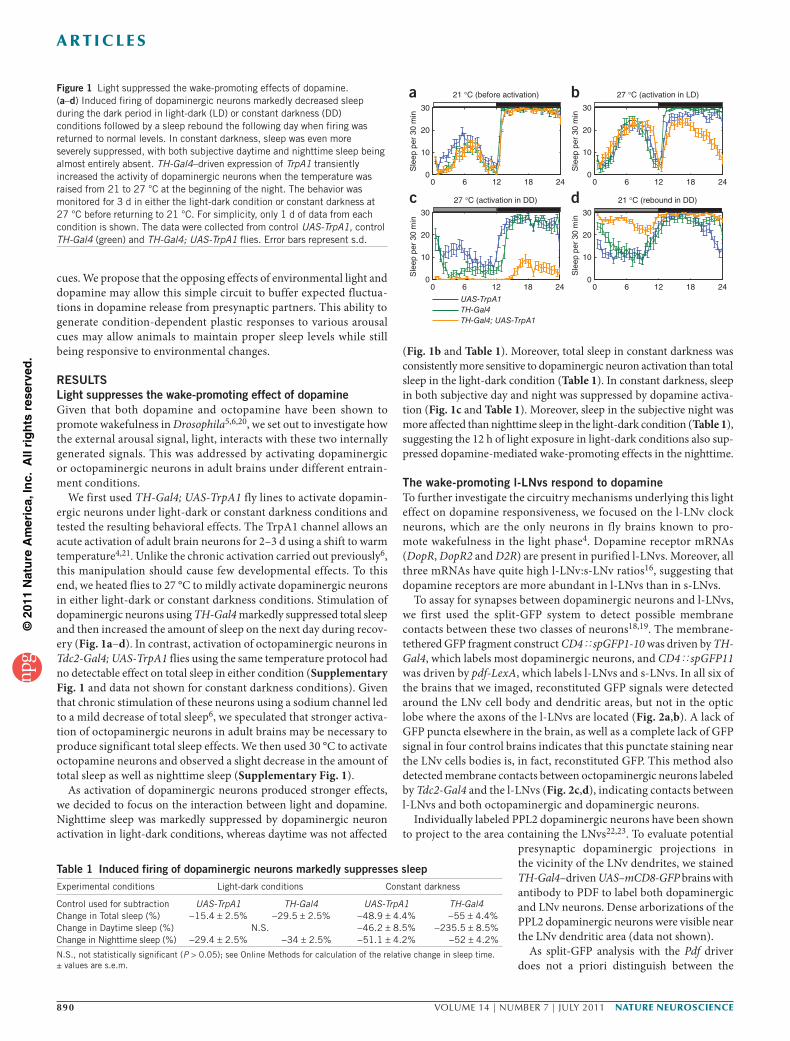
RESULTSLight suppresses the wake-promoting effect of dopamineGiven that both dopamine and octopamine have been shown to promote wakefulness in Drosophila5,6,20, we set out to investigate how the external arousal signal, light, interacts with these two internally generated signals. This was addressed by activating dopaminergic or octopaminergic neurons in adult brains under different entrain-ment conditions.
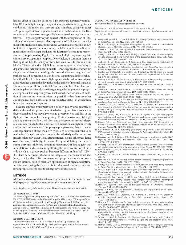
We first used TH-Gal4; UAS-TrpA1 fly lines to activate dopamin-ergic neurons under light-dark or constant darkness conditions and tested the resulting behavioral effects. The TrpA1 channel allows an acute activation of adult brain neurons for 2–3 d using a shift to warm temperature4,21. Unlike the chronic activation carried out previously6, this manipulation should cause few developmental effects. To this end, we heated flies to 27 °C to mildly activate dopaminergic neurons in either light-dark or constant darkness conditions. Stimulation of dopaminergic neurons using TH-Gal4 markedly suppressed total sleep and then increased the amount of sleep on the next day during recov-ery (Fig. 1a–d). In contrast, activation of octopaminergic neurons in Tdc2-Gal4; UAS-TrpA1 flies using the same temperature protocol had no detectable effect on total sleep in either condition (Supplementary Fig. 1 and data not shown for constant darkness conditions). Given that chronic stimulation of these neurons using a sodium channel led to a mild decrease of total sleep6, we speculated that stronger activa-tion of octopaminergic neurons in adult brains may be necessary to produce significant total sleep effects. We then used 30 °C to activate octopamine neurons and observed a slight decrease in the amount of total sleep as well as nighttime sleep (Supplementary Fig. 1).
As activation of dopaminergic neurons produced stronger effects, we decided to focus on the interaction between light and dopamine. Nighttime sleep was markedly suppressed by dopaminergic neuron activation in light-dark conditions, whereas daytime was not affected
(Fig. 1b and Table 1). Moreover, total sleep in constant darkness was consistently more sensitive to dopaminergic neuron activation than total sleep in the light-dark condition (Table 1). In constant darkness, sleep in both subjective day and night was suppressed by dopamine activa-tion (Fig. 1c and Table 1). Moreover, sleep in the subjective night was more affected than nighttime sleep in the light-dark condition (Table 1), suggesting the 12 h of light exposure in light-dark conditions also sup-pressed dopamine-mediated wake-promoting effects in the nighttime.
The wake-promoting l-LNvs respond to dopamineTo further investigate the circuitry mechanisms underlying this light effect on dopamine responsiveness, we focused on the l-LNv clock neurons, which are the only neurons in fly brains known to pro-mote wakefulness in the light phase4. Dopamine receptor mRNAs (DopR, DopR2 and D2R) are present in purified l-LNvs. Moreover, all three mRNAs have quite high l-LNv:s-LNv ratios16, suggesting that dopamine receptors are more abundant in l-LNvs than in s-LNvs.
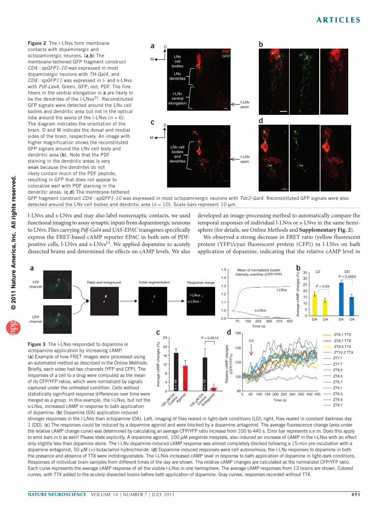
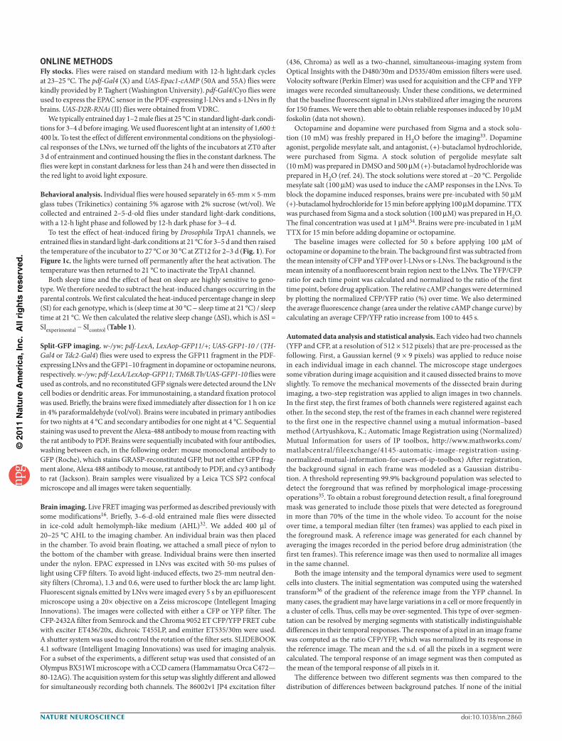
To assay for synapses between dopaminergic neurons and l-LNvs, we first used the split-GFP system to detect possible membrane contacts between these two classes of neurons18,19. The membrane- tethered GFP fragment construct CD4øspGFP1-10 was driven by TH-Gal4, which labels most dopaminergic neurons, and CD4øspGFP11 was driven by pdf-LexA, which labels l-LNvs and s-LNvs. In all six of the brains that we imaged, reconstituted GFP signals were detected around the LNv cell body and dendritic areas, but not in the optic lobe where the axons of the l-LNvs are located (Fig. 2a,b). A lack of GFP puncta elsewhere in the brain, as well as a complete lack of GFP signal in four control brains indicates that this punctate staining near the LNv cells bodies is, in fact, reconstituted GFP. This method also detected membrane contacts between octopaminergic neurons labeled by Tdc2-Gal4 and the l-LNvs (Fig. 2c,d), indicating contacts between l-LNvs and both octopaminergic and dopaminergic neurons.
Individually labeled PPL2 dopaminergic neurons have been shown to project to the area containing the LNvs22,23. To evaluate potential
presynaptic dopaminergic projections in the vicinity of the LNv dendrites, we stained TH-Gal4–driven UAS–mCD8-GFP brains with antibody to PDF to label both dopaminergic and LNv neurons. Dense arborizations of the PPL2 dopaminergic neurons were visible near the LNv dendritic area (data not shown).
As split-GFP analysis with the Pdf driver does not a priori distinguish between the
Table 1 Induced firing of dopaminergic neurons markedly suppresses sleep Experimental conditions Light-dark conditions Constant darkness
Control used for subtraction UAS-TrpA1 TH-Gal4 UAS-TrpA1 TH-Gal4Change in Total sleep (%) −15.4 ± 2.5% −29.5 ± 2.5% −48.9 ± 4.4% −55 ± 4.4%Change in Daytime sleep (%) N.S. −46.2 ± 8.5% −235.5 ± 8.5%Change in Nighttime sleep (%) −29.4 ± 2.5% −34 ± 2.5% −51.1 ± 4.2% −52 ± 4.2%
N.S., not statistically significant (P > 0.05); see Online Methods for calculation of the relative change in sleep time. ± values are s.e.m.
UAS-TrpA1TH-Gal4TH-Gal4; UAS-TrpA1
a 21 °C (before activation)
Sle
ep p
er 3
0 m
in
0 6 12 18 240
10
20
30
0 6 12 18 240
10
20
30
c
0 6 12 18 240
10
20
30
Sle
ep p
er 3
0 m
in
27 °C (activation in DD)
b
d
Sle
ep p
er 3
0 m
in
0 6 12 18 240
10
20
30
Sle
ep p
er 3
0 m
in
27 °C (activation in LD)
21 °C (rebound in DD)
Figure 1 Light suppressed the wake-promoting effects of dopamine. (a–d) Induced firing of dopaminergic neurons markedly decreased sleep during the dark period in light-dark (LD) or constant darkness (DD) conditions followed by a sleep rebound the following day when firing was returned to normal levels. In constant darkness, sleep was even more severely suppressed, with both subjective daytime and nighttime sleep being almost entirely absent. TH-Gal4–driven expression of TrpA1 transiently increased the activity of dopaminergic neurons when the temperature was raised from 21 to 27 °C at the beginning of the night. The behavior was monitored for 3 d in either the light-dark condition or constant darkness at 27 °C before returning to 21 °C. For simplicity, only 1 d of data from each condition is shown. The data were collected from control UAS-TrpA1, control TH-Gal4 (green) and TH-Gal4; UAS-TrpA1 flies. Error bars represent s.d.
©20
11 N
atu
re A
mer
ica,
Inc.
All
rig
hts
res
erve
d.
NATURE NEUROSCIENCE VOLUME 14 | NUMBER 7 | JULY 2011 891
A R T I C L E S
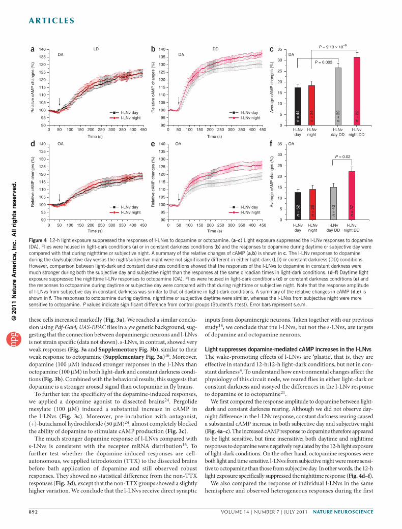
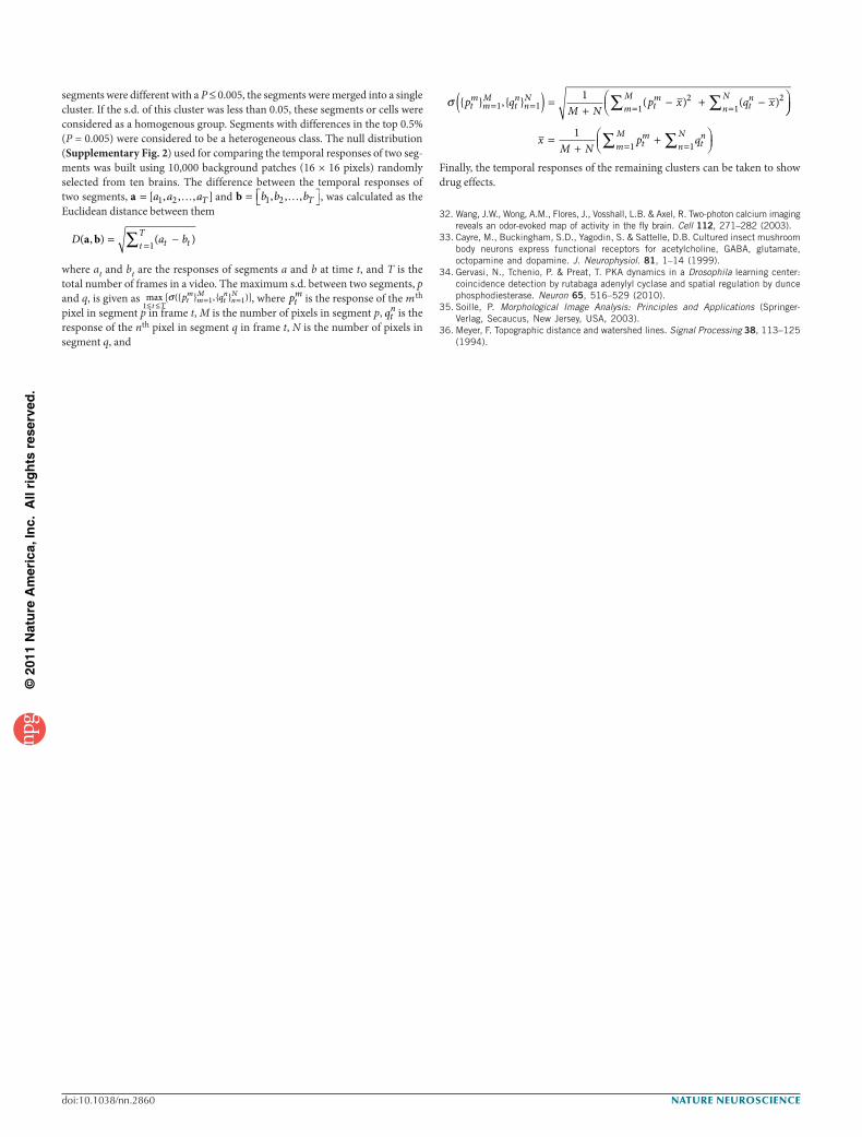
l-LNvs and s-LNvs and may also label nonsynaptic contacts, we used functional imaging to assay synaptic inputs from dopaminergic neurons to LNvs. Flies carrying Pdf-Gal4 and UAS-EPAC transgenes specifically express the FRET-based cAMP reporter EPAC in both sets of PDF- positive cells, l-LNvs and s-LNvs15. We applied dopamine to acutely dissected brains and determined the effects on cAMP levels. We also
developed an image-processing method to automatically compare the temporal responses of individual l-LNvs or s-LNvs in the same hemi-sphere (for details, see Online Methods and Supplementary Fig. 2).
We observed a strong decrease in FRET ratio (yellow fluorescent protein (YFP)/cyan fluorescent protein (CFP)) in l-LNvs on bath application of dopamine, indicating that the relative cAMP level in
1.5
1.4
1.3
1.2
1.1
1.0
0.975 150 225 300 375 450
a
DA
Ratio and foreground Response merge
l-LNvs
s-LNvs
Mean of normalized clusterintensity overtime (CFP/YFP)
Time (s)
YFPchannel
CFPchannel
b
l-LNvs
s-LNvs
Initial segmentation
c
P = 0.0003
P = 0.0014
Ave
rage
cA
MP
cha
nges
(%
)
Ave
rage
cA
MP
cha
nges
(%
)
100
µM D
A
Contro
lAgo
nist
100
µM D
A
Contro
l
Antag
onist
d 160
140
120
100
800 50 100 200
Time (s)
300 400 450
ZT8.1 TTX
ZT8.7 TTX
ZT9.3 TTX
ZT10.2 TTX
ZT7.1
ZT7.7
ZT8.2
ZT8.5
ZT8.7
ZT9.1
ZT9.3
ZT9.5
ZT9.7
250 350150
Rel
ativ
e cA
MP
cha
nges
(CF
P/Y
FP
%)
35
24
20
16
12
8
n =
44
n =
6
n = 7n =
104
0
P = 0.04
DDLD
25
15
10
5
DA OA DA OA0
30
20
Figure 3 The l-LNvs responded to dopamine or octopamine application by increasing cAMP. (a) Example of how FRET images were processed using an automated method as described in the Online Methods. Briefly, each video had two channels (YFP and CFP). The responses of a cell to a drug were computed as the mean of its CFP/YFP ratios, which were normalized by signals captured under the untreated condition. Cells without statistically significant response differences over time were merged as a group. In this example, the l-LNvs, but not the s-LNvs, increased cAMP in response to bath application of dopamine. (b) Dopamine (DA) application induced stronger responses in the l-LNvs than octopamine (OA). Left, imaging of flies reared in light-dark conditions (LD); right, flies reared in constant darkness day 1 (DD). (c) The responses could be induced by a dopamine agonist and were blocked by a dopamine antagonist. The average fluorescence change (area under the relative cAMP change curve) was determined by calculating an average CFP/YFP ratio increase from 100 to 445 s. Error bar represents s.e.m. Does this apply to error bars in b as well? Please state explicitly. A dopamine agonist, 100 µM pergolide mesylate, also induced an increase of cAMP in the l-LNvs with an effect only slightly less than dopamine alone. The l-LNv dopamine-induced cAMP response was almost completely blocked following a 15-min pre-incubation with a dopamine antagonist, 50 µM (+)-butaclamol hydrochloride. (d) Dopamine-induced responses were cell autonomous; the l-LNv responses to dopamine in both the presence and absence of TTX were indistinguishable. The l-LNvs increased cAMP level in response to bath application of dopamine in light-dark conditions. Responses of individual brain samples from different times of the day are shown. The relative cAMP changes are calculated as the normalized CFP/YFP ratio. Each curve represents the average cAMP response of all the visible l-LNvs in one hemisphere. The average cAMP responses from 13 brains are shown. Colored curves, with TTX added to the acutely dissected brains before bath application of dopamine. Gray curves, responses recorded without TTX.
Figure 2 The l-LNvs form membrane contacts with dopaminergic and octopaminergic neurons. (a,b) The membrane-tethered GFP fragment construct CD4øspGFP1-10 was expressed in most dopaminergic neurons with TH-Gal4, and CD4øspGFP11 was expressed in l- and s-LNvs with Pdf-LexA. Green, GFP; red, PDF. The fine fibers in the ventral elongation in a are likely to be the dendrites of the l-LNvs31. Reconstituted GFP signals were detected around the LNv cell bodies and dendritic area but not in the optical lobe around the axons of the l-LNvs (n = 6). The diagram indicates the orientation of the brain. D and M indicate the dorsal and medial sides of the brain, respectively. An image with higher magnification shows the reconstituted GFP signals around the LNv cell body and dendritic area (b). Note that the PDF staining in the dendritic areas is very weak because the dendrites do not likely contain much of the PDF peptide, resulting in GFP that does not appear to colocalize well with PDF staining in the dendritic areas. (c,d) The membrane-tethered GFP fragment construct CD4øspGFP1-10 was expressed in most octopaminergic neurons with Tdc2-Gal4. Reconsitituted GFP signals were also detected around the LNv cell bodies and dendritic area (n = 10). Scale bars represent 10 µm.
a b
LNvdendrites
l-LNvaxon
l-LNvaxon
LNvcell
bodies
l-LNvventral
elongation
D
M
d
LNv cellbodies
anddendrites
D
M
c
PDFGFP
©20
11 N
atu
re A
mer
ica,
Inc.
All
rig
hts
res
erve
d.
892 VOLUME 14 | NUMBER 7 | JULY 2011 NATURE NEUROSCIENCE
A R T I C L E S
these cells increased markedly (Fig. 3a). We reached a similar conclu-sion using Pdf-Gal4; UAS-EPAC flies in a yw genetic background, sug-gesting that the connection between dopaminergic neurons and l-LNvs is not strain specific (data not shown). s-LNvs, in contrast, showed very weak responses (Fig. 3a and Supplementary Fig. 3b), similar to their weak response to octopamine (Supplementary Fig. 3a)16. Moreover, dopamine (100 µM) induced stronger responses in the l-LNvs than octopamine (100 µM) in both light-dark and constant darkness condi-tions (Fig. 3b). Combined with the behavioral results, this suggests that dopamine is a stronger arousal signal than octopamine in fly brains.
To further test the specificity of the dopamine-induced responses, we applied a dopamine agonist to dissected brains24. Pergolide mesylate (100 µM) induced a substantial increase in cAMP in the l-LNvs (Fig. 3c). Moreover, pre-incubation with antagonist, (+)-butaclamol hydrochloride (50 µM)24, almost completely blocked the ability of dopamine to stimulate cAMP production (Fig. 3c).
The much stronger dopamine response of l-LNvs compared with s-LNvs is consistent with the receptor mRNA distribution16. To further test whether the dopamine-induced responses are cell- autonomous, we applied tetrodotoxin (TTX) to the dissected brains before bath application of dopamine and still observed robust responses. They showed no statistical difference from the non-TTX responses (Fig. 3d), except that the non-TTX groups showed a slightly higher variation. We conclude that the l-LNvs receive direct synaptic
inputs from dopaminergic neurons. Taken together with our previous study16, we conclude that the l-LNvs, but not the s-LNvs, are targets of dopamine and octopamine neurons.
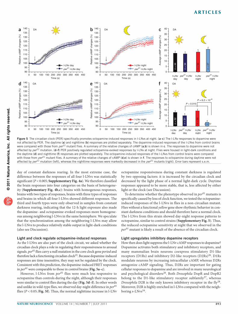
Light suppresses dopamine-mediated cAMP increases in the l-LNvsThe wake-promoting effects of l-LNvs are ‘plastic’, that is, they are effective in standard 12-h:12-h light-dark conditions, but not in con-stant darkness4. To understand how environmental changes affect the physiology of this circuit node, we reared flies in either light-dark or constant darkness and assayed the differences in the l-LNv response to dopamine or to octopamine21.
We first compared the response amplitude to dopamine between light-dark and constant darkness rearing. Although we did not observe day-night difference in the l-LNv response, constant darkness rearing caused a substantial cAMP increase in both subjective day and subjective night (Fig. 4a–c). The increased cAMP response to dopamine therefore appeared to be light sensitive, but time insensitive; both daytime and nighttime responses to dopamine were negatively regulated by the 12-h light exposure of light-dark conditions. On the other hand, octopamine responses were both light and time sensitive. l-LNvs from subjective night were more sensi-tive to octopamine than those from subjective day. In other words, the 12-h light exposure specifically suppressed the nighttime response (Fig. 4d–f).
We also compared the response of individual l-LNvs in the same hemisphere and observed heterogeneous responses during the first
a 140 LD
135
130
125
120
Rel
ativ
e cA
MP
cha
nges
(%
)
115
110
105
100
95
900 50 100 150
DA
200 250
Time (s)
300
I-LNv dayI-LNv night
350 400 450
b 140 DD
135
130
125
120
Rel
ativ
e cA
MP
cha
nges
(%
)
115
110
105
100
95
900 50 100 150
DA
200 250
Time (s)
300
I-LNv dayI-LNv night
350 400 450
d 140
135
130
125
120
Rel
ativ
e cA
MP
cha
nges
(%
)
115
110
105
100
95
900 50 100 150
OA
200 250
Time (s)
300
I-LNv dayI-LNv night
350 400 450
e 140
135
130
125
120
Rel
ativ
e cA
MP
cha
nges
(%
)
115
110
105
100
95
900 50 100 150
OA
200 250
Time (s)
300
I-LNv dayI-LNv night
350 400 450
c 35
30
25
20
Ave
rage
cA
MP
cha
nges
(%
)
10
5
0I-LNvday
DA
P = 0.003
P = 9.13 × 10⁻⁶
15
I-LNvday DD
I-LNvnight
I-LNvnight DD
n =
44
n =
34
n =
39
n =
22
f 35
30
25
20
Ave
rage
cA
MP
cha
nges
(%
)
10
5
0I-LNvday
OA
P = 0.02
15
I-LNvday DD
I-LNvnight DD
I-LNvnight
n =
52
n =
26
n =
40
n =
37
Figure 4 12-h light exposure suppressed the responses of l-LNvs to dopamine or octopamine. (a–c) Light exposure suppressed the l-LNv responses to dopamine (DA). Flies were housed in light-dark conditions (a) or in constant darkness conditions (b) and the responses to dopamine during daytime or subjective day were compared with that during nighttime or subjective night. A summary of the relative changes of cAMP (a,b) is shown in c. The l-LNv responses to dopamine during the day/subjective day versus the night/subjective night were not significantly different in either light-dark (LD) or constant darkness (DD) conditions. However, comparison between light-dark and constant darkness conditions showed that the responses of the l-LNvs to dopamine in constant darkness were much stronger during both the subjective day and subjective night than the responses at the same circadian times in light-dark conditions. (d–f) Daytime light exposure suppressed the nighttime l-LNv responses to octopamine (OA). Flies were housed in light-dark conditions (d) or constant darkness conditions (e) and the responses to octopamine during daytime or subjective day were compared with that during nighttime or subjective night. Note that the response amplitude of l-LNvs from subjective day in constant darkness was similar to that of daytime in light-dark conditions. A summary of the relative changes in cAMP (d,e) is shown in f. The responses to octopamine during daytime, nighttime or subjective daytime were similar, whereas the l-LNvs from subjective night were more sensitive to octopamine. P values indicate significant difference from control groups (Student’s t test). Error bars represent s.e.m.
©20
11 N
atu
re A
mer
ica,
Inc.
All
rig
hts
res
erve
d.
NATURE NEUROSCIENCE VOLUME 14 | NUMBER 7 | JULY 2011 893
A R T I C L E S
day of constant darkness rearing. In the most extreme case, the difference between the responses of all four l-LNvs was statistically significant (P < 0.005; Supplementary Fig. 4a). We therefore classified the brain responses into four categories on the basis of heterogene-ity (Supplementary Fig. 4b,c): brains with homogenous responses, brains with two types of responses, brains with three types of responses and brains in which all four l-LNvs showed different responses. The third and fourth types were only observed in samples from constant darkness rearing, indicating that the 12-h light exposure also made the dopamine- and octopamine-evoked responses more homogene-ous among neighboring l-LNvs in the same hemisphere. We speculate that the synchronization among the neighboring l-LNvs may allow the l-LNvs to produce relatively stable output in light-dark conditions (also see Discussion).
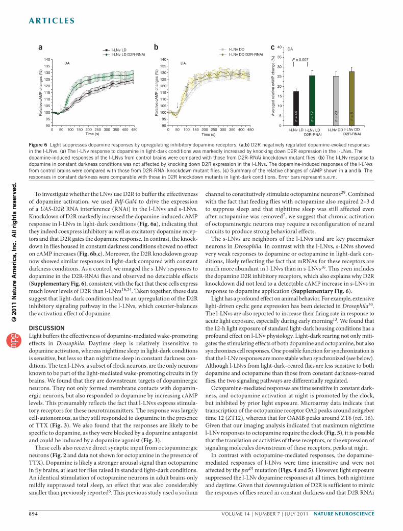
Light and clock regulate octopamine-induced responsesAs the l-LNvs are also part of the clock circuit, we asked whether the circadian clock plays a role in regulating their responsiveness to arousal signals. per01 flies carry a null mutation in the core clock gene period and therefore lack a functioning circadian clock25. Because dopamine-induced responses are time insensitive, they may not be regulated by the clock. Consistent with this prediction, the dopamine-induced FRET responses in per01 were comparable to those in control brains (Fig. 5a–c).
However, l-LNvs from per01 flies were much less responsive to octopamine than controls during the night, although their responses were similar to control flies during the day (Fig. 5d–f). In other words and unlike in wild-type flies, we observed day-night difference in per01 flies (P < 0.05; Fig. 5f). Thus, the normal nighttime increase in l-LNv
octopamine responsiveness during constant darkness is regulated by two opposing factors: it is increased by the circadian clock and decreased by the light phase of a normal light-dark cycle. Daytime responses appeared to be more stable, that is, less affected by either light or the clock (see Discussion).
To determine whether the phenotype observed in per01 mutants is specifically caused by loss of clock function, we tested the octopamine-induced responses of the l-LNvs in flies in a non-circadian mutant. Flies without a functional yellow gene show rhythmic behavior in con-stant darkness conditions and should therefore have a normal clock. The l-LNvs from this strain showed day-night response patterns to octopamine, similar to control strains (Supplementary Fig. 5). Thus, the reduced octopamine sensitivity at night that we observed in the per01 mutant is likely a result of the absence of the circadian clock.
Light upregulates inhibitory dopamine receptorsHow then does light suppress the l-LNv cAMP responses to dopamine? Dopamine activates both stimulatory and inhibitory receptors, and many mammalian brain neurons coexpress stimulatory D1-like receptors (D1Rs) and inhibitory D2-like receptors (D2Rs)26. D1Rs modulate neurons by increasing intracellular cAMP, whereas D2Rs antagonize cAMP signaling. Thus, D2Rs are important for gating cellular responses to dopamine and are involved in many neurological and psychological disorders26. Both Drosophila DopR and DopR2 belong to the D1-like stimulatory receptor subfamily27, whereas Drosophila D2R is the only known inhibitory receptor in the fly28. Moreover, D2R is highly enriched in l-LNvs compared with the neigh-boring s-LNvs16.
140
135a DA
I-LNv dayper01 I-LNv day
0
130
125
120
115
Rel
ativ
e cA
MP
cha
nges
(%)
110
105
100
95
9050 100 150 200 250 300 350 400 450
Time (s)
c DA
I-LNvday
per01 I-LNvday
I-LNvnight
per01 I-LNvnight
Ave
rage
cA
MP
cha
nges
(%)
40
35
30
25
20
15
10
5
0
n =
44
n =
34
n =
27
n =
34
f OA
I-LNvday
per01 I-LNvday
I-LNvnight
per01 I-LNvnight
Ave
rage
cA
MP
cha
nges
(%)
40
35
P = 0.015P = 6.2 × 10�6
30
25
20
15
10
5
0
n =
52
n =
31
n =
26
n = 27
d OA
I-LNv dayper01 I-LNv day
140
135
130
125
120
115
Rel
ativ
e cA
MP
cha
nges
(%)
110
105
100
95
900 50 100 150 200 250 300 350 400 450
Time (s)
e OA
I-LNv nightper01 I-LNv night
140
135
130
125
120
115
Rel
ativ
e cA
MP
cha
nges
(%)
110
105
100
95
900 50 100 150 200 250 300 350 400 450
Time (s)
b DA
I-LNv nightper01 I-LNv night
140
135
130
125
120
115
Rel
ativ
e cA
MP
cha
nges
(%)
110
105
100
95
900 50 100 150 200 250 300 350 400 450
Time (s)
Figure 5 The circadian clock (PER) specifically promotes octopamine-induced responses in l-LNvs at night. (a–c) The l-LNv responses to dopamine were not affected by PER. The daytime (a) and nighttime (b) responses are plotted separately. The dopamine-induced responses of the l-LNvs from control brains were compared with those from per01 mutant flies. A summary of the relative changes of cAMP (a,b) is shown in c. The responses to dopamine were not affected by per01 mutation. (d–f) PER positively regulated octopamine-evoked responses by l-LNv at night. Flies were housed in light-dark conditions and the daytime (d) and nighttime (f) responses are plotted separately. The octopamine-induced responses of the l-LNvs from control brains were compared with those from per01 mutant flies. A summary of the relative changes of cAMP (d,e) is shown in f. The responses to octopamine during daytime were not affected by per01 mutation (left), whereas the nighttime responses were markedly decreased in the per01 mutants (right). Error bars represent s.e.m.
©20
11 N
atu
re A
mer
ica,
Inc.
All
rig
hts
res
erve
d.
894 VOLUME 14 | NUMBER 7 | JULY 2011 NATURE NEUROSCIENCE
A R T I C L E S
To investigate whether the LNvs use D2R to buffer the effectiveness of dopamine activation, we used Pdf-Gal4 to drive the expression of a UAS-D2R RNA interference (RNAi) in the l-LNvs and s-LNvs. Knockdown of D2R markedly increased the dopamine-induced cAMP response in l-LNvs in light-dark conditions (Fig. 6a), indicating that they indeed coexpress inhibitory as well as excitatory dopamine recep-tors and that D2R gates the dopamine response. In contrast, the knock-down in flies housed in constant darkness conditions showed no effect on cAMP increases (Fig. 6b,c). Moreover, the D2R knockdown group now showed similar responses in light-dark compared with constant darkness conditions. As a control, we imaged the s-LNv responses to dopamine in the D2R-RNAi flies and observed no detectable effects (Supplementary Fig. 6), consistent with the fact that these cells express much lower levels of D2R than l-LNvs16,24. Taken together, these data suggest that light-dark conditions lead to an upregulation of the D2R inhibitory signaling pathway in the l-LNvs, which counter-balances the activation effect of dopamine.
DISCUSSIONLight buffers the effectiveness of dopamine-mediated wake-promoting effects in Drosophila. Daytime sleep is relatively insensitive to dopamine activation, whereas nighttime sleep in light-dark conditions is sensitive, but less so than nighttime sleep in constant darkness con-ditions. The ten l-LNvs, a subset of clock neurons, are the only neurons known to be part of the light-mediated wake-promoting circuits in fly brains. We found that they are downstream targets of dopaminergic neurons. They not only formed membrane contacts with dopamin-ergic neurons, but also responded to dopamine by increasing cAMP levels. This presumably reflects the fact that l-LNvs express stimula-tory receptors for these neurotransmitters. The response was largely cell-autonomous, as they still responded to dopamine in the presence of TTX (Fig. 3). We also found that the responses are likely to be specific to dopamine, as they were blocked by a dopamine antagonist and could be induced by a dopamine agonist (Fig. 3).
These cells also receive direct synaptic input from octopaminergic neurons (Fig. 2 and data not shown for octopamine in the presence of TTX). Dopamine is likely a stronger arousal signal than octopamine in fly brains, at least for flies raised in standard light-dark conditions. An identical stimulation of octopamine neurons in adult brains only mildly suppressed total sleep, an effect that was also considerably smaller than previously reported6. This previous study used a sodium
channel to constitutively stimulate octopamine neurons29. Combined with the fact that feeding flies with octopamine also required 2–3 d to suppress sleep and that nighttime sleep was still affected even after octopamine was removed7, we suggest that chronic activation of octopaminergic neurons may require a reconfiguration of neural circuits to produce strong behavioral effects.
The s-LNvs are neighbors of the l-LNvs and are key pacemaker neurons in Drosophila. In contrast with the l-LNvs, s-LNvs showed very weak responses to dopamine or octopamine in light-dark con-ditions, likely reflecting the fact that mRNAs for these receptors are much more abundant in l-LNvs than in s-LNvs16. This even includes the dopamine D2R inhibitory receptors, which also explains why D2R knockdown did not lead to a detectable cAMP increase in s-LNvs in response to dopamine application (Supplementary Fig. 6).
Light has a profound effect on animal behavior. For example, extensive light-driven cyclic gene expression has been detected in Drosophila30. The l-LNvs are also reported to increase their firing rate in response to acute light exposure, especially during early morning13. We found that the 12-h light exposure of standard light-dark housing conditions has a profound effect on l-LNv physiology. Light-dark rearing not only miti-gates the stimulating effects of both dopamine and octopamine, but also synchronizes cell responses. One possible function for synchronization is that the l-LNv responses are more stable when synchronized (see below). Although l-LNvs from light-dark–reared flies are less sensitive to both dopamine and octopamine than those from constant darkness–reared flies, the two signaling pathways are differentially regulated.
Octopamine-mediated responses are time sensitive in constant dark-ness, and octopamine activation at night is promoted by the clock, but inhibited by prior light exposure. Microarray data indicate that transcription of the octopamine receptor OA2 peaks around zeitgeber time 12 (ZT12), whereas that for OAMB peaks around ZT6 (ref. 16). Given that our imaging analysis indicated that maximum nighttime l-LNv responses to octopamine require the clock (Fig. 5), it is possible that the translation or activities of these receptors, or the expression of signaling molecules downstream of these receptors, peaks at night.
In contrast with octopamine-mediated responses, the dopamine-mediated responses of l-LNvs were time insensitive and were not affected by the per01 mutation (Figs. 4 and 5). However, light exposure suppressed the l-LNv dopamine responses at all times, both nighttime and daytime. Given that downregulation of D2R is sufficient to mimic the responses of flies reared in constant darkness and that D2R RNAi
140DA
Rel
ativ
e cA
MP
cha
nges
(%
)
a
135
130
125
120
115
110
105
100
95
900 50 100 150 200 250 300 350 400 450
Time (s)
I-LNv LDI-LNv LD D2R-RNAi
40 DA
P = 0.007
Ave
rage
d re
lativ
e cA
MP
cha
nge
(%)
c35
25
20
15
10
5
0I-LNv LD I-LNv DDI-LNv LD
D2R-RNAiI-LNv DDD2R-RNAi
30
140DA
Rel
ativ
e cA
MP
cha
nges
(%
)
b
135
130
125
120
115
110
105
100
95
900 50 100 150 200 250 300 350 400 450
Time (s)
I-LNv DDI-LNv DD D2R-RNAi
n =
44
n =
17
n =
20
n =
8
Figure 6 Light suppresses dopamine responses by upregulating inhibitory dopamine receptors. (a,b) D2R negatively regulated dopamine-evoked responses in the l-LNvs. (a) The l-LNv response to dopamine in light-dark conditions was markedly increased by knocking down D2R expression in the l-LNvs. The dopamine-induced responses of the l-LNvs from control brains were compared with those from D2R-RNAi knockdown mutant flies. (b) The l-LNv response to dopamine in constant darkness conditions was not affected by knocking down D2R expression in the l-LNvs. The dopamine-induced responses of the l-LNvs from control brains were compared with those from D2R-RNAi knockdown mutant flies. (c) Summary of the relative changes of cAMP shown in a and b. The responses in constant darkness were comparable with those in D2R knockdown mutants in light-dark conditions. Error bars represent s.e.m.
©20
11 N
atu
re A
mer
ica,
Inc.
All
rig
hts
res
erve
d.
NATURE NEUROSCIENCE VOLUME 14 | NUMBER 7 | JULY 2011 895
A R T I C L E S
had no effect in constant darkness, light exposure apparently upregu-lates D2R activity to dampen dopamine responsiveness in light-dark conditions. This implies that there are light-stimulated changes in either D2R gene expression or regulation, such as a modification of the D2R receptor or its downstream targets. Light may also downregulate stimu-latory D1R signaling pathways in concert with the upregulation of D2R, although our results suggest that expression of D2R can account for most of the reduction in responsiveness. Given that there are no known inhibitory receptors for octopamine, the l-LNvs must use a different mechanism to effect light-mediated modulation of octopamine respon-siveness (Supplementary Fig. 7). For example, light may downregulate stimulatory octopamine receptors. Nonetheless, a common theme is that light inhibits the ability of these two chemicals to stimulate the l-LNvs. The fact that the 12-h light exposure suppressed the ability of dopamine and octopamine to stimulate l-LNvs suggests that they do not simply sum different arousal signals. Instead, they are integrated and perhaps scaled depending on conditions, suggesting a link to behav-ioral flexibility. In this scenario, light appears to be a dominant signal, as its presence during the day reduces the ability of internal signals to stimulate arousal. However, the l-LNvs use a number of mechanisms, including the circadian clock to integrate signals and produce appropri-ate responses. The surprisingly weak behavioral effects of acute stimula-tion of octopamine neurons raises the possibility that there are other circumstances (age, nutritional or reproductive status) in which these inputs become more important.
Because animals must maintain a proper quality and quantity of daily wake and sleep time, counter-balancing mechanisms such as those described here may also serve to preserve sleep stability in the fly brain. For example, the opposing effects of environmental light and dopamine may allow the l-LNvs and perhaps other arousal-sleep– relevant neurons to buffer unexpected fluctuations in light intensity and/or dopamine release from presynaptic partners; that is, the cir-cuit organization allows the activity of sleep-relevant neurons to be maintained in a physiological range with a relatively stable output. We imagine that only exceptional circumstances would take precedence over sleep-wake stability; for example, by modulating the ratio of stimulatory and inhibitory dopamine receptors. Our data suggest that modulation could also occur by altering the synchronization of indi-vidual cells in a group, such as between different individual l-LNvs. It will not be surprising if additional integration mechanisms are also important for the l-LNvs to generate appropriate signals to down-stream circuits, both to maintain optimal sleep at night and optimal wakefulness during the day, that is, for sleep-wake homeostasis, and for appropriate responses to emergency circumstances.
METHODSMethods and any associated references are available in the online version of the paper at http://www.nature.com/natureneuroscience/.
Note: Supplementary information is available on the Nature Neuroscience website.
ACKNOWLEDGMENTSWe thank P. Taghert for kindly providing pdfGal4 (X) and UAS-EPAC flies. We obtained UAS-D2R-RNAi lines from the Vienna Drosophila RNAi center. We are grateful to O. Shafer for technical help with cAMP imaging. We also thank E. Dougherty for assistance in confocal microscopy, K. Palm and S. Pescatore for administrative assistance and C. Vecsey for comments on the manuscript. The work was supported in part by grants from the US National Institutes of Health (PO1 NS044232-06 to M.R., R01 MH067284 to L.C.G. and NIH R01 EB007042 to P. Hong).
AUTHOR CONTRIBUTI ONSY.S. conceived the project. Y.S., P. Haynes, N.P. and F.G. performed the experiments. K.I.H., J.P. and P. Hong developed the algorithm for the automated imaging analysis. Y.S., L.C.G. and M.R. wrote the paper.
COMPETI NG FINANCIA L INTERESTSThe authors declare no competing financial interests.
Published online at http://www.nature.com/natureneuroscience/. Reprints and permissions information is available online at http://www.nature.com/reprints/index.html.
1. Ganguly-Fitzgerald, I., Donlea, J. & Shaw, P.J. Waking experience affects sleep need in Drosophila. Science 313, 1775–1781 (2006).
2. Ho, K.S. & Sehgal, A. Drosophila melanogaster: an insect model for fundamental studies of sleep. Methods Enzymol. 393, 772–793 (2005).
3. Keene, A.C. et al. Clock and cycle limit starvation-induced sleep loss in Drosophila. Curr. Biol. 20, 1209–1215 (2010).
4. Shang, Y., Griffith, L.C. & Rosbash, M. Light-arousal and circadian photoreception circuits intersect at the large PDF cells of the Drosophila brain. Proc. Natl. Acad. Sci. USA 105, 19587–19594 (2008).
5. Andretic, R., van Swinderen, B. & Greenspan, R.J. Dopaminergic modulation of arousal in Drosophila. Curr. Biol. 15, 1165–1175 (2005).
6. Crocker, A. & Sehgal, A. Octopamine regulates sleep in Drosophila through protein kinase A–dependent mechanisms. J. Neurosci. 28, 9377–9385 (2008).
7. Crocker, A., Shahidullah, M., Levitan, I.B. & Sehgal, A. Identification of a neural circuit that underlies the effects of octopamine on sleep:wake behavior. Neuron 65, 670–681 (2010).
8. Parisky, K.M. et al. PDF cells are a GABA-responsive wake-promoting component of the Drosophila sleep circuit. Neuron 60, 672–682 (2008).
9. Hendricks, J.C. et al. Rest in Drosophila is a sleep-like state. Neuron 25, 129–138 (2000).
10. Shaw, P.J., Cirelli, C., Greenspan, R.J. & Tononi, G. Correlates of sleep and waking in Drosophila melanogaster. Science 287, 1834–1837 (2000).
11. Sheeba, V. et al. Large ventral lateral neurons modulate arousal and sleep in Drosophila. Curr. Biol. 18, 1537–1545 (2008).
12. Donlea, J.M., Ramanan, N. & Shaw, P.J. Use-dependent plasticity in clock neurons regulates sleep need in Drosophila. Science 324, 105–108 (2009).
13. Sheeba, V., Gu, H., Sharma, V.K., O’Dowd, D.K. & Holmes, T.C. Circadian- and light-dependent regulation of resting membrane potential and spontaneous action potential firing of Drosophila circadian pacemaker neurons. J. Neurophysiol. 99, 976–988 (2008).
14. Renn, S.C., Park, J.H., Rosbash, M., Hall, J.C. & Taghert, P.H. A pdf neuropeptide gene mutation and ablation of PDF neurons each cause severe abnormalities of behavioral circadian rhythms in Drosophila. Cell 99, 791–802 (1999).
15. Shafer, O.T. et al. Widespread receptivity to neuropeptide PDF throughout the neuronal circadian clock network of Drosophila revealed by real-time cyclic AMP imaging. Neuron 58, 223–237 (2008).
16. Kula-Eversole, E. et al. Surprising gene expression patterns within and between PDF-containing circadian neurons in Drosophila. Proc. Natl. Acad. Sci. USA 107, 13497–13502 (2010).
17. Shakiryanova, D. & Levitan, E.S. Prolonged presynaptic posttetanic cyclic GMP signaling in Drosophila motoneurons. Proc. Natl. Acad. Sci. USA 105, 13610–13613 (2008).
18. Feinberg, E.H. et al. GFP reconstitution across synaptic partners (GRASP) defines cell contacts and synapses in living nervous systems. Neuron 57, 353–363 (2008).
19. Gordon, M.D. & Scott, K. Motor control in a Drosophila taste circuit. Neuron 61, 373–384 (2009).
20. Crocker, A. & Sehgal, A. Genetic analysis of sleep. Genes Dev. 24, 1220–1235 (2010).
21. Hamada, F.N. et al. An internal thermal sensor controlling temperature preference in Drosophila. Nature 454, 217–220 (2008).
22. Chiang, A.S. et al. Three-dimensional reconstruction of brain-wide wiring networks in Drosophila at single-cell resolution. Curr. Biol. 21, 1–11 (2011).
23. Mao, Z. & Davis, R.L. Eight different types of dopaminergic neurons innervate the Drosophila mushroom body neuropil: anatomical and physiological heterogeneity. Front. Neural. Circuits 3, 5 (2009).
24. Sugamori, K.S., Demchyshyn, L.L., McConkey, F., Forte, M.A. & Niznik, H.B. A primordial dopamine D1-like adenylyl cyclase–linked receptor from Drosophila melanogaster displaying poor affinity for benzazepines. FEBS Lett. 362, 131–138 (1995).
25. Hall, J.C. Systems approaches to biological rhythms in Drosophila. Methods Enzymol. 393, 61–185 (2005).
26. Bonci, A. & Hopf, F.W. The dopamine D2 receptor: new surprises from an old friend. Neuron 47, 335–338 (2005).
27. Han, K.A., Millar, N.S., Grotewiel, M.S. & Davis, R.L. DAMB, a novel dopamine receptor expressed specifically in Drosophila mushroom bodies. Neuron 16, 1127–1135 (1996).
28. Hearn, M.G. et al. A Drosophila dopamine 2–like receptor: molecular characterization and identification of multiple alternatively spliced variants. Proc. Natl. Acad. Sci. USA 99, 14554–14559 (2002).
29. Nitabach, M.N., Sheeba, V., Vera, D.A., Blau, J. & Holmes, T.C. Membrane electrical excitability is necessary for the free-running larval Drosophila circadian clock. J. Neurobiol. 62, 1–13 (2005).
30. Wijnen, H., Naef, F., Boothroyd, C., Claridge-Chang, A. & Young, M.W. Control of daily transcript oscillations in Drosophila by light and the circadian clock. PLoS Genet. 2, e39 (2006).
31. Helfrich-Förster, C. et al. Development and morphology of the clock-gene-expressing lateral neurons of Drosophila melanogaster. J. Comp. Neurol. 500, 47–70 (2007).
©20
11 N
atu
re A
mer
ica,
Inc.
All
rig
hts
res
erve
d.
NATURE NEUROSCIENCEdoi:10.1038/nn.2860
segments were different with a P ≤ 0.005, the segments were merged into a single cluster. If the s.d. of this cluster was less than 0.05, these segments or cells were considered as a homogenous group. Segments with differences in the top 0.5% (P = 0.005) were considered to be a heterogeneous class. The null distribution (Supplementary Fig. 2) used for comparing the temporal responses of two seg-ments was built using 10,000 background patches (16 × 16 pixels) randomly selected from ten brains. The difference between the temporal responses of two segments, a = …[ , , , ]a a aT1 2 and b b b bT1 2, , , , was calculated as the Euclidean distance between them
D a bt ttT( , ) ( )a b = −=∑ 1
where at and bt are the responses of segments a and b at time t, and T is the total number of frames in a video. The maximum s.d. between two segments, p and q, is given as max [ ({ } ,{ } )]
11 1
t Ttm
mM
tnnNp qs , where pt
m is the response of the m th pixel in segment p in frame t, M is the number of pixels in segment p, qt
n is the response of the nth pixel in segment q in frame t, N is the number of pixels in segment q, and
s { } ,{ } ( ) ( )p qM N
p x q xtm
mM
tnnN
tm
mM
tn
nN
�� �� �� ���� � �� ��� �� � �⁄
�ƒ�¥�‡�£ �£1 1
21
21
1�µ�µ�·
xM N
p qtm
mM
tn
nN=
++
= =∑ ∑1
1 1
Finally, the temporal responses of the remaining clusters can be taken to show drug effects.
32. Wang, J.W., Wong, A.M., Flores, J., Vosshall, L.B. & Axel, R. Two-photon calcium imaging reveals an odor-evoked map of activity in the fly brain. Cell 112, 271–282 (2003).
33. Cayre, M., Buckingham, S.D., Yagodin, S. & Sattelle, D.B. Cultured insect mushroom body neurons express functional receptors for acetylcholine, GABA, glutamate, octopamine and dopamine. J. Neurophysiol. 81, 1–14 (1999).
34. Gervasi, N., Tchenio, P. & Preat, T. PKA dynamics in a Drosophila learning center: coincidence detection by rutabaga adenylyl cyclase and spatial regulation by dunce phosphodiesterase. Neuron 65, 516–529 (2010).
35. Soille, P. Morphological Image Analysis: Principles and Applications (Springer-Verlag, Secaucus, New Jersey, USA, 2003).
36. Meyer, F. Topographic distance and watershed lines. Signal Processing 38, 113–125 (1994).