Embed Size (px)
Citation preview

Image Gallery: Lesion detection on low dose head CT
Sarabjeet Singh, MD
Mannudeep K. Kalra, MD
*Eugene J. Mark, MD
*James Stone, MD
James H. Thrall, MD
Web Chapter 4
Department of Radiology and *Department of Pathology
Massachusetts General Hospital
Harvard Medical School, Boston

Do you see any abnormal findings in this transverse CT image?

Do you see any periventricular hypoattenuation?

Periventricular hypoattenuation
300 mAs:CTDI vol: 45.8 mGy
150 mAs22.9 mGy
60 mAs9.1 mGy
90 mAs13.7 mGy
Do you see any additional findings on higher dose images?

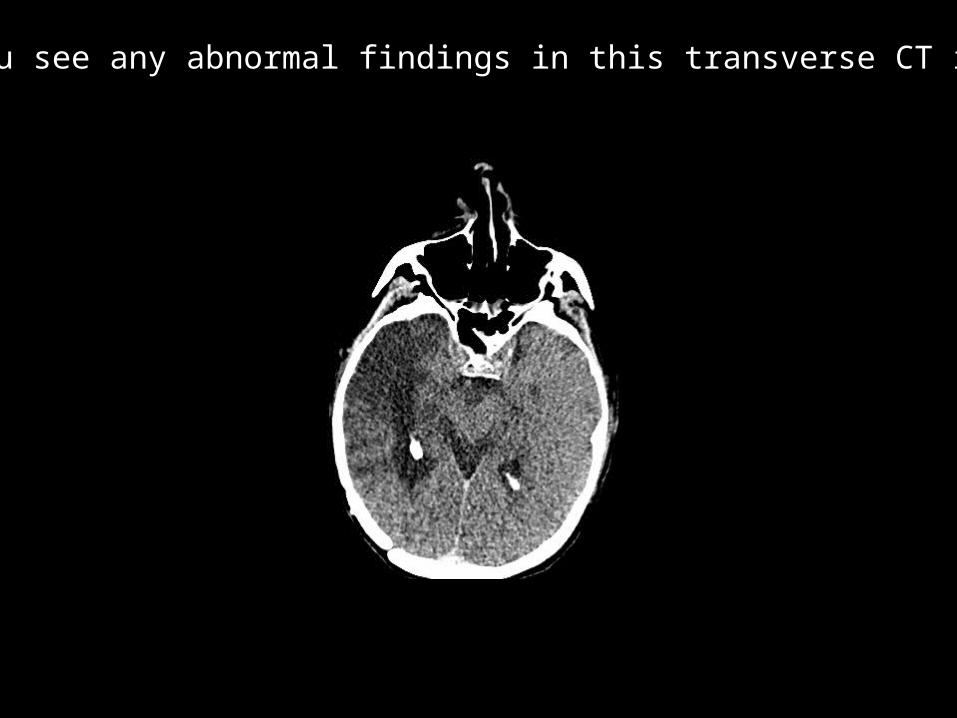
Do you see any abnormal findings in this transverse CT image?

Do you see tiny bright hemorrhagic or calcified spec?

Tiny bright, high contrast structure seen at all dose levels
300 mAs:CTDI vol: 45.8 mGy
150 mAs22.9 mGy
60 mAs9.1 mGy
90 mAs13.7 mGy
Do you see any additional findings on higher dose images?

Tiny bright, high contrast structure seen at all dose levels
300 mAs:CTDI vol: 45.8 mGy
150 mAs22.9 mGy
60 mAs9.1 mGy
90 mAs13.7 mGy
Do you see any additional findings on higher dose images?
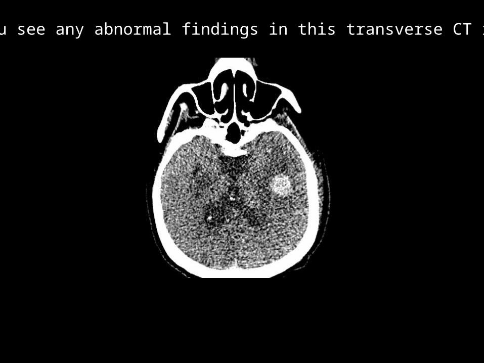
Do you see any abnormal findings in this transverse CT image?
Do you see bright hemorrhagic metastatic nodule ?

Bright hemorrhagic metastatic nodule seen at all dose levels
150 mA:CTDI vol: 45.8 mGy
75 mA22.9 mGy
20 mA9.1 mGy
38 mA13.7 mGy
Do you see any additional findings on higher dose images?
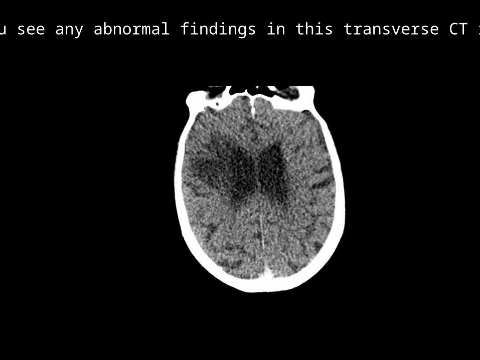
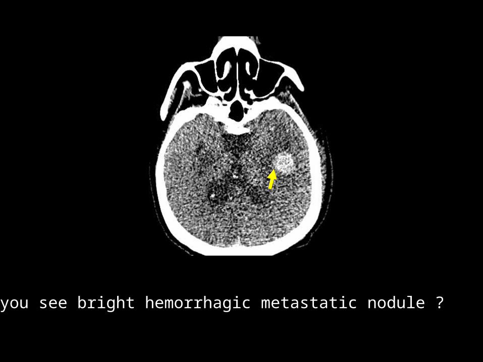
Do you see any abnormal findings in this transverse CT image?

Do you see a hypodensity in right fronto parietal peri ventricular white matter?

hypodensity in right frontoparietal peri ventricular white matter seen at all dose levels
150 mA:CTDI vol: 45.8 mGy
75 mA22.9 mGy
20 mA9.1 mGy
38 mA13.7 mGy
Do you see any additional findings on higher dose images?

Do you see any abnormal findings in this transverse CT image?
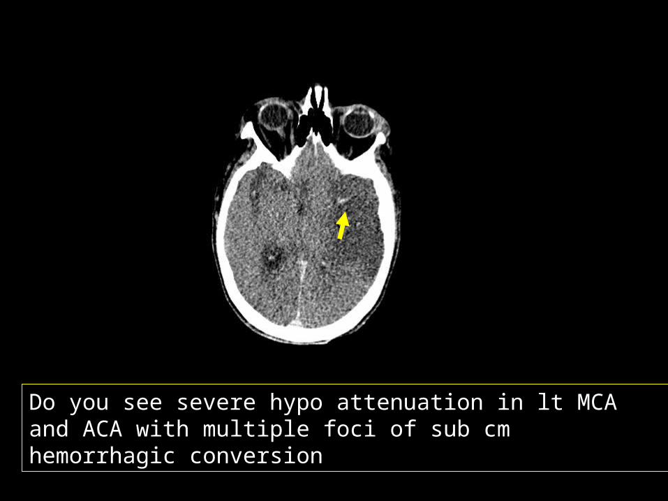
Do you see severe hypo attenuation in lt MCA and ACA with multiple foci of sub cm hemorrhagic conversion

Loss of grey-white matter differentiation with marked hypoattenuation in left MCA and ACA territory with sub-centimeter size hemorrhagic conversion
150 mA:CTDI vol: 45.8 mGy
75 mA22.9 mGy
20 mA9.1 mGy
38 mA13.7 mGy
Do you see any additional findings on higher dose images?
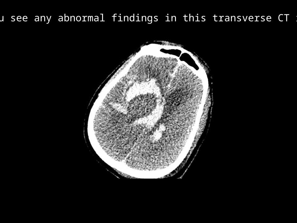
Do you see any abnormal findings in this transverse CT image?

Do you see massive subarachnoid hemorrhage
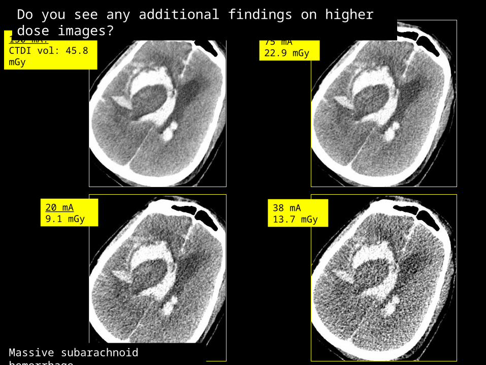
Massive subarachnoid hemorrhage
150 mA:CTDI vol: 45.8 mGy
75 mA22.9 mGy
20 mA9.1 mGy
38 mA13.7 mGy
Do you see any additional findings on higher dose images?

Do you see any abnormal findings in this transverse CT image?

Do you see extensive areas of hypoattenuation in left middle cerebral artery vascular territory?

Extensive low attenuation in left MCA territory
300 mAsCTDI vol: 46 mGy
150 mAs 23 mGy
40 mAs6 mGy
75 mAs12 mGy
Do you see any additional findings on higher dose images?

Extensive low attenuation in left MCA territory
300 mAsCTDI vol: 46 mGy
150 mAs 23 mGy
40 mAs6 mGy
75 mAs12 mGy
Do you see any additional findings on higher dose images?

Do you see any abnormal findings in this transverse CT image?

Do you see left frontal lobe infarct and
left temporal ventricle dilation?
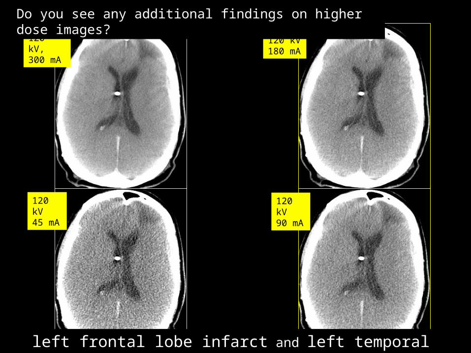
120 kV, 300 mA
120 kV180 mA
120 kV45 mA
120 kV90 mA
Do you see any additional findings on higher dose images?
left frontal lobe infarct and left temporal ventricle dilation
Do you see any abnormal findings in this transverse CT image?
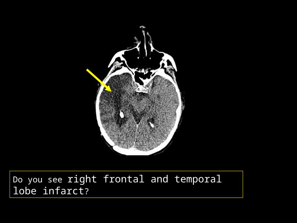
Do you see right frontal and temporal lobe infarct?

400 mAs61 mGy
Do you see any additional findings on higher dose images?
Right frontal and temporal lobe infarct
300 mAs46 mGy
200 mAs31 mGy
100 mAs16 mGy
60mAs9 mGy
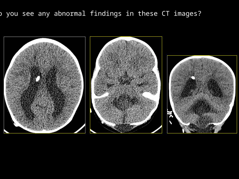
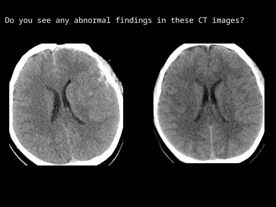
Do you see any abnormal findings in these CT images?
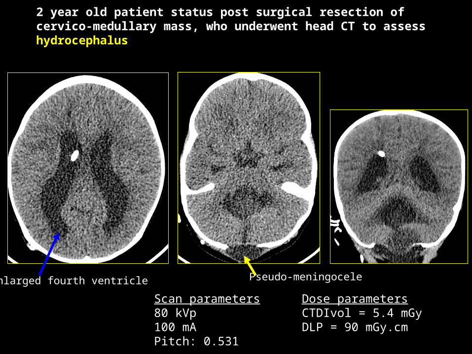
2 year old patient status post surgical resection of cervico-medullary mass, who underwent head CT to assess hydrocephalus
Scan parameters80 kVp100 mAPitch: 0.531
Dose parametersCTDIvol = 5.4 mGyDLP = 90 mGy.cm
Enlarged fourth ventricle Pseudo-meningocele
Do you see any abnormal findings in these CT images?

18yr old boy, with subarachnoid blood along the b/l frontal convexities and small SDH along the left frontotemporal convexity
Follow up: (ASIR)
Scan parameters120 kVp231 mA
Estimated dose: CTDIvol: 25.4 mGyDLP: 459.8 mGy.cm0.9 mSv
Scan parameters120 kVp159 mA, NI: 42Pitch: 0.531ASIR:90
Prior scan: (FBP)
Estimated dose: CTDIvol: 65.6 mGyDLP: 1223.6 mGy.cm2.4 mSv

1 yr girl, 10kg, with abnormal facies, underwent Head CT for osseous dysmorphism: Craniosynostosis protocol
Technique: (ASIR)
Scan parameters80 kVp50 mAEstimated dose: 0.1 mSv
CT evaluation of bony abnormalities such as craniostenosis should be performed at lowest possible radiation dose levels. Note that brain parenchyma is hard to see at such dose levels but bony details and reformatted images show exquisite bony details.

Amelia of right upper limb
Hypoplastic left scapula
Hypoplastic left chest wall muscles
No scoliosis or spinal abnormality
9 yr old boy underwent whole spine CT with scoliosis protocol
DLP= 10 mGy.cm
Estimated Dose: 0.15 mSv
Scan parametersHigh pitch: 3.0:1kVp: 100Table speed: 115.2Ref mAs: 10

Thank You for your kind attention
Contact for any queries
Sarabjeet Singh, MD: [email protected]
Mannudeep K Kalra, MD: [email protected]