Embed Size (px)
Citation preview
2362014
Anicteric leptospirosis with pneumonitis, pericarditis and acalculous cholecystitis Leptospirosi anitterica con polmonite, pericardite e colecistite alitiasica
Vitorino Modesto dos Santos1,2, Uliana Medeiros dos Santos1, Daniela Gomes Gebrim1, Alessandra Maria Rodrigues Oliveira Santos1,Ana Carolina Vieira Cançado1
1Medicine Department from Armed Forces Hospital (HFA);2Catholic University of Brasília (UCB), Brasil
n INTRODUCTION
Leptospirosis is a spirochetal infection dueto pathogenic serovars of the Leptospira in-terrogans species, and this cosmopolitan
zoonosis seems to be often underestimated [1-10]. Weil’s syndrome is the association of jaun-dice, hemorrhages and renal failure [1, 3-5, 7]. Arterial hypotension in addition to cardiacand lung injury can herald poor outcomes [3-5,10]. Wild and domestic animals are the nat-ural reservoirs of the spirochetes, and profes-sional, sportive and leisure activities may fa-vor the indirect contact with the infectiousagent [1-9]. Worthy of note, concomitant dengue and Han-tavirus infection may occur in people with lep-tospirosis in endemic areas for all thesepathogens, like Brasilia-DF periphery region [6,8]. The incubation period of leptospirosis isvariable (from 2 to 30 days) after infection [5]. Diagnostic challenges are enhanced by non-specific manifestations and late developmentof serum antibodies, which can establish thediagnosis from the second phase of disease [1-9]. Arrhythmias, myocarditis, endocarditisand pericarditis are described in severe lep-tospirosis. The exact pathogenesis of the ECGchanges should be better cleared by further re-search [7].
n CASE REPORT
A 19-year-old-man had a left retrosternal pain,which worsened with respiratory efforts, andwas associated with cough, yellowish expectora-tion, and frontal headache during eight days. Inaddition, he had fever, intense myalgias, arthral-gias, abdominal pain, nausea and vomiting.With initial hypothesis of dengue he received in-travenous hydration and symptomatic drugs.Five days later, he was admitted with intense as-thenia, diarrhea, hypotension and syncope. Pre-viously healthy, he denied tobacco smoking, al-cohol abuse and use of any illicit drugs. There was contact with dogs and, a month pre-viously, he visited a forest region wheredengue, yellow fever, and infections caused byLeptospira and Hantavirus had occurred. Onphysical examination, BMI: 21.85 kg/m2, heartrate: 102 bpm, and blood pressure: 109/60mmHg; the lung sounds were reduced on theright lower third, with respiratory rate: 22 rpm.There was mild conjunctival suffusion, and dis-crete ecchymosis on the left cubital fossa. Peripheral lymph nodes, liver, and spleen wereconsidered of normal aspect on clinical evalua-tion. Laboratory tests showed neutrophilia withhigh band counts (up to 35%), low platelets, hi-gh creatine kinase levels, without hyperbiliru-binemia or renal failure. The urinalysis revealed hemoglobin (3+), redcells (213/ HPF) and negative nitrite. Routineblood determinations are shown in Table 1.Echocardiographic studies ruled out hypothe-ses of myocardiopathy and pericardial effusion.
Casoclinico
Casereport
Le Infezioni in Medicina, n. 3, 236-240, 2014
Corresponding authorVitorino Modesto dos SantosE-mail: [email protected]
InfMed3_10_Dos_Santos_InfMed_IBAT_2005.qxp 01/10/14 10.07 Pagina 236
2372014
The tomographic images of chest and abdomenrevealed a right basal pneumonia and acalcu-lous cholecystitis, and electrocardiogramsshowed typical changes of acute pericarditis(Figures 1 and 2). Blood cultures for microorganisms and tests forHantaviruses, dengue, infectious mononucleo-sis, viral hepatitis, cytomegalovirus, HIV,syphilis, and toxoplasmosis were negative. Positive results of specific serologic tests forleptospirosis (IgM by enzyme immunoassay)were found in the second and third weeks of di-sease. Arterial gasometry on D4 showed: pH7.51 (7.35-7.45), PaCO2 23.6 (35-45 mmHg), PO2
143 (80-95 mmHg), HCO3 18.9 (22-26 mEq/L),BE -3.7 (+/- 1), and O2 saturation 99.5 (95-99%);
control findings on D6 were: pH 7.39, PaCO2
37.2, PO2 28.7, HCO3 22.0, BE -2.2, and O2 satu-ration 95%. Undergoing supportive care andceftriaxone 1g IV twice daily, he becameasymptomatic.
n DISCUSSION
Leptospirosis is worldwide distributed zoono-sis, which poses heavy burdens on the publichealth and on the industries of animal products[9]. It is considered a globally re-emerging con-dition, with an estimated incidence of approxi-mately 500,000 severe cases per year [9]. Lep-tospirosis occur in rural as well as in urban en-
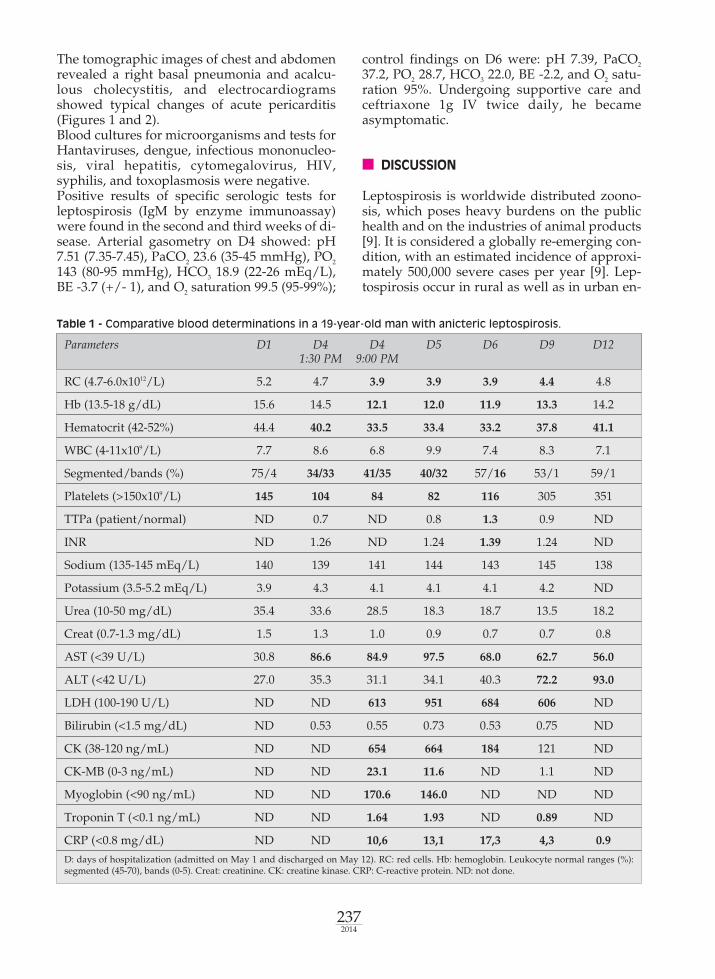
Table 1 - Comparative blood determinations in a 19-year-old man with anicteric leptospirosis.
Parameters D1 D4 D4 D5 D6 D9 D121:30 PM 9:00 PM
RC (4.7-6.0x10¹²/L) 5.2 4.7 3.9 3.9 3.9 4.4 4.8
Hb (13.5-18 g/dL) 15.6 14.5 12.1 12.0 11.9 13.3 14.2
Hematocrit (42-52%) 44.4 40.2 33.5 33.4 33.2 37.8 41.1
WBC (4-11x109/L) 7.7 8.6 6.8 9.9 7.4 8.3 7.1
Segmented/bands (%) 75/4 34/33 41/35 40/32 57/16 53/1 59/1
Platelets (>150x109/L) 145 104 84 82 116 305 351
TTPa (patient/normal) ND 0.7 ND 0.8 1.3 0.9 ND
INR ND 1.26 ND 1.24 1.39 1.24 ND
Sodium (135-145 mEq/L) 140 139 141 144 143 145 138
Potassium (3.5-5.2 mEq/L) 3.9 4.3 4.1 4.1 4.1 4.2 ND
Urea (10-50 mg/dL) 35.4 33.6 28.5 18.3 18.7 13.5 18.2
Creat (0.7-1.3 mg/dL) 1.5 1.3 1.0 0.9 0.7 0.7 0.8
AST (<39 U/L) 30.8 86.6 84.9 97.5 68.0 62.7 56.0
ALT (<42 U/L) 27.0 35.3 31.1 34.1 40.3 72.2 93.0
LDH (100-190 U/L) ND ND 613 951 684 606 ND
Bilirubin (<1.5 mg/dL) ND 0.53 0.55 0.73 0.53 0.75 ND
CK (38-120 ng/mL) ND ND 654 664 184 121 ND
CK-MB (0-3 ng/mL) ND ND 23.1 11.6 ND 1.1 ND
Myoglobin (<90 ng/mL) ND ND 170.6 146.0 ND ND ND
Troponin T (<0.1 ng/mL) ND ND 1.64 1.93 ND 0.89 ND
CRP (<0.8 mg/dL) ND ND 10,6 13,1 17,3 4,3 0.9
D: days of hospitalization (admitted on May 1 and discharged on May 12). RC: red cells. Hb: hemoglobin. Leukocyte normal ranges (%):segmented (45-70), bands (0-5). Creat: creatinine. CK: creatine kinase. CRP: C-reactive protein. ND: not done.
InfMed3_10_Dos_Santos_InfMed_IBAT_2005.qxp 01/10/14 10.07 Pagina 237
2382014
vironments, more frequently affecting peoplefrom poor communities both in industrializedand in developing regions [5, 9]. Infections occur during occupational, sportiveand leisure activities, either by direct contactwith the infective agent, or indirectly by meansof animal sources as pets, rats and mice [5, 9].Moreover, ticks and other parasitic arthropodsmay play a role in disease transmission [9].Outbreaks of disease usually follow periods ofintense rainfalls, as occur in Brazil [4-6, 8, 9],and the mortality rate of the more severe infec-tions have ranged from 5% to 50% [9]. Lep-tospirosis may be anicteric (up to 90%) or withvariable jaundice, and evolve in two phases,
with a subclinical or symptom-free interval ofone to three days between them [9]. The first (septicemic), with blood born distribu-tion of leptospiras for about seven days; and thesecond (tissue), characterized by diverse signsand symptoms related to organ injury [5, 9]. Inthis phase, the antibody response develops,acalculous cholecystitis, respiratory and cardiacchanges, hemoglobinuria and hematuria canoccur, as is described in the present report [5].The patient herein described had anicteric lep-tospirosis with some less common features.But, despite of the absence of jaundice or renalfailure, the occurrence of fever, headache, hem-orrhagic phenomena, myalgia and arthralgia
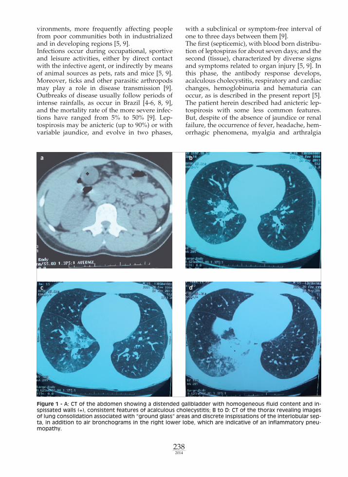
Figure 1 - A: CT of the abdomen showing a distended gallbladder with homogeneous fluid content and in-spissated walls (+), consistent features of acalculous cholecystitis; B to D: CT of the thorax revealing imagesof lung consolidation associated with “ground glass” areas and discrete inspissations of the interlobular sep-ta, in addition to air bronchograms in the right lower lobe, which are indicative of an inflammatory pneu-mopathy.
a b
c d
InfMed3_10_Dos_Santos_InfMed_IBAT_2005.qxp 01/10/14 10.07 Pagina 238
2392014
after exposure to a rural environment raised theclinical suspicion about leptospirosis. Dengue and Hantavirus infection were the maindifferential diagnoses because these pathogensare endemic in the same environment [5, 6, 8].The initial elevated creatinine level was proba-bly due to pre-renal mechanisms, but the associ-ation with lowering platelet counts can be mis-taken for Hantavirus hemorrhagic fever with re-nal syndrome, a condition not yet reported in
Brazilian Central Plateau [6, 7]. Imaging studiesof chest and abdomen showed normal heart,unilateral lung parenchymal infiltrates, andacalculous cholecystitis. Moreover, the chestpain and typical ECG changes led to diagnosisof acute pericarditis, which is an uncommonmanifestation of leptospirosis. Transient ECGchanges may be found (up to 68%) in the initialphase, due to the action of Leptospira and meta-bolic or electrolyte disorders, and are associatedwith jaundice, high alanine aminotransferase,hypokalemia, low platelets, and abnormal pul-monary images [7, 10]. The patient of this casereport had anicteric leptospirosis but presentedwith precordial pain and ECG abnormalitiesconsistent with acute pericarditis, even in ab-sence of hypokalemia. There was an excess ofband forms, moderate anemia, thrombocytope-nia, elevated cardiac and muscle injury markers,disorder in coagulation tests, and mild changesin liver enzymes. These findings are usually de-scribed in patients with Hantavirus pulmonarysyndrome, and coinfections can be under diag-nosed because of similar clinical and laboratorydata [6, 8]. Concomitant infections were alsoruled out, with base on the negative tests fordengue (immunochromatography and MAC-ELISA), and Hantavirus (antibody-captureELISA) [5, 8]. Yellow fever, malaria and sepsiswere other initial concerns as alternative diag-noses, but these entities were ruled out byanalysis of clinical features and complementarydata [8]. Clinical improvement occurred in spiteof antibiotic therapy starting after the first week[2, 4]. In fact, several challenges have played arole in the late diagnosis of leptospirosis [1-4,6].In addition to the sub-clinical infections, the lackof laboratory resources may also occur [2].
n CONCLUSION
The role of the suspicion index about leptospi-rosis should be emphasized, especially in anic-teric patients, as well as the possible pulmo-nary, cardiac and gallbladder involvement inthe course of this cosmopolitan zoonosis, whichmay affect previously healthy travellers.
Competing interests: The authors declare thatthey have no competing interests.Source of support: Nil
Keywords: anicteric leptospirosis, non-lithiasiccholecystitis, pericarditis, pneumonia.
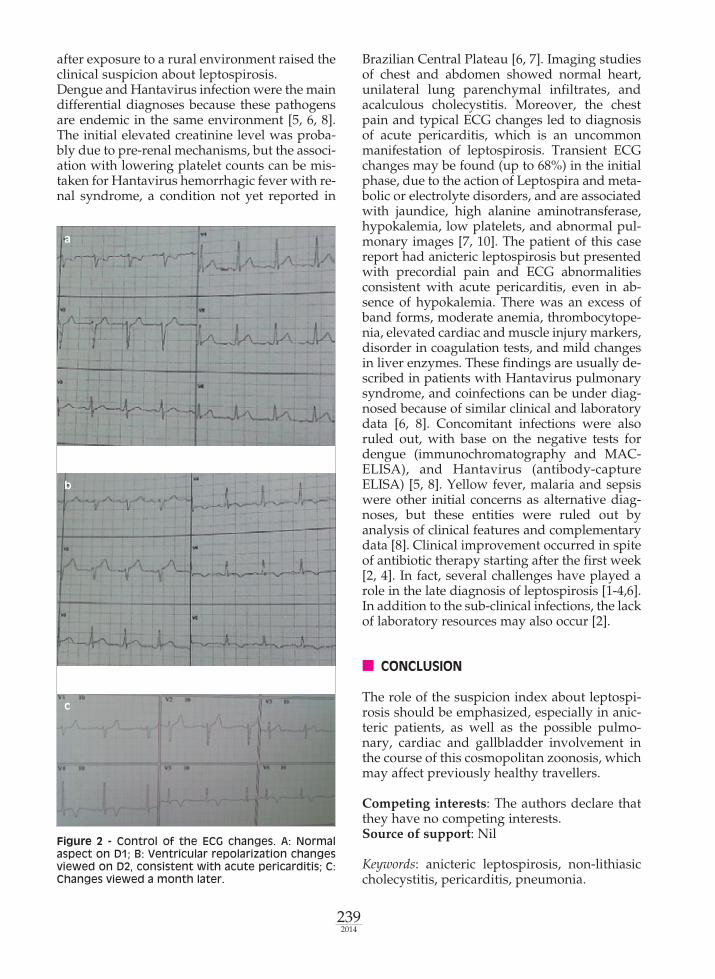
Figure 2 - Control of the ECG changes. A: Normalaspect on D1; B: Ventricular repolarization changesviewed on D2, consistent with acute pericarditis; C:Changes viewed a month later.
a
b
c
InfMed3_10_Dos_Santos_InfMed_IBAT_2005.qxp 01/10/14 10.07 Pagina 239
2402014
n REFERENCES
[1] Anselmo M., De Leo P., Calcagno G., Donelli E.,Tirotta A., Menardo G. Ceftazidime in the treat-ment of Weil’s disease. Infez. Med. 8, 2, 106-109,2000.[2] Conti E., Lazzarini L., Reatto P., Tositti G., de Lal-la F. Human leptospirosis in the Vicenza area (Italy)from 1990 to 2003: an epidemiological and clinicalstudy. Infez. Med. 13, 4, 235-240, 2005.[3] De Brito T., Aiello V.D., da Silva L.F., et al. Cor-rection: Human hemorrhagic pulmonary leptospiro-sis: pathological findings and pathophysiologicalcorrelations. PLoS One. 8, 9, 2013.[4] dos Santos V.M., dos Santos J.A., Sugai T.A., dosSantos L.A. Weil’s syndrome. Rev. Cubana Med. Trop.55, 1, 44-46, 2003.[5] Russo R., Panarello G. Leptospirosis: epidemiolo-gy, diagnosis and clinical aspects. Infez. Med. 7, 2, 74-84, 1999.
[6] Santos V.M., Rocha de Sá D.A., Turra T.Z., Ferrei-ra Borges N.M., Nascimento U.M., Damasceno E.A.Hantavirus pulmonary syndrome in Brasilia periph-ery: a diagnostic challenge. J. Infect. Dev. Ctries. 3, 8,639-643, 2009.[7] Skerk V., Markotic A., Puljiz I., et al. Electrocar-diographic changes in hospitalized patients with lep-tospirosis over a 10-year period. Med. Sci. Monit. 17,7, CR369-375, 2011.[8] dos Santos V.M., de Sá D.A., Martins R.R., PazB.C., de Oliveira E.R., Barcelos M. dos S. Hantaviruspulmonary syndrome coexistent with dengue. IndianJ. Chest Dis. Allied Sci. 52, 4, 249-251, 2010.[9] WasiDski B., Dutkiewicz J. Leptospirosis-currentrisk factors connected with human activity and theenvironment. Ann. Agric. Environ. Med. 20, 2, 239-244, 2013.[10] Navinan M.R., Rajapakse S. Cardiac involve-ment in leptospirosis. Trans. R. Soc. Trop. Med. Hyg.106, 9, 515-520, 2012.
Riportiamo il caso di un paziente maschio di 19 anni ri-coverato in ospedale per febbre, astenia, vomito, doloreaddominale e toracico, tosse ed escreato giallastro, eipotensione arteriosa. Gli esami di laboratorio eviden-ziavano leucocitosi, aumento della creatinfosfochinasi,senza iperbilirubinemia né insufficienza renale. Le im-magini tomografiche di torace e addome evidenziavanouna polmonite basale destra e colecistite alitiasica. Glielettrocardiogrammi mostravano segni caratteristici di
pericardite acuta. Le prove sierologiche specifiche per laleptospirosi eseguite nella seconda e terza settimana dimalattia hanno mostrato risultati positivi. La terapiacon ceftriaxone 1g per via endovenosa, due volte algiorno per un periodo di 7 giorni, ha determinato unmiglioramento clinico complessivo. Si sottolinea l’im-portanza del sospetto diagnostico di leptospirosi nei pa-zienti senza ittero, con compromissione polmonare epericardico.
RIASSUNTO
We report the case of a 19-year-old male patient ad-mitted to hospital with fever, asthenia, vomiting,abdominal and chest pains, cough with yellowishsputum, and hypotension. Laboratory tests showedleukocytosis and high creatine phosphokinase le-vels, without hyperbilirubinaemia or renal failure.The tomographic images of the chest and abdomenshowed a right basal pneumonia and acalculouscholecystitis. The electrocardiograms revealed si-
gnificant characteristics of acute pericarditis. Speci-fic serology for leptospirosis done in the second andthird weeks of disease showed positive results. Theuse of ceftriaxone 1g intravenously, twice a day for7 days, resulted in an overall clinic improvement.The role of the suspicion index for diagnosis of lep-tospirosis is emphasized in anicteric patients, aswell as the unsuspected possibility of pulmonary,pericardial and gallbladder involvement.
SUMMARY
InfMed3_10_Dos_Santos_InfMed_IBAT_2005.qxp 01/10/14 10.07 Pagina 240


![Case Report Taeniasaginata:ARareCauseofGallBladderPerforationdownloads.hindawi.com/journals/cris/2012/572484.pdf · bladder causing acalculous cholecystitis [6–11]. To the best](https://img.pdfslide.us/doc/110x75/5fdead341c0daa158f3896fc/case-report-taeniasaginataararecauseofgallbladder-bladder-causing-acalculous-cholecystitis.jpg)