Embed Size (px)
Citation preview

of May 16, 2018.This information is current as
Promoting CXCR-2-Dependent AngiogenesisCell Lung Cancer in SCID Mice throughand In Vivo Growth of Human Non-Small IL-17 Enhances the Net Angiogenic Activity
Kolls and Hidetada SasakiTakanori Hishinuma, Junichi Goto, Michael T. Lotze, Jay K.Hidenori Takahashi, Akira Nakamura, Florencia McAllister, Muneo Numasaki, Mika Watanabe, Takashi Suzuki,
http://www.jimmunol.org/content/175/9/6177doi: 10.4049/jimmunol.175.9.6177
2005; 175:6177-6189; ;J Immunol
Referenceshttp://www.jimmunol.org/content/175/9/6177.full#ref-list-1
, 30 of which you can access for free at: cites 61 articlesThis article
average*
4 weeks from acceptance to publicationFast Publication! •
Every submission reviewed by practicing scientistsNo Triage! •
from submission to initial decisionRapid Reviews! 30 days* •
Submit online. ?The JIWhy
Subscriptionhttp://jimmunol.org/subscription
is online at: The Journal of ImmunologyInformation about subscribing to
Permissionshttp://www.aai.org/About/Publications/JI/copyright.htmlSubmit copyright permission requests at:
Email Alertshttp://jimmunol.org/alertsReceive free email-alerts when new articles cite this article. Sign up at:
Print ISSN: 0022-1767 Online ISSN: 1550-6606. Immunologists All rights reserved.Copyright © 2005 by The American Association of1451 Rockville Pike, Suite 650, Rockville, MD 20852The American Association of Immunologists, Inc.,
is published twice each month byThe Journal of Immunology
by guest on May 16, 2018
http://ww
w.jim
munol.org/
Dow
nloaded from
by guest on May 16, 2018
http://ww
w.jim
munol.org/
Dow
nloaded from

IL-17 Enhances the Net Angiogenic Activity and In VivoGrowth of Human Non-Small Cell Lung Cancer in SCID Micethrough Promoting CXCR-2-Dependent Angiogenesis1
Muneo Numasaki,2* Mika Watanabe,‡ Takashi Suzuki,† Hidenori Takahashi,*Akira Nakamura,¶ Florencia McAllister,� Takanori Hishinuma,§ Junichi Goto,§
Michael T. Lotze,# Jay K. Kolls,� and Hidetada Sasaki*
In this study, we examined the biological action of IL-17 on human non-small cell lung cancer (NSCLC). Although IL-17 had nodirect effect on the in vitro growth rate of NSCLC, IL-17 selectively augmented the secretion of an array of angiogenic CXCchemokines, including CXCL1, CXCL5, CXCL6, and CXCL8 but not angiostatic chemokines, by three different NSCLC lines.Endothelial cell chemotactic activity (as a measure of net angiogenic potential) was increased in response to conditioned mediumfrom NSCLC stimulated with IL-17 compared with those from unstimulated NSCLC. Enhanced chemotactic activity was sup-pressed by neutralizing mAb(s) to CXCL1, CXCL5, and CXCL8 or to CXCR-2 but not to vascular endothelial growth factor-A.Transfection with IL-17 into NSCLC had no effect on the in vitro growth, whereas IL-17 transfectants grew more rapidlycompared with controls when transplanted in SCID mice. This IL-17-elicited enhancement of NSCLC growth was associated withincreased tumor vascularity. Moreover, treatment with anti-mouse CXCR-2-neutralizing Ab significantly attenuated the growthof both neomycin phosphotransferase gene-transfected and IL-17-transfected NSCLC tumors in SCID mice. A potential role forIL-17 in modulation of the human NSCLC phenotype was supported by the findings that, in primary NSCLC tissues, IL-17expression was frequently detected in accumulating and infiltrating inflammatory cells and that high levels of IL-17 expressionwere associated with increased tumor vascularity. These results demonstrate that IL-17 increases the net angiogenic activity andin vivo growth of NSCLC via promoting CXCR-2-dependent angiogenesis and suggest that targeting CXCR-2 signaling may bea novel promising strategy to treat patients with NSCLC. The Journal of Immunology, 2005, 175: 6177–6189.
I nterleukin 17, initially termed CTLA-8 (1), is producedmainly by activated CD4 T cells (2) and has sequence ho-mology with the open reading frame 13 of the T lympho-
tropic Herpesvirus saimiri (2). Several recently discovered homol-ogous proteins with similar or different biological profilesincluding IL-17B, IL-17C, IL-17D, IL-17E, and IL-17F form anovel cytokine family (3–5). IL-17 has pleiotropic biological ac-tivities including induction of IL-6, CXCL8/IL-8 and vascular en-dothelial growth factor-A (VEGF-A),3 as well as enhancement of
ICAM-1 expression (2–11). In addition, IL-17 induces the secre-tion of TNF-� and IL-1� by activated macrophages (12). IL-17Ris a type 1 transmembrane protein with an extraordinarily longintracellular domain (2, 13). Although the expression of IL-17mRNA is restricted to activated T cells, the expression of IL-17RmRNA has been detected in virtually all cells and tissues (2, 13).
In its role as a proinflammatory cytokine, IL-17 levels have beenfound to significantly increase in rheumatoid arthritis synovium,asthmatic airways, during allograft rejection, and in other chronicinflammatory diseases, including multiple sclerosis and psoriasis(14–18). IL-17 has been also implicated in the tumors. Ciree et al.(19) reported that cutaneous T cell lymphomas spontaneously se-crete IL-17, which is associated with infiltration of polymorpho-nuclear neutrophils into inflamed tissues. Kato et al. (20) reportedthat a considerable proportion of ovarian carcinomas naturally ex-press IL-17 and its expression significantly correlates with the in-creased vascularity. However, until now, the possible role forIL-17 in non-small cell lung cancer (NSCLC) has not beenelucidated.
The salient feature of solid tumor growth such as NSCLC is thestrict dependence on the sustained new vessel growth (21). Hana-han and Folkman (22) proposed that quiescent endothelium existsin a net balance of angiostatic factors over angiogenic ones,thereby maintaining homeostasis. Conversely, tumors themselves,interacting stromal cells and/or responding inflammatory cells,may cause an imbalance to increase the secretion of angiogenicinducers or decrease the production or effect of angiogenic sup-pressors. Although there are a wide variety of putative mediatorsof angiogenesis, certain tumors have been found to produce factors
*Department of Geriatric and Respiratory Medicine and †Department of AnatomicPathology, Tohoku University School of Medicine, Sendai, Japan; ‡Department ofPathology and §Department of Pharmaceutical Sciences, Tohoku University Hospital,Sendai, Japan; ¶Department of Experimental Immunology, Institute of Development,Aging, and Cancer, Tohoku University, Sendai, Japan; �Laboratory of Lung Immu-nology and Host Defense, Department of Pediatrics, University of Pittsburgh, Pitts-burgh, PA 15213; and #Translational Research, Molecular Medicine Institute, Uni-versity of Pittsburgh School of Medicine, Pittsburgh, PA 15219
Received for publication May 20, 2004. Accepted for publication August 3, 2005.
The costs of publication of this article were defrayed in part by the payment of pagecharges. This article must therefore be hereby marked advertisement in accordancewith 18 U.S.C. Section 1734 solely to indicate this fact.1 This work was supported in part by a grant-in-aid for Scientific Research from theJapan Society for the Promotion of Science (Grant 15590791; to M.N.).2 Address correspondence and reprint requests to Dr. Muneo Numasaki, Departmentof Geriatric and Respiratory Medicine, Tohoku University School of Medicine,1-1 Seiryo-machi, Aoba-ku, Sendai 980-8574, Japan. E-mail address:[email protected] Abbreviations used in this paper: VEGF-A, vascular endothelial growth factor-A;NSCLC, non-small cell lung cancer; FGF, fibroblast growth factor; HGF, hepatocytegrowth factor; CM, conditioned medium; NEAA, nonessential amino acid; LMVEC,lung microvascular endothelial cell; HPF, high-powered field; PDGF, platelet-derivedgrowth factor; Neo, neomycin phosphotransferase gene; DAB, 3,3�-diaminobenzi-dine; ELR, glutamic acid-leucine-arginine; aFGF, acidic FGF.
The Journal of Immunology
Copyright © 2005 by The American Association of Immunologists, Inc. 0022-1767/05/$02.00
by guest on May 16, 2018
http://ww
w.jim
munol.org/
Dow
nloaded from

that are directly angiogenic while others may depend upon vascu-larization induced by products of infiltrating inflammatory cells(23).
NSCLC cells have been reported to produce a wide variety ofangiogenic factors, including CXCL1/growth related oncogene-�,CXCL5/epithelial cell-derived neutrophil-activating protein-78,CXCL6/granulocyte chemotactic protein-2, CXCL8/IL-8,VEGF-A, fibroblast growth factors (FGFs), hepatocyte growth fac-tor (HGF), platelet-derived growth factor (PDGF)-BB, TGF-�,CXCL12/stromal derived factor-1�, angiopoietin-2, and PGE2
over the course of their growth (24–32). Especially, multiple stud-ies have established that angiogenic CXC chemokines are criti-cally involved in the angiogenic activity of NSCLC (22, 33, 34).Arenberg et al. (33) reported that inhibition of the action ofCXCL8 markedly suppressed the in vivo growth of NSCLC inSCID mice model. Arenberg et al. (34) also showed that neutral-ization of CXCL5 in vivo significantly suppressed the growth ofNSCLC. In contrast, tumor-derived CXCL10/IFN-inducible pro-tein-10 is an important endogenous angiostatic factor in NSCLC,which negatively regulates tumor angiogenesis and growth (35).
IL-17 induces CXCL8 production from various cell types andCXCL8 is involved in the angiogenic activity of NSCLC. There-fore, we hypothesized that IL-17 might enhance the net angiogenicactivity and promote the tumorigenicity of NSCLC. To addressthis issue, we examined the biological effect of IL-17 on NSCLCand found that IL-17 selectively enhanced the production of anarray of angiogenic CXC chemokines, including CXCL1, CXCL5,CXCL6, and CXCL8 but not angiostatic chemokines, fromNSCLC lines in vitro. Therefore, these characteristic biologic ac-tivities of IL-17 prompted us to further investigate the role for thiscytokine in in vivo biologic action of NSCLC. In this study, weshow that IL-17 significantly enhances the net angiogenic activityand in vivo growth of NSCLC through promoting CXCR-2-de-pendent angiogenesis.
Materials and MethodsMice and reagents
Male SCID mice, 5–7 wk of age, were obtained from Charles River Lab-oratories. Human IL-17 cDNA was supplied by Schering-Plough. Recom-binant human IL-17 and IL-23 proteins and anti-human IL-17, IL-17R,CXCL1, CXCL5, CXCL8, VEGF-A, HGF, CXCR-1, and CXCR-2 mAbswere purchased from R&D Systems. Anti-human IFN-� mAb was suppliedby Mabtech. Goat anti-human IL-17R polyclonal Ab was from Santa CruzBiotechnology. Rabbit anti-mouse CXCR-2-neutralizing polyclonal Abwas supplied by Dr. K. Matsushima (University of Tokyo, Tokyo, Japan).
Human NSCLC cell lines, cell cultures, and conditioned medium(CM)
Human NSCLC lines Sq-19 (squamous cell carcinoma), A549 (alveolarcell carcinoma), and LK-87 (adenocarcinoma) were supplied from the CellResource Center for Biomedical Research, Institute of Development, Ag-ing and Cancer (Tohoku University). These cells were maintained in RPMI1640 with 10% FCS, 2 mM L-glutamine, 0.1 mM nonessential amino acid(NEAA), 100 IU/ml penicillin, and 100 �g/ml streptomycin (all from In-vitrogen Life Technologies). Human lung microvascular endothelial cells(LMVECs) were purchased from Cambrex and maintained in HuMedia-EB2 with 10 ng/ml epidermal growth factor, 1 �g/ml hydrocortisone, 5ng/ml basic FGF, 10 �g/ml heparin, 39.3 �g/ml dibutyryl cAMP, 50 �g/mlgentamicin, 50 ng/ml amphotericin B, and 5% FCS (all from Kurabo). CMwas generated as follows. Cells (1 � 105/ml) were cultured in HuMedia-EB2 containing 3% FCS with or without 50 ng/ml IL-17 for 60 h. Cell-freesupernatants were collected and stored at �70°C until use.
Clinical materials
Tumor tissues (n � 77) were obtained from patients with NSCLC, whounderwent complete resection at surgery without any preoperative therapy.
Flow cytometry
Cells (1 � 106), washed twice with PBS containing 2% FCS and 0.1%NaN3, were incubated with mouse anti-human IL-17R mAb or with irrel-evant normal mouse IgG1 (R&D Systems) at 4°C for 60 min. After wash-ing, the cells were incubated with PE-conjugated goat anti-mouse IgG1 Ab(Caltag Laboratories) at 4°C for 30 min. The cells were analyzed withFACSCalibur (BD Biosciences).
MTT assay
Cells (1 � 103) were seeded into 96-well flat-bottom plates and cultured inRPMI 1640 containing 3% FCS, 2 mM L-glutamine, 0.1 mM NEAA, 100IU/ml penicillin, and 100 �g/ml streptomycin with or without 0.1–1000ng/ml human IL-17. On day 5 or 7, cells were washed with RPMI 1640,and 100 �l of MTT (Sigma-Aldrich) solution (2.5 mg/ml in RPMI 1640with 5% FCS) were added to each well. Plates were incubated for 50 min.Next, MTT solution was removed, and 50 �l of DMSO (Sigma-Aldrich)were added to each well to solubilize formazan crystals formed in viablecells. The absorbance was read at a wavelength of 590 nm on an ELISAplate reader.
Cytokine assay
Cells (1 � 105/ml) were cultured in RPMI 1640 containing 5% FCS, 2 mML-glutamine, 0.1 mM NEAA, 100 IU/ml penicillin, and 100 �g/ml strep-tomycin with or without IL-17 or IL-17 plus 5 �g/ml anti-IL-17 mAb atindicated concentrations for 48 h. Cell-free supernatants were collected andstored at �70°C. Concentrations of cytokines were measured using com-mercially available ELISA kits (R&D Systems). PGE2 concentration wasmeasured as reported previously (36).
Migration assay
Migration of LMVECs was evaluated using a modified Boyden chamberassay, as described previously (37). Briefly, LMVECs were cultured inHuMedia-EB2 with 2% FCS for 8 h and plated at 12 � 104 cells/cm2 ontothe polycarbonate filter with 5-�m pores (Kurabo) coated with 10 �g/mlfibronectin (Sigma-Aldrich). CM from NSCLC unstimulated or stimulatedwith 50 ng/ml IL-17 (NSCLC/IL-17) was applied in the lower compart-ments of the chamber. LMVECs were cultured for 4 h at 37°C. The filterswere fixed and stained with Diff-Quick (Harleco), and the number of mi-grating cells was quantified by counting cells in five randomly selectedhigh-powered fields (HPFs) (�200) in each well. For the inhibitory assay,neutralizing mAb(s) (5 �g/ml each) was added in the upper and lowercompartments of the chamber.
Construction of expression vector carrying human IL-17 cDNA
A BamHI-XbaI fragment of the PCR product of IL-17 cDNA was ligatedinto the EcoRI site of pCRTM2.1. The insert DNA was excised from theplasmid with BamHI/XbaI and inserted into the multiple cloning site ofpRc/CMV. A549 and Sq-19 cells were transfected with the human IL-17gene expression plasmid using LipofectAMINE (Invitrogen Life Technol-ogies) and selected in RPMI 1640 with 10% FCS, 2 mM L-glutamine, 1mM sodium pyruvate, 0.1 mM NEAA, 100 IU/ml penicillin, 100 �g/mlstreptomycin, and 1000 �g/ml G418. As a control, a vector carrying onlyneomycin phosphotransferase gene (Neo) was used.
In vitro cell growth assay
To examine the in vitro growth, cells (5 � 104) were seeded in 10-cmculture plates on day 0, and the cell number was counted on days 2, 3, 4,5, and 6.
Human NSCLC-SCID mouse chimeras
Eight- to 10-wk-old male SCID mice were inoculated s.c. with 7 � 106
Sq-19WT, Sq-19Neo, or Sq-19IL-17 cells or with 1 � 107 A549WT,A549Neo, or A549IL-17 cells into the right flank. The animals were main-tained under sterile conditions in laminar flow rooms. In some experiments,mice were treated with either control Ab or rabbit polyclonal Ab specificfor mouse CXCR-2 every 4 days. On day 25 for Sq-19 or on day 40 forA549, blood was collected from the mice, kept overnight at 4°C, and thencentrifuged. The serum was stored at �70°C until use. Tumor volume(mm3) was calculated using the formula a � b2/2 (a � largest diameter;b � smallest diameter) (38).
Immunohistochemical staining for Ki-67 in human NSCLCtissues from SCID mice
Analysis of the biological effect of IL-17 on the cell proliferation ofNSCLC in vivo was performed with Ki-67 immunostaining, using MIB-1
6178 ENHANCED ANGIOGENIC ACTIVITY OF HUMAN NSCLC BY IL-17
by guest on May 16, 2018
http://ww
w.jim
munol.org/
Dow
nloaded from

Ab (Immunotech). Sections were deparaffinized and autoclaved in acetatebuffer at 120°C for 5 min. After cooling, slides were incubated overnightat 4°C with MIB-1 or with normal mouse IgG (R&D Systems). Afterwashing, sections were then incubated with biotinylated rabbit anti-mouseIgG and HRP-conjugated streptavidin (Nichirei, Tokyo, Japan). Sectionswere developed with 3,3�-diaminobenzidine (DAB) and counterstainedwith hematoxylin. The proliferating cells were estimated by the percentageof Ki-67-positive staining cells in 10 randomly chosen HPFs for each sec-tion (�400).
In situ detection of apoptosis
To detect apoptotic cells, the TUNEL method was performed using theWAKO in situ apoptosis detection kit (WAKO). Briefly, after deparaf-finization, sections were incubated with a protein digestion enzyme for 5min at 37°C. TdT with biotin-11-dUTP and dATP was then applied to theslides for 10 min at 37°C in a moist chamber. Endogenous peroxidaseactivity was blocked with 3% hydrogen peroxide for 5 min at room tem-perature. Then, anti-biotin-11-dUTP labeling was conducted for 10 min at37°C, followed by exposure to DAB. Sections were counterstained withmethyl green. The apoptotic cells were estimated by the percentage ofpositive staining cells visualized in 10 randomly chosen HPFs for eachsection (�400).
Immunohistochemical staining for vascular endothelial cells inhuman NSCLC tissues and quantitation of vessel density
Immunostaining for CD31 was performed on frozen sections with rat anti-mouse CD31 mAb (BD Pharmingen). Briefly, frozen sections of NSCLCtissues from SCID mice were air-dried and incubated with 1% normal goatserum for 30 min at room temperature. Endogenous peroxidase activitywas blocked using DakoCytomation peroxidase blocking reagent (Dako-Cytomation). Then, the primary Ab was applied overnight at 4°C. Afterwashing, the sections were incubated with biotinylated rabbit anti-rat IgGAb (DakoCytomation) for 30 min. After washing, peroxidase-conjugatedstreptavidin solution (DakoCytomation) was applied. Immunoreactivitywas visualized by DAB and counterstained with hematoxylin.
Immunostaining for CD34 was performed on sections of NSCLC tissuesfrom surgery, using the streptavidin-biotin amplification method (Histofinekit). Briefly, after deparaffinization, slides were treated with 3% hydrogenperoxidase in methanol for 10 min. Sections were then incubated with 1%normal goat serum for 30 min, followed by the application of mouse anti-human CD34 mAb (Nichirei) overnight at 4°C. Sections were treated withbiotinylated anti-mouse IgG for 30 min, followed by peroxidase-conju-gated streptavidin for 30 min. Immunoreactivity was visualized by DABand counterstained with hematoxylin.
Sections were examined in a blinded fashion for the presence of CD31or CD34 immunolocalization. According to the method described byWeidner et al. (39), specimens were scanned at low magnification (�40),and the 10 most vascularized tumor areas within a section were selected forevaluation of angiogenesis. Blood vessels stained with anti-mouse CD31mAb or anti-human CD34 mAb were counted under light microscopy at�200. Any distinct area of positive staining for CD31 or CD34 wascounted as a single regardless of size. The results are shown as mean � SDper 10 HPFs per tumor section.
Immunohistochemical staining for IL-17, IL-17R, and CD3 inhuman NSCLC tissues
Immunohistochemical staining for IL-17 and CD3 was performed on sec-tions of NSCLC tissues from surgery, using the streptavidin-biotin ampli-fication method (Histofine kit). Goat polyclonal Ab for human IL-17 andrabbit polyclonal Ab for human CD3 were used as a primary Ab. Thedilution of primary Ab used in this study was 1/500 for IL-17 and 1/100 forCD3. Ag retrieval was performed by heating the slides in a microwaveoven for 15 min for IL-17 or by treatment with protease K (Sigma-Aldrich)for 10 min for CD3. The Ag-Ab complex was visualized with DAB andcounterstained with hematoxylin. As a positive control, we used the sec-tions from blocks of Formalin-fixed, paraffin-embedded, IL-23-stimulatedhuman CD4 T cells. As a negative control, an immunohistochemical pre-absorption test was performed in these specimens.
For IL-17R staining, goat anti-human IL-17R polyclonal Ab (SantaCruz Biotechnology) was used to characterize the expression of IL-17R onprimary NSCLC tissues. As a control, sections were incubated with normalgoat IgG (R&D Systems). The blocking of nonspecific proteins was con-ducted with PBS containing 2% BSA. The staining was conducted usingCy-3-conjugated rabbit anti-goat Ab as a secondary Ab (Sigma-Aldrich)and ProLong Gold antifade reagent with 4�,6�-diamidino-2-phenylindole(Molecular Probes) as a mounting medium. The pictures were captured by
a camera attached to an Axioplan 2 universal imaging microscope (Intel-ligent Imaging Innovations) and were further analyzed with SlideBook 4.1(Intelligent Imaging Innovations).
RT-PCR and quantitative RT-PCR
Total cellular RNA was extracted using Isogen (Nippon Gene) according tothe manufacturer’s instructions. Five micrograms of total RNA was usedfor the synthesis of cDNAs with SuperScript RNaseH-Reverse Transcrip-tase (Invitrogen Life Technologies). PCR was performed in a DNA Ther-mal Cycler (PerkinElmer/Cetus) using Taq polymerase (Boehringer Mann-heim). The primer sequences of the oligonucleotides used for PCR andPCR product sizes were as follows: �-actin, sense, 5�-TCTGGTCAATGGAAGCCTGT-3�, and antisense, 5�-CTGTGGTGGTGAAGCTGTAC-3�,436 bp; and human IL-17, sense, 5�-ACTCCTGGGAAGACCTCATTG-3�, and antisense, 5�-GGCCACATGGTGGACAATCG-3�, 461 bp. Real-time quantitative PCR (TaqMan PCR) using an ABI PRISM 7700 Se-quence Detection System and TaqMan PCR Core Reagent kit(PerkinElmer) was performed according to the manufacturer’s protocol.Oligonucleotides and a probe specific for human IL-17 were purchasedfrom PerkinElmer Biosystems. Internal standard gene expression was ex-amined by using TaqMan �-actin control reagents (PerkinElmer Biosys-tems). The amount of amplified IL-17 mRNA was correlated to that of�-actin mRNA.
Statistical analysis
Statistical analysis was performed using an unpaired two-tailed Student’s ttest with confirmation by parametric and F tests. Differences were consid-ered to be statistically significant when the p value was �0.05.
ResultsExpression of IL-17R on human NSCLC lines Sq-19, A549, andLK-87
The expression of IL-17R on three NSCLC cells was examined byflow cytometry using anti-human IL-17R mAb. These NSCLCcells expressed IL-17R on the surface at the protein level (Fig. 1).No staining was observed when an isotype matched control mAbwas used (Fig. 1). These results are in accordance with the ubiq-uitous expression of IL-17R (2).
IL-17 has no direct effect on the in vitro growth of NSCLC cells
Because NSCLC cells expressed IL-17R, we investigated whetherIL-17 can modulate the phenotype of these cells. We first exam-ined the possibility that IL-17 might modulate the in vitro growthrate of NSCLC. To determine whether IL-17 affects the in vitrogrowth, cells were cultured in the presence or absence of humanIL-17 for 5 or 7 days. A wide range of doses of IL-17 had no directeffect on the in vitro growth of Sq-19 and A549 cells as shown inFig. 2, A and B. These results indicate that IL-17 has no biologicalaction to promote or suppress the growth of NSCLC cells.
IL-17 selectively up-regulates the production of angiogenic CXCchemokines by three different NSCLC lines
We next examined the possibility that IL-17 might modulate thesecretion of angiogenic and angiostatic factors by NSCLC, whichpromote or suppress tumor angiogenesis and growth. NSCLC cellshave been reported to naturally produce diverse angiogenic factors.Interestingly, IL-17 selectively up-regulated the production ofNSCLC-derived several angiogenic CXC chemokines, includingCXCL1, CXCL5, CXCL6 and CXCL8 (Fig. 3, A–C, and Table I).Especially, IL-17 markedly augmented the production of CXCL1,CXCL5, and CXCL8 (Fig. 3, A–C). To control these results, wedemonstrated that anti-IL-17 mAb inhibited the up-regulated pro-duction (Fig. 3, A–C), whereas an isotype-matched control mAbhad no effect (data not shown). In contrast, IL-17 did not signifi-cantly enhance the production of acidic FGF (aFGF), bFGF,VEGF-A, PDGF-BB, HGF, TGF-�, angiopoietin-2, CXCL12, andPGE2 (Fig. 3, A–C, and Table I). Moreover, we could not detectthe up-regulated secretion of angiostatic CXC chemokines
6179The Journal of Immunology
by guest on May 16, 2018
http://ww
w.jim
munol.org/
Dow
nloaded from

CXCL9/monokine induced by IFN-�, CXCL10, and CXCL11/IFN-inducible T cell � chemoattractant from NSCLC lines withIL-17 (Table I). These results indicate that IL-17 selectively up-regulates the production of an array of angiogenic CXC chemo-kines, but not angiostatic CXC chemokines, from NSCLC.
IL-17 significantly augments the net angiogenic activity ofNSCLC
IL-17 selectively up-regulated the production of important angio-genic CXC chemokines of NSCLC. We therefore hypothesizedthat IL-17 might enhance the net angiogenic activity of NSCLC.To address this question, we performed endothelial cell chemo-
taxis assays on CM from NSCLC or NSCLC/IL-17. As shown inFig. 4A, in comparison to CM from Sq-19, CM from Sq-19/IL-17demonstrated a marked increase in angiogenic activity as assessedby endothelial cell chemotaxis. Similarly, CM from A549 demon-strated less angiogenic activity than CM from A549/IL-17 (Fig.4B). These data clearly indicate that IL-17 significantly augmentsthe net angiogenic activity of NSCLC measured as endothelial cellchemotactic activity.
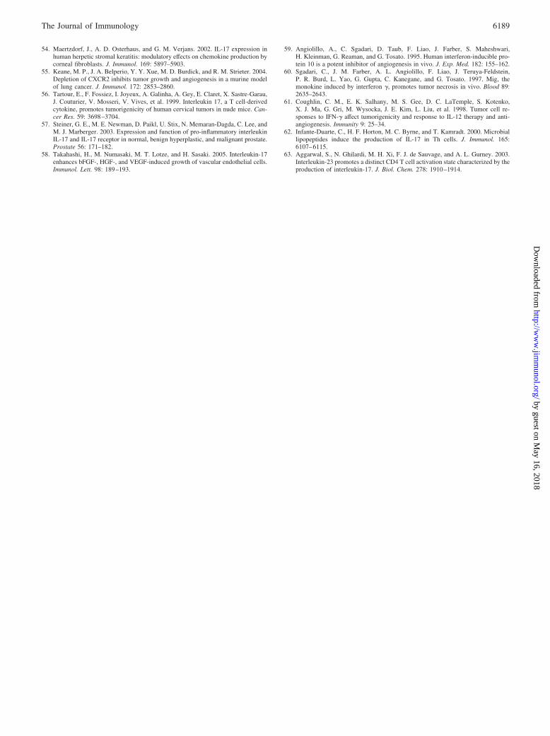
Increased endothelial cell chemotaxis in response to CM fromNSCLC/IL-17 is suppressed in the presence of neutralizingmAb(s) to CXCL1, CXCL5, and CXCL8 or to CXCR-2
We hypothesized that the increased endothelial cell chemotaxis toCM from NSCLC/IL-17 was attributable to the observed increasedsecretion of angiogenic CXC chemokines by NSCLC with IL-17.To test this postulate, endothelial cell chemotaxis was performedwith CM from NSCLC/IL-17 in the presence of neutralizingmAb(s) against CXCL1, CXCL5 and CXCL8, VEGF-A, HGF,IFN-�, CXCR-1, or CXCR-2. We found that endothelial cell che-motaxis to CM from Sq-19/IL-17 was reduced significantly in thepresence of neutralizing mAb(s) to CXCL1, CXCL5, and CXCL8or to CXCR-2 as compared with control IgG (Fig. 5A). On thecontrary, adding a neutralizing mAb against VEGF-A, HGF,IFN-�, or CXCR-1 did not significantly inhibit the increased en-dothelial cell chemotaxis. Similarly, endothelial cell chemotaxis toCM from A549/IL-17 in the presence of neutralizing mAb(s) toCXCL1, CXCL5, and CXCL8 or to CXCR-2 was significantly lessthan that in the presence of control mouse IgG (Fig. 5B). Thesedata demonstrate that IL-17 increases the net angiogenic activity ofNSCLC through angiogenic CXC chemokine- and CXCR-2-de-pendent fashion.
Establishment and characterization of the IL-17-producingNSCLC lines
To evaluate the biological effect of IL-17 on NSCLC in detail, wegenerated the human IL-17 gene expression vector as described inthe Materials and Methods. Two NSCLC lines, Sq-19 and A549,were transfected with human IL-17 gene using LipofectAMINE.No expression of either IL-17 mRNA or protein could be detectedin Sq-19 and A549 cells before transfection (data not shown). Af-ter G418 selection, the expression of human IL-17 mRNA by sta-ble transfectants was determined by RT-PCR (data not shown).Sq-19IL-17 or A549IL-17 secretes 67 or 35 ng/1 � 106 cells/48 h
FIGURE 2. IL-17 has no direct effect on the in vitro growth of humanNSCLC cells. Cells (1 � 103) were seeded into 96-well flat-bottom platesand cultured in RPMI 1640 with 3% FCS in the presence or absence of0.1–1000 ng/ml human IL-17. On day 5 or 7, cells were washed with RPMI1640, and 100 �l of MTT solution were added to each well. A wide rangeof doses of human IL-17 has no direct effect on the in vitro growth of Sq-19and A549 NSCLC cells. Each value represents mean � SD (n � 7). Theresult is representative of two independent experiments.
FIGURE 1. IL-17R expression on human NSCLC lines. Three different human NSCLC lines, Sq-19, A549, and LK-87, were incubated with mouseanti-human IL-17R mAb (solid line) or irrelevant mouse IgG1 (dotted line), followed by PE-conjugated goat anti-mouse IgG1 Ab. The expression of IL-17Ron the surface of NSCLC cells was analyzed by flow cytometry.
6180 ENHANCED ANGIOGENIC ACTIVITY OF HUMAN NSCLC BY IL-17
by guest on May 16, 2018
http://ww
w.jim
munol.org/
Dow
nloaded from

FIGURE 3. Effects of increasingconcentrations of IL-17 on CXCL1,CXCL5, CXCL8, and VEGF-A pro-duction from human NSCLC lines.NSCLC lines, Sq-19 (A), A549 (B),and LK-87 (C), were stimulated withor without 0.1, 0.5 1, 5, 10, 50, 100,and 500 ng/ml IL-17 for 48 h.CXCL1, CXCL5, CXCL8, andVEGF-A concentrations in cell cul-ture supernatants determined byELISA are shown. Data are ex-pressed as mean � SD (n � 3). Theresult is representative of two inde-pendent experiments. (control vs 0.1–500 ng/ml IL-17, �, p � 0.05; 50ng/ml IL-17 vs 50 ng/ml IL-17 � 5�g/ml anti-human IL-17 mAb, ��,p � 0.003).
6181The Journal of Immunology
by guest on May 16, 2018
http://ww
w.jim
munol.org/
Dow
nloaded from
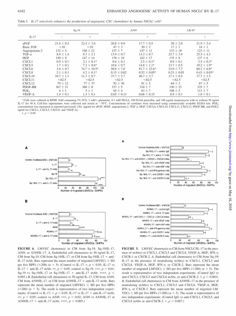
FIGURE 4. LMVEC chemotaxis to CM from Sq-19, Sq-19/IL-17,A549, or A549/IL-17. A, Endothelial cell chemotaxis to 50 ng/ml IL-17,CM from Sq-19, CM from Sq-19/IL-17, or CM from Sq-19/IL-17 � anti-IL-17 mAb. Bars represent the mean number of migrated LMVECs � SDper five HPFs (�200) (n � 5). (Control vs IL-17, �, p � 0.05; IL-17 vsIL-17 � anti-IL-17 mAb, ��, p � 0.05; control vs Sq-19, ���, p � 0.01;Sq-19 vs Sq-19/IL-17 or Sq-19/IL-17 � anti-IL-17 mAb; ����, p �0.003.) B, Endothelial cell chemotaxis to 50 ng/ml IL-17, CM from A549,CM from A549/IL-17, or CM from A549/IL-17 � anti-IL-17 mAb. Barsrepresent the mean number of migrated LMVECs � SD per five HPFs(�200) (n � 5). The result is representative of two independent experi-ments. (Control vs IL-17, �, p � 0.05; IL-17 vs IL-17 � anti-IL-17 mAb,��, p � 0.05; control vs A549, ���, p � 0.02; A549 vs A549/IL-17 orA549/IL-17 � anti-IL-17 mAb, ����, p � 0.005.)
FIGURE 5. LMVEC chemotaxis to CM from NSCLC/IL-17 in the pres-ence of mAb(s) to CXCL1, CXCL5 and CXCL8, VEGF-A, HGF, IFN-�,CXCR-1 or CXCR-2. A, Endothelial cell chemotaxis to CM from Sq-19/IL-17 in the presence of neutralizing mAb(s) to CXCL1, CXCL5 andCXCL8, VEGF-A, HGF, IFN-�, or CXCR-2. Bars represent the meannumber of migrated LMVECs � SD per five HPFs (�200) (n � 5). Theresult is representative of two independent experiments. (Control IgG vsanti-CXCL1, CXCL5 and CXCL8 mAbs, or anti-CXCR-2, �, p � 0.003).B, Endothelial cell chemotaxis to CM from A549/IL-17 in the presence ofneutralizing mAb(s) to CXCL1, CXCL5 and CXCL8, VEGF-A, HGF,IFN-�, or CXCR-2. Bars represent the mean number of migrated LM-VECs � SD per five HPFs (�200) (n � 5). The result is representative oftwo independent experiments. (Control IgG vs anti-CXCL1, CXCL5, andCXCL8 mAbs or anti-CXCR-2, �, p � 0.007.)
Table I. IL-17 selectively enhances the production of angiogenic CXC chemokines by human NSCLC cellsa
IL-17
Sq-19 A549 LK-87
� � � � � �
aFGF 21.6 � 0.5 22.4 � 3.6 20.8 � 0.9 17.7 � 0.5 20 � 2.0 21.9 � 2.4Basic FGF �10 �10 47 � 3 50 � 3 17 � 1 18 � 1Angiopoietin-2 132 � 4 140 � 21 157 � 7 147 � 11 133 � 18 123 � 11TGF-� 8.9 � 1.4 9.3 � 2.1 13.9 � 0.7 13.3 � 0.7 23.7 � 3.9 25.5 � 4.3HGF 150 � 9 147 � 14 178 � 10 162 � 17 135 � 8 137 � 6CXCL1 0.9 � 0.1 2.1 � 0.1* 0.6 � 0.1 2.5 � 0.1* 0.9 � 0.1 2.5 � 0.3*CXCL5 1.7 � 0.1 7.3 � 0.6* 10.6 � 0.7 14.8 � 1.1* 13.7 � 0.5 19.2 � 1.9*CXCL6 3.6 � 0.7 74.7 � 10.5* 50.8 � 7.0 91.7 � 22.6* 23.0 � 7.7 60.2 � 6.0*CXCL8 2.1 � 0.1 4.2 � 0.1* 0.15 � 0.02 0.52 � 0.05* 0.23 � 0.03 0.43 � 0.03*CXCL10 10.7 � 1.1 11.1 � 0.7 37.7 � 2.7 40.7 � 3.7 17.1 � 0.5 17.7 � 1.5CXCL11 �62.5 �62.5 �62.5 �62.5 �62.5 �62.5CXCL12 79 � 12 77 � 37 93 � 30 91 � 5 64 � 3 67 � 5PDGF-BB 367 � 21 360 � 8 337 � 5 334 � 7 330 � 23 335 � 7PGE2 4 � 1 5 � 1 65 � 4 63 � 7 108 � 5 111 � 7VEGF-A 1.4 � 0.1 1.3 � 0.1 0.65 � 0.13 0.66 � 0.15 0.9 � 0.1 1.0 � 0.1
a Cells were cultured in RPMI 1640 containing 5% FCS, 2 mM L-glutamine, 0.1 mM NEAA, 100 IU/ml penicillin, and 100 �g/ml streptomycin with or without 50 ng/mlIL-17 for 48 h. Cell-free supernatants were collected and stored at �70°C. Concentrations of cytokines were measured using commercially available ELISA kits. PGE2
concentration was measured as reported previously (36). (pg/ml for aFGF, bFGF, angiopoietin-2, TGF-�, HGF, CXCL6, CXCL10, CXCL11, CXCL12, PDGF-BB, and PGE2;ng/ml for CXCL1, CXCL5, CXCL8, and VEGF-A).
�, p � 0.05.
6182 ENHANCED ANGIOGENIC ACTIVITY OF HUMAN NSCLC BY IL-17
by guest on May 16, 2018
http://ww
w.jim
munol.org/
Dow
nloaded from

IL-17, respectively, determined by a commercially availableELISA kit (R&D Systems).
IL-17 significantly promotes the in vivo growth of NSCLC inSCID mice
Significant changes were not observed in the in vitro growth ofIL-17 transfectants when compared with that of parental cells orNeo transfectants (Fig. 6A). When Sq-19WT, Sq-19Neo, or Sq-19IL-17 cells were implanted s.c. in SCID mice, they all formedsolid tumors. However, Sq-19IL-17 developed tumors with a strik-ingly increased growth rate compared with controls (Sq-19WT vsSq-19 IL-17: p � 0.0003; Sq-19Neo vs Sq-19IL-17: p � 0.0002,on day 30) (Fig. 6, B and D). A similar increase in in vivo tumorgrowth was observed when A549IL-17 was implanted in SCIDmice as compared with controls (A549WT vs A549 IL-17: p �0.0007; A549Neo vs A549 IL-17: p � 0.0006, on day 45) (Fig. 6,C and D). In in vitro cultures, A549 proliferated more rapidly thanSq-19, whereas Sq-19 grew much faster in vivo than A549 whentransplanted in SCID mice. The representative photographs of thegross NSCLC tumors in SCID mice were shown in Fig. 6D.
IL-17 significantly increases the intratumoral microvesseldensity
Although IL-17 enhanced the in vivo growth of NSCLC, the resultraised the question as to how IL-17 promotes the in vivo growth ofthese tumors, inasmuch as IL-17 has no direct effect on the pro-liferation of NSCLC cells in vitro. To address this question andfurther investigate the mechanism by which the in vivo growth ofNSCLC was enhanced with IL-17, NSCLC tumors were excised,and the proliferation index of tumor cells in situ was quantified by
immunohistochemical staining for Ki-67 (Fig. 7, A–D). Althoughthe volume of NSCLC tumors transduced with IL-17 increased to�170% of the controls, the proliferation index did not significantlychange (Fig. 7M). Thus, we speculated that IL-17 increases the invivo growth by promoting tumor angiogenesis rather than stimu-lating the proliferation of NSCLC cells. To examine this possibil-ity, we evaluated the vascular density by immunohistochemicalexamination of NSCLC tissues. Immunostaining for CD31 showedthat tumor tissues of IL-17 transfectants were more markedly vas-cularized when compared with those of controls (Fig. 7, I–L). Tocompare the vascular density, the mean number of blood vessels ofCD31-stained sections obtained from five independent tumors wasdetermined. The mean number of microvessels of Sq-19IL-17 tu-mors was significantly higher than that of Sq-19WT or Sq-19Neoon day 25 ( p � 0.007) (Fig. 7M). Similar results were obtained inA549 tumors on day 40 ( p � 0.001) (Fig. 7M). These resultsindicate that the enhanced in vivo growth of IL-17 transfectantsclosely correlated with increased vascularity. Furthermore, theTUNEL assay showed that IL-17 led to a 0.34-fold decrease forSq-19 and 0.47-fold decrease for A549 in the number of apoptoticcells (Fig. 7M). These findings are consistent with previous stud-ies, which showed that angiogenesis stimulators or inhibitors canmodulate the in vivo tumor growth by increasing or decreasingapoptosis of tumor cells (40).
The elevated levels of circulating serum angiogenic chemokinesduring tumorigenesis of NSCLC cells transfected with IL-17 inSCID mice correlate with increased growth
Because IL-17 selectively augments the secretion of angiogenicCXC chemokines by NSCLC and promotes tumor angiogenesis,
FIGURE 6. Expression of IL-17 markedlypromotes the in vivo NSCLC growth in SCIDmice. A, Transduction with the IL-17 gene hasno direct effect on the in vitro growth of NSCLCcells. Each value represents mean � SD (n � 3).The result is representative of two independentexperiments. B, The time course of the in vivogrowth for Sq-19WT, Sq-19Neo, and Sq-19IL-17 in SCID mice. Data are mean tumorvolume � SD for seven mice per group. Theresult is representative of two independent ex-periments. C, The time course of the in vivogrowth for A549WT, A549Neo, and A549IL-17in SCID mice. Data are mean tumor volume �SD for seven mice per group. The result is rep-resentative of two independent experiments. D,These are representative photographs of thegross NSCLC tumors in SCID mice.
6183The Journal of Immunology
by guest on May 16, 2018
http://ww
w.jim
munol.org/
Dow
nloaded from

we postulated that the increased growth of NSCLC with IL-17 wasmediated by an enhanced production of angiogenic chemokines.Thus, we initially examined the serum concentrations of angio-genic CXC chemokines in SCID mice bearing NSCLC. As shownin Table II, the serum concentrations of CXCL1, CXCL5, andCXCL8 in mice bearing IL-17 transfectants increased in directcorrelation with enlarged tumor size.
Administration of anti-mouse CXCR-2-neutralizing Ab intotumor-bearing SCID mice markedly abrogates the IL-17-inducedincreased growth and vascularity
To delineate the role for up-regulated production of angiogenicchemokines during tumorigenesis of IL-17 transfectants, animals
were subjected to a strategy to block the biological action of an-giogenic CXC chemokines. Because the CXC chemokine receptor2, CXCR-2, has been reported to be the receptor accounting forCXC chemokine-induced angiogenic activity on human and mouseendothelium (41), SCID mice were treated with either control Abor neutralizing Ab against mouse CXCR-2 at the time of inocula-tion and every 4 days for a period of 4 or 5 wk. As illustrated inFig. 8A, Sq-19IL-17 tumor-bearing SCID mice treated with anti-CXCR-2 Ab demonstrated a marked reduction in tumor growth ascompared with animals that were treated with control irrelevant Ab(control Ab vs anti-CXCR-2 Ab; p � 0.0001, on day 30). In ad-dition, Sq-19Neo tumors in SCID mice treated with anti-CXCR-2Ab grew more slowly than those in animals that were treated withcontrol Ab (control Ab vs anti-CXCR-2 Ab; p � 0.007, on day30). Similarly, administration of anti-CXCR-2 Ab into SCID micewith A549IL-17 tumors resulted in a significant reduction of tumorgrowth (control Ab vs anti-CXCR-2 Ab; p � 0.0003, on day 55)(Fig. 8B). Moreover, treatment with anti-CXCR-2 Ab significantlysuppressed the growth of A549Neo in SCID mice (control Ab vsanti-CXCR-2 Ab; p � 0.01, on day 55). We also investigated themicrovessel density and found that the treatment with anti-CXCR-2 Ab significantly reduced the tumor vascularity of Neotransfectants as well as IL-17 transfectants (Table III). These re-sults indicate that the increased growth of NSCLC elicited byIL-17 is mediated by CXCR-2 signaling and that NSCLC growth
FIGURE 7. Immunohistochemical exam-ination of NSCLC tissues. Tumor tissueswere excised from SCID mice, fixed in 4%buffered paraformaldehyde, embedded inparaffin or immediately soaked in OCT com-pound, and frozen in liquid nitrogen. Repre-sentative photomicrographs show a typicalimmunochemical appearance in tumor tis-sues from Neo transfectants (A, C, E, G, I,and K) and IL-17 transfectants (B, D, F, H, J,and I). Proliferation, apoptosis and angiogen-esis were detected using anti-Ki-67 Ab (A–D), the TUNEL method (E–H), and anti-CD31 mAb (I–L), respectively. M, Graphsshow the change in Ki-67-positive cells,TUNEL-positive cells, and blood vesselnumber in the tumor tissues. Data representthe mean � SD (n � 5).
Table II. Markedly elevated serum concentrations of angiogenic CXCchemokines in SCID mice bearing tumors transfected with IL-17a
Tumors CXCL1 CXCL5 CXCL8
Sq-19Neo 475.5 � 117.7 717.5 � 171.7 117.5 � 37.1Sq-19IL-17 1577.2 � 317.1� 1877.8 � 517.5� 233.8 � 51.7�A549Neo 175.7 � 48.7 1657.7 � 177.5 51.7 � 13.7A549IL-17 575.7 � 138.7� 2146.7 � 217.7� 205.2 � 34.3�
a On day 25 for Sq-19 or on day 40 for A549, peripheral blood was collected fromSCID mice, kept over night at 4°C, and centrifuged. The serum was stored at �70°Cuntil use. The circulating levels of angiogenic CXC chemokines were measured usingcommercially available ELISA kits. Data are the mean � SD (pg/ml) (n � 3). Theresult is representative of two independent experiments. �, p � 0.05.
6184 ENHANCED ANGIOGENIC ACTIVITY OF HUMAN NSCLC BY IL-17
by guest on May 16, 2018
http://ww
w.jim
munol.org/
Dow
nloaded from

is partly dependent on angiogenic CXC chemokines, which com-monly bind to CXCR-2. These findings also suggest that the block-ing of CXCR-2 signaling might be a promising strategy to treatpatients with NSCLC.
IL-17 production and vascularity in primary NSCLC tissues
To further explore the possible role for IL-17 in promoting angio-genic activity of NSCLC, we investigated the IL-17 mRNA ex-pression in freshly isolated surgical specimens of NSCLC andfound that its expression was detected in 57% of the cases (44 of77 samples) (Fig. 9A and data not shown). The levels of IL-17mRNA expression assessed by quantitative RT-PCR were classi-fied as follows: high 0.15, low � 0.15, and undetectable �0.001. High levels of IL-17 expression were found in 17 cases, lowin 27, and undetectable in 33. To investigate what types of cellsproduce IL-17 in primary NSCLC tissues, we performed an im-munohistochemical analysis using anti-human IL-17 Ab. Thestrong immunoreactivity for IL-17 was found when high levels ofIL-17 expression were detected, whereas the immunoreactivitywas not observed when IL-17 expression was not detected (Fig. 9,
D and E). Furthermore, in NSCLC tissues, the immunoreactivityfor IL-17 was found only in the accumulating and infiltrating in-flammatory cells but not in tumor cells (Fig. 9E). Fossiez et al. (7)reported that IL-17 transcripts are detected only in T cells uponactivation. To examine whether the infiltrating T cells are the or-igin of IL-17 production, we stained the serial sections of NSCLCtissues using Ab against IL-17 or CD3. Surprisingly, the cellsstained positively with anti-IL-17 Ab were not necessarily stainedwith anti-CD3 Ab (Fig. 9, F and G). These cells, which werestained positively with anti-IL-17 Ab and negatively with anti-CD3 Ab, had polymorphonuclear morphology (Fig. 9H). Thesefindings indicated that, in NSCLC tissues, the IL-17-producingcells are T cells and polymorphonuclear neutrophils. In addition,we found that the presence of T cells in NSCLC tissues had nodirect correlation with IL-17 expression. In some NSCLC cases,infiltrating T cells were stained positively with anti-IL-17 Ab,whereas, in other cases, T cells were not stained at all (data notshown). Moreover, there was no significant relationship betweenIL-17 expression and any clinicopathologic parameter such asstage or histological type (data not shown).
We also examined the expression of IL-17R in NSCLC tissuesfrom surgery by immunohistochemical staining. The strong immu-noreactivity for IL-17R on NSCLC cells was detected (Fig. 9, Iand J). We further examined the vascular density of primaryNSCLC tissues by immunostaining using anti-human CD34 mAb.A statistically significant association was found between high lev-els of IL-17 expression and tumor vascularity (Fig. 9, K and L).The vascular density of NSCLC tissues with high levels of IL-17expression was significantly higher than that of NSCLC tissueswith undetectable levels of IL-17 (Table IV).
DiscussionIn the present study, we demonstrate that IL-17, a T cell cytokine,markedly increases the net angiogenic activity and promotes the invivo growth of NSCLC transplanted in SCID mice through aCXCR-2-dependent mechanism. We found that IL-17 stimulatesNSCLC to selectively up-regulate the production of an array ofangiogenic, but not angiostatic, CXC chemokines and strikinglyenhances the NSCLC-derived net angiogenic activity. Althoughtwo NSCLC lines transfected with the IL-17 gene grew more rap-idly when compared with controls in SCID mice, administration ofanti-mouse CXCR-2-neutralizing Ab into mice largely abolished
FIGURE 8. Administration of anti-mouse CXCR-2-neutralizing Abinto SCID mice significantly attenuates the in vivo growth of NSCLC.SCID mice were inoculated s.c. with NSCLC cells and treated with eithercontrol Ab or anti-mouse CXCR-2 Ab at 4-day intervals. A, The timecourse of in vivo growth for Sq-19Neo and Sq-19IL-17 in SCID micetreated with either control Ab or anti-mouse CXCR-2 Ab. Data are meantumor volume � SD for five mice per group. The result is representativeof two independent experiments. B, The time course of in vivo growth forA549Neo and A549IL-17 in SCID mice treated with either control Abs oranti-mouse CXCR-2 Ab. Data are mean tumor volume � SD for five miceper group. The result is representative of two independent experiments.
Table III. Treatment with anti-mouse CXCR-2-neutralizing Absignificantly reduced the intratumoral microvessel densitya
Treatment No. of Tumor Vessels/10 HPFs
Sq-19Neo � control Ab 81 � 11Sq-19Neo � anti-CXCR-2 Ab 57 � 7�Sq-19IL-17 � control Ab 171 � 31Sq-19IL-17 � anti-CXCR-2 Ab 64 � 8��A549Neo � control Ab 75 � 10A549Neo � anti-CXCR-2 Ab 47 � 5���A549IL-17 � control Ab 130 � 17A549IL-17 � anti-CXCR-2 Ab 56 � 6����
a Tumor tissues were harvested from SCID mice on day 25 for Sq-19 or on day40 for A549, immediately soaked in OCT compound, and frozen in liquid nitrogen.Sections were stained for CD31. Specimens (n � 4) were evaluated by quantifying thenumber of stained blood vessels in 10 selected most vascularized HPFs per tumorsection. The mean number of microvessels/10 HPFs/tumor section (�200) from micetreated with anti-mouse CXCR-2-neutralizing Ab was much less than that from micetreated with control Ab. Data represent the mean number of vessels � SD. (Sq-19Neo � control Ab versus Sq-19Neo � anti-CXCR-2 Ab, �, p � 0.01; Sq-19IL-17 � control Ab vs Sq-19IL-17 � anti-CXCR-2 Ab, ��, p � 0.002; A549Neo �control Ab vs A549Neo � anti-CXCR-2 Ab, ���, p � 0.01; A549IL-17 � control Abvs A549IL-17 � anti-CXCR-2 Ab, ����, p � 0.005).
6185The Journal of Immunology
by guest on May 16, 2018
http://ww
w.jim
munol.org/
Dow
nloaded from

this enhanced NSCLC growth. A direct effect of IL-17 on the invivo growth of NSCLC cells seems unlikely because a wide rangeof doses of IL-17 did not affect the in vitro growth rate and wild-type, Neo-, or IL-17-transfected tumors exhibited the same in vitroproliferation rate. Immunostaining for CD31 revealed that the vas-cular elements within tumor tissues of IL-17 transfectants signif-icantly increased when compared with those of controls. Becauseangiogenesis is an essential process in the development and pro-gression of malignant solid tumors, our findings strongly suggestthat IL-17 might accelerate the in vivo NSCLC growth via en-hancing the net angiogenic activity.
The CXC chemokines can be divided into two groups on thebasis of the presence or absence of the glutamic acid-leucine-argi-nine (ELR) motif. CXC chemokines with the ELR motif are potentangiogenic factors that directly promote endothelial cell prolifer-ation, chemotaxis, and tubular morphogenesis (42, 43). ELR�
CXC chemokines are also major angiogenic factors in NSCLC andmurine models of human NSCLC (33–34, 44). In contrast, CXCchemokines without the ELR motif such as CXCL9 and CXCL10are potent angiostatic factors (45). Especially, CXCL10 is an im-portant endogenous angiostatic factor in NSCLC, which negativelyregulates the NSCLC-derived net angiogenic activity (35). Therelative expression of angiogenic, as compared with angiostatic,members of the CXC chemokine family is an important determi-nant of the net angiogenic activity in NSCLC (24, 46). The IFNssuch as IFN-�, IFN-�, and IFN-� are potent agonists for the ex-pression of CXCL10 from a variety of cells, including keratino-cytes, fibroblasts, endothelial cells, mononuclear cells, and tumorcells (47–49). The IFNs are also potent inhibitors of the productionof CXCL8 by monocytes, fibroblasts, and endothelial cells (50–
FIGURE 9. IL-17 and IL-17R expression and vascularity in humanNSCLC tissues. A, Total cellular RNA was extracted from human NSCLCtissues. Five micrograms of total RNA were applied for the synthesis ofcDNAs. IL-17 mRNA expression was detected in five of seven NSCLC
Table IV. Relation between vascular density and IL-17 mRNAexpression in NSCLC tissuesa
IL-17 mRNA Expression Vascular Density
Undetectable (n � 33) 175.4 � 15.7High (n � 17) 217.7 � 27.4�
a NSCLC sections were stained for CD34. Specimens were evaluated by quanti-fying the number of stained blood vessels/10 HPFs/section (�200). Data represent themean number of vessels � SD. (Undetectable levels of IL-17 mRNA expressionversus high levels of IL-17 mRNA expression; �, p � 0.03).
cases by RT-PCR. Lanes 1, 3, 4, 5, and 7 are IL-17 mRNA positive. B andC, As a positive control, we stained the sections from blocks of formalin-fixed and paraffin-embedded, IL-23-stimulated CD4 T cells with anti-human IL-17 Ab. As a negative control, immunohistochemical preabsorp-tion test was performed in these sections. D, Anti-IL-17 reactivity inNSCLC tissues with undetectable levels of IL-17 mRNA expression. Theimmunoreactivity for IL-17 is not observed. E, Anti-IL-17 reactivity inNSCLC tissues with high levels of IL-17 mRNA expression. The strongimmunoreactivity for IL-17 is observed in infiltrating inflammatory cellsbut not in NSCLC cells. F and G, The serial sections of NSCLC tissueswere stained with Ab against IL-17 (F) or CD3 (G). Not all cells, which arestained positively with anti-IL-17 Ab, are stained positively with anti-CD3Ab. H, Some of the cells stained positively with anti-IL-17 Ab have poly-morphonuclear morphology. I and J, IL-17R expression in NSCLC tissuesfrom surgery was examined by immunohistochemical staining using goatanti-human IL-17R polyclonal Ab. The strong immunoreactivity for IL-17R is detected on the surface of NSCLC cells. K and L, NSCLC tissuesfrom surgery were fixed in 4% buffered paraformaldehyde and embeddedin paraffin. NSCLC sections were stained with anti-CD34 mAb and coun-terstained with hematoxylin. NSCLC tissues with high levels of IL-17mRNA expression are more markedly vascularized than those with unde-tectable levels of IL-17 expression.
6186 ENHANCED ANGIOGENIC ACTIVITY OF HUMAN NSCLC BY IL-17
by guest on May 16, 2018
http://ww
w.jim
munol.org/
Dow
nloaded from

53). Conversely, IL-17 is a potent inducer of angiogenic chemo-kines from a number of cells, including keratinocytes, fibroblasts,epithelial cells, and tumor cells (2–8). IL-17 also inhibits TNF-�-induced CXCL10 secretion by fibroblasts (54). In this study, weindicated that IL-17 has the capability to selectively enhance theproduction of angiogenic CXC chemokines from NSCLC. Takentogether, the IFNs may shift the local biologic balance betweenangiogenic and angiostatic CXC chemokines toward a predomi-nance of angiostatic chemokines to reduce the net angiogenic ac-tivity, whereas IL-17 may promote the angiogenic activity ofNSCLC by causing a biological imbalance to increase the produc-tion of angiogenic CXC chemokines and to suppress the secretionof angiostatic CXC chemokines.
To confirm the action of IL-17 in enhancing the net angiogenicactivity of NSCLC via selectively up-regulated production of an-giogenic CXC chemokines, we performed an endothelial cell che-motaxis assay on CM from NSCLC/IL-17. IL-17 markedly pro-moted NSCLC-induced endothelial chemotactic activity. To assesswhether this biological property of IL-17 could be caused by an-giogenic chemokines, we tested the inhibitory effects of neutral-izing mAb(s) against angiogenic molecule(s) or their receptor andfound that the inhibition of CXCL1, CXCL5, and CXCL8 orCXCR-2 abolished the enhanced endothelial cell chemotaxis toCM from NSCLC/IL-17. On the contrary, neutralization ofVEGF-A, HGF, IFN-�, or CXCR-1 did not significantly affect theincreased endothelial chemotaxis in the presence of IL-17. Thus, itis confirmed that the enhanced angiogenic activity of NSCLCstimulated with IL-17 is mediated by an array of angiogenic CXCchemokines.
Because neutralization of angiogenic CXC chemokines or theirreceptor CXCR-2 in vivo could constitute the direct evidence ofthe role for these factors as mediators of IL-17, we administeredthe neutralizing anti-mouse CXCR-2 Ab into mice. Treatment withanti-CXCR-2 Ab led to a marked reduction in in vivo growth ofIL-17 transfectants, confirming the findings in in vitro endothelialcell chemotaxis assay. Interestingly, treatment with anti-CXCR-2Ab significantly suppressed the neovascularization and in vivogrowth of Neo transfectants as well as IL-17 transfectants. Thesefindings are consistent with the previous report demonstrating thatthe in vivo growth of Lewis lung cancer primary tumors signifi-cantly reduced in CXCR-2�/� mice as compared with that in con-trol mice (55). For antiangiogenic therapy to be successful, it mustinhibit diverse angiogenic stimuli produced by the tumor and itsmicroenvironment. Thus, the attractive feature of targeting thecommon receptor CXCR-2 for the angiogenic CXC chemokines isthe fact that it results in the inhibition of binding of several ELR�
chemokine ligands at once. From this point of view, targetingCXCR-2 signaling may be more effective than other monothera-pies, which target a single mediator such as CXCL8, to treat pa-tients with NSCLC. Moreover, combination antiangiogenesis pro-tocols, which target both CXCR-2 and VEGF-A signalings, couldgreatly improve the therapeutic efficacy of NSCLC. Taken collec-tively, our results indicate that CXCR-2 signaling mainly mediatesthe angiogenic activity of NSCLC and highlights the importance ofdeveloping the novel strategies to target CXCR-2 signaling.
In the current study, we showed that IL-17 expression is fre-quently detected in NSCLC tissues. Moreover, our immunohisto-chemical analysis provided an additional evidence that, in NSCLCtissues, the infiltrating inflammatory cells, but not tumor cells, areproducing IL-17. There have been reports demonstrating that Tcells infiltrating into cervical and prostate carcinoma tissues areexpressing IL-17 (56, 57). Interestingly, in NSCLC tissues, poly-morphonuclear neutrophils in addition to T cells are producingIL-17. In addition, IL-17R is expressed on the surface of primary
NSCLC cells. Thus, it is likely that IL-17, produced by both T cellsand polymorphonuclear neutrophils in situ, may have some bio-logical action on NSCLC cells in a paracrine fashion. Immuno-staining for CD34 revealed that NSCLC tissues with high levels ofIL-17 mRNA expression had significantly higher microvessel den-sity than those in which IL-17 mRNA expression was not detected.These findings raised the possibility that the infiltrating inflamma-tory cells such as T cells and polymorphonuclear neutrophils mayoccasionally stimulate the production of angiogenic CXC chemo-kines by NSCLC via secretion of IL-17.
We recently reported that IL-17 is an angiogenic factor, whichstimulates the migration and cord formation of vascular endothe-lial cells in vitro and elicits neovessel formation in vivo (37, 58).Thus, it is a little strange that the enhanced NSCLC growth me-diated by IL-17 was largely impaired in mice treated with anti-CXCR-2 Ab. Although the elucidation of a precise mechanism bywhich IL-17 mediates angiogenesis in vivo is far from complete,neovessel formation elicited by IL-17 may be mostly mediated bythe angiogenic CXC chemokine family, which shares a commonchemokine receptor CXCR-2.
Although our study indicates that CD4 T cell cytokine may pro-mote the in vivo NSCLC growth through enhancing CXCR-2-de-pendent angiogenesis, CD4 T cells have been thought, in general,to regulate angiogenesis negatively via production of IFN-�, whichin turn induces the production of angiostatic CXC chemokines (59,60). CD4 T cells stimulated by IL-12 secrete IFN-� and inhibitangiogenesis (61). In contrast, physiological regulation of IL-17production by CD4 T cells has not been fully elucidated (62, 63).Taken collectively, our results suggest that CD4 T cells may havethe regulatory ability to promote or suppress angiogenesis depend-ing on the stimuli.
In conclusion, our findings illustrate the biological action ofIL-17 on human NSCLC. IL-17 markedly enhances the net angio-genic activity and promotes the in vivo growth of NSCLC viaselectively up-regulated production of an array of angiogenic CXCchemokines, which lead to an imbalance between angiogenesispromoters and inhibitors present within the vascular microenvi-ronment. Our results also demonstrate that targeting CXCR-2 sig-naling may represent a potential therapeutic strategy againstNSCLC. Further analyses are needed to elucidate the precise rolefor IL-17 in tumor angiogenesis and growth of NSCLC.
AcknowledgmentsWe thank Eri Takahashi and Toshie Suzuki for their excellent technicalassistance in carrying out this study.
DisclosuresThe authors have no financial conflict of interest.
References1. Rouvier, E., M.-F. Luciani, M.-G. Mattei, F. Denizot, and P. Golstein. 1993.
CTLA-8, cloned from an activated T cell, bearing AU-rich messenger RNA in-stability sequences, and homologous to a herpesvirus saimiri gene. J. Immunol.150: 5445–5456.
2. Yao, Z., W. C. Fanslow, M. F. Seldin, A. M. Rousseau, S. J. Painter,M. R. Comeau, J. I. Cohen, and M. K. Spriggs. 1995. Herpesvirus Saimiri en-codes a new cytokine, IL-17, which binds to a novel cytokine receptor. Immunity3: 811–821.
3. Starnes, T., M. J. Robertson, G. Sledge, S. Kelich, H. Nakshatri,H. E. Broxmeyer, and R. Hromas. 2001. IL-17F, a novel cytokine selectivelyexpressed in activated T cells and monocytes, regulates angiogenesis and endo-thelial cell cytokine production. J. Immunol. 167: 4137–4140.
4. Hymowitz, S. G., E. H. Filvaroff, J. P. Yin, J. Lee, L. Cai, P. Risser, M. Maruoka,W. Mao, J. Foster, R. F. Kelley, et al. 2001. IL-17s adopt a cystine knot fold:structure and activity of a novel cytokine, IL-17F, and implications for receptorbinding. EMBO J. 20: 5332–5341.
6187The Journal of Immunology
by guest on May 16, 2018
http://ww
w.jim
munol.org/
Dow
nloaded from

5. Antonysamy, M. A., and M. Numasaki. 2003. Interleukin-17 (IL-17, IL-25). InThe Cytokine Handbook, 4th Ed. A. W. Thomson and M. T. Lotze, eds. AcademicPress, London, pp. 475–502.
6. Fossiez, F., O. Djossou, P. Chomarat, L. Flores-Romo, S. Ait-Yahia, C. Maat,J. J. Pin, P. Garrone, E. Garcia, S. Saeland, et al. 1996. T cell interleukin-17induces stromal cells to produce proinflammatory and hematopoietic cytokines. J.Exp. Med. 183: 2593–2603.
7. Yao, Z., S. L. Painter, W. C. Fanslow, D. Ulrich, B. M. Macduff, M. K. Spriggs,and R. J. Armitage. 1995. Human IL-17: a novel cytokine derived from T cells.J. Immunol. 155: 5483–5486.
8. Numasaki, M., Y. Tomioka, H. Takahashi, and H. Sasaki. 2004. IL-17 and IL-17F modulate GM-CSF production by lung microvascular endothelial cells stim-ulated with IL-1� and/or TNF-�. Immunol. Lett. 95: 175–184.
9. Numasaki, M., H. Takahashi, Y. Tomioka, and H. Sasaki. 2004. Regulatory rolesof IL-17 and IL-17F in G-CSF production by lung microvascular endothelial cellsstimulated with IL-1� and/or TNF-�. Immunol. Lett. 95: 97–104.
10. Numasaki, M., M. T. Lotze, and H. Sasaki. 2004. Interleukin-17 augments tumornecrosis factor �-induced elaboration of proangiogenic factors from fibroblasts.Immunol. Lett. 93: 39–43.
11. Aarvak, T., M. Chabaud, P. Miossec, and J. B. Natvig. 1999. IL-17 is producedby some proinflammatory Th1/Th0 cells but not by Th2 cells. J. Immunol. 162:1246–1251.
12. Jovanovic, D. V., J. A. Di Battista, J. Martel-Pelletier, F. C. Jolicoeur, Y. He,M. Zhang, F. Mineau, and J. P. Pelletier. 1998. IL-17 stimulates the productionand expression of proinflammatory cytokines, IL-1� and TNF-�, by human mac-rophages. J. Immunol. 160: 3513–3521.
13. Yao, Z., M. K. Spriggs, J. M. Derry, L. Strockbine, L. S. Park, T. VandenBos,J. D. Zappone, S. L. Painter, and R. J. Armitage. 1997. Molecular characterizationof the human interleukin (IL)-17 receptor. Cytokine 9: 794–800.
14. Kotake, S., N. Udagawa, N. Takahashi, K. Matsuzaki, K. Itoh, S. Ishiyama,S. Saito, K. Inoue, N. Kamatani, M. T. Gillespie, T. J. Martin, and T. Suda. 1999.IL-17 in synovial fluids from patients with rheumatoid arthritis is a potent stim-ulator of osteoclast genesis. J. Clin. Invest. 103: 1345–1352.
15. Chabaud, M., E. Lubberts, L. Joosten, W. van Den Berg, and P. Miossec. 2001.IL-17 derived from juxta-articular bone and synovium contributes to joint deg-radation in rheumatoid arthritis. Arthritis Res. 3: 168–177.
16. Antonysamy, M. A., W. C. Fanslow, F. Fu, W. Li, S. Qian, A. B. Troutt, andA. W. Thomson. Evidence for a role of IL-17 in organ allograft rejection: IL-17promotes the functional differentiation of dendritic cell progenitors. J. Immunol.162: 577–584.
17. Teunissen, M. B., C. W. Koomen, R. de Waal Malefyt, E. A. Wierenga, andJ. D. Bos. 1998. Interleukin-17 and interferon � synergize in the enhancement ofproinflammatory cytokine production by human keratinocytes. J. Invest. Derma-tol. 111: 645–649.
18. Molet, S., Q. Hamid, F. Davoine, E. Nutku, R. Taha, N. Page, R. Olivenstein,J. Elias, and J. Chakir. 2001. IL-17 is increased in asthmatic airways and induceshuman bronchial fibroblasts to produce cytokines. J. Allergy Clin. Immunol. 108:430–438.
19. Ciree, A., L. Michel, S. Camilleri-Broet, F. Jean Louis, M. Oster, B. Flageul,P. Senet, F. Fossiez, W. H. Fridman, H. Bachelez, and E. Tartour. 2004. Expres-sion and activity of IL-17 in cutaneous T cell lymphomas (mycosis fungoides andSezary syndrome). Int. J. Cancer. 112: 113–120.
20. Kato, T., H. Furumoto, T. Ogura, Y. Onishi, M. Irahara, S. Yamano, M. Kamada,and T. Aono. 2001. Expression of IL-17 mRNA in ovarian cancer. Biochem.Biophys. Res. Commun. 282: 735–738.
21. Folkman, J., K. Watson, D. Ingber, and D. Hanahan. 1989. Induction of angio-genesis during the transition from hyperplasia to neoplasia. Nature 339: 58–61.
22. Hanahan, D., and J. Folkman. 1996. Patterns and emerging mechanisms of theangiogenic switch during tumorigenesis. Cell 86: 353–364.
23. Leek, R. D., A. L. Harris, and C. E. Lewis. 1994. Cytokine networks in solidhuman tumors: regulation of angiogenesis. J. Leukocyte Biol. 56: 423–435.
24. Arenberg, D. A., P. J. Polverini, S. L. Kunkel, A. Shanafelt, J. Hesselgesser,R. Horuk, and R. M. Strieter. 1997. The role of CXC chemokines in the regu-lation of angiogenesis in non-small cell lung cancer. J. Leukocyte Biol. 62:554–562.
25. Fontanini, G., S. Vignati, L. Boldrini, S. Chine, V. Silvestri, M. Lucchi, A. Mussi,C. A. Angeletti, and G. Bevilacqua. 1997. Vascular endothelial growth factor isassociated with neovascularization and influences progression of non-small celllung carcinoma. Clin. Cancer Res. 3: 861–865.
26. Walz, A., R. Burgener, B. Car, M. Baggiolini, S. L. Kunkel, and R. M. Strieter.1991. Structure and neutrophil-activating properties of a novel inflammatory pep-tide (ENA-78) with homology to interleukin-8. J. Exp. Med. 174: 1355–1362.
27. Tanaka, F., S. Ishikawa, K. Yanagihara, R. Miyahara, Y. Kawano, M. Li,Y. Otake, and H. Wada. 2002. Expression of angiopoietins and its clinical sig-nificance in non-small cell lung cancer. Cancer Res. 62: 7124–7129.
28. Kawai, T., S. Hiroi, and C. Torikata. 1997. Expression in lung carcinomas ofplatelet-derived growth factor and its receptors. Lab. Invest. 77: 431–436.
29. Hsieh, E. T., F. A. Shepherd, and M. S. Tsao. 2000. Co-expression of epidermalgrowth factor receptor and transforming growth factor � is independent of rasmutations in lung adenocarcinoma. Lung Cancer 29: 151–157.
30. Ogawa, T., K. Takayama, N. Takakura, S. Kitano, and H. Ueno. 2002. Antitumorangiogenesis therapy using soluble receptors: enhanced inhibition of tumorgrowth when soluble fibroblast growth factor receptor-1 is used with solublevascular endothelial growth factor receptor. Cancer Gene Ther. 9: 633–640.
31. Tsao, M. S., Y. Yang, A. Marcus, N. Liu, and L. Mou. 2001. Hepatocyte growthfactor is predominantly expressed by the carcinoma cells in non-small cell lungcancer. Hum. Pathol. 32: 57–65.
32. Oonakahara, K., W. Matsuyama, I. Higashimoto, M. Kawabata, K. Arimura, andM. Osame. 2004. Stromal-derived factor-1�/CXCL12-CXCR 4 axis is involvedin the dissemination of NSCLC cells into pleural space. Am. J. Respir. Cell Mol.Biol. 30: 671–677.
33. Arenberg, D. A., S. L. Kunkel, P. J. Polverini, M. Glass, M. D. Burdick, andR. M. Strieter. 1996. Inhibition of interleukin-8 reduces tumorigenesis of humannon-small cell lung cancer in SCID mice. J. Clin. Invest. 97: 2792–2802.
34. Arenberg, D. A., M. P. Keane, B. DiGiovine, S. L. Kunkel, S. B. Morris,Y. Y. Xue, M. D. Burdick, M. C. Glass, M. D. Iannettoni, and R. S. Strieter. 1998.Epithelial-neutrophil activating peptide (ENA-78) is an important angiogenic fac-tor in non-small cell lung cancer. J. Clin. Invest. 102: 465–472.
35. Arenberg, D. A., S. L. Kunkel, P. J. Polverini, S. B. Morris, M. D. Burdick,M. C. Glass, D. T. Taub, M. D. Iannettoni, R. I. Whyte, and R. M. Strieter. 1996.Interferon �-inducible protein 10 (IP-10) is an angiostatic factor that inhibitshuman non-small cell lung cancer (NSCLC) tumorigenesis and spontaneous me-tastases. J. Exp. Med. 184: 981–992.
36. Takabatake, M., T. Hishinuma, N. Suzuki, S. Chiba, H. Tsukamoto,H. Nakamura, T. Saga, Y. Tomioka, A. Kurose, T. Sawai, and M. Mizugaki.2002. Simultaneous quantification of prostaglandins in human synovial cell-cul-tured medium using liquid chromatography/tandem mass spectrometry. Prosta-glandins Leukot. Essent. Fatty Acids 67: 51–56.
37. Numasaki, M., J. Fukushi, M. Ono, S. K. Narula, P. J. Zavodny, T. Kudo,P. D. Robbins, H. Tahara, and M. T. Lotze. 2003. Interleukin-17 promotes an-giogenesis and tumor growth. Blood 101: 2620–2627.
38. Hashimoto, W., T. Osaki, H. Okamura, P. D. Robbins, M. Kurimoto, S. Nagata,M. T. Lotze, and H. Tahara. 1999. Differential antitumor effects of administrationof recombinant IL-18 or recombinant IL-12 are mediated primarily by Fas-Fasligand- and perforin-induced tumor apoptosis, respectively. J. Immunol. 163:583–589.
39. Weidner, N., J. P. Semple, W. R. Welch, and J. Folkman. 1991. Tumor angio-genesis and metastasis: correlation in invasive breast carcinoma. N. Engl. J. Med.324: 1–8.
40. Holmgren, L., M. S. O’Reilly, and J. Folkman. 1995. Dormancy of micrometas-tases: balanced proliferation and apoptosis in the presence of angiogenesis sup-pression. Nat. Med. 1: 149–153.
41. Addison, C. L., T. O. Daniel, M. D. Burdick, H. Liu, J. E. Ehlert, Y. Y. Xue,L. Buechi, A. Walz, A. Richmond, and R. M. Strieter. 2000. The CXC chemokinereceptor 2, CXCR2, is the putative receptor for ELR� CXC chemokine-inducedangiogenic activity. J. Immunol. 165: 5269–5277.
42. Koch, A. E., P. J. Polverini, S. L. Kunkel, L. A. Harlow, L. A. DiPietro,V. M. Elner, S. G. Elner, and R. M. Strieter. 1992. Interleukin-8 as a macrophage-derived mediator of angiogenesis. Science 268: 447–448.
43. Loukinova, E., G. Dong, I. Enamorado-Ayalya, G. R. Thomas, Z. Chen,H. Schreiber, and C. Van Waes. 2000. Growth regulated oncogene-� expressionby murine squamous cell carcinoma promotes tumor growth, metastasis, leuko-cyte infiltration and angiogenesis by a host CXC receptor-2 dependent mecha-nism. Oncogene 19: 3477–3486.
44. Smith, D. R., P. J. Polverini, S. L. Kunkel, M. B. Orringer, R. I. Whyte,M. D. Burdick, C. A. Wilke, and R. M. Strieter. 1994. Inhibition of interleukin8 attenuates angiogenesis in bronchogenic carcinoma. J. Exp. Med. 179:1409–1415.
45. Strieter, R. M., P. J. Polverini, D. A. Arenberg, A. Walz, G. Opdenakker,J. Van Damme, and S. L. Kunkel. 1995. Role of C-X-C chemokines as regulatorsof angiogenesis in lung cancer. J. Leukocyte Biol. 57: 752–762.
46. Kaplan, G., A. D. Luster, G. Hancock, and Z. A. Cohn. 1987. The expression ofa � interferon-induced protein (IP-10) in delayed immune responses in humanskin. J. Exp. Med. 166: 1098–1108.
47. Boorsma, D. M., P. de Haan, R. Willemze, and T. J. Stoof. 1994. Human growthfactor (huGRO), interleukin-8 (IL-8) and interferon �-inducible protein (�-IP-10)gene expression in cultured normal human keratinocytes. Arch. Dermatol. Res.286: 471–475.
48. Gomez-Chiarri, M., T. A. Hamilton, J. Egido, and S. N. Emancipator. 1993.Expression of IP-10, a lipopolysaccharide- and interferon �-inducible protein, inmurine mesangial cells in culture. Am. J. Pathol. 142: 433–439.
49. Gattass, C. R., L. B. King, A. D. Luster, and J. D. Ashwell. 1994. Constitutiveexpression of interferon �-inducible protein 10 in lymphoid organs and inducibleexpression in T cells and thymocytes. J. Exp. Med. 179: 1373–1378.
50. Rathanaswami, P., M. Hachicha, M. Sadick, T. J. Schall, and S. R. McColl. 1993.Expression of the cytokine RANTES in human rheumatoid synovial fibroblasts:differential regulation of RANTES and interleukin-8 genes by inflammatory cy-tokines. J. Biol. Chem. 268: 5834–5839.
51. Schnyder-Candrian, S., R. M. Strieter, S. L. Kunkel, and A. Walz. 1995. Inter-feron � and interferon � down-regulate the production of interleukin-8 andENA-78 in human monocytes. J. Leukocyte Biol. 57: 929–935.
52. Gusella, G. L., T. Musso, M. C. Bosco, I. Espinoza-Delgado, K. Matsushima, andL. Varesio. 1993. IL-2 up-regulates but IFN-� suppresses IL-8 expression inhuman monocytes. J. Immunol. 151: 2725–2732.
53. Nyhlen, K., M. Linden, R. Andersson, and S. Uppugunduri. 2000. Corticosteroidsand interferons inhibit cytokine-induced production of IL-8 by human endothelialcells. Cytokine 12: 355–360.
6188 ENHANCED ANGIOGENIC ACTIVITY OF HUMAN NSCLC BY IL-17
by guest on May 16, 2018
http://ww
w.jim
munol.org/
Dow
nloaded from
54. Maertzdorf, J., A. D. Osterhaus, and G. M. Verjans. 2002. IL-17 expression inhuman herpetic stromal keratitis: modulatory effects on chemokine production bycorneal fibroblasts. J. Immunol. 169: 5897–5903.
55. Keane, M. P., J. A. Belperio, Y. Y. Xue, M. D. Burdick, and R. M. Strieter. 2004.Depletion of CXCR2 inhibits tumor growth and angiogenesis in a murine modelof lung cancer. J. Immunol. 172: 2853–2860.
56. Tartour, E., F. Fossiez, I. Joyeux, A. Galinha, A. Gey, E. Claret, X. Sastre-Garau,J. Couturier, V. Mosseri, V. Vives, et al. 1999. Interleukin 17, a T cell-derivedcytokine, promotes tumorigenicity of human cervical tumors in nude mice. Can-cer Res. 59: 3698–3704.
57. Steiner, G. E., M. E. Newman, D. Paikl, U. Stix, N. Memaran-Dagda, C. Lee, andM. J. Marberger. 2003. Expression and function of pro-inflammatory interleukinIL-17 and IL-17 receptor in normal, benign hyperplastic, and malignant prostate.Prostate 56: 171–182.
58. Takahashi, H., M. Numasaki, M. T. Lotze, and H. Sasaki. 2005. Interleukin-17enhances bFGF-, HGF-, and VEGF-induced growth of vascular endothelial cells.Immunol. Lett. 98: 189–193.
59. Angiolillo, A., C. Sgadari, D. Taub, F. Liao, J. Farber, S. Maheshwari,H. Kleinman, G. Reaman, and G. Tosato. 1995. Human interferon-inducible pro-tein 10 is a potent inhibitor of angiogenesis in vivo. J. Exp. Med. 182: 155–162.
60. Sgadari, C., J. M. Farber, A. L. Angiolillo, F. Liao, J. Teruya-Feldstein,P. R. Burd, L. Yao, G. Gupta, C. Kanegane, and G. Tosato. 1997. Mig, themonokine induced by interferon �, promotes tumor necrosis in vivo. Blood 89:2635–2643.
61. Coughlin, C. M., E. K. Salhany, M. S. Gee, D. C. LaTemple, S. Kotenko,X. J. Ma, G. Gri, M. Wysocka, J. E. Kim, L. Liu, et al. 1998. Tumor cell re-sponses to IFN-� affect tumorigenicity and response to IL-12 therapy and anti-angiogenesis. Immunity 9: 25–34.
62. Infante-Duarte, C., H. F. Horton, M. C. Byrne, and T. Kamradt. 2000. Microbiallipopeptides induce the production of IL-17 in Th cells. J. Immunol. 165:6107–6115.
63. Aggarwal, S., N. Ghilardi, M. H. Xi, F. J. de Sauvage, and A. L. Gurney. 2003.Interleukin-23 promotes a distinct CD4 T cell activation state characterized by theproduction of interleukin-17. J. Biol. Chem. 278: 1910–1914.
6189The Journal of Immunology
by guest on May 16, 2018
http://ww
w.jim
munol.org/
Dow
nloaded from