Embed Size (px)
Citation preview

2005).
Xylariales play a primary role because, they act as
decomposers. They found in associate with algae to form
lichens, act as parasites to cause damage to plants, and in
association with roots of plants as mycorrhizae. Additionally,
they may play an unquantified but crucial role in the carbon
cycle (Senkowsky, 2006). All the members of Xylariales have
the capacity to degrade lignin and cellulose. The enzymatic
activity of these fungi is widely exploited except for the
endophytic species ( Pande, 2005).
Important morphological characteristics of Xylariales
are: stomata usually well developed, made up of only fungal
tissue; ascocarps perithecial; rarely clestothecial, globose,
superficial or immersed in stroma; generally black, thick walled
with plane, irregular, or wrinkled surface; perithecia spherical,
obovoid, or tubular; ostiolate; paraphyses simple; asci
cylindrical, persistant, thick walled, apical structure complex;
ascospores are light brown, brown, dark brown, or blackish
brown, ellipsoid-inequilateral or crescent shaped; germ slit
conspicuous, inconspicuous straight, or sigmoid with spore
length or less than spore length; conidiogenous structure are
Sporothrix, Virgariella, Nodulisporium, or Perioniella like
(Pande, 2008).
The present study attempts a detail account of 33 species
of Xylariales, belongs to genera Xylaria, Hypoxylon,
Rosellinia and Nemania. Telomorphic details were given for
all the species, along with cultural details for few species.
Xylariales of Sharavathi wild life sanctuary Karnataka
Xylariales is oldest group belongs to the class
Pyrenomycetes. It is considered as one of the specialized
order in Ascomycetes exhibits a wide range of morphological
variations. It includes 48 genera and 386 species (Kirk et al.,
2001). The history of Xylariales is as old as the history of
fungi in general. The earliest record of Xylariaceous fungus
in India seems to be Sphaeria (Cordyceps) nigripes Klotzsch,
who described it on the basis of a collection sent to him by
Wright from Belgaum (Karnataka). The floristics of Xylariales
in India has never been specifically studied, and whatever
was known is through the scattered reports of different
workers of fungi in general. Many members of the family were
reported with different names, from time to time (Dargan, 2006).
Most of the members of this order are grow in wet and shady
places, either on dead wood or litter. Some members are known
to be pathogenic inciting wood rots and cankers ( Pande,
DAYANAND NEJAKAR, POORNAPRAJNA BELUR, SUJATA MALI AND RAHUL PATIL
SUMMARYA detailed floristic monograph of 33 Xylariales species of Sharavathi wild life sanctuary (SWLS) was presented along with cultural
nature of few taxa. These taxa belongs to 4 genera viz., Xylaria(13), Hypoxylon(10), Rosellinia(5) and Nemania(5). It includes both
annulated and non-annulated/erect taxa.
Key Words : Xylaria, Xylariales, Hypoxylon, Rosellinia, Nemania
How to cite this article : Nejakar, Dayanand, Belur, Poornaprajna, Mali, Sujata, and Patil, Rahul (2012). Xylariales of Sharavathi
wild life sanctuary Karnataka. Internat. J. Plant Sci., 7 (1) : 97-110.
Article chronicle : Received : 15.07.2011; Sent for revision : 20.08.2011; Accepted : 30.11.2011
M E M B E R OF R E S E A R C H T H E F O R U M
Author to be contacted :DAYANAND NEJAKAR, Hongirana Independent College, Amatekoppa, SAGAR
(KARNATAKA) INDIA
E-mail: [email protected]
Address of the co-authors:POORNAPRAJNA BELUR, Naturalist and Freelance Journalist, Sagar, BELUR
(KARNATAKA) INDIA
SUJATA MALI, P.G. Department of Economics, Mount Carmel Coll ege,
BENGALURU (KARNATAKA) INDIA
RAHUL PATIL, Department of Botany, Karnataka Science College, DHARWAD
(KARNATAKA) INDIA
M E M B E R S OF T H E R E S E A R C H F O R U M
INTERNATIONAL JOURNAL OF PLANT SCIENCES
Volume 7 | Issue 1 | January, 2012 | 97-110IIII S S S SJJJJ P P P PRESEARCH ARTICLE
HIND AGRICULTURAL RESEARCH AND TRAINING INSTITUTE

Hind Agricultural Research and Training InstituteInternat. J. Plant Sci., 7 (1) Jan, 2012:98
MATERIALS AND METHODS
The survey for Xylariales was carried out from January
to June in Sharavathi Wild Life Sanctuary (SWLS). SWLS
located very near to world famous Jog falls and Linganamakki
dam. It having 431 Sq. Kms of total area with core zone
(74.33 Sq. Kms), buffer zone (170.67 Sq. Kms), and tourism
zone (57.53 Sq. Kms) and remaining area submerged under
Sharavathi back water. It is situated between latitude 130 to
140 15´ North and longitude 740 30´ to 750 with rich diverse,
evergreen and moist deciduous forest. SWLS divided in to
Kargal and Kogar as two zones. During survey the average
temperature 27.30C, humidity 70-80 per cent and light
intensity 1123 (x10) Lux. Plants like Dipterocarpus indicus,
Calophyllum tomentosum, Alstonia scholaris, Atrocarpus
sp., Syzigium sp., and animals like Carvus unicalour,
Tragulus maninna, Canis aurus, Varanus grisena, Refuta
indica, Macaca radiate, Acridontheres fuscus, Upupa cops
are important species. Loin-Tailed Macaques and Malabar
Giant Squirrel are the threatened species of wild life
sanctuary.
Repeated field tours were conducted to collect
specimens in telomorphic condition. Detail field observations
were made such as habitat/substrata, colour, shape and size
of the specimens noted immediately since they are sure to be
drying and shrinking by the time of specimen reach the
laboratory. Local ity and col lect ion number also
documented. Specimens were collected in polythene/tea
bags, dried in shade/low temperature heat using electric
bulbs. After processing specimens were preserved in thick
khaki packets with herbarium details like- species name,
substrate, family, altitude, locality, date of collection,
collection number, collectors name, herbarium number (like
230(XH)) and important note. The essential data of specimen
recorded using standard diagnostic sheet (Ju and Roger,
1996) and deposited in college herbarium. Microscopic
observations were done under 400x magnification power in
bright field microscope. By using Melzer’s iodine reagent
bluing or non bluing nature of apical ring was also tested.
Conidiogenous structures of few species were seen on oat
meal agar (OA).
RESULTS AND DISCUSSION
Thirty three species belongs to 4 genera were identified
using standard manual and papers. Conidiogenous nature of
14 species was seen on OA. Author citations of isolated
species were abbreviated according to http://
www.indexfungorum.org.
Taxonomic descriptions :
Hypoxylon nicaraguense ellis and everh. (Fig. D) :
Stromata peltate, sessile, with entire margins,
inconspicuous perithecial mounds, 1-3cm diam x 6-8mm high;
surface brown; dark brown granules immediately beneath
surface. Perithecia long tubular, 0.3-0.4mm diam x 1-1.5mm high.
Ostioles slightly higher than the stromatal surface. Asci
fragmentary, apical ring bluing in Melzer’s iodine reagent,
discoid, 1µm high x 2-3µm broad. Ascospores brown to dark
brown, unicellular, ellipsoid-inequilateral, with narrowly
rounded ends, (11-) 12-15 (-16) x 5-6µm, with straight germ slit
nearly spore length.
Specimens examined :
Kargal RF (range forest):
Aralagodu, on dead wood of Diapterocarpous sp.,
Dayanand Nejakar, 229 (XH), May 2011, Elevation (E): 42104 .́
Muppane, on the bark of dead dicot wood, Dayanand
Nejakar, 232 (XH), January 2011, E: 39809 .́
Kogar RF :
Kanur, on the bark of Hopea sp., Dayanand Nejakar, 231
(XH), April 2011, E: 31407 .́
Xylaria cubensis (Mont.) Fr. :
Stromata stipitate, cylindrical, unbranched, discoid base,
2.8cm long x 1cm diam; surface brown-black, wrinkled;
interperithecial tissue white, solid. Perithecia globose,
completely immersed. Ostioles papillate. Asci cylindrical, apical
ring bluing in Melzer’s iodine reagent, 167-172µm high x 6-
7µm broad. Ascospores brown, ellipsoid-inequilateral,
rounded ends, (8-) 9.2-9.6 (-10.5) x 4-5µm, straight germ slit
spore length.
Culture :
Mycelium grey, powdery; conidia colourless, (4-) 4.6-5
(-6) x 1.5-2 µm on OA.
Specimens examined :
Kargal RF:
Vatemakki, on wood of Myristica sp., Dayanand Nejakar,
239 (XH), June 2011, E: 40901 .́
Hypoxylon fragiforme Pers. (Fig. A) :
Stromata spherical, sessile with conspicuous perithecial
mounds, 2-10mm x 1.5-5mm thick; surface dark brick; white
granules between perithecia. Perithecia obovoid, 0.2-0.4mm
diam x 0.5-0.6mm high. Ostioles lower than the stromatal
surface. Asci 155-170µm length x 6.5-8µm broad, the spore
bearing parts about 85µm long, stipes 80-90µm long, with apical
ring bluing in Melzer’s iodine reagent, discoid, 1µm x 2.4-2.8µm
broad. Ascospores dark brown, ellipsoid-inequilateral, with
narrowly rounded ends, (10.5-) 11-15 x 5-6(-7)µm, with straight
germ slit spore length.
Culture :
The conidiogenous structure Nodulisporium like on OA.
DAYANAND NEJAKAR, POORNAPRAJNA BELUR, SUJATA MALI AND RAHUL PATIL
97-110

Hind Agricultural Research and Training InstituteInternat. J. Plant Sci., 7 (1) Jan, 2012:99
XYLARIALES OF SHARAVATHI WILD LIFE SANCTUARY KARNATAKA
97-110
Fig. B : Hypoxylon michelianum Ces. and De Not.
Fig. C : Hypoxylon michelianum Mont.
Specimens examined :
Kargal RF:
On the bark of Bombax ceiba, Dayanand Nejakar, 241
(XH), February 2011, E: 42705 .́
Kogar RF:
In between Kanur fort and Hebbainakere, on dead dicot
wood, Dayanand Nejakar, 244 (XH), April 2011.
Fig. A : Hypaxylon fragiforme Pers.
Specimens examined :
Kargal RF:
Near to Kalamanji, on dead angiospermic stem, Dayanand
Nejakar, February and March 2011, 236 (XH).
Hypoxylon monticulosum Mont. (Fig. C) :
Stromata pulvinate, conspicuous to perithecial mounds,
0.2-4cm long x 0.2-1cm broad x 0.5-1(-1.5)mm thick; surface
blackish, carbonaceous tissue immediately beneath surface;
tissue below the perithecial layer inconspicuous. Perithecia
spherical 0.2-0.5mm diam x 0.3-0.5mm high. Ostioles higher
than the stromatal surface. Asci 100-120µm length x 4.5-6µm
broad, apical ring bluing in Melzer’s iodine reagent, discoid,
1µm high x 2µm broad. Ascospores brown to dark brown,
unicellular, ellipsoid-inequilateral, with narrowly rounded ends,
7-11 x 3.5-4.5 (-5)µm with straight germ slit spore length.
Culture:
The conidiogenous structure Virgariella like on OA.
Specimens examined :
Kargal RF:
Near the circle of Linganamakki and Kargal, on dead
stem of Terminalia sp., Dayanand Nejakar, February and
March 2011, 238 (XH), E: 50802 .́
Hypoxylon michelianum Ces. and De Not. (Fig. B) :
Stromata pulvinate, with perithecial mound, 1-2mm thick;
surface more or less whitish; tissue below the perithecial layer
dark brown. Perithecia spherical, 0.5-0.6mm diam. Ostioles
conical, truncatum- type disc 0.2 mm diam. Asci 200-250µm
length x 5-6µm broad, apical ring bluing in Melzer’s iodine
reagent, discoid, 1µm high x 2µm broad. Ascospores brown to
dark brown, unicellular, ellipsoid-inequilateral, with narrowly
rounded ends, 11-15 x 4.5-5.5 (-6)µm with straight germ slit
spore length.

Hind Agricultural Research and Training InstituteInternat. J. Plant Sci., 7 (1) Jan, 2012:100
DAYANAND NEJAKAR, POORNAPRAJNA BELUR, SUJATA MALI AND RAHUL PATIL
97-110
Fig. D : Hypoxylon nicaraguense ellis and everh
Hypoxylon perforatum (Schwein.: Fr.) Fr. (Fig. E)
Stromata hemispherical, pulvinate , with inconspicuous
perithecial mounds, 1-20mm long x 1-10mm broad x 0.5-1mm
thick; surface dark brick; the tissue below the perithecial layer
dark brown, about 1mm thick. Perithecia spherical 0.1-0.3mm
diam. Ostioles lower than the stromatal surface. Asci 110-
130µm length x 6-9µm broad, with apical ring bluing in Melzer’s
iodine reagent, discoid, 0.5- 1.8µm high x 2-2.5µm broad.
Ascospores brown to dark brown, unicellular, ellipsoid-
inequilateral, with narrowly rounded ends, (8-) 10-12 (-13) x 4-
6µm with sigmoid germ slit spore length.
Fig. E : Hypoxylon perforatum (Schwein. : Fr.) Fr.
Culture:
The conidiogenous structure Virgariella like on OA.
Specimens examined :
Kargal RF:
On the way of Muppane nature camp, on dead Vitex
stem, Dayanand Nejakar, 245 (XH), February 2011.
Hypoxylon petriniae stadler and fournier. (Fig. F) :
Stromata elongate, with inconspicuous perithecial
mounds, 10-50mm long x 5- 21 (-23)mm broad x 0.3-0.8mm
thick; surface yellowish brown; the tissue below the perithecial
layer inconspicuous. Perithecia spherical, 307-365µm diam x
227-432µm high. Ostioles umbilicate. Asci 119-138µm length x
7-9.5 broad, discoid, 1µm high x 3-3.4µm broad. Ascospores
brown, ellipsoid-inequilateral, 8.8-11 (-13) x 4.8-6µm, with
straight germ slit spore length.
Fig. F : Hypoxylon petriniae stadler and fournier
Culture:
The conidiogenous structure Virgariella like on OA.
Specimens examined :
Kargal RF:
On the way of Kanur fort, on dead Olea stem, Dayanand
Nejakar, 243 (XH), February 2011, E: 31409 .́
Xylaria tuberiformis Berk :
Stromata stipitate, gregarious, cylindrical, sometimes
slightly curved, discoid base, 6-10mm high x 5-8mm diam;
surface blackish brown, wrinkled; interperithecial tissue white,
solid. Perithecia oval-slightly round, completely immersed. Asci

Hind Agricultural Research and Training InstituteInternat. J. Plant Sci., 7 (1) Jan, 2012:101
XYLARIALES OF SHARAVATHI WILD LIFE SANCTUARY KARNATAKA
Fig. G : Hypoxylon salicicola Granmo
97-110
cylindrical, apical ring bluing in Melzer’s iodine reagent, 4-6µm
high x 3-4µm wide. Ascospores brown, ellipsoid-inequilateral,
rounded ends, (20-) 21.2-24.6 (-26) x (6-)7.5-9.4(-10)µm, straight
germ slit spore length.
Culture :
White colony, conidia hyaline, 6-10 x 3-4 µm on OA.
Specimens examined :
Kargal RF:
Biligar, on wood of Randia sp., Dayanand Nejakar, 267
(XH), April 2011, E: 37206 .́
Hypoxylon salicicola Granmo. (Fig. G) :
Stromata effused, inconspicuous perithecial mounds, 42-
57mm long x 7-13mm broad x 0.4-0.5mm thick; surface dark
brick; the tissue below the perithecial layer inconspicuous.
Perithecia spherical 230-334µm diam. Ostioles lower than the
stromatal surface. Asci 110-121µm length x 6µm broad, discoid,
0.6µm high x 2µm broad. Ascospores brown, ellipsoid, 7.5-9 x
3.5-4µm, with straight germ slit spore length.
Culture:
The conidiogenous structure Nodulisporium like on OA.
Specimens examined :
Kargal RF:
Iduvaani, Aralagodu and on way to Tumbri, on dead
angiospermic wood, Dayanand Nejakar, March and April 2011,
231 (XH).
Kogar RF :
Biligar, on bark of dead dicot stem, Dayanand Nejakar,
229 (XH), May 2011, E: 35703 .́
Hypoxylon subticinense Y. M. Ju and J. D. Rogers. (Fig. H) :
Stromata effused-pulvinate, with inconspicuous
perithecial mounds, 2-4cm diam x 0.7 mm thick; surface dark
brick; the tissue below the perithecial layer black. Perithecia
obovoid, 0.3mm diam x 0.4mm high. Ostioles same level as the
stromatal surface. Asci 140-152µm length x 5µm broad, with
apical ring bluing in Melzer’s iodine reagent, discoid, 1µm
high x 2µm broad. Ascospores brown to dark brown, unicellular,
ellipsoid, 7-9 (-11) x 4-5µm, with straight germ slit spore length.
Fig. H : Hypoxylon subticinese
Culture:
The conidiogenous structure Virgariella like on OA.
Specimens examined :
Near the main road of Kogar, on bark of Terminalia sp.,
Dayanand Nejakar, 268 (XH), February 2011.
Nemania effusa (Nitschke) Pouzar. (Fig. J) :
Stromata effused, conspicuous perithecial mounds 20-
27mm long x 5-9mm broad x 0.5-0.8 mm thick, carbonaceous;
surface blackish brown; interperithecial tissue white.
Perithecia subglobose, 0.4-0.6mm diam x 0.5mm high. Ostioles
papillate. Asci cylindrical, apical apparatus amyloid, 2µm high
x 1.5-1.7µm broad. Ascospores brown, ellipsoid-inequilateral
with broadly rounded ends, 6-7.3 (-8) x 3-3.5µm, with straight
germ slit spore length.

Hind Agricultural Research and Training InstituteInternat. J. Plant Sci., 7 (1) Jan, 2012:102
DAYANAND NEJAKAR, POORNAPRAJNA BELUR, SUJATA MALI AND RAHUL PATIL
97-110
Specimens examined :
Kogar RF:
Hebbainakere, on dead wood, Dayanand Nejakar, 242
(XH), February 2011, E: 31104 .́
Xylaria apiculata Cooke :
Stromata stipitate, cylindrical, branched or unbranched,
acute, discoid base, 4-6cm high x 1.5-2cm diam; surface brown,
wrinkled; interperithecial tissue white, solid. Perithecia
globose, completely immersed. Ostioles non-papillate. Asci
cylindrical, apical ring bluing in Melzer’s iodine reagent, 4-
5µm high x 3-4µm wide. Ascospores brown, ellipsoid-
inequilateral, rounded ends, (16-) 18.4-22.8 (-26) x 6-8µm,
sigmoid germ slit.
Culture:
Dull black mycelium, light brown conidiophores, linear
chain conidiogenous cells; conidia brown, elliptic with blunt
ends.
Specimens examined :
Kargal RF:
Kalasavalli, on dead angiospermous stump, Dayanand
Nejakar, 260 (XH), June 2011.
Xylaria schreuderiana van der Bijl :
Stromata stipitate, gregarious, branched or unbranched,
club shape, acute, discoid base, 1-2cm high x 0.5-1cm diam;
surface grey or dull black, wrinkled; interperithecial tissue
white, solid. Perithecia oval, completely immersed. Ostioles
papillate. Asci cylindrical, apical ring bluing in Melzer’s iodine
reagent, 3-4µm high x 2µm wide. Ascospores brown, ellipsoid-
inequilateral, rounded ends, 16-20 x 6-8µm, straight germ slit
spore length.
Culture:
White cottony mycelium, conidia hyaline, 5-6 x 1-2µm
on OA.
Specimens examined :
Kargal RF:
Kalasavalli, on dead angiospermous stump, Dayanand
Nejakar, 247 (XH), February 2011.
Nemania gwyneddii (Whalley, Edwards and Francis) Pouzar.
(Fig. K) :
Stromata superficial, ellipsoid - elongate, inconspicuous
Fig. J : Nemania effusa (Nitschke) Pouzar
Fig. I : Nemania atropurpurea
Fig. K : Nemania gwyneddii
Hind Agricultural Research and Training InstituteInternat. J. Plant Sci., 7 (1) Jan, 2012:103
XYLARIALES OF SHARAVATHI WILD LIFE SANCTUARY KARNATAKA
97-110
perithecial mounds, 13 - 19mm long x 3-4mm broad x 0.6mm
thick carbonaceous; surface blackish brown; interperithecial
tissue soft. Perithecia subglobose, 0.5mm diam. Ostioles
papillate. Asci cylindrical, apical apparatus amyloid, 7µm high
x 3-4µm broad. Ascospores brown, ellipsoid-inequilateral with
broadly rounded ends, 23-26 (-28) x 8-11µm, with straight germ
slit spore length.
Specimens examined :
Kargal RF:
Bidaruru, on Mangifera bark, Dayanand Nejakar, 237
(XH), April 2011, E: 41308 .́
Kogar RF :
Near Kanur RF office, on dead Atrocarpous stem,
Dayanand Nejakar, March 2011, E: 48801 .́
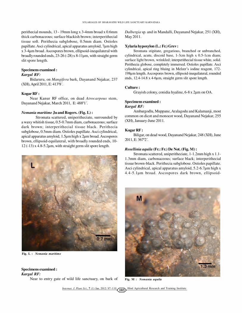
Nemania maritime Ju and Rogers. (Fig. L) :
Stromata scattered, uniperitheciate, surrounded by
a waxy whitish tissue, 0.5-0.7mm diam, carbonaceous; surface
dark brown; interperithecial tissue black. Perithecia
subglobose, 0.5mm diam. Ostioles papillate. Asci cylindrical,
apical apparatus amyloid, 1.5µm high x 2µm broad. Ascospores
brown, ellipsoid-equilateral, with broadly rounded ends, 10-
12 (-13) x 4.8-5.2µm, with straight germ slit spore length.
Dalbergia sp. and in Mandalli, Dayanand Nejakar, 251 (XH),
May 2011.
Xylaria hypoxylon (L.: Fr.) Grev :
Stromata stipitate, gregarious, branched or unbranched,
cylindrical, acute, discoid base, 1-3cm high x 0.5-1cm diam;
surface light brown, wrinkled; interperithecial tissue white, solid.
Perithecia globose, completely immersed. Ostioles papillate. Asci
cylindrical, apical ring bluing in Melzer’s iodine reagent, 172-
198µm length. Ascospores brown, ellipsoid-inequilateral, rounded
ends, 12.4-14.8 x 4-6µm, straight germ slit spore length.
Culture :
Grayish colony, conidia hyaline, 6-8 x 2µm on OA.
Specimens examined :
Kargal RF:
Ambargodlu, Muppane, Aralagodu and Kalamanji, most
common on dicot and monocot wood, Dayanand Nejakar, 255
(XH), January-June 2011.
Kogar RF :
Biligar, on dead wood, Dayanand Nejakar, 248 (XH), June
2011, E: 36702 .́
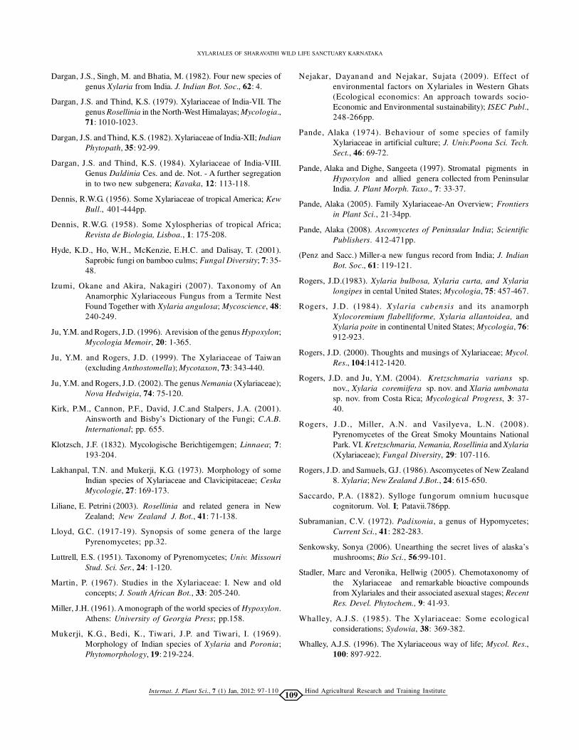
Rosellinia aquila (Fr.: Fr.) De Not. (Fig. M) :
Stromata scattered, uniperitheciate, 1-1.2mm high x 1.1-
1.3mm diam, carbonaceous; surface black; interperithecial
tissue brown-black. Perithecia subglobose. Ostioles papillate.
Asci cylindrical, apical apparatus amyloid, 5.2-6.7µm high x
4.4-5.1µm broad. Ascospores dark brown, ellipsoid-
Fig. M : Nemania aquila
Fig. L : Nemania maritime
Specimens examined :
Kargal RF:
Near to entry gate of wild life sanctuary, on bark of

Hind Agricultural Research and Training InstituteInternat. J. Plant Sci., 7 (1) Jan, 2012:104
DAYANAND NEJAKAR, POORNAPRAJNA BELUR, SUJATA MALI AND RAHUL PATIL
inequilateral with narrow rounded ends, (17-) 19.7-21.2 x 6-
8µm, with straight germ slit spore length.
Culture :
The conidiogenous structure is Geniculosporium like
on OA.
Specimens examined :
Kargal RF:
Way to Nagavalli, on dead stem of monocot, Dayanand
Nejakar, 264 (XH), April 2011, E: 42307 .́
Rosellenia mammaeformis (Pers.: Fr.) Ces. and De Not. (Fig.
N) :
Stromata scattered, subglobose, uniperitheciate,
carbonaceous, 0.9mm diam x 0.6mm high; surface dark brown;
interperithecial tissue black. Perithecia subglobose. Ostioles
papillate. Asci cylindrical, apical apparatus amyloid, 3.8-4µm
high x 3.2-3.5µm broad. Ascospores brown, ellipsoid-
inequilateral, (18-) 19.3-20.8 x 6.6-7µm, with straight germ slit
spore length.
Xylaria castorea Berk :
Stromata stipitate, solitary, cylindrical-flattened slightly,
unbranched, obtuse, (1-) 2.5-3.4 (-6)cm long x 7-13mm wide x
4-6mm thick; surface dull black, wrinkled; interperithecial tissue
white, solid. Perithecia globose, complete immersed. Ostioles
papillate. Asci cylindrical, bluing in Melzer’s iodine reagent,
(130-) 132-136 (-140)µm high x 5-7µm broad. Ascospores dark
brown, ellipsoid-inequilateral, rounded ends, (8-) 10-11 (-12) x
5-6µm, straight germ slit spore length.
Culture :
White cottony mycelium with hyaline conidia on OA.
Specimens examined :
Kargal RF:
Muppane, on angiospermous stump, Dayanand Nejakar,
235 (XH), June 2011, E: 45103 .́
Kogar RF:
Megane, on dead monocot stem, Dayanand Nejakar, 240
(XH), February 2011.
Xylaria anisopleura (Mont.) Fr. :
Stromata stipitate, gregarious, club shape-slightly
flattened, discoid base, 3-4cm high x 1.5-2cm diam; surface
brown-black, wrinkled; interperithecial tissue white, solid.
Perithecia oval, completely immersed. Ostioles papillate. Asci
cylindrical, apical ring bluing in Melzer’s iodine reagent, 5-
6µm high x 4-5µm wide. Ascospores brown, ellipsoid-
inequilateral, rounded ends, (20-) 23.5- 24.5 (-26) x 6-8µm,
sigmoid germ slit.
Culture:
White cottony mycelium with hyaline conidia, 11-12 x 3-
4 µm on OA.
Specimens examined :
Kargal RF:
Ambaragodlu, on dead dicot wood, Dayanand Nejakar,
250 (XH), May 2011, E: 40302 .́
Rosellinia mycophila (Fr.: Fr.) Sacc. (Fig. O) :
Stromata fused, uniperitheciate, subglobose, 0.7mm high
x 1mm diam; surface dark brown-black; interperithecial tissue
brown. Perithecia subglobose. Ostioles conical. Asci
cylindrical, apical apparatus amyloid, 6µm high x 4.8µm broad.
Ascospores dark brown, ellipsoid-inequilateral, narrow
rounded ends, (17-) 19-21 x 5.5-6.1µm, with straight germ slit
spore length.
Specimens examined :
Kargal RF:
Vatemakki, on dead wood of Carryota sp., Dayanand
Fig. N : Rosellenia mammaeformis
Specimens examined :
Kogar RF:
Karani, on dead dicot wood, Dayanand Nejakar, 261 (XH),
April 2011, E: 36302 .́
97-110

Hind Agricultural Research and Training InstituteInternat. J. Plant Sci., 7 (1) Jan, 2012:105
XYLARIALES OF SHARAVATHI WILD LIFE SANCTUARY KARNATAKA
97-110
Nejakar, 246 (XH), February 2011, E: 40209 .́
Rosellinia necatrix Prillieux. (Fig. P) :
Stromata gregarious, subglobose, uniperitheciate,
carbonaceous, 1.5mm high x 1.6mm diam; surface black;
interperithecial tissue dark brown. Perithecia subglobose.
Ostioles papillate. Asci cylindrical, apical apparatus amyloid,
6-8µm high x 4.7-5.2µm broad. Ascospores brown-dark brown,
ellipsoid-inequilateral, rounded ends, 36.7-40.1 x 5.5-5.9 (-8)µm,
with straight germ slit spore length.
Culture:
The conidiogenous structure is Dematophora like on
OA.
Specimens examined :
Kargal RF:
Aralagodu, Mandalli, Muppane and Kalamanji, on
angiospermic wood, Dayanand Nejakar, 259 (XH), January,
February, April and June 2011.
Kogar RF:
Hebbainakere, Kanur and Nagavalli, on dead dicot wood,
Dayanand Nejakar, 256 (XH), February, March and June 2011.
Xylaria filiformis (Alb. and Schw.: Fr.) Fr. :
Stromata stipitate, solitary, unbranched, cylindrical
thread like, acute, 6-8cm high x <0.5 diam; surface black;
interperithecial tissue white, solid. Perithecia globose,
completely immersed. Ostioles papillate. Asci cylindrical, apical
ring bluing in Melzer’s iodine reagent, (166-) 167.5- 168.2 (-
169)µm high x 88.3-102µm wide. Ascospores brown, ellipsoid-
inequilateral, rounded ends, 13.6-18 x 5-8 µm, straight germ
slit spore length.
Specimens examined :
Kargal RF:
Vatemakki, on dead leaf of dicot, Dayanand Nejakar, 258
(XH), June 2011, E: 40106 .́
Rosellinia submilis Karsten and Starb. (Fig. Q) :
Stromata scattered, uniperitheciate, subglobose, 0.5 mm
high x 0.7mm diam; surface black; interperithecial tissue dark
brown. Perithecia subglobose. Ostioles papillate. Asci
cylindrical, apical apparatus amyloid, 7.3-8.4µm high x 4.2-
Fig. O : Rosellenia mycophila
Fig. P : Rosellenia necatrix Prillieux
Fig. Q : Rosellenia submilis Karsten and Starb

Hind Agricultural Research and Training InstituteInternat. J. Plant Sci., 7 (1) Jan, 2012:106
DAYANAND NEJAKAR, POORNAPRAJNA BELUR, SUJATA MALI AND RAHUL PATIL
97-110
5.1µm broad. Ascospores brown, ellipsoid-inequilateral with
broadly rounded ends, 26 µm x 7.1-7.8µm, with straight germ
slit spore length.
Specimens examined :
Kogar RF:
Hebbainakere, on dead wood of Careya sp., Dayanand
Nejakar, 263 (XH), February 2011.
Rosellinia callosa G. Winter. (Fig. R) :
Stromata scattered, subglobose, uniperitheciate, 0.6-
0.7mm high x 0.8mm diam; surface dark brown; interperithecial
tissue light brown. Perithecia subglobose, 0.5-0.6mm diam.
Ostioles papillate. Asci cylindrical, apical apparatus amyloid,
6µm high x 3.7-3.9µm broad. Ascospores dark brown, ellipsoid-
inequilateral, narrow rounded ends, (21-) 23-27 (-28) x (6.5-)
6.8-7.2 (-7.5)µm, with sigmoid germ slit.
Specimens examined :
Kargal RF:
Muppane nature camp, on dead stem of Xylia sp.,
Dayanand Nejakar, 253 (XH), March 2011, E: 43702 .́
Xylaria theissenii Lloyd :
Stromata stipitate, solitary, cylindrical, unbranched,
fertile part appears as beaded thread, 4-5cm high x 1-2cm diam;
surface dull black or grey, wrinkled; interperithecial tissue
white, solid. Perithecia globose, completely immersed. Ostioles
papillate. Asci cylindrical, apical ring bluing in Melzer’s iodine
reagent. Ascospores dark brown, ellipsoid-inequilateral,
rounded ends, 35.7-38.3 x 10.6-12.4 µm, straight germ slit spore
length.
Specimens examined :
Kogar RF:
Karani, on dead dicot wood, Dayanand Nejakar, 252
Fig. R : Rosellenia callosa
Fig. S : Xylaria aenea
Fig. T : Xylaria arbuscula Sacc.
Specimens examined :
Kargal RF:
Vatemakki and Muppane, on dead dicot wood, Dayanand
Nejakar, 265 (XH), February and June 2011.
Xylaria arbuscula Sacc. (Fig. T) :
Stromata solitary, unbranched, wrinkled, glabrous, 10-
20mm high x 1-1.5mm diam; surface black; interperithecial tissue
white. Perithecia completely immersed. Ostioles non papillate.
(XH), March 2011.
Xylaria aenea Mont. (Fig. S) :
Stromata stipitate, constricted at the base, 4-6cm high x
1.5-2cm thick, smooth, slender stalk; surface black and wrinkled
on drying; interperithecial tissue white. Perithecia oval,
completely immersed. Ostioles non papillate. Asci cylindrical.
Ascospores brown, inequilateral, slightly curved ends, (33-)
35.2-38.9 (-40) x 6.7-7.5µm, sigmoid germ slit.

Hind Agricultural Research and Training InstituteInternat. J. Plant Sci., 7 (1) Jan, 2012:107
XYLARIALES OF SHARAVATHI WILD LIFE SANCTUARY KARNATAKA
97-110
Asci cylindrical, 171.4-192µm length x 5.6-6.8µm broad.
Ascospores brown, ellipsoid-inequilateral, rounded ends,
(12-) 13.4-15.7 (-17) x 5-6µm, sigmoid germ slit.
Specimens examined :
Kargal RF:
Vatemakki, on dead dicot wood, Dayanand Nejakar, 234
(XH), June 2011.
Xylaria grammica Mont. (Fig. U) :
Stromata stipitate, unbranched, cylindrical, obtuse apex,
smooth, 13.5cm high x 1.5-2cm diam; surface grey-black strips;
interperithecial tissue solid, white. Perithecia completely
immersed. Ostioles papillate. Asci cylindrical. Ascospores
brown, ellipsoid-inequilateral, rounded ends, (11-)12.2-14.7(-
15) x 3.5-4µm, sigmoid germ slit.
Specimens examined :
Kargal RF:
Aralagodu, on wood of Mangifera sp., Dayanand
Fig. U : Xylaria gramumica Mont.
Fig. V : Xylaria polymorpha
Nejakar, 262 (XH), May 2011, E: 42807 .́
Xylaria polymorpha (Pres. ex Fr.) Grev. (Fig. V) :
Stromata stipitate, solitary, club shape, carbonaceous,
slightly pointed or obtuse apex, smooth, 4cm high x 1.5-2cm
diam; surface black; interperithecial tissue solid, white.
Perithecia completely immersed. Ostioles papillate, black. Asci
cylindrical. Ascospores dark brown, ellipsoid-inequilateral,
rounded ends, (20-) 22.5-28.3 (-30) x (6-)7.1-9.6 (-12)µm, sigmoid
germ slit.
Specimens examined :
Kargal RF:
Mandalli and Vatemakki, on dead wood, Dayanand
Nejakar, 257 (XH), February and March 2011.
Kogar RF:
Nagavalli, on angiospermic stump, Dayanand Nejakar,
270 (XH).

Hind Agricultural Research and Training InstituteInternat. J. Plant Sci., 7 (1) Jan, 2012:108
DAYANAND NEJAKAR, POORNAPRAJNA BELUR, SUJATA MALI AND RAHUL PATIL
97-110
Xylaria longipes Nits :
Stromata stipitate, solitary, slender, flattened,
unbranched, slightly acute or obtuse, 3.4-6cm high x 2-2.5cm
diam; surface black, wrinkled; interperithecial tissue white,
solid. Perithecia subglobose, completely immersed, 427-502µm
diameter. Ostioles papillate. Asci cylindrical, apical ring bluing
in Melzer’s iodine reagent. Ascospores dark brown, ellipsoid,
rounded ends, (9-) 10.4-11.8 (-12.5) x 4-5µm, straight germ slit
spore length.
Specimens examined :
Kargal RF:
Ambargodlu, on dicot wood, Dayanand Nejakar, 254
(XH), February 2011, E: 41609 .́
Nemania atropurpurea (Fr.:Fr.) Pouzar. (Fig. I) :
Stromata superficial, carbonaceous, host surface black,
20cm long x 6-8cm broad x 1mm thick; surface dull black;
conspicuous perithecial mounds forming a polyhedral pattern.
Perithecia subglobose, 0.6-0.9mm diam. Ostioles papillate. Asci
cylindrical, apical apparatus amyloid, 2.2-2.4µm high x 2µm broad.
Ascospores dark brown, ellipsoid-inequilateral, narrow rounded
ends, (9.6-) 10-11(-11.5) x 4.2-4.8µm, conspicuous short germ slit.
Specimens examined :
Kargal RF:
Hebbainakere, on dead dicot wood, Dayanand Nejakar,
233 (XH), January 2011, E: 30604 .́
Xylaria multiplex (Kze) Fr.:
Stromata gregarious, acute tip, unequal middle portion,
discoid base, 3-5cm high x 2cm diam; surface black, rough;
interperithecial tissue white, solid. Perithecia subglobose, dark
brown, completely immersed. Ostioles papillate. Asci
cylindrical, apical ring bluing in Melzer’s iodine reagent, (80-
)83.4-88.6(-90)µm high x 4-5µm wide. Ascospores dark brown,
fabiform, rounded ends,(8-) 9.5-10(-12) x 3-4(-4.5)µm, straight
germ slit spore length.
Specimens examined :
Kargal RF:
Muppane, on dead dicot wood, Dayanand Nejakar, 249
(XH), June 2011, E: 44601 .́
Kogar RF:
Karani, on dead wood of Syzigium sp., Dayanand
Nejakar, 270 (XH), February 2011.
In present study totally 33 species belongs to 4 genera
were collected. Xylaria was the dominant genera and available
in all study area. Xylaria hypoxylon (L.: Fr.) Grev. was the
dominant species and found in all the months, followed by
Rosellinia necatrix Prillieux. Xylaria filiformis (Alb. and Schw.:
Fr.) Fr. was the only species collected on dead dicot leaf and
remaining from dead dicot and monocot stems.
Conclusion :
Majority of the mushroom are available only in rainy
season but almost all Xylariales members available throughout
the year. As per our earlier study September to February was
the perfect time to get telomorphs (Nejakar and Nejakar, 2009).
Thus, the study provides magnificent opening for further
study. The results reveal that their still a lot of varied
investigated groups. In the word of Rogers (2000) “Almost
every collecting box of specimens received from
correspondents reveals new taxa and other surprises. Although
I have not collected in India, Xylaria collections from both areas
contain a high percentage of taxa unknown to me”.
Acknowledgement :
The work was carried out with the grants from ATREE,
Bangalore under the Scheme of Small Grants-2010. The authors
are thankful to faculty members of Kuvempu University,
Karnataka University, Hongirana and L. B. College Sagar.
Special thanks to Karnataka Forest Department and staff
members of SWLS. Thanks to Dr. J. S. Dargan, Retd. Professor,
Punjabi University, Patiala for providing useful monographs.
REFERENCES
Agnihothrudu, V. (1965). Fungi from North India XXII. Mycopath.
Mycol. Appl., 23: 111-115.
Dargan, J.S. (1980). The family Xylariaceae in India- A Review; J.
Indian Bot. Soc., 59: 53-59.
Dargan, J.S. (1982). Xylaria mussooriensis-a new species from
India. Mycologia, 64:523-525.
Dargan, J.S. (1983). Xylariaceae of India-XIV. Two new species of
genus Xylaria from India. J. Biol. Res., 3:43-49.
Dargan, J.S. (1984). Xylariaceae of India-XIII. Plant and Nature;
2:136-138.
Dargan, J.S. (1987a). Distribution of Xylariaceae in western
Himalayas. J. Indian Bot. Soc., 66: 40-42.
Dargan, J.S. (2006). Family Xylariaceae – Status and Progress in
India. Kavaka; 34:1-16.
Dargan, J.S. and Bhatia, M. (1988b). Genus Eutypella from W.
Himalayas. Nova Hedwigia, 47: 111-117.
Dargan, J.S. and Mann, S.K. (1985). Pyrenomycetous fungi of
Punjab-IV. The family Xylariaceae. Biologica, 1: 146-156.
Dargan, J.S. and Singh, M. (1982c). Pyrenomycetous fungi of
Punjab-III Xylaria punjabensis, sp.nov. from India. Nordic
J. Bot., 2: 71-73.
Dargan, J.S. and Singh, M. (1986). Hypoxylon nummularium var.
exutans and Hypoxylon albostictum: Two unrecorded
pyrenomycetes from India. J. Indian Bot. Soc.,65: 411-
415.
Hind Agricultural Research and Training InstituteInternat. J. Plant Sci., 7 (1) Jan, 2012:109
XYLARIALES OF SHARAVATHI WILD LIFE SANCTUARY KARNATAKA
97-110
Dargan, J.S., Singh, M. and Bhatia, M. (1982). Four new species of
genus Xylaria from India. J. Indian Bot. Soc., 62: 4.
Dargan, J.S. and Thind, K.S. (1979). Xylariaceae of India-VII. The
genus Rosellinia in the North-West Himalayas; Mycologia.,
71: 1010-1023.
Dargan, J.S. and Thind, K.S. (1982). Xylariaceae of India-XII; Indian
Phytopath, 35: 92-99.
Dargan, J.S. and Thind, K.S. (1984). Xylariaceae of India-VIII.
Genus Daldinia Ces. and de. Not. - A further segregation
in to two new subgenera; Kavaka, 12: 113-118.
Dennis, R.W.G. (1956). Some Xylariaceae of tropical America; Kew
Bull., 401-444pp.
Dennis, R.W.G. (1958). Some Xylospherias of tropical Africa;
Revista de Biologia, Lisboa., 1: 175-208.
Hyde, K.D., Ho, W.H., McKenzie, E.H.C. and Dalisay, T. (2001).
Saprobic fungi on bamboo culms; Fungal Diversity; 7: 35-
48.
Izumi, Okane and Akira, Nakagiri (2007). Taxonomy of An
Anamorphic Xylariaceous Fungus from a Termite Nest
Found Together with Xylaria angulosa; Mycoscience, 48:
240-249.
Ju, Y.M. and Rogers, J.D. (1996). A revision of the genus Hypoxylon;
Mycologia Memoir, 20: 1-365.
Ju, Y.M. and Rogers, J.D. (1999). The Xylariaceae of Taiwan
(excluding Anthostomella); Mycotaxon, 73: 343-440.
Ju, Y.M. and Rogers, J.D. (2002). The genus Nemania (Xylariaceae);
Nova Hedwigia, 74: 75-120.
Kirk, P.M., Cannon, P.F., David, J.C.and Stalpers, J.A. (2001).
Ainsworth and Bisby’s Dictionary of the Fungi; C.A.B.
International; pp. 655.
Klotzsch, J.F. (1832). Mycologische Berichtigemgen; Linnaea; 7:
193-204.
Lakhanpal, T.N. and Mukerji, K.G. (1973). Morphology of some
Indian species of Xylariaceae and Clavicipitaceae; Ceska
Mycologie, 27: 169-173.
Liliane, E. Petrini (2003). Rosellinia and related genera in New
Zealand; New Zealand J. Bot., 41: 71-138.
Lloyd, G.C. (1917-19). Synopsis of some genera of the large
Pyrenomycetes; pp.32.
Luttrell, E.S. (1951). Taxonomy of Pyrenomycetes; Univ. Missouri
Stud. Sci. Ser., 24: 1-120.
Martin, P. (1967). Studies in the Xylariaceae: I. New and old
concepts; J. South African Bot., 33: 205-240.
Miller, J.H. (1961). A monograph of the world species of Hypoxylon.
Athens: University of Georgia Press; pp.158.
Mukerji, K.G., Bedi, K., Tiwari, J.P. and Tiwari, I. (1969).
Morphology of Indian species of Xylaria and Poronia;
Phytomorphology, 19: 219-224.
Nejakar, Dayanand and Nejakar, Sujata (2009). Effect of
environmental factors on Xylariales in Western Ghats
(Ecological economics: An approach towards socio-
Economic and Environmental sustainability); ISEC Publ.,
248-266pp.
Pande, Alaka (1974). Behaviour of some species of family
Xylariaceae in artificial culture; J. Univ.Poona Sci. Tech.
Sect., 46: 69-72.
Pande, Alaka and Dighe, Sangeeta (1997). Stromatal pigments in
Hypoxylon and allied genera collected from Peninsular
India. J. Plant Morph. Taxo., 7: 33-37.
Pande, Alaka (2005). Family Xylariaceae-An Overview; Frontiers
in Plant Sci., 21-34pp.
Pande, Alaka (2008). Ascomycetes of Peninsular India; Scientific
Publishers. 412-471pp.
(Penz and Sacc.) Miller-a new fungus record from India; J. Indian
Bot. Soc., 61: 119-121.
Rogers, J.D.(1983). Xylaria bulbosa, Xylaria curta, and Xylaria
longipes in cental United States; Mycologia, 75: 457-467.
Rogers, J .D. (1984). Xylaria cubensis and its anamorph
Xylocoremium flabelliforme, Xylaria allantoidea, and
Xylaria poite in continental United States; Mycologia, 76:
912-923.
Rogers, J.D. (2000). Thoughts and musings of Xylariaceae; Mycol.
Res., 104:1412-1420.
Rogers, J.D. and Ju, Y.M. (2004). Kretzschmaria varians sp.
nov., Xylaria coremiifera sp. nov. and Xlaria umbonata
sp. nov. from Costa Rica; Mycological Progress, 3: 37-
40.
Rogers, J .D., Miller, A.N. and Vasilyeva, L.N. (2008).
Pyrenomycetes of the Great Smoky Mountains National
Park. VI. Kretzschmaria, Nemania, Rosellinia and Xylaria
(Xylariaceae); Fungal Diversity, 29: 107-116.
Rogers, J.D. and Samuels, G.J. (1986). Ascomycetes of New Zealand
8. Xylaria; New Zealand J.Bot., 24: 615-650.
Saccardo, P.A. (1882). Sylloge fungorum omnium hucusque
cognitorum. Vol. I; Patavii.786pp.
Subramanian, C.V. (1972). Padixonia, a genus of Hypomycetes;
Current Sci., 41: 282-283.
Senkowsky, Sonya (2006). Unearthing the secret lives of alaska’s
mushrooms; Bio Sci., 56:99-101.
Stadler, Marc and Veronika, Hellwig (2005). Chemotaxonomy of
the Xylariaceae and remarkable bioactive compounds
from Xylariales and their associated asexual stages; Recent
Res. Devel. Phytochem., 9: 41-93.
Whalley, A.J .S. (1985). The Xylariaceae: Some ecological
considerations; Sydowia, 38: 369-382.
Whalley, A.J.S. (1996). The Xylariaceous way of life; Mycol. Res.,
100: 897-922.

Hind Agricultural Research and Training InstituteInternat. J. Plant Sci., 7 (1) Jan, 2012:110
DAYANAND NEJAKAR, POORNAPRAJNA BELUR, SUJATA MALI AND RAHUL PATIL
************
Whalley, A.J.S. and Edwards, R.L. and Francis, S.M. (1983).
Hypoxylon gwynedii sp. nov. from Wales Trans; Br. Mycol.
Soc., 81: 389-392.
WEBLIOGRAPHY
www.indexfungorum.org.
www.pyrenomycetes.free.fr.
97-110


![News9/Newson6 – Pri ry Election 2018 May, 2018 · 2018. 5. 23. · 4. Fo ve unfavo ble, p ss 4 5. If you’ unsu , p ss 5 To have these choices peated, p ss 6 [RE9˛AT] [ANOTHER](https://img.pdfslide.us/doc/110x75/609989cee7cbf251f2164584/news9newson6-a-pri-ry-election-2018-may-2018-2018-5-23-4-fo-ve-unfavo.jpg)


![p[;¢W ‡ te ) ¨m; sS]ITvSy S]ITvk;le p[;¢W](https://img.pdfslide.us/doc/110x75/5e874826911e5f034c3ab9c4/-pw-a-te-m-ssitvsy-sitvkle-pw.jpg)