Embed Size (px)
Citation preview

Instructions for Use
IJ Catheter Ultrasound Model Version 2300
3601 Sagamore Parkway North, Suite B Lafayette, IN 47904
Phone 765.742.4813
www.hemocleanse.com
Instructions Revision 1.0
© 2012

IJ Catheter Ultrasound Model 2300 Page 2 Revision 1.0
Page Component Photos Torso 3 Heart Bulb, Reservoir and Tubes 3 Vascular Insert 4 Neck 4 Equipment and Supplies 5 Operating Procedures Overview 6 Subcutaneous Layer installation 7 Vein replacement 8 Install Vascular Insert 10 Install Neck 11 Fill and connect Heart Bulb 12 Install Water Reservoir 13 Install Subcutaneous Tunnels 14 Install Skin Assembly 15 Cannulation 16 Anatomy of Neck Vasculature 19 Replacement Component Details 20
Table of Contents

IJ Catheter Ultrasound Model 2300 Page 3 Revision 1.0
Torso
Heart Bulb, Reservoir, and Connector Tubes
Catheter Tunneling
Sites
Heart Bulb & Tube
Water Reservoir
Torso Spine
Component Photos
Neck - Reservoir
Tube
Water Reservoir
Holder
Skin Assembly

IJ Catheter Ultrasound Model 2300 Page 4 Revision 1.0
Vascular Insert
Neck
Subcutaneous Layer
Spine
Spine
Neck Latch (1 of 3)
Trachea
Carotid Artery (1 of 2)
Internal Jugular Veins
Heart Bulb Tube
Connector
Component Photos
Latches for Vascular
Insert
Neck - Reservoir
Tube Connector
Clamp
Clamp Latches
Superior Vena Cava
Left Brachio-cephalic Vein

IJ Catheter Ultrasound Model 2300 Page 5 Revision 1.0
Quantity Description
Equipment 1 Torso 1 Skin Assembly 1 Neck 1 Vascular Insert (inside Neck) 1 Heart Bulb with tube 1 Water Reservoir 1 Neck - Reservoir Tube 1 400 mL beaker (water container) Spare Parts IJ Veins Latex Penrose tubing, 6 inches long Skin Assembly Subcutaneous Layer Rubber Sheet Arteries, thick wall Latex tubing, 6 inches long O-rings for holding IJ Veins and Arteries Tunneling site cover — Velcro® Supplies Needle Guide, 18 gauge Needle Guide, 21 gauge Conductivity Gel Syringes
Equipment and Supplies
IJ Catheter Ultrasound Model 2300 Page 6 Revision 1.0
Overview
1. This device is designed to train medical personnel to cannulate the internal jugular vein with large catheters for hemodialysis or other procedures requiring large catheters placed for long duration.
2. Preparing to use the model requires the following steps:
• Replace the subcutaneous layer in the neck
• Replace IJ veins and, if necessary, arteries
• Install Vascular Insert into neck
• Install neck into torso
• Install heart bulb and water reservoir
• Install Velcro for subcutaneous tunnel
• Apply ultrasound gel between the skin assembly and the subcutaneous layer.
3. Each subcutaneous layer and vein should last for several cannulations. It is not necessary to change the subcutaneous layer and vein after each cannulation. Longevity of the subcutaneous layer is mostly determined by the availability of suitable places to insert the catheter. Each puncture of the subcutaneous layer will leak a small amount of water; much of the water will collect inside the torso.
4. Replacing an artery should not be necessary unless it is punctured during the cannulation procedure.
5. Model 2300 is asymmetric distal to the vascular insert. The right side has a straight tube to simulate the superior vena cava. The left side angles across to the superior vena cava, simulating the left brachiocephalic vein.
6. The model is shipped with new subcutaneous layer, new veins, and new arteries installed.

IJ Catheter Ultrasound Model 2300 Page 7 Revision 1.0
Subcutaneous Layer Installation 1. Open the clamp latches, open the clamp,
and remove the rubber sheet (subcutaneous layer ). Here the neck is shown with the subcutaneous layer re-moved.
2. Place the subcutaneous layer on the neck. Center
it on the hole in the neck. 3. Close the clamp on the subcutaneous layer and
close the latches.
4. Check the subcutaneous layer placement by look-ing from the inside. The subcutaneous layer should have at approximately the same amount of overlap of the neck all the way around the hole.
IJ Catheter Ultrasound Model 2300 Page 8 Revision 1.0
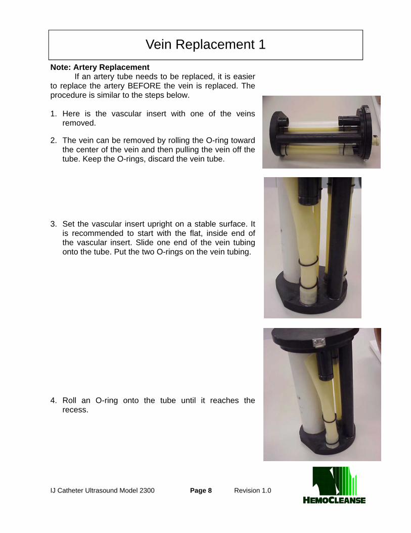
Note: Artery Replacement If an artery tube needs to be replaced, it is easier to replace the artery BEFORE the vein is replaced. The procedure is similar to the steps below. 1. Here is the vascular insert with one of the veins
removed.
2. The vein can be removed by rolling the O-ring toward the center of the vein and then pulling the vein off the tube. Keep the O-rings, discard the vein tube.
3. Set the vascular insert upright on a stable surface. It
is recommended to start with the flat, inside end of the vascular insert. Slide one end of the vein tubing onto the tube. Put the two O-rings on the vein tubing.
4. Roll an O-ring onto the tube until it reaches the
recess.
Vein Replacement 1

IJ Catheter Ultrasound Model 2300 Page 9 Revision 1.0
4. Installing the other end of the vein is easiest if the
vascular insert is on a stable surface. Here it is shown inverted on top of the neck. The other end of the veins has been installed over the tube.
Vein should be stretched so that it is taut. 5. Roll the O-ring onto the tube until it reaches the
recess. Vein installation is now complete. Notes on Artery Replacement
• The arteries may be replaced using the same procedure as veins.
• Artery installation is easier if it is done with the adjacent vein removed, before the vein is installed.
• Wetting the inside of the artery tube will make installation easier.
Vein Replacement 2

IJ Catheter Ultrasound Model 2300 Page 10 Revision 1.0
1. Add about 1300 mL water to the neck. This is best
done where overflow from the neck can be con-tained.
2. Install the vascular insert in the neck. Align the
spine of the vascular insert with the spine in the neck.
3. Slowly lower the vascular insert into the neck. A
small amount of water should overflow out of the neck. Here is the vascular insert ready to be latched to the neck.
4. Latch the vascular insert to the neck. A small
amount of water may squirt out of the top of the vascular insert as the air is purged from the arter-ies and veins.
Install Vascular Insert

IJ Catheter Ultrasound Model 2300 Page 11 Revision 1.0
1. Align the spine of the neck with the spine in the
torso. Slide the neck into the torso, being careful to get the latches inside the torso.
2. Neck is fully installed when the stop is seated in
the slot in the torso.
Install Neck
Neck locating stop

IJ Catheter Ultrasound Model 2300 Page 12 Revision 1.0
1. Fill reservoir about 3/4 full of water. 2. Connect heart tubing to reservoir. 3. Install reservoir in holder. The heart tubing goes
into interior of holder so that it does not kink. 4. Holding the bulb with the tube upright, pump it
several times until bulb is completely filled, and air bubbles no longer come out.
5. Install light tan colored connector on the heart
bulb tube into the light tan colored connector on the vascular insert of the neck.
Fill and connect Heart Bulb

IJ Catheter Ultrasound Model 2300 Page 13 Revision 1.0
1. Fill water reservoir at least half full of water. 2. Install neck-reservoir tube in the reservoir. Close
the clamp on the tube. 3. Place reservoir in its holder with the cap down
inside. 4. Purge air from the tube by opening the clamp.
Clamp to stop water flow. 5. Install the white connector of the neck-reservoir tube in white connector on vascular insert. 6. Open the clamp. 7. Replenish water in the reservoir as necessary.
Install Water Reservoir

IJ Catheter Ultrasound Model 2300 Page 14 Revision 1.0
1. Model without Velcro top pieces. Velcro tunnels
simulate the subcutaneous tissue that the catheter must traverse.
2. Place a Velcro piece on model in the desired location
of the catheter exit site. 3. Repeat on the other side of the model. Notes: Spare Velcro pieces can be used to make custom subcutaneous tunnels. The standard subcutaneous tunnel is 4 3/16 inch (105mm) long with a centered catheter insertion hole 3/16 by 1/2 inch (5 x 12 mm).
Install Subcutaneous Tunnels

IJ Catheter Ultrasound Model 2300 Page 15 Revision 1.0
1. Align the skin assembly with the neck and the skin
exit sites. 2. Attach the Velcro tabs on the chest.. 3. Add ultrasound gel to surface of neck at the catheter
insertion site. 3. Attach skin assembly to Velcro patches adjacent to
each side of the neck. 4. Note that the skin assembly already has a slit on
each side of the model for access to the deeper lay-ers of the neck. If an additional slit in the skin is nec-essary, please use a scissors to cut the skin.
Install Skin Assembly

IJ Catheter Ultrasound Model 2300 Page 16 Revision 1.0
1. To improve realism of ultrasound location of the IJ
vein, have an assistant pump the heart bulb. The arteries will pulsate.
2. Put ultrasound gel on the outside of the skin.
Insert the needle into the vein using ultrasound to guide the placement.
3. Test needle placement by removing the syringe
from the needle and watch the fluid drip out of the needle hub.
4. Advance the guide wire into vein as far as desired. 5. Remove the needle from the vein, leaving the
guide wire installed.
Cannulation, 1

IJ Catheter Ultrasound Model 2300 Page 17 Revision 1.0
6. Insert the tunneling tool through the hole in the
Velcro toward the vein. 7. Advance the catheter into the tunnel until the hub
is at the skin incision. 8. Insert the first dilator over the guidewire into vein.
Remove this dilator. Repeat with next size dilator if desired.
9. Insert the largest dilator and catheter sheath over
the guide wire.
Cannulation, 2

IJ Catheter Ultrasound Model 2300 Page 18 Revision 1.0
10. Remove the dilator and guidewire. 11. Insert the catheter into the sheath. Split the sheath
hub. 12. Pull the two halves of the sheath apart until the
sheath is completely removed. 13. Fully installed catheter.
Cannulation, 3

IJ Catheter Ultrasound Model 2300 Page 19 Revision 1.0
Illustration from: http://accweb.itr.maryville.edu/myu/image/ThyroidVein.gif
Anatomy of the Neck Vasculature
Neck Vasculature

IJ Catheter Ultrasound Model 2300 Page 20 Revision 1.0
IJ Vein Penrose tubing, 3/4 inch diameter x 6 inches long (about 1¼ in wide when flat)
[Kendall Argyle Latex Penrose Tubing # 515601] O-ring 3/4” ID by 1/16” thick, extra hard Buna-N, Size 018
[McMaster-Carr part number 5308T127] Carotid Artery, Thick Latex tubing, super soft, 5/16 inch ID, 1/16 inch wall, 6 inches long [McMaster-Carr part 5234K34] O-ring: 3/8” ID by 1/16” thick, extra hard Buna-N, Size 012 [McMaster-Carr part number 5308T121] Carotid Artery, Thin Latex tubing, super soft, 1/4 inch ID, 1/32 inch wall, 6 inches long [McMaster-Carr part 5234K981] O-ring: 5/16” ID by 1/16” thick, extra hard Buna-N, Size 011 [McMaster-Carr part number 5308T12] Subcutaneous Layer Natural latex rubber film, 0.050 inch thick x 6.0 in wide x 4.5 in [McMaster-Carr part number 85995K32] Seal for vascular insert O-ring: 3-7/8” ID by 1/8” thick, medium soft Buna-N, Size 241 [McMaster-Carr part number 2418T194] Subcutaneous Tunnel Velcro®, 2 inch wide, sew-on Hole for inserting catheter is 3/16 x 1/2 inch (5 x 12 mm) Insertion site is 2.0 inches (50 mm) from end of supplied cover Skin Assembly Natural latex rubber film, 0.020 inch thick x 13.0 in wide x 10.0 in [McMaster-Carr part number 8611K16] Velcro loop patches are attached with heavy duty staples
Most replacement parts can be purchased from McMaster-Carr Supply Company, Inc. whose web site is www.mcmaster.com.
Penrose tubing for the IJ vein can be purchased from medical supply companies. HemoCleanse can supply replacement parts. Contact David Carr at
Replacement Component Details


![Using Bayesian Causal Forest Models to Examine Treatment ...y ij = j + (x ij)+[ (w ij)+ j] z ij + ij Coloring outside the lines: Multilevel Bayesian Causal Forests We replace linear](https://img.pdfslide.us/doc/110x75/6043fc95e860f968ce356f89/using-bayesian-causal-forest-models-to-examine-treatment-y-ij-j-x-ij.jpg)