Embed Size (px)
Citation preview
Mogens VybergProfessor of Clinical PathologyDirector of NordiQCAalborg University Hospital, Aalborg, Denmark
IHC Classification of undifferentiated tumors –
the primary panel
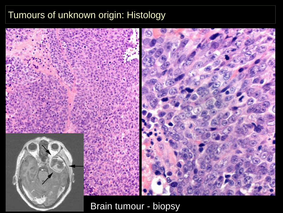
Tumours of unknown origin: Histology
Brain tumour - biopsy
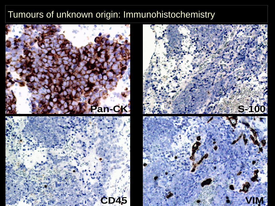
Tumours of unknown origin: Immunohistochemistry
Pan-CK S-100
CD45 VIM
UPT: A tumour appearing in metastatic setting without a histologically proven primary tumour.
UPT pose an increasing challenge for the pathologist - due to the progress in surgical and oncological treatment possibilities.
IHC classification of the Unknown Primary Tumour
New, relatively specific antibodies give the pathologist more and better diagnostic tools.
But the diagnostic work also become more complex in terms of planning, optimization of protocols, interpretation of reaction patterns and error trapping.
IHC classification of the Unknown Primary Tumour
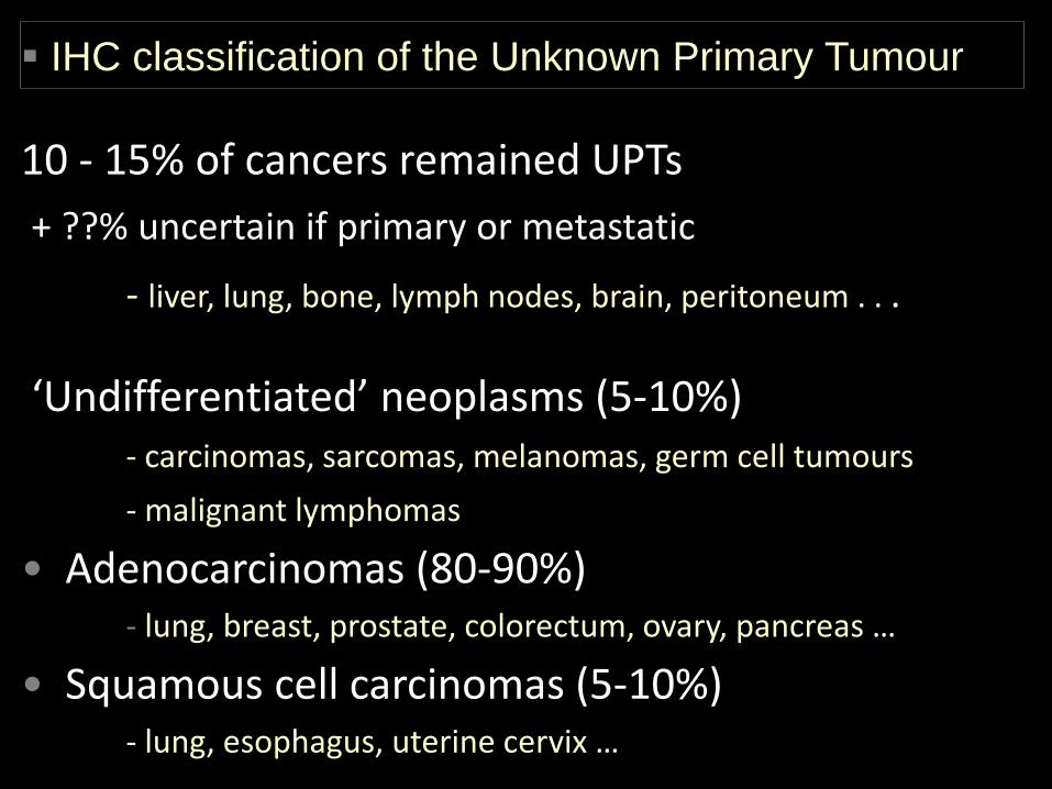
10 - 15% of cancers remained UPTs+ ??% uncertain if primary or metastatic
- liver, lung, bone, lymph nodes, brain, peritoneum . . .
‘Undifferentiated’ neoplasms (5-10%)- carcinomas, sarcomas, melanomas, germ cell tumours- malignant lymphomas
• Adenocarcinomas (80-90%)- lung, breast, prostate, colorectum, ovary, pancreas …
• Squamous cell carcinomas (5-10%)- lung, esophagus, uterine cervix …
IHC classification of the Unknown Primary Tumour
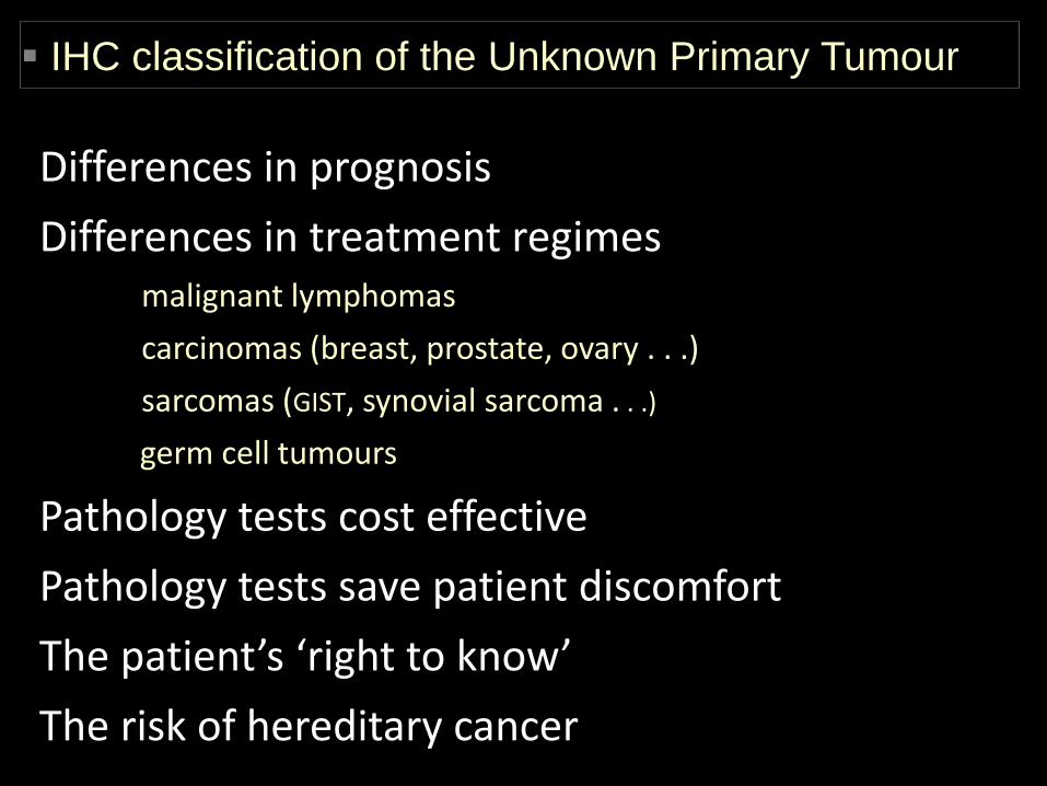
Differences in prognosisDifferences in treatment regimes
malignant lymphomascarcinomas (breast, prostate, ovary . . .)sarcomas (GIST, synovial sarcoma . . .)
germ cell tumours
Pathology tests cost effectivePathology tests save patient discomfortThe patient’s ‘right to know’The risk of hereditary cancer
IHC classification of the Unknown Primary Tumour
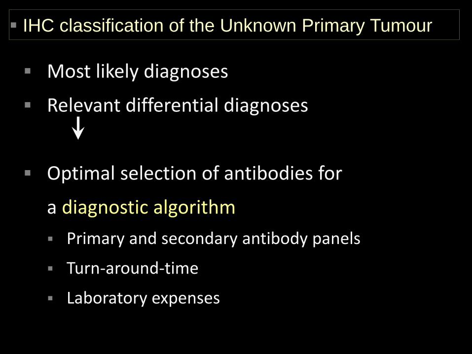
Most likely diagnoses
Relevant differential diagnoses
Optimal selection of antibodies for
a diagnostic algorithm Primary and secondary antibody panels
Turn-around-time
Laboratory expenses
IHC classification of the Unknown Primary Tumour
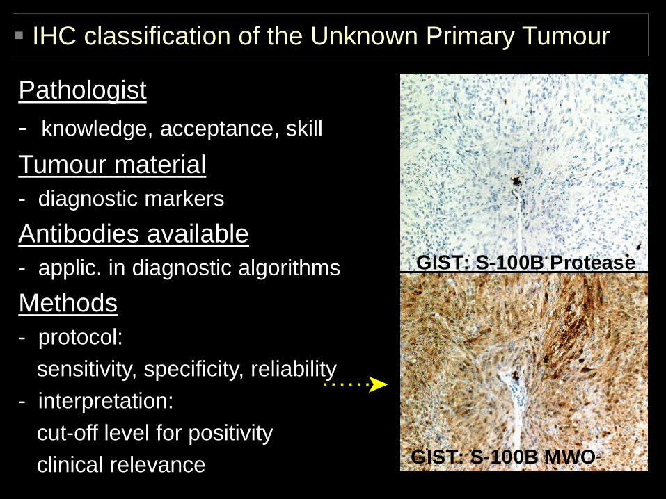
Pathologist- knowledge, acceptance, skill
Tumour material- diagnostic markersAntibodies available- applic. in diagnostic algorithmsMethods- protocol:
sensitivity, specificity, reliability- interpretation:
cut-off level for positivityclinical relevance
GIST: S-100B Protease
GIST: S-100B MWO
IHC classification of the Unknown Primary Tumour
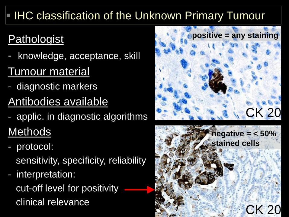
CK 20
CK 20
positive = any staining
negative = < 50% stained cells
Pathologist- knowledge, acceptance, skill
Tumour material- diagnostic markersAntibodies available- applic. in diagnostic algorithmsMethods- protocol:
sensitivity, specificity, reliability- interpretation:
cut-off level for positivityclinical relevance
IHC classification of the Unknown Primary Tumour
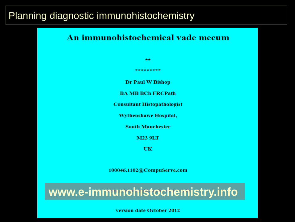
www.e-immunohistochemistry.info
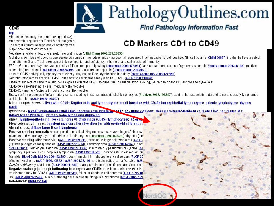
Planning diagnostic immunohistochemistry
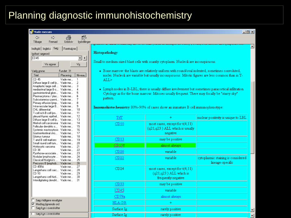
Planning diagnostic immunohistochemistry
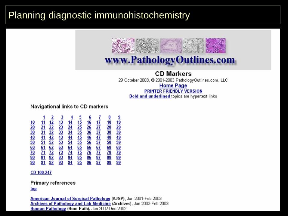
Planning diagnostic immunohistochemistry

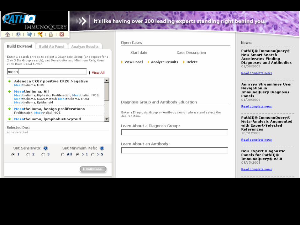
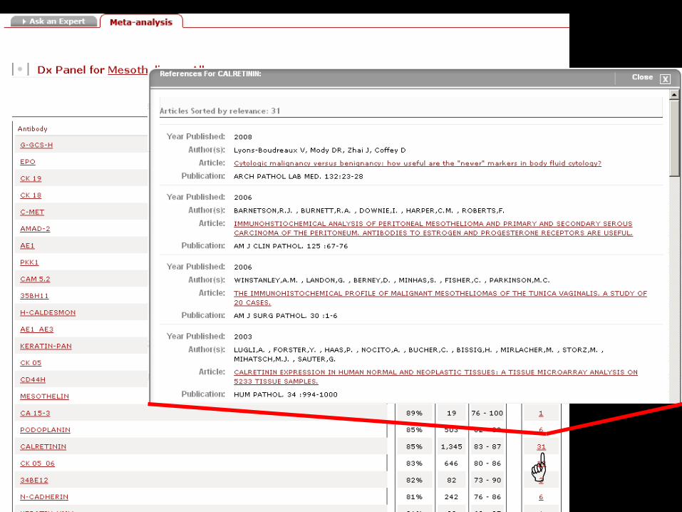
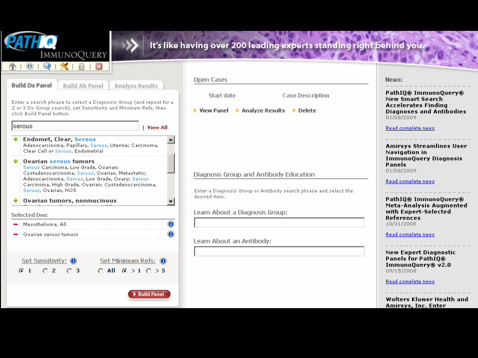
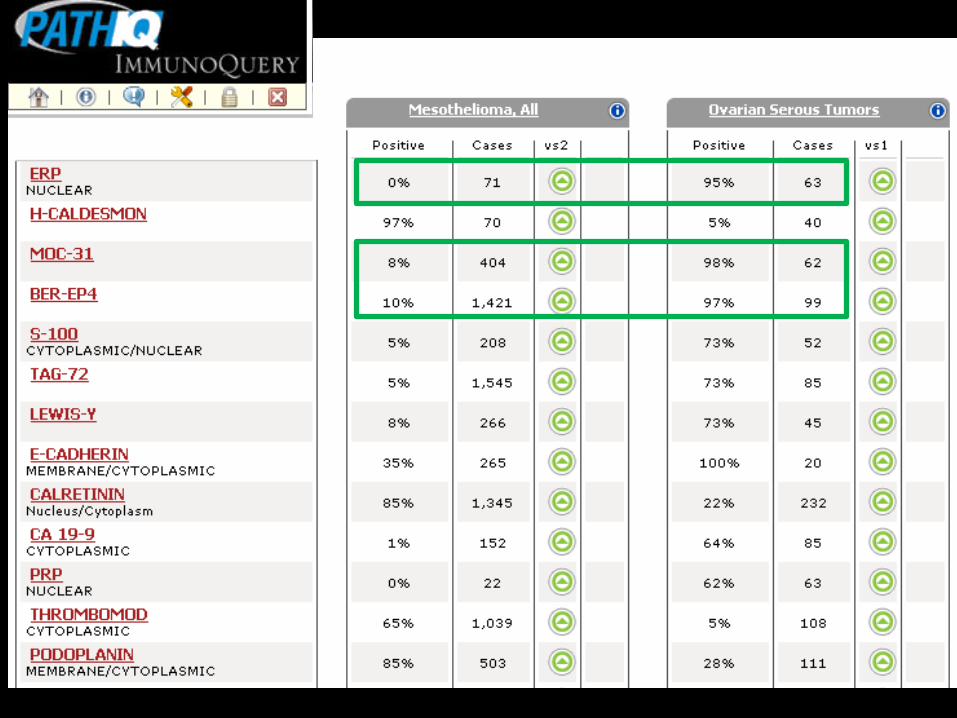
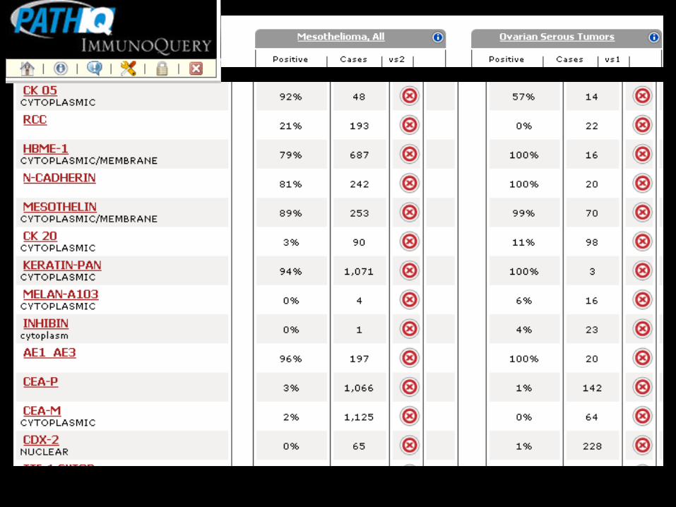
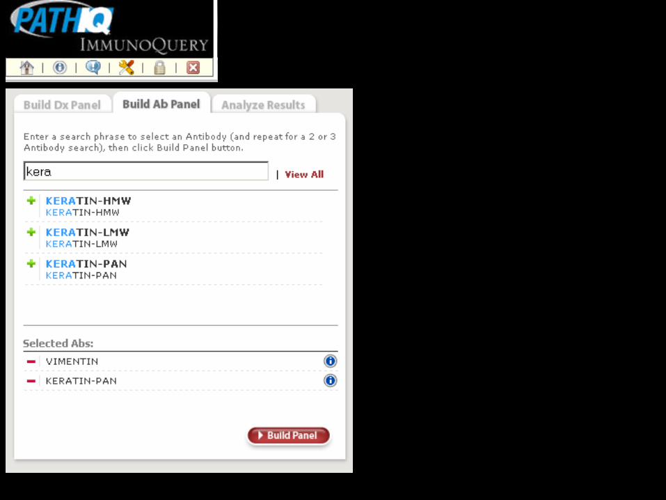
my.statdxpathiq.com
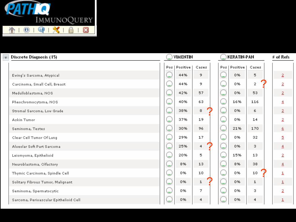
?
?
?
?
?
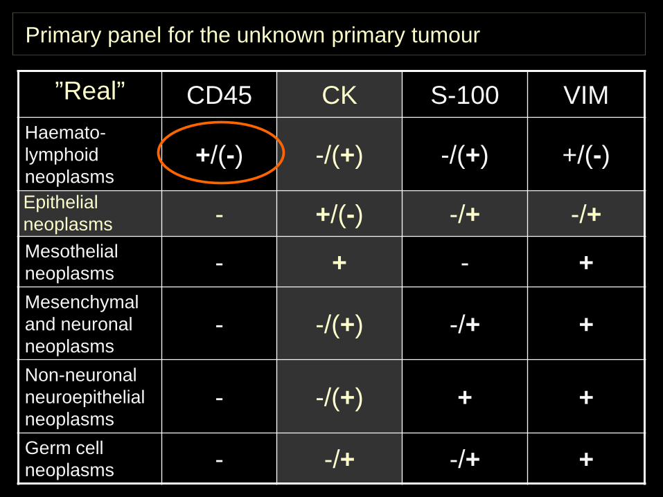
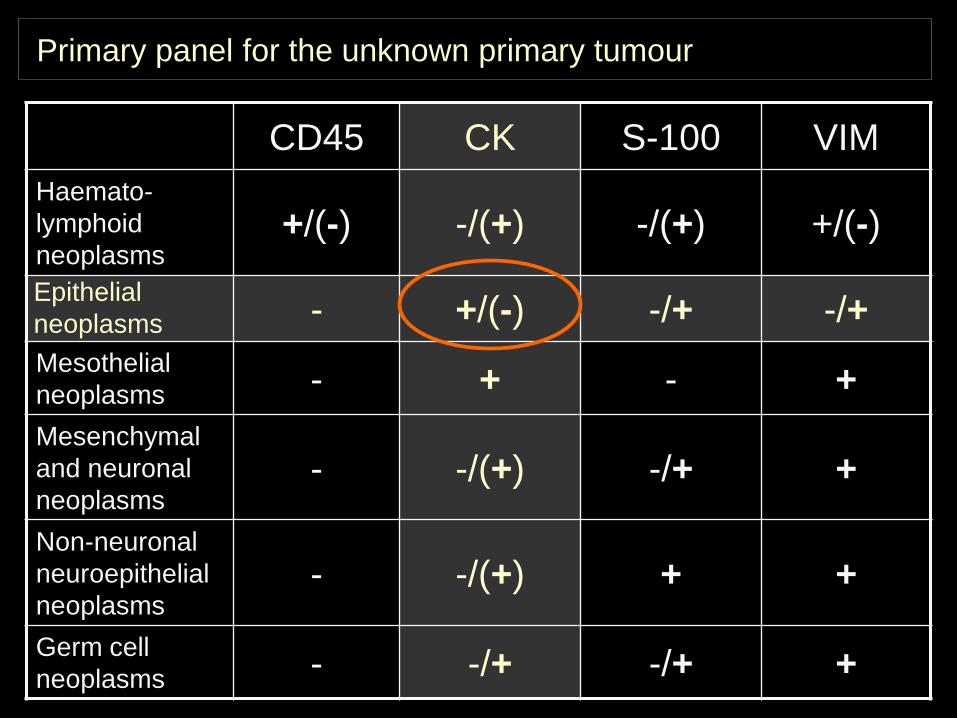
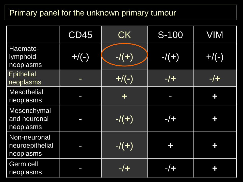
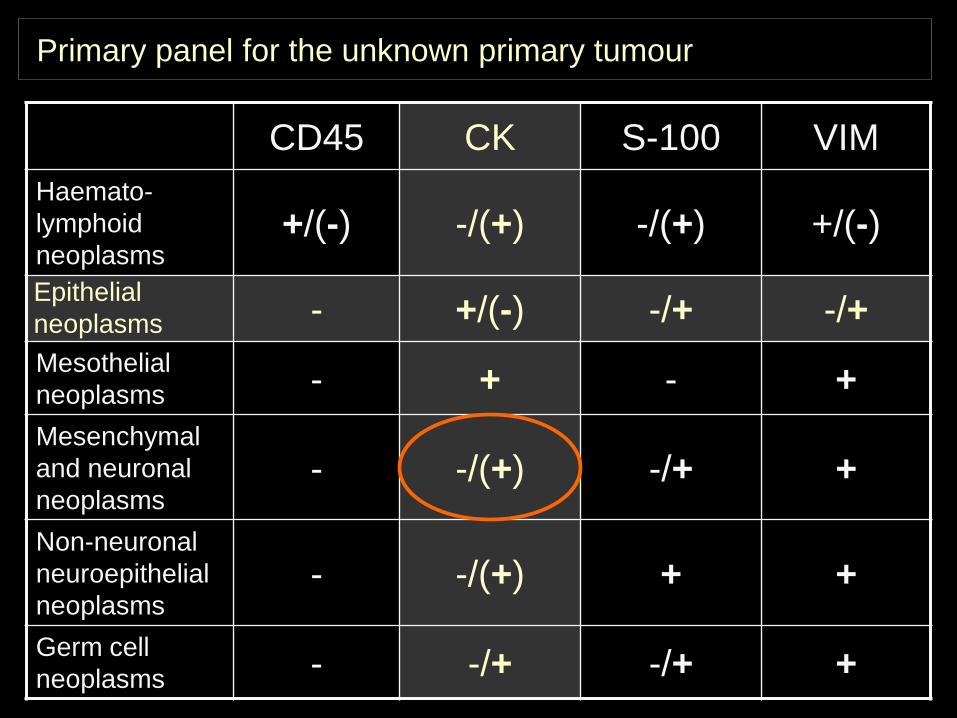
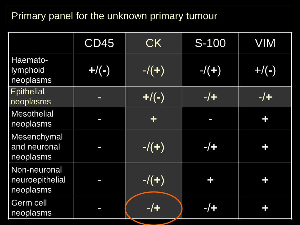
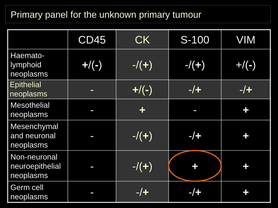
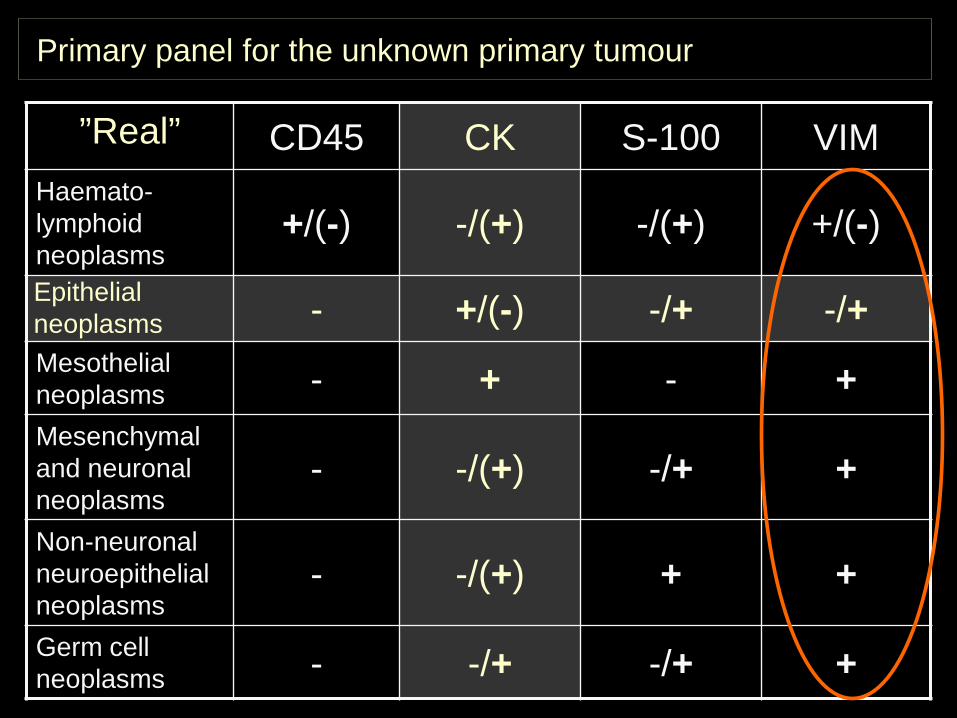
Primary panel for the unknown primary tumour
”Real” CD45 CK S-100 VIMHaemato-lymphoid neoplasms
+/(-) -/(+) -/(+) +/(-)Epithelialneoplasms - +/(-) -/+ -/+Mesothelial neoplasms - + - +Mesenchymal and neuronal neoplasms
- -/(+) -/+ +
Non-neuronal neuroepithelial neoplasms
- -/(+) + +
Germ cell neoplasms - -/+ -/+ +
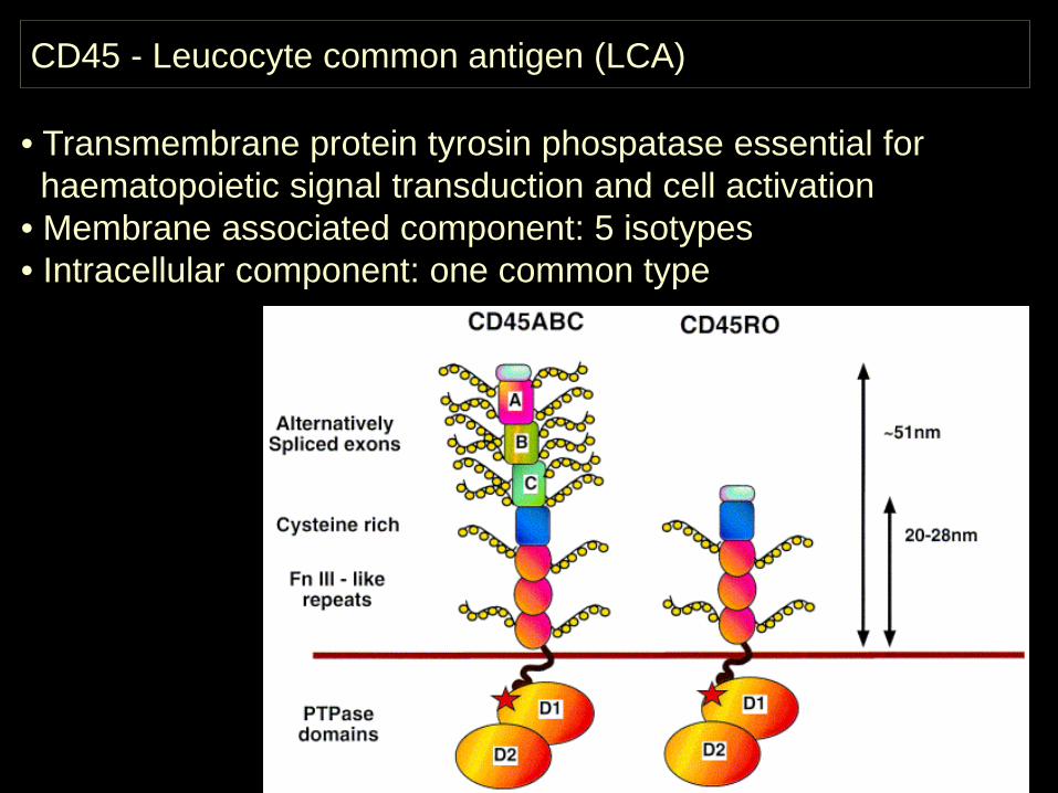
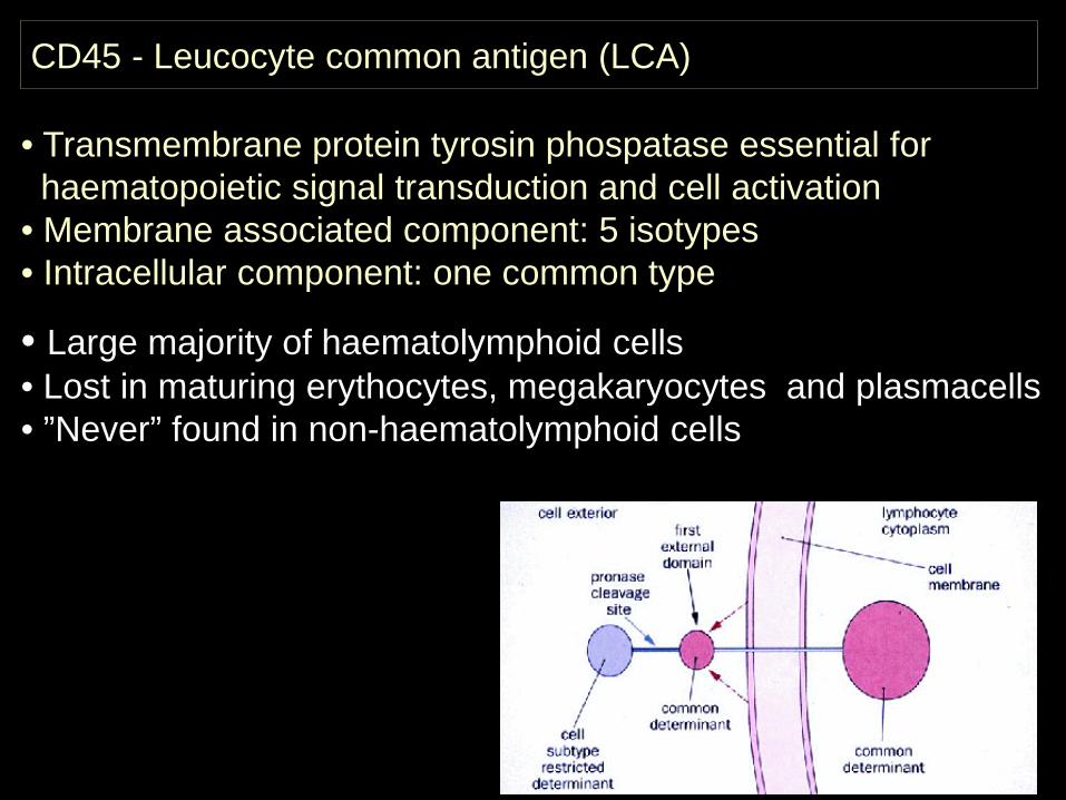
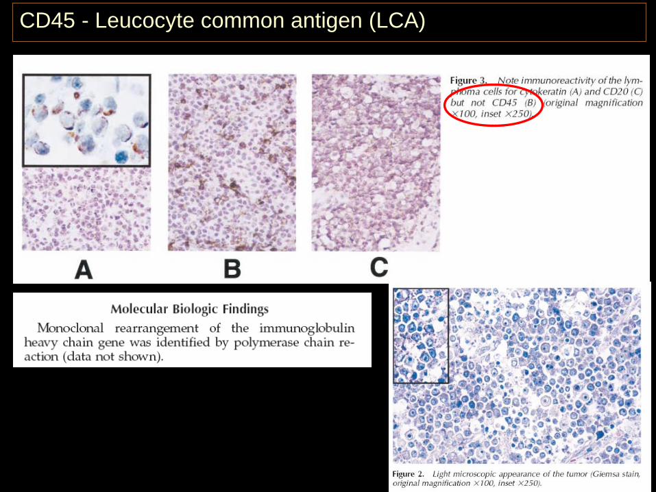
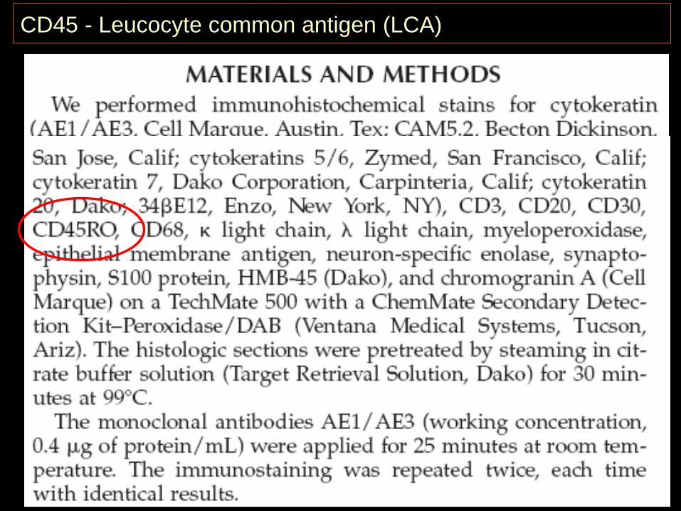
CD45 - Leucocyte common antigen (LCA)
• Transmembrane protein tyrosin phospatase essential for haematopoietic signal transduction and cell activation
• Membrane associated component: 5 isotypes• Intracellular component: one common type
CD45 - Leucocyte common antigen (LCA)
• Transmembrane protein tyrosin phospatase essential for haematopoietic signal transduction and cell activation
• Membrane associated component: 5 isotypes• Intracellular component: one common type
• Large majority of haematolymphoid cells• Lost in maturing erythocytes, megakaryocytes and plasmacells• ”Never” found in non-haematolymphoid cells
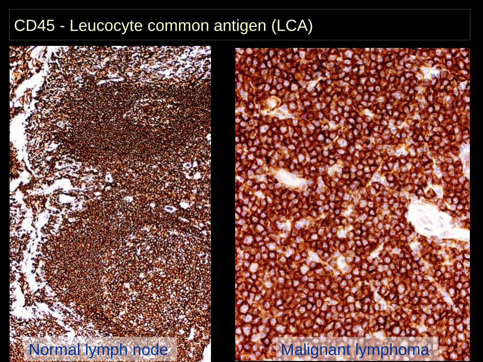
CD45 - Leucocyte common antigen (LCA)
Normal lymph node Malignant lymphoma
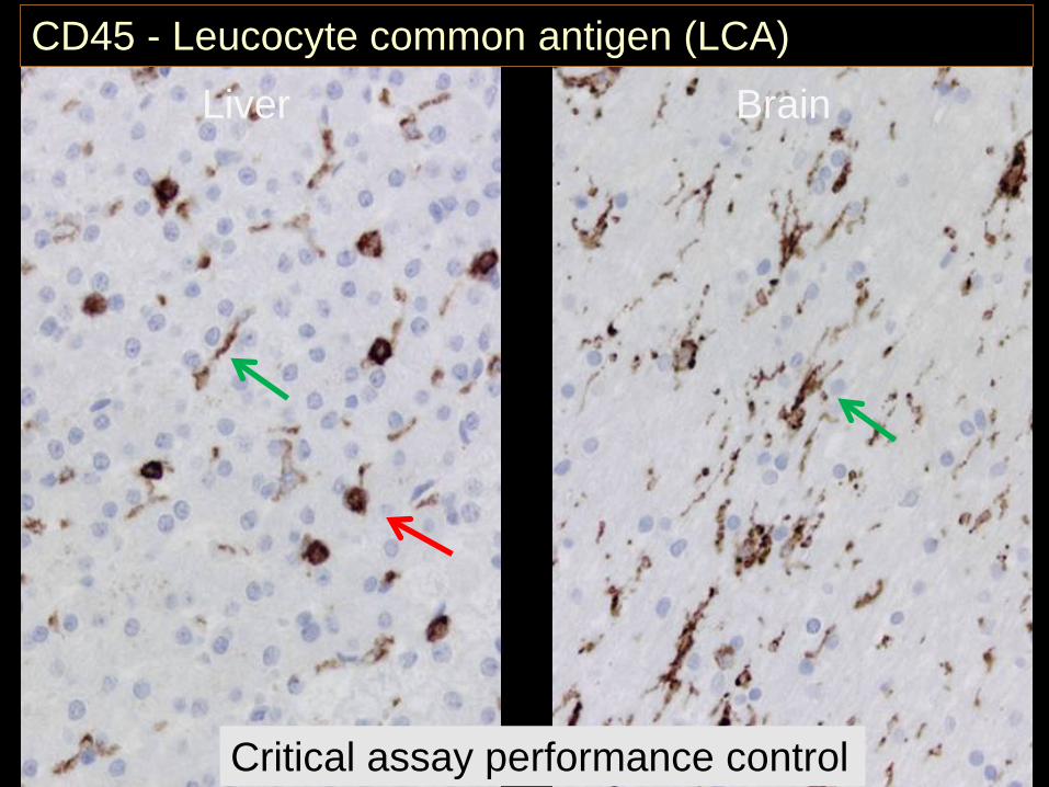
CD45 - Leucocyte common antigen (LCA)
Liver Brain
Critical assay performance control
www.nordiqc.org/Run-15/Assessment/assessment-CD45.htm
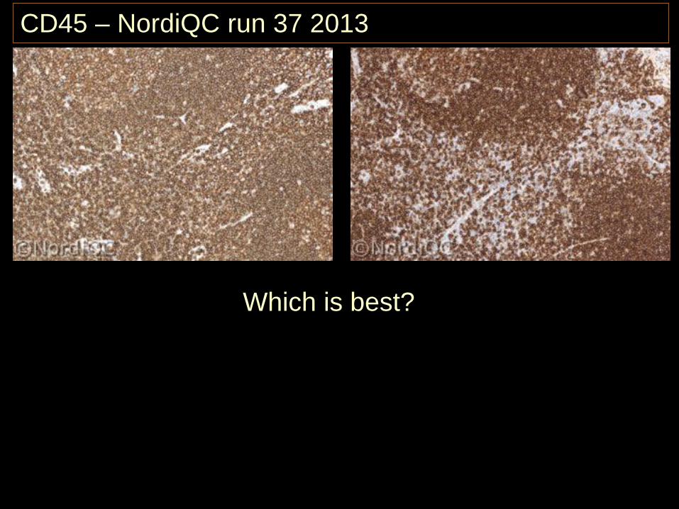
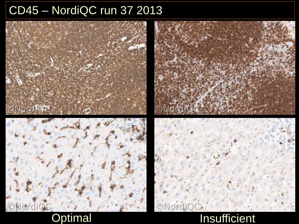
CD45 – NordiQC run 37 2013
Which is best?
www.nordiqc.org/Run-15/Assessment/assessment-CD45.htm
CD45 – NordiQC run 37 2013
Optimal Insufficient
www.nordiqc.org/Run-15/Assessment/assessment-CD45.htm
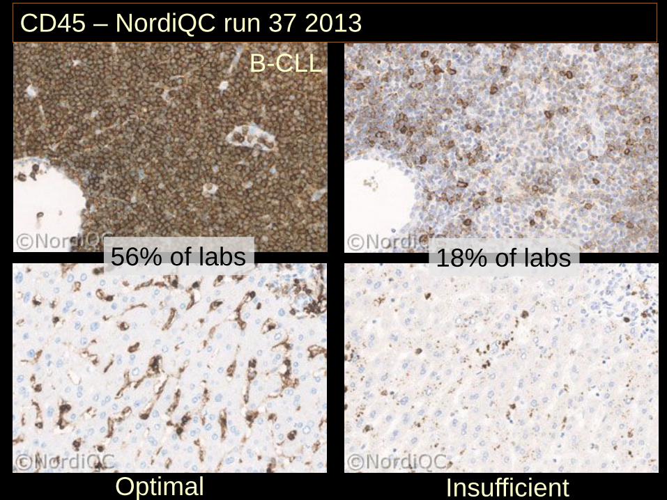
CD45 – NordiQC run 37 2013
Optimal Insufficient
B-CLL
56% of labs 18% of labs
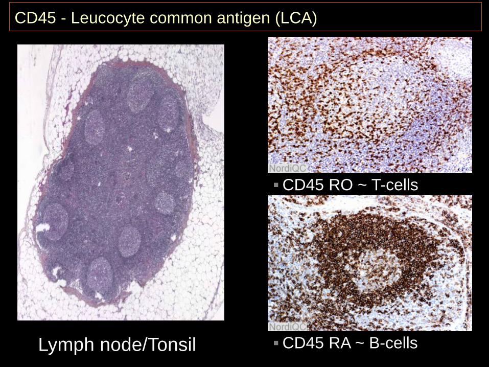
CD45 - Leucocyte common antigen (LCA)
Lymph node/Tonsil
CD45 RO ~ T-cells
CD45 RA ~ B-cells
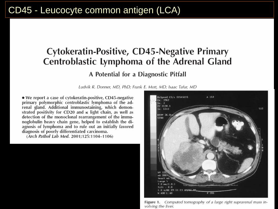
CD45 - Leucocyte common antigen (LCA)
CD45 - Leucocyte common antigen (LCA)
CD45 - Leucocyte common antigen (LCA)
Primary panel for the unknown primary tumour
CD45 CK S-100 VIMHaemato-lymphoid neoplasms
+/(-) -/(+) -/(+) +/(-)Epithelialneoplasms - +/(-) -/+ -/+Mesothelial neoplasms - + - +Mesenchymal and neuronal neoplasms
- -/(+) -/+ +
Non-neuronal neuroepithelial neoplasms
- -/(+) + +
Germ cell neoplasms - -/+ -/+ +
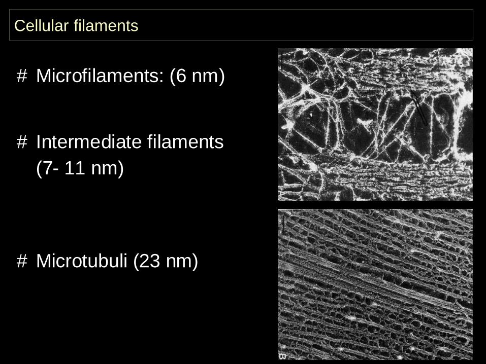
Cellular filaments
# Microfilaments: (6 nm)
# Intermediate filaments (7- 11 nm)
# Microtubuli (23 nm)
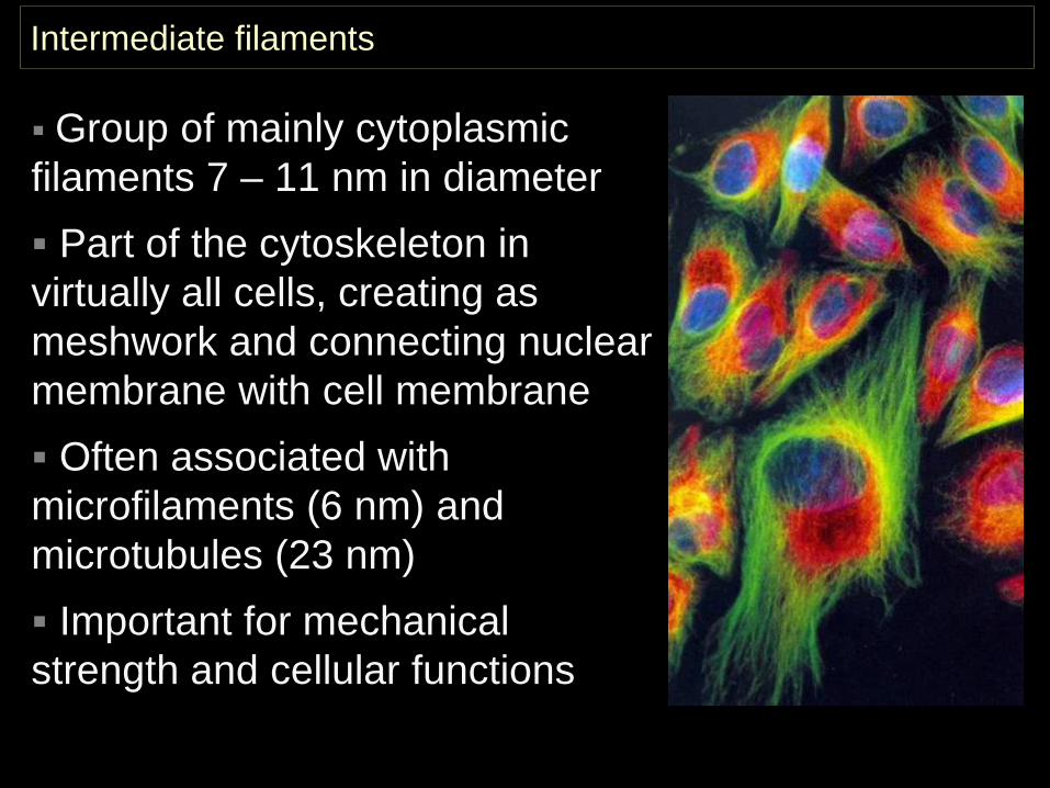
Intermediate filaments
Group of mainly cytoplasmic filaments 7 – 11 nm in diameter Part of the cytoskeleton in virtually all cells, creating as meshwork and connecting nuclear membrane with cell membrane Often associated with microfilaments (6 nm) and microtubules (23 nm) Important for mechanical strength and cellular functions
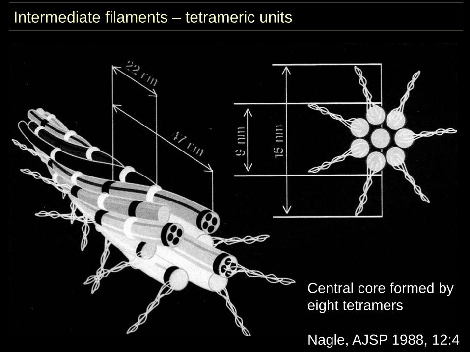
Intermediate filaments – tetrameric units
Central core formed by eight tetramers
Nagle, AJSP 1988, 12:4
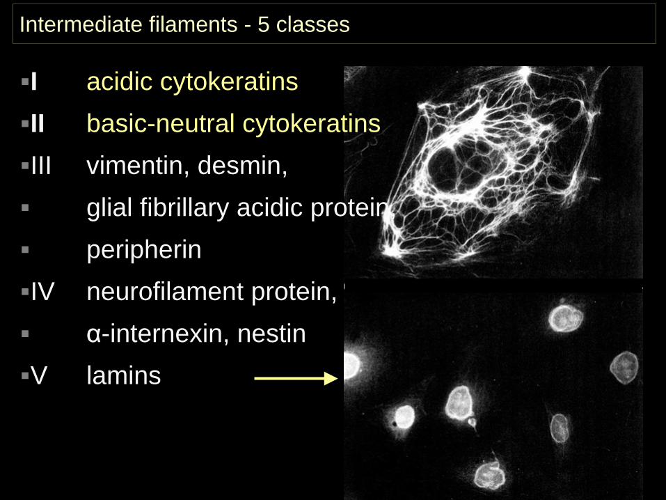
Intermediate filaments - 5 classes
I acidic cytokeratinsII basic-neutral cytokeratinsIII vimentin, desmin, glial fibrillary acidic protein, peripherinIV neurofilament protein, α-internexin, nestinV lamins
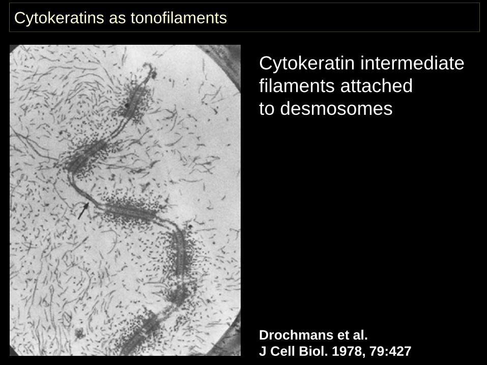
Cytokeratins as tonofilaments
Drochmans et al. J Cell Biol. 1978, 79:427
Cytokeratin intermediate filaments attached to desmosomes
Cytokeratins (CKs) belong to the most fundamental markers of epithelial differentiation CKs comprise a large family of subtypes. Different cell types express different patterns of CK subtypes Cancers generally express CK patterns that at least in part represent the pattern of the putative cell of origin Metastases express CK patterns fairly concordant with those of the primary tumours
Cytokeratins in diagnostic pathology
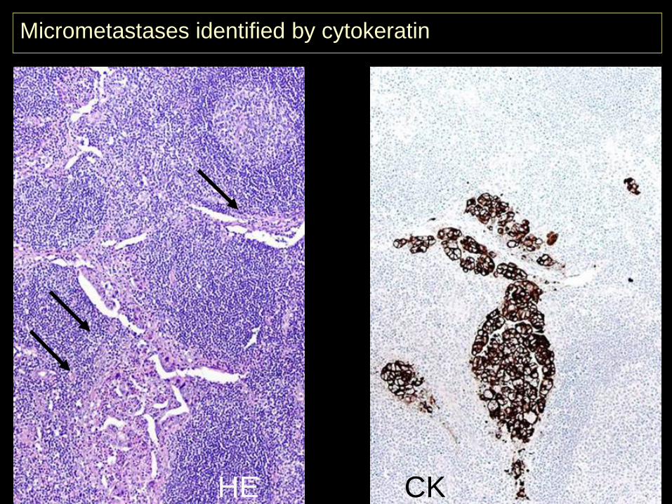
HE CK
Micrometastases identified by cytokeratin
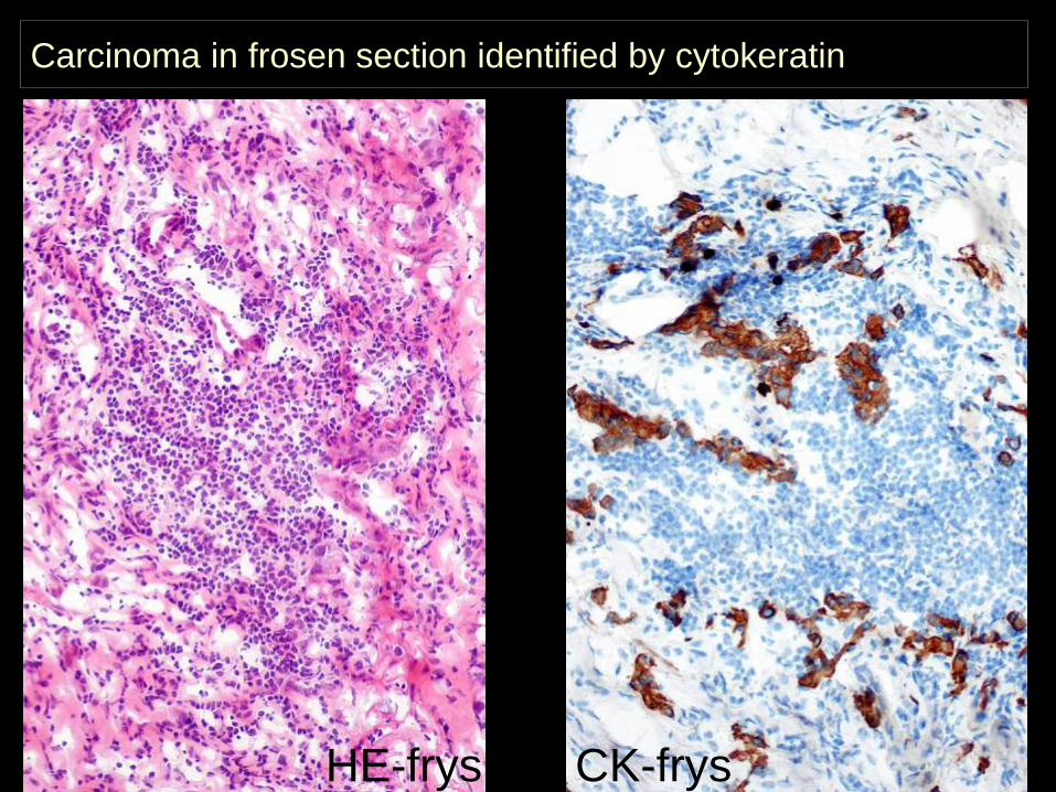
Carcinoma in frosen section identified by cytokeratin
HE-frys CK-frys
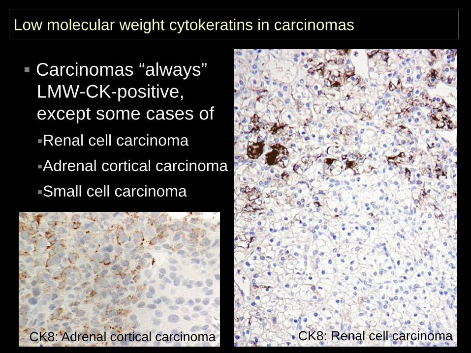
Low molecular weight cytokeratins in carcinomas
CK: renal cell carcinoma
Carcinomas “always” LMW-CK-positive, except some cases of Renal cell carcinomaAdrenal cortical carcinomaSmall cell carcinoma
CK8: Adrenal cortical carcinoma CK8: Renal cell carcinoma
Primary panel for the unknown primary tumour
CD45 CK S-100 VIMHaemato-lymphoid neoplasms
+/(-) -/(+) -/(+) +/(-)Epithelialneoplasms - +/(-) -/+ -/+Mesothelial neoplasms - + - +Mesenchymal and neuronal neoplasms
- -/(+) -/+ +
Non-neuronal neuroepithelial neoplasms
- -/(+) + +
Germ cell neoplasms - -/+ -/+ +
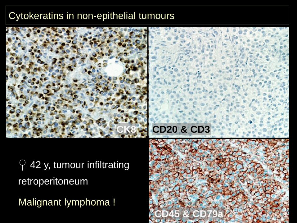
CK8 CD20 & CD3
♀ 42 y, tumour infiltrating retroperitoneum
CD45 & CD79a
Cytokeratins in non-epithelial tumours
Malignant lymphoma !
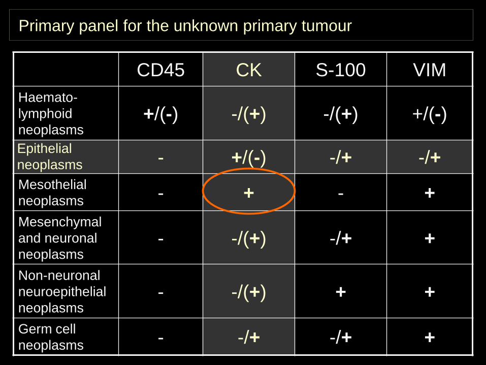
Primary panel for the unknown primary tumour
CD45 CK S-100 VIMHaemato-lymphoid neoplasms
+/(-) -/(+) -/(+) +/(-)Epithelialneoplasms - +/(-) -/+ -/+Mesothelial neoplasms - + - +Mesenchymal and neuronal neoplasms
- -/(+) -/+ +
Non-neuronal neuroepithelial neoplasms
- -/(+) + +
Germ cell neoplasms - -/+ -/+ +
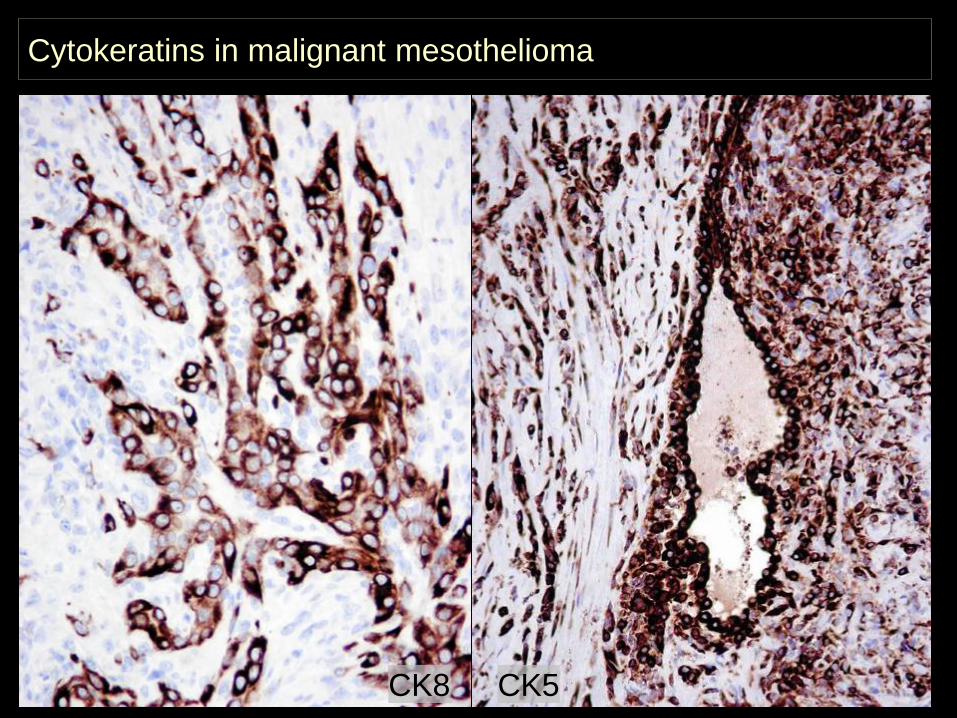
Cytokeratins in malignant mesothelioma
CK8 CK5
Primary panel for the unknown primary tumour
CD45 CK S-100 VIMHaemato-lymphoid neoplasms
+/(-) -/(+) -/(+) +/(-)Epithelialneoplasms - +/(-) -/+ -/+Mesothelial neoplasms - + - +Mesenchymal and neuronal neoplasms
- -/(+) -/+ +
Non-neuronal neuroepithelial neoplasms
- -/(+) + +
Germ cell neoplasms - -/+ -/+ +
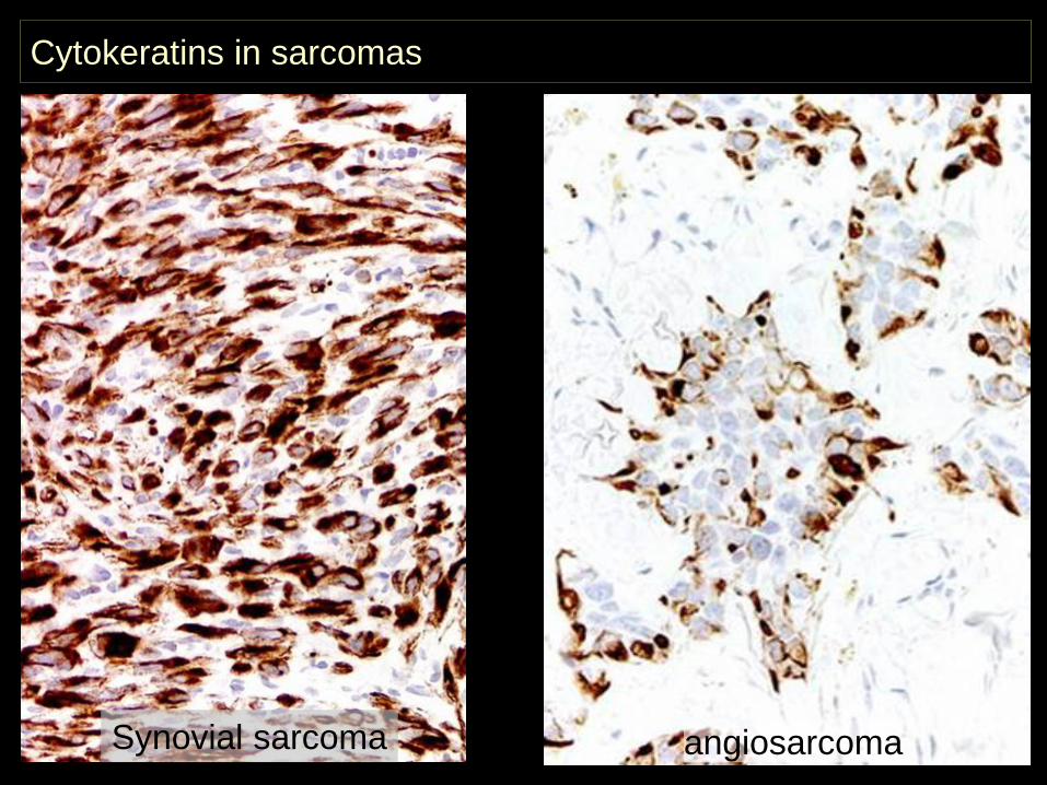
Cytokeratins in sarcomas
angiosarcomaSynovial sarcoma
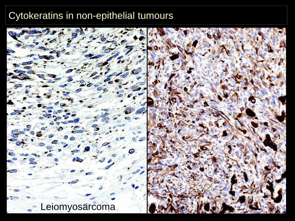
Leiomyosarcoma
Cytokeratins in non-epithelial tumours
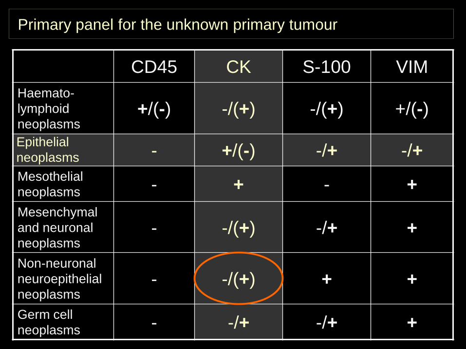
Primary panel for the unknown primary tumour
CD45 CK S-100 VIMHaemato-lymphoid neoplasms
+/(-) -/(+) -/(+) +/(-)Epithelialneoplasms - +/(-) -/+ -/+Mesothelial neoplasms - + - +Mesenchymal and neuronal neoplasms
- -/(+) -/+ +
Non-neuronal neuroepithelial neoplasms
- -/(+) + +
Germ cell neoplasms - -/+ -/+ +
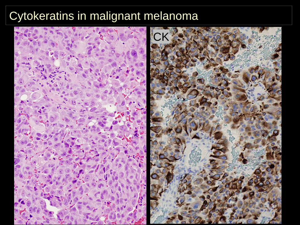
Cytokeratins in malignant melanoma
CK
Primary panel for the unknown primary tumour
CD45 CK S-100 VIMHaemato-lymphoid neoplasms
+/(-) -/(+) -/(+) +/(-)Epithelialneoplasms - +/(-) -/+ -/+Mesothelial neoplasms - + - +Mesenchymal and neuronal neoplasms
- -/(+) -/+ +
Non-neuronal neuroepithelial neoplasms
- -/(+) + +
Germ cell neoplasms - -/+ -/+ +
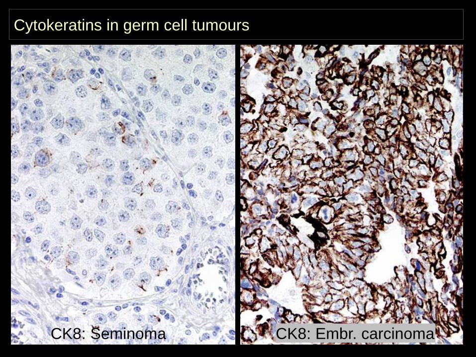
Cytokeratins in germ cell tumours
CK8: Seminoma CK8: Embr. carcinoma
CK: renal cell carcinoma
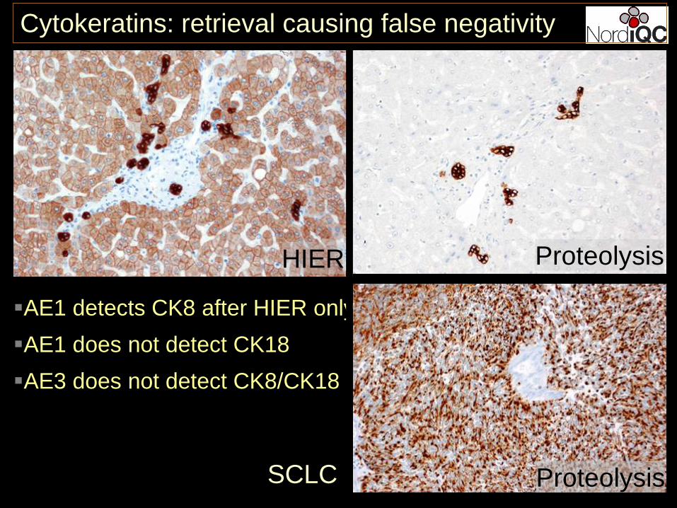
HIER Proteolysis
AE1 detects CK8 after HIER onlyAE1 does not detect CK18AE3 does not detect CK8/CK18
Proteolysis
Cytokeratins: retrieval causing false negativity
SCLC
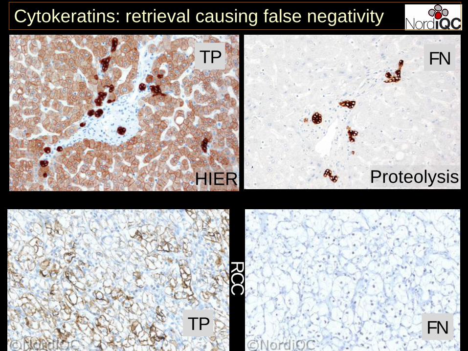
CK: renal cell carcinoma
Cytokeratins: retrieval causing false negativity
RCC
FNTP
HIER Proteolysis
FNTP
Primary panel for the unknown primary tumour
CD45 CK S-100 VIMHaemato-lymphoid neoplasms
+/(-) -/(+) -/(+) +/(-)Epithelialneoplasms - +/(-) -/+ -/+Mesothelial neoplasms - + - +Mesenchymal and neuronal neoplasms
- -/(+) -/+ +
Non-neuronal neuroepithelial neoplasms
- -/(+) + +
Germ cell neoplasms - -/+ -/+ +
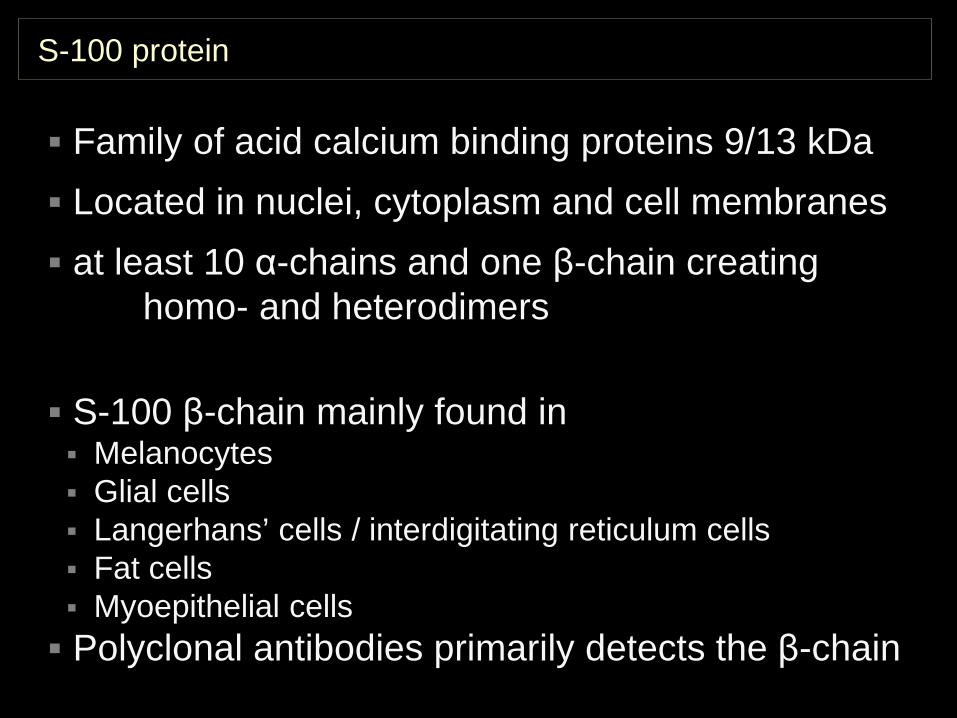
S-100 protein
Family of acid calcium binding proteins 9/13 kDa Located in nuclei, cytoplasm and cell membranes at least 10 α-chains and one β-chain creating
homo- and heterodimers
S-100 β-chain mainly found in Melanocytes Glial cells Langerhans’ cells / interdigitating reticulum cells Fat cells Myoepithelial cells Polyclonal antibodies primarily detects the β-chain
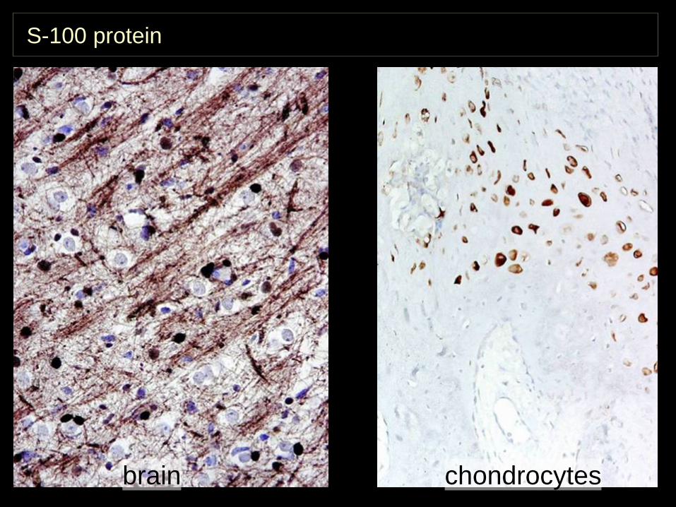
S-100 protein
chondrocytesbrain
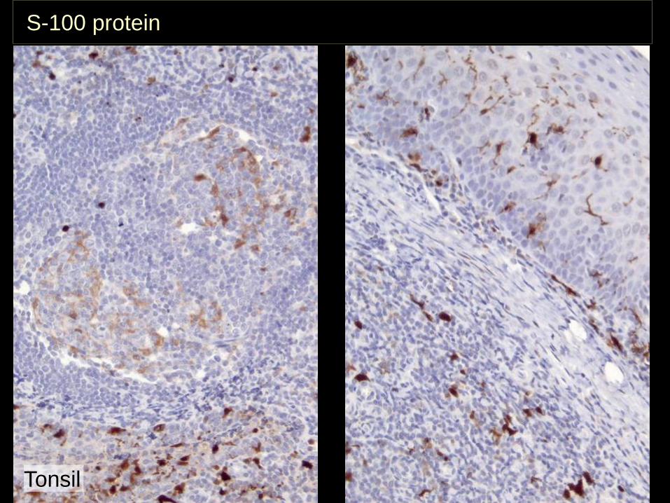
Tonsil
S-100 protein

S-100 protein – pancreas
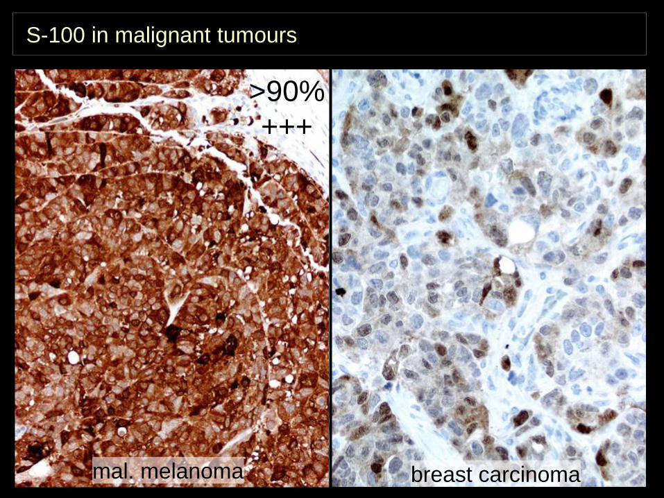
S-100 in malignant tumours
mal. melanoma breast carcinoma
>90%+++
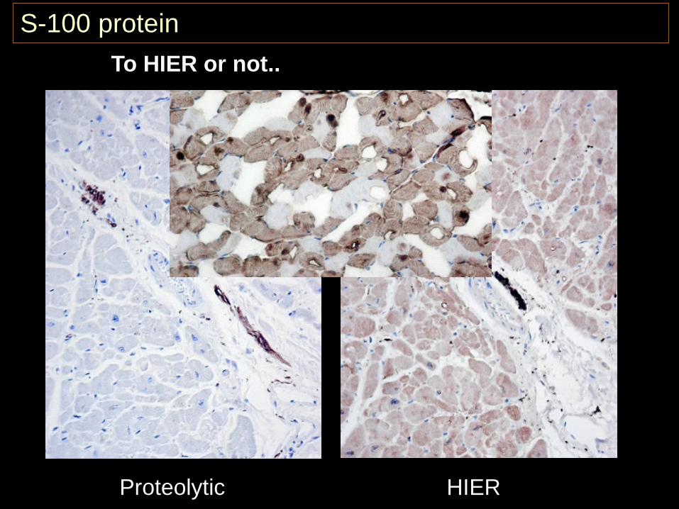
To HIER or not..
S100B Frozen IHC
Proteolytic HIER
S-100 protein
Primary panel for the unknown primary tumour
”Real” CD45 CK S-100 VIMHaemato-lymphoid neoplasms
+/(-) -/(+) -/(+) +/(-)Epithelialneoplasms - +/(-) -/+ -/+Mesothelial neoplasms - + - +Mesenchymal and neuronal neoplasms
- -/(+) -/+ +
Non-neuronal neuroepithelial neoplasms
- -/(+) + +
Germ cell neoplasms - -/+ -/+ +
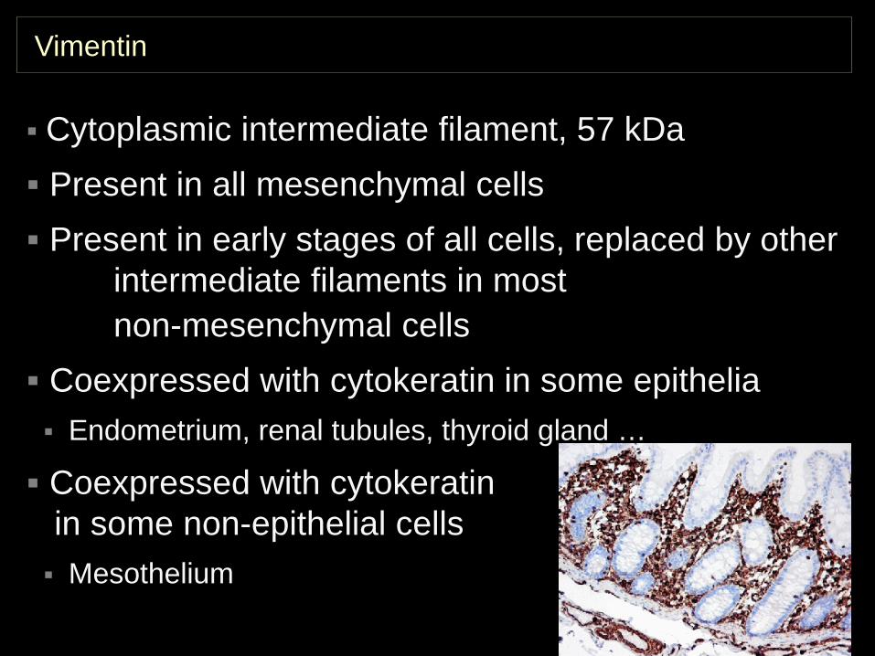
Vimentin
Cytoplasmic intermediate filament, 57 kDa Present in all mesenchymal cells Present in early stages of all cells, replaced by other
intermediate filaments in most non-mesenchymal cells
Coexpressed with cytokeratin in some epithelia Endometrium, renal tubules, thyroid gland …
Coexpressed with cytokeratinin some non-epithelial cells Mesothelium
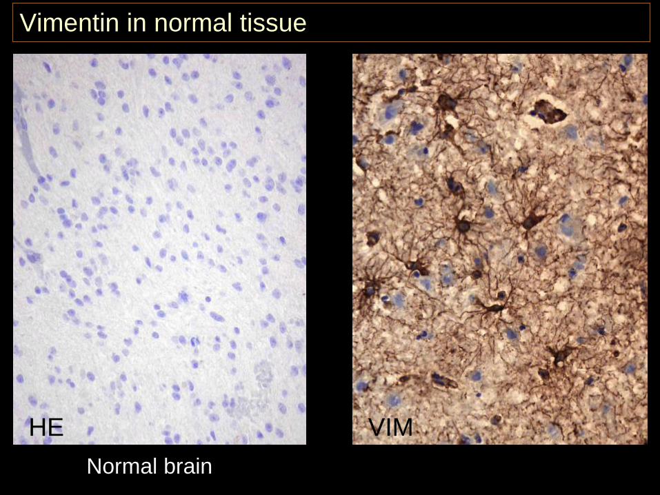
Vimentin in normal tissue
Normal brain
HE VIM
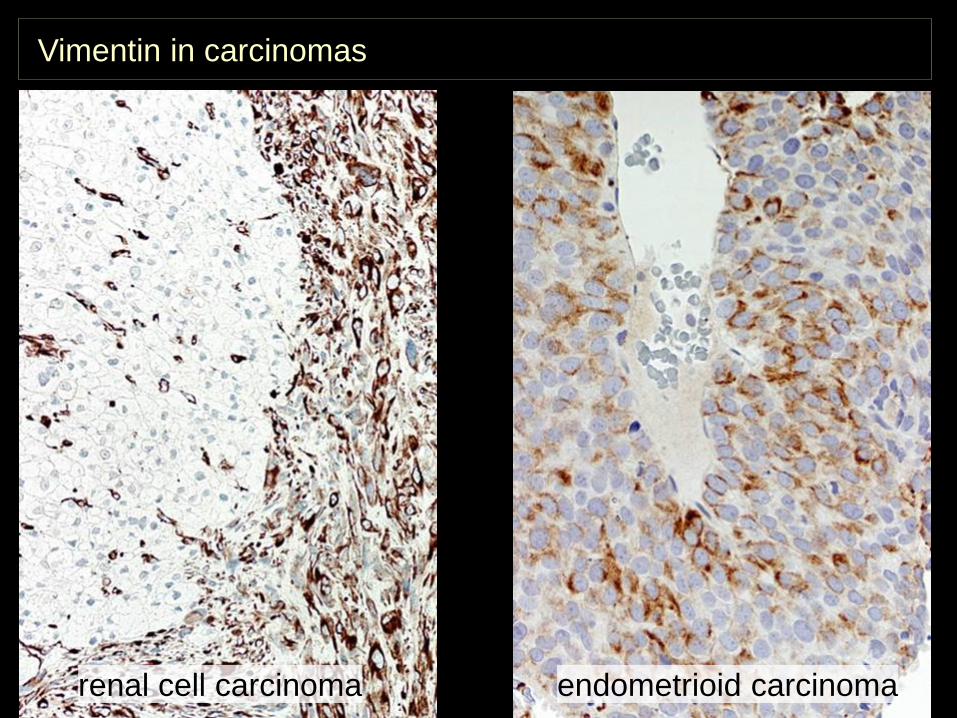
Vimentin in carcinomas
renal cell carcinoma endometrioid carcinoma
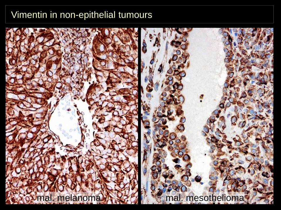
Vimentin in non-epithelial tumours
mal. mesotheliomamal. melanoma
Mogens VybergProfessor of Clinical PathologyDirector of NordiQCAalborg University Hospital, Aalborg, Denmark
IHC Classification of undifferentiated tumors –
the primary panelThank you
for your attention


![IHC PPT Ancillary Productsmy1hr-public.s3.amazonaws.com/documents/enroll/IHC PPT Ancillary Products[3].pdfAncillary Products From The IHC Group. The IHC Group Corporate Overview Ø](https://img.pdfslide.us/doc/110x75/5e38c9b5e1bb9a3e4e5b3bd8/ihc-ppt-ancillary-productsmy1hr-publics3-ppt-ancillary-products3pdf-ancillary.jpg)