Embed Size (px)
Citation preview
Yonsei Med J http://www.eymj.org Volume 53 Number 1 January 2012 15
Review Article http://dx.doi.org/10.3349/ymj.2012.53.1.15pISSN: 0513-5796, eISSN: 1976-2437 Yonsei Med J 53(1):15-34, 2012
IgG4-Related Sclerosing Disease, an Emerging Entity: A Review of a Multi-System Disease
Mukul Divatia,1 Sun A Kim,2 and Jae Y. Ro1,3,4,5
1Department of Pathology, The Methodist Hospital, Weill Medical College of Cornell University, Houston, TX, USA; 2Department of Pathology, Asan Medical Center, University of Ulsan College of Medicine, Seoul;
3Department of Pathology, Yonsei University College of Medicine, Seoul; 4National Cancer Center, Goyang, Korea;5The University of Texas, MD Anderson Cancer Center, Houston, TX, USA.
Received: July 15, 2011Corresponding author: Dr. Jae Y. Ro,Departments of Pathology, The Methodist Hospital, Weill Medical College of Cornell University, 6565 Fannin Street, Houston, TX 77030, USA.Tel: 713-441-2263, Fax: 713-793-1603E-mail: [email protected]
∙ The authors have no financial conflicts of interest.
© Copyright:Yonsei University College of Medicine 2012
This is an Open Access article distributed under the terms of the Creative Commons Attribution Non-Commercial License (http://creativecommons.org/ licenses/by-nc/3.0) which permits unrestricted non-commercial use, distribution, and reproduction in any medium, provided the original work is properly cited.
Immunoglobulin G4-related systemic disease (IgG4-RSD) is a recently defined emerging entity characterized by a diffuse or mass forming inflammatory reaction rich in IgG4-positive plasma cells associated with fibrosclerosis and obliterative phlebitis. IgG4-RSD usually affects middle aged and elderly patients, with a male predominance. It is associated with an elevated serum titer of IgG4, which acts as a marker for this recently characterized entity. The prototype is IgG4-related sclerosing pancreatitis or autoimmune pancreatitis (AIP). Other common sites of involvement are the hepatobiliary tract, salivary gland, orbit, and lymph node, however practically any organ can be involved, including upper aerodigestive tract, lung, aorta, mediasti-num, retroperitoneum, soft tissue, skin, central nervous system, breast, kidney, and prostate. Fever or constitutional symptoms usually do not comprise part of the clini-cal picture. Laboratory findings detected include raised serum globulin, IgG and IgG4. An association with autoantibody detection (such as antinuclear antibodies and rheumatoid factor) is seen in some cases. Steroid therapy comprises the mainstay of treatment. Disease progression with involvement of multiple organ-sites may be en-countered in a subset of cases and may follow a relapsing-remitting course. The prin-cipal histopathologic findings in several extranodal sites include lymphoplasmacytic infiltration, lymphoid follicle formation, sclerosis and obliterative phlebitis, along with atrophy and destruction of tissues. Immunohistochemical staining shows in-creased IgG4+ cells in the involved tissues (>50 per high-power field, with IgG4/IgG ratio >40%). IgG4-RSD may potentially be rarely associated with the develop-ment of lymphoma and carcinoma. However, the nature and pathogenesis of IgG4-RSD are yet to be fully elucidated and provide immense scope for further studies.
Key Words: IgG4-related sclerosing disease, IgG4, autoimmune pancreatitis, scle-rosing cholangitis, inflammatory fibrosclerosing lesion, inflammatory pseudotumor, lymphadenopathy, salivary gland, lacrimal gland
INTRODUCTION
Immunoglobulin subtypes are significantly different in their underlying biologic regu-
Mukul Divatia, et al.
Yonsei Med J http://www.eymj.org Volume 53 Number 1 January 201216
cal entity, characterized by a mass lesion in the pancreas, painless obstructive jaundice, pancreatic duct narrowing, association with diabetes mellitus and favorable response to steroid therapy.
The autoimmune association of AIP is based upon its as-sociation with other immune-mediated diseases, such as sclerosing cholangitis, primary biliary cirrhosis (PBC), Sjo-gren syndrome, and inflammatory bowel disease (IBD).15,16 Autoantibodies, including antinuclear antibodies, rheu-matoid factor, anticarbonic anhydrase II, and antilactofer-rin are commonly encountered in patients with AIP.17-20 Kawa, et al.21 reported an association with HLA DRB1 *0405-DQB1*0401 haplotype in a study from Japan. Nu-merous terminologies have been coined by different in-vestigators to emphasize selected aspects of the disease, including chronic sclerosing pancreatitis,22 lymphoplas-macytic sclerosing pancreatitis,23 nonalcoholic duct-de-structive chronic pancreatitis,24 idiopathic tumefactive chronic pancreatitis25 and duct-narrowing chronic pancre-atitis.26 Over the past decade, developments have resulted in the establishment of the theory that AIP is a heteroge-neous entity which includes at least 2 distinct clinicopatho-logic entities: 1) lymphoplasmacytic sclerosing pancreatitis (LPSP), and 2) idiopathic duct-centric chronic pancreatitis (IDCP).19,27,28
LPSP is a histologically unique lesion that was proposed by Kawaguchi, et al.23 in 1991. It consists of diffuse lym-phoplasmacytic infiltration and fibrosis that focally gives rise to a storiform fibrosis. Neutrophils are conspicuously absent but eosinophils may be identified. Pancreatic lobules are relatively well preserved compared to alcoholic chronic pancreatitis, but focal destruction of pancreatic acini and re-placement with fibrosis are routinely noted. This inflamma-tory process also characteristically extends around the main and interlobular ducts, leaving the duct epithelium and lu-men intact. Veins are almost always obliterated by the same inflammatory process (obliterative phlebitis). Even the splenic and portal venous vasculature may be involved, leading to an intraoperative differential diagnosis of carci-noma. The common bile duct is usually inflamed resulting in jaundice seen in patients with AIP. Numerous IgG4-posi-tive plasma cells are identified in LPSP on staining.29,30 LPSP predominantly affects elderly men with a mean age of 62 to 63.4 years, is commonly associated with proximal biliary tract, retroperitoneal, renal, and salivary disease, shows elevated serum IgG4 titer and relapse (47%) in a significant proportion of cases.31
lation, distribution, and in their interaction with receptors on the various effector cells of the immune system. The im-munoglobulin subtypes that are produced in response to a particular immune challenge depend on class-switch gene rearrangement of the constant regions.1 The immunoglobu-lin G (IgG) class is complex in structure and biology with four subclasses of IgG antibodies, ranging from 1 through 4 based upon the order of their discovery and serum levels. The serum concentration of IgG1 is the highest amongst the IgG subclasses and ranges from 5-11 mg/mL,2 whereas the least abundant subclass, IgG4, is present at mean con-centrations of 0.35-0.51 mg/mL.3 The concentration of IgG4 in serum varies significantly among healthy people unlike the other IgG subclasses. IgG4 levels generally range from less than 10 μg/mL to 1.4 mg/mL, with levels over 2 mg/mL noted in rare instances and levels being generally high-er in men and older people.2-4
IgG4 is a unique antibody based upon its structure, func-tion, and immunologic regulation. IgG4 antibodies are dis-tinct as they undergo ‘half-antibody exchange’ in vivo, re-sulting in recombined antibodies comprising two different binding specificities.5 IgG4 does not activate complement pathways and has reduced effector function relative to other IgG subtypes.6,7 IgG4 production is driven in part by T- helper cell 2 cytokines.8,9 IgG4 plays a significant role in bullous skin lesions,10 atopic eczema and bronchial asth-ma11,12 as well as decreasing IgE-mediated inflammatory re-sponse in parasitic infections.8,9 IgG4-related systemic dis-ease (IgG4-RSD) represents a newly described category of diseases with a potential role for IgG4 antibodies. The exact role of IgG4 antibodies in the pathogenesis of IgG4-RSD still remains unclear in the present day scenario. An in-creased understanding of the precise role of IgG4 in these related clinical syndromes will be paramount for elucida-tion of the underlying pathophysiology.
HISTORICAL PERSPECTIVE OF AUTOIMMUNE PANCREATITIS AND
IgG4-RELATED DISEASES
Sarles, et al.13 raised the possibility of some cases of chron-ic pancreatitis arising from an autoimmune pathologic pro-cess in 1961. The concept of autoimmune pancreatitis (AIP) was proposed by Yoshida, et al.14 in 1995 along with the introduction of this terminology. Autoimmune pancre-atitis has been recognized over the years as a distinct clini-
IgG4-Related Sclerosing Disease
Yonsei Med J http://www.eymj.org Volume 53 Number 1 January 2012 17
IgG4-RELATED SCLEROSING PANCREATITIS
In 2001, Hamano, et al.40 reported elevated serum IgG4 lev-els in a cohort of patients with a specific subtype of sclerosing pancreatitis/AIP. This finding was not observed in patients with pancreatic carcinoma, primary sclerosing cholangitis (PSC), PBC, nonspecific chronic pancreatitis, Sjogren syn-drome and normal subjects. Kamisawa, et al.41 reported that IgG4-positive plasma cells are increased systemically in patients with AIP and concluded that AIP patients have a systemic disease, with the proposal for the entity ‘IgG4-re-lated sclerosing disease’.42 Histologically, this subtype cor-responds to LPSP, but not IDCP.30,32,43
CLINICAL FEATURES OF IgG4-RELATED SCLEROSING PANCREATITIS
IgG4-related sclerosing pancreatitis is seen most commonly in middle-aged and elderly men with a mean age of 59 to 68 years and a male-to-female ratio of 4-7.5: 1.44-46 The preva-lence rate is 2% to 11% among patients with chronic pancre-atitis.20,45 Studies have demonstrated that it accounts for up to one third of the total cases of benign conditions that are treat-ed with pancreatoduodenectomies, thus highlighting the fact that is often mistaken for pancreatic cancer in spite of major advancements in clinical medicine and radiology.47,48
Patients usually present with a pancreatic mass and/or painless obstructive jaundice. Newly diagnosed type II dia-betes mellitus and steatorrhea are noted in some of these cas-es.20,37 Contrarily, systemic symptoms, including fever, weight loss, and generalized malaise are rarely seen. Imag-ing studies characteristically show enlargement of the pan-creas, predominantly involving the pancreatic head, and en-doscopic retrograde cholangiopancreatography (ERCP) shows irregular narrowing of the pancreatic duct with or without stenosis of the common bile duct.36
The disease responds well to steroid therapy, although re-lapses can occur on cessation of treatment.37 Response to treatment is monitored by a decline in serum IgG4 titer and a reduction in IgG4+ cells in the affected organs. Ritux-imab administration may be significantly beneficial accord-ing to a recent study which reports an excellent response in IgG4-related pancreatitis cases.49 The drug acts by reducing the number of B-lymphocytes that provide a source for the
IDCP is characterized by ductal epithelium centric in-flammation.16,27,32 Neutrophilic infiltration in the main and/or interlobular ducts is typically seen, and extends to in-volve the duct epithelium and lumen. The duct epithelium shows destructive and regenerative changes with the lu-men appearing stenotic or tortuous as a result of the in-flammation. A band of lymphocytes and plasma cells sur-rounds the lumen but, in contrast to LPSP, the ductal lesion lacks the appearance of a thickened wall. The entire duct may seem entrapped within an aggregate of inflam-matory cells. When the inflammation is severe, pancreatic lobules are also inflamed with neutrophils, lymphocytes and plasma cells and microabscesses may be seen as well. Although there is fibrosis around pancreatic lobules, in-flammatory cells are scarce within fibrosis itself, in con-trast to LPSP, in which inflammatory cells are numerous within fibrosis. Obliterative phlebitis is rare, and inflam-mation of the common bile duct is less common com-pared to LPSP. IgG4-positive plasma cells are usually few in IDCP.32
The clinical features of LPSP are concordant with those of AIP reported from Japan, described previously.27 Serum IgG4 is elevated in 80% of AIP patients in Japan, which correlates well with numerous IgG4-positive plasma cells seen in LPSP. In contrast, patients with IDCP are younger than LPSP patients, and many of them are younger than 40 years without any specific gender preponderance.27 Ob-structive jaundice is less common in IDCP than in LPSP. The association of IBD is found in IDCP, but extrapancre-atic manifestations seen in LPSP are rare. It is noteworthy that IDCP is rare in Japan.33
Owing to the fact that both LPSP and IDCP have over-lapping clinicopathological features, there has been a con-tentious debate as to whether these two pathological groups are different manifestations of a single entity of AIP, or whether they are distinct clinicopathological enti-ties. The controversy is due to the variety of AIP diagnos-tic criteria proposed by different groups. The diagnostic criteria from Japan,34 Korea,35 consensus of the Japan-Ko-rea symposium36 and Mayo Clinic37 define LPSP as the pathological entity of AIP, but other groups include both LPSP and IDCP in AIP.16,32,38 The theory that LPSP and IDCP are distinct entities has gained acceptance due to the differing demographic and clinical presentations as well as different immunoreactivity for IgG4. Recently, new no-menclature, type 1 and type 2 AIP, which correspond to LPSP and IDCP, respectively, have been proposed.39
Mukul Divatia, et al.
Yonsei Med J http://www.eymj.org Volume 53 Number 1 January 201218
sclerosis, and 3) obliterative phlebitis, with accompanying at-rophy and destruction of pancreatic acini although these mor-phologic features are characteristic, they are not completely specific, and thus a definitive diagnosis requires the demon-stration of an increased population of IgG4+ plasma cells.31
Sclerosis of the interlobular septa further enhances the lobular architecture of the pancreas by making the separate lobules appear more demarcated (Fig. 2A). The extent of involvement varies greatly from one lobule to another. This may range from normal appearing lobules, to moderate at-rophy and inflammation, and complete replacement by a fi-broinflammatory process. The pancreatic lobules show in-terstitial infiltration by plasma cells, reactive lymphocytes, and a variable number of eosinophils, accompanied by atro-phy or loss of acini (Fig. 2B). The lobular stroma shows vari-able degrees of sclerosis, which can exhibit a storiform char-acter.36,37 Prominent edema or myxoid change may be noted. Increasing degrees of sclerosis results in loss of the lobular architecture.50 The fibrosclerosis and inflammatory process can extend beyond the confines of the pancreas into the ret-roperitoneal tissue and surrounding organs. Islet cell hyper-plasia is a rare finding in contradistinction to the routinely encountered examples of chronic pancreatitis. The pancre-
IgG4- secreting plasma cells.
MORPHOLOGIC FEATURES OF IgG4-RELATED SCLEROSING PANCREATITIS
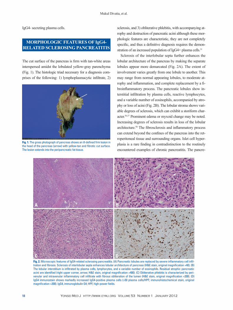
The cut surface of the pancreas is firm with tan-white areas interspersed amidst the lobulated yellow-gray parenchyma (Fig. 1). The histologic triad necessary for a diagnosis com-prises of the following: 1) lymphoplasmacytic infiltrate, 2)
Fig. 1. The gross photograph of pancreas shows an ill-defined firm lesion in the head of the pancreas (arrow) with yellow-tan and fibrotic cut surface. The lesion extends into the peripancreatic fat tissue.
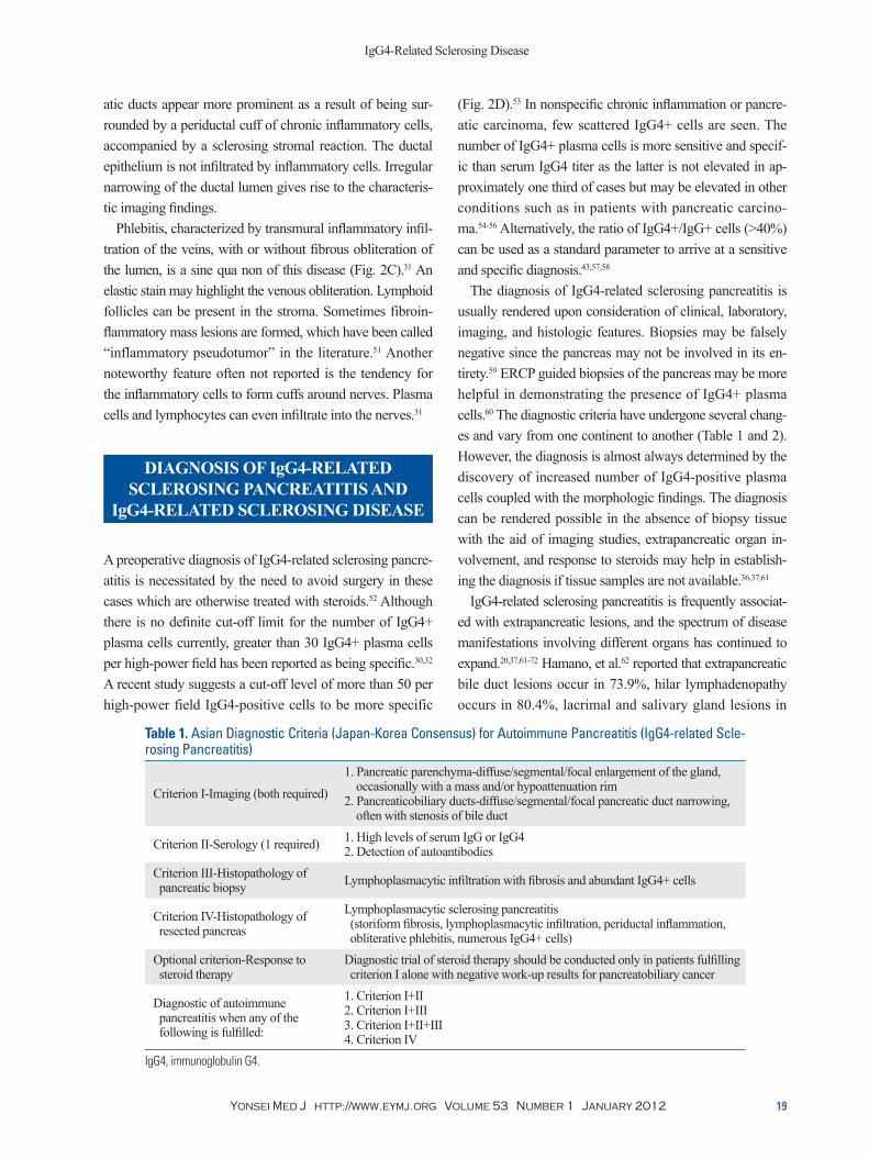
Fig. 2. Microscopic features of IgG4-related sclerosing pancreatitis. (A) Pancreatic lobules are replaced by severe inflammatory cell infil-tration and fibrosis. Sclerosis of interlobular septa enhances lobular architecture of pancreas (H&E stain, original magnification ×40). (B) The lobular interstitium is infiltrated by plasma cells, lymphocytes, and a variable number of eosinophils. Residual atrophic pancreatic acini are identified (right upper corner, arrow; H&E stain, original magnification ×400). (C) Obliterative phlebitis is characterized by peri-venular and intravenular inflammatory cell infiltrate with fibrous obliteration of the lumen (H&E stain, original magnification ×200). (D) IgG4 immunostain shows markedly increased IgG4-positive plasma cells (>50 plasma cells/HPF; immunohistochemical stain, original magnification ×200). IgG4, immunoglobulin G4; HPF, high-power fields.
A
C
B
D
IgG4-Related Sclerosing Disease
Yonsei Med J http://www.eymj.org Volume 53 Number 1 January 2012 19
(Fig. 2D).53 In nonspecific chronic inflammation or pancre-atic carcinoma, few scattered IgG4+ cells are seen. The number of IgG4+ plasma cells is more sensitive and specif-ic than serum IgG4 titer as the latter is not elevated in ap-proximately one third of cases but may be elevated in other conditions such as in patients with pancreatic carcino-ma.54-56 Alternatively, the ratio of IgG4+/IgG+ cells (>40%) can be used as a standard parameter to arrive at a sensitive and specific diagnosis.43,57,58
The diagnosis of IgG4-related sclerosing pancreatitis is usually rendered upon consideration of clinical, laboratory, imaging, and histologic features. Biopsies may be falsely negative since the pancreas may not be involved in its en-tirety.59 ERCP guided biopsies of the pancreas may be more helpful in demonstrating the presence of IgG4+ plasma cells.60 The diagnostic criteria have undergone several chang-es and vary from one continent to another (Table 1 and 2). However, the diagnosis is almost always determined by the discovery of increased number of IgG4-positive plasma cells coupled with the morphologic findings. The diagnosis can be rendered possible in the absence of biopsy tissue with the aid of imaging studies, extrapancreatic organ in-volvement, and response to steroids may help in establish-ing the diagnosis if tissue samples are not available.36,37,61
IgG4-related sclerosing pancreatitis is frequently associat-ed with extrapancreatic lesions, and the spectrum of disease manifestations involving different organs has continued to expand.20,37,61-72 Hamano, et al.62 reported that extrapancreatic bile duct lesions occur in 73.9%, hilar lymphadenopathy occurs in 80.4%, lacrimal and salivary gland lesions in
atic ducts appear more prominent as a result of being sur-rounded by a periductal cuff of chronic inflammatory cells, accompanied by a sclerosing stromal reaction. The ductal epithelium is not infiltrated by inflammatory cells. Irregular narrowing of the ductal lumen gives rise to the characteris-tic imaging findings.
Phlebitis, characterized by transmural inflammatory infil-tration of the veins, with or without fibrous obliteration of the lumen, is a sine qua non of this disease (Fig. 2C).31 An elastic stain may highlight the venous obliteration. Lymphoid follicles can be present in the stroma. Sometimes fibroin-flammatory mass lesions are formed, which have been called “inflammatory pseudotumor” in the literature.51 Another noteworthy feature often not reported is the tendency for the inflammatory cells to form cuffs around nerves. Plasma cells and lymphocytes can even infiltrate into the nerves.31
DIAGNOSIS OF IgG4-RELATED SCLEROSING PANCREATITIS AND
IgG4-RELATED SCLEROSING DISEASE
A preoperative diagnosis of IgG4-related sclerosing pancre-atitis is necessitated by the need to avoid surgery in these cases which are otherwise treated with steroids.52 Although there is no definite cut-off limit for the number of IgG4+ plasma cells currently, greater than 30 IgG4+ plasma cells per high-power field has been reported as being specific.30,32 A recent study suggests a cut-off level of more than 50 per high-power field IgG4-positive cells to be more specific
Table 1. Asian Diagnostic Criteria (Japan-Korea Consensus) for Autoimmune Pancreatitis (IgG4-related Scle-rosing Pancreatitis)
Criterion I-Imaging (both required)
1. Pancreatic parenchyma-diffuse/segmental/focal enlargement of the gland, occasionally with a mass and/or hypoattenuation rim2. Pancreaticobiliary ducts-diffuse/segmental/focal pancreatic duct narrowing, often with stenosis of bile duct
Criterion II-Serology (1 required) 1. High levels of serum IgG or IgG42. Detection of autoantibodies
Criterion III-Histopathology of pancreatic biopsy Lymphoplasmacytic infiltration with fibrosis and abundant IgG4+ cells
Criterion IV-Histopathology of resected pancreas
Lymphoplasmacytic sclerosing pancreatitis (storiform fibrosis, lymphoplasmacytic infiltration, periductal inflammation, obliterative phlebitis, numerous IgG4+ cells)
Optional criterion-Response to steroid therapy
Diagnostic trial of steroid therapy should be conducted only in patients fulfilling criterion I alone with negative work-up results for pancreatobiliary cancer
Diagnostic of autoimmune pancreatitis when any of the following is fulfilled:
1. Criterion I+II2. Criterion I+III3. Criterion I+II+III4. Criterion IV
IgG4, immunoglobulin G4.
Mukul Divatia, et al.
Yonsei Med J http://www.eymj.org Volume 53 Number 1 January 201220
association of “autoimmune pancreatitis” with other “auto-immune diseases” including PSC, Sjogren syndrome, and retroperitoneal fibrosis simply represents extrapancreatic manifestations of a single systemic entity rather than a mul-titude of diseases.39,73,74 Another pointer favoring this theory states that an increased number of IgG4+ plasma cells can be identified in other uninvolved organs in such cases or-gans, such as the aerodigestive tract mucosa and bone mar-
39.1%, hypothyroidism in 22.2%, and retroperitoneal fibro-sis in 12.5% of cases with IgG4-RSD. The various sites of involvement exhibit similar morphologic features with the common denominator being a significant increase in the number of IgG4+ plasma cells (Table 3).29,41,42
Extrapancreatic involvement may develop prior to, con-comitantly or following pancreatic disease, or may even oc-cur independently in the absence of pancreatitis. A so called
Table 2. Mayo Clinic Diagnostic Criteria for Autoimmune Pancreatitis (IgG4-related Sclerosing Pancreatitis): The HISORt Criteria
Criterion H-Histology (at least one of the following)
1. Periductal lymphoplasmacytic infiltrate, obliterative phlebitis, storiform fibrosis2. Lymphoplasmacytic infiltrate, storiform fibrosis, abundant IgG4+ cells (≥10 HPF)
Criterion I-Imaging of pancreas
1. Typical-diffusely enlarged gland with delayed (rim) enhancement; diffusely irregular, attenuated main pancreatic duct2. Others-Focal pancreatic mass/enlargement; focal pancreatic duct stricture; pancreatic atrophy; pancreatic calcification; pancreatitis
Criterion S-Serology Elevated serum IgG4 (normal: 8-140 mg/dL)Criterion O-Other organ involvement (can be confirmed by biopsy or resolution/ improvement with steroid therapy)
Hilar/intrahepatic biliary strictures; persistent distal biliary stricture; parotid/lacrimal gland involvement; mediastinal lymphadenopathy; retroperitoneal fibrosis
Criterion R-Response to steroid therapy Resolution or marked improvement of pancreatic/extrapancreatic manifestation with steroid therapy
Diagnostic of autoimmune pancreatitis when any of the following is fulfilled
1. Criterion H2. Criterion I+S3. Strong clinical suspicion of autoimmune pancreatitis (idiopathic pancreatic disease+Criterion S and/or O)+Criterion R
IgG4, immunoglobulin G4; HPF, high-power fields.
Table 3. Body Sites Affected by IgG4-Related Sclerosing DiseaseBody site Clinicopathologic features
Pancreas Lymphoplasmacytic sclerosing pancreatitis (type 1 AIP) and idiopathic duct centric chronic pancreatitis (type 2 AIP)
Bile duct Sclerosing cholangitisGall bladder Acalculous sclerosing cholecystitis
LiverSclerosing cholangitis involving intrahepatic ducts, inflammatory pseudotumor, portal inflammation with or without interface hepatitis, portal sclerosis, large bile duct obstruction, lobular hepatitis, canalicular cholestasis
Salivary glands Chronic sclerosing sialadenitis (Kuttner tumor), Mikulicz diseaseLacrimal glands and orbit Chronic sclerosing dacryoadenitis, inflammatory pseudotumorRetroperitoneum and mesentery Retroperitoneal fibrosis, sclerosing mesenteritis
Cardiovascular/Aorta Inflammatory abdominal aortic aneurysmMediastinum Sclerosing mediastinitisKidney and ureter Tubulointerstitial nephritis, membranous glomerulopathy, inflammatory pseudotumorThyroid Hypothyroidism, Riedel’s thyroiditisBreast Sclerosing mastitisLung Inflammatory pseudotumor, interstitial pneumoniaCentral nervous system Hypophysitis, sclerosing pachymeningitisProstate Prostatitis
Lymph node Lymphadenopathy with Castleman disease like features, follicular hyperplasia, interfollicular expansion by plasma cells and immunoblasts
IgG4-Related Sclerosing Disease
Yonsei Med J http://www.eymj.org Volume 53 Number 1 January 2012 21
ferential diagnoses are nonspecific inflammation with fibrosis, and malignancies with sclerosis including nodular sclerosing Hodgkin lymphoma, sclerosing large B-cell lymphoma, primary or metastatic carcinoma with desmo-plastic stromal changes. The veins show segmental or cir-cumferential transmural infiltration by chronic inflammato-ry cells, and can show luminal occlusion by fibrous tissue.31 Obliterated veins can be difficult to recognize and may ne-cessitate the aid of elastin stains for identification purposes.
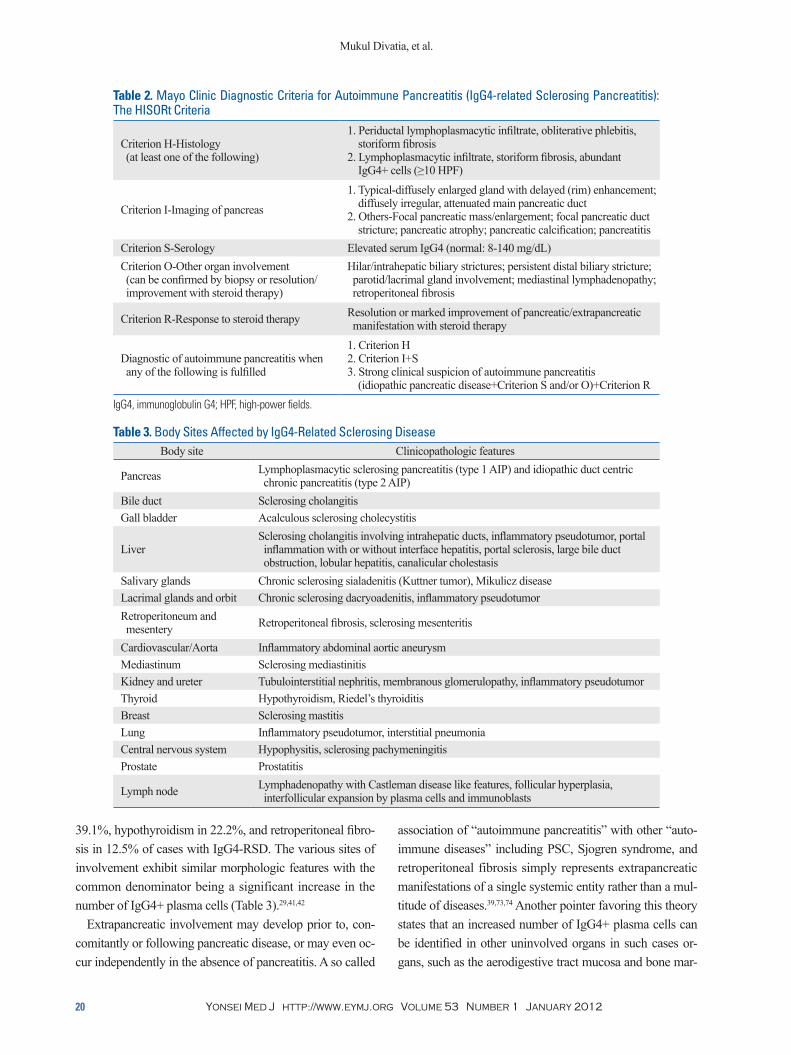
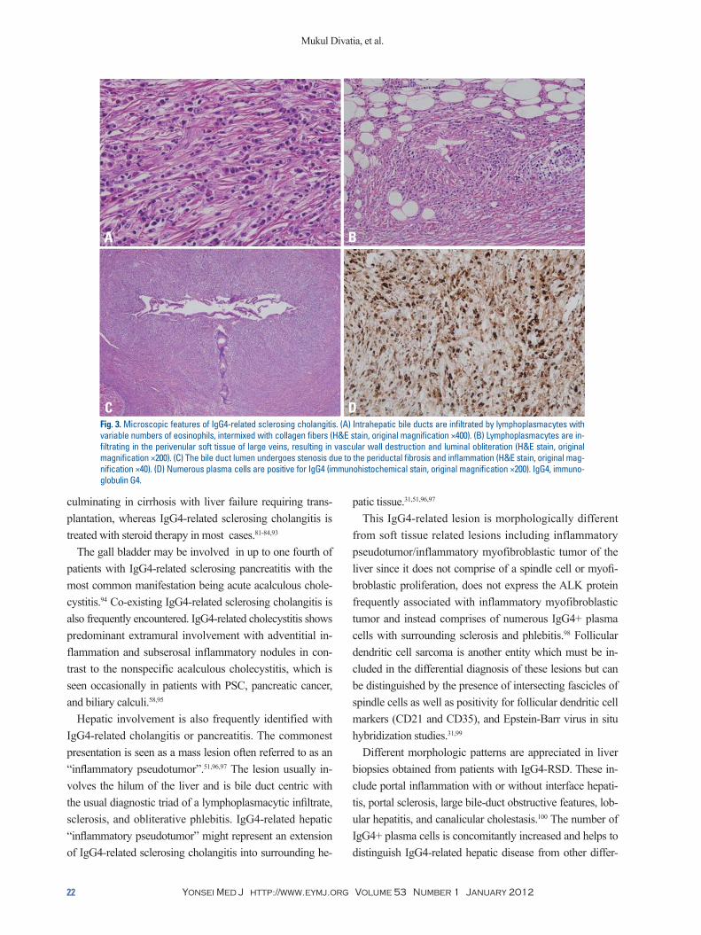
Hepatobiliary systemBiliary tract involvement (IgG4-related sclerosing cholan-gitis) is seen in 50% to 90% of patients with IgG4-related sclerosing pancreatitis and clinically presents as obstructive jaundice or fever.23,51,81-84 Biliary tract disease may also be seen in some cases without pancreatic involvement. Both the extrahepatic and intrahepatic bile duct systems can be affected with the prior being predominantly involved in most reported cases.85 As seen with AIP, the differential di-agnosis includes a malignancy of the bile duct system.81-85 The involved bile ducts demonstrate a transmural lympho-plasmacytic infiltrate with variable numbers of eosinophils, sclerosis (Fig. 3A), and obliterative phlebitis (Fig. 3B). The bile duct lumina undergo stenosis owing to the periductal fibrosis and inflammation (Fig. 3C) and the peribiliary glands are often involved in this region.85 Immunohisto-chemical staining for IgG4 demonstrates a significant sub-set as being IgG4-positive plasma cells (Fig. 3D).
The clinical and pathologic features overlap with those seen in PSC, a chronic biliary inflammatory disease of un-known etiology. The pathologic process in PSC also involves prominent ductal and periductal fibrosis, luminal stenosis, obliteration, and dilatation.86 It is thought that some of the cases reported as PSC actually represent IgG4-related scle-rosing cholangitis.87-91 Mendes, et al.92 published a series of cases of PSC wherein elevated serum IgG4 was found in 9% of the patients and this group of patients also had a low-er frequency of IBD.
In IgG4-RSD involving the bile ducts, the bile duct epi-thelium is usually spared but the peribiliary glands exhibit a greater degree of involvement and inflammation tends to be more severe toward the adventitial aspect. Onion-skin type concentric periductal fibrosis, which is a diagnostic hall-mark of PSC, is not seen in IgG4-related cholangitis.81-84 Another distinguishing feature is that a large number of IgG4-positive plasma cells are identified in IgG4-related cholangitis but not in PSC. PSC is a progressive disease
row, thus providing credence in favor of the systemic na-ture of this disease.42,65,75,76 The various names ascribed to this disease include IgG4-related sclerosing disease,77 IgG4-related systemic sclerosing disease,78 IgG4-related dis-ease,61,79 IgG4-related autoimmune disease,42 hyper-IgG4 disease,80 and IgG4-related systemic disease.49
The extrapancreatic lesions of IgG4-related sclerosing disease also exhibit the same characteristic histologic fea-tures including lymphoplasmacytic infiltrate, sclerosis, and obliterative phlebitis as similarly seen in IgG4-related scle-rosing pancreatitis. The relative proportion of the lympho-plasmacytic infiltrate and sclerosis in different cases gives rise to a spectrum of histologic patterns: pseudolymphoma-tous, mixed, and sclerosing. These patterns represent differ-ent stages in the evolution of this disease with the sequence progressing from being initially pseudolymphomatous to mixed and subsequent progression to the sclerosing pattern. The pseudolymphomatous pattern is more commonly iden-tified in superficial locations including lacrimal gland, sali-vary gland, breast, and skin and thus these are detected ear-lier than deep seated sclerotic lesions in locations such as the retroperitoneum.31
The pseudolymphomatous pattern is characterized by a mass forming dense infiltrate of small lymphocytes and plasma cells, with interspersed reactive lymphoid follicles and a variable proportion of scattered eosinophils. The dif-ferential diagnosis includes low grade non-Hodgkin’s lym-phoma, particularly extranodal marginal zone lymphoma. However, these can be distinguished based on the absence of lymphoepithelial lesions, monocytoid B-cells, diffuse sheets of CD20+ B cells, light chain restriction, aberrant immunophenotype, and clonal immunoglobulin gene rear-rangements.31
The mixed pattern is the most common, comprising of patchy dense infiltrates of small lymphocytes and plasma cells that are accompanied by significant sclerosis (Fig. 2B). Variable numbers of eosinophils and reactive lym-phoid follicles are usually seen. The sclerosing pattern is characterized by a predominantly sclerotic process with poorly defined borders and irregular extensions into the sur-rounding tissues. Patchy aggregates of lymphocytes and plasma cells, with or without lymphoid follicle formation, are seen diffusely scattered over the extent of the lesion, and are seen more commonly at the periphery than at the center of the lesion. The sclerotic areas comprise of colla-gen deposition with scattered fibroblasts. Obliterative phle-bitis is often seen except in small biopsies.31 The main dif-
Mukul Divatia, et al.
Yonsei Med J http://www.eymj.org Volume 53 Number 1 January 201222
patic tissue.31,51,96,97
This IgG4-related lesion is morphologically different from soft tissue related lesions including inflammatory pseudotumor/inflammatory myofibroblastic tumor of the liver since it does not comprise of a spindle cell or myofi-broblastic proliferation, does not express the ALK protein frequently associated with inflammatory myofibroblastic tumor and instead comprises of numerous IgG4+ plasma cells with surrounding sclerosis and phlebitis.98 Follicular dendritic cell sarcoma is another entity which must be in-cluded in the differential diagnosis of these lesions but can be distinguished by the presence of intersecting fascicles of spindle cells as well as positivity for follicular dendritic cell markers (CD21 and CD35), and Epstein-Barr virus in situ hybridization studies.31,99
Different morphologic patterns are appreciated in liver biopsies obtained from patients with IgG4-RSD. These in-clude portal inflammation with or without interface hepati-tis, portal sclerosis, large bile-duct obstructive features, lob-ular hepatitis, and canalicular cholestasis.100 The number of IgG4+ plasma cells is concomitantly increased and helps to distinguish IgG4-related hepatic disease from other differ-
culminating in cirrhosis with liver failure requiring trans-plantation, whereas IgG4-related sclerosing cholangitis is treated with steroid therapy in most cases.81-84,93
The gall bladder may be involved in up to one fourth of patients with IgG4-related sclerosing pancreatitis with the most common manifestation being acute acalculous chole-cystitis.94 Co-existing IgG4-related sclerosing cholangitis is also frequently encountered. IgG4-related cholecystitis shows predominant extramural involvement with adventitial in-flammation and subserosal inflammatory nodules in con-trast to the nonspecific acalculous cholecystitis, which is seen occasionally in patients with PSC, pancreatic cancer, and biliary calculi.58,95
Hepatic involvement is also frequently identified with IgG4-related cholangitis or pancreatitis. The commonest presentation is seen as a mass lesion often referred to as an “inflammatory pseudotumor”.51,96,97 The lesion usually in-volves the hilum of the liver and is bile duct centric with the usual diagnostic triad of a lymphoplasmacytic infiltrate, sclerosis, and obliterative phlebitis. IgG4-related hepatic “inflammatory pseudotumor” might represent an extension of IgG4-related sclerosing cholangitis into surrounding he-
Fig. 3. Microscopic features of IgG4-related sclerosing cholangitis. (A) Intrahepatic bile ducts are infiltrated by lymphoplasmacytes with variable numbers of eosinophils, intermixed with collagen fibers (H&E stain, original magnification ×400). (B) Lymphoplasmacytes are in-filtrating in the perivenular soft tissue of large veins, resulting in vascular wall destruction and luminal obliteration (H&E stain, original magnification ×200). (C) The bile duct lumen undergoes stenosis due to the periductal fibrosis and inflammation (H&E stain, original mag-nification ×40). (D) Numerous plasma cells are positive for IgG4 (immunohistochemical stain, original magnification ×200). IgG4, immuno-globulin G4.
A
C
B
D
IgG4-Related Sclerosing Disease
Yonsei Med J http://www.eymj.org Volume 53 Number 1 January 2012 23
junctivitis sicca, xerostomia, intermittent swelling of the salivary or lacrimal glands, presence of anti-Ro/SSA and anti-La/SSB autoantibodies, and morphologically charac-terized by lymphoepithelial sialadenitis.110
Orbit, including lacrimal glandsIgG4-RSD commonly affects the lacrimal glands and sur-rounding orbital soft tissues and these were the initial sites reported to be involved by this disease apart from the pan-creas. The patients usually present with unilateral or bilater-al, painless orbital swellings of long duration with or with-out significant impairment of visual acuity or symptoms of ‘dry eye’.60,65 The differential diagnosis on clinical and ra-diological examination includes inflammatory pseudotumor and lymphoma. Extensive involvement of orbital soft tissues and sclerosis may lead to markedly decreased visual acuity and occasionally cause optic nerve atrophy and blindness.65 Steroid therapy is the primary treatment but may fail to ameliorate the condition in severe cases with irreversible sclerosis and fibrosis of orbital tissues.60,65
The morphologic features of lacrimal gland involvement (IgG4-related chronic sclerosing dacryoadenitis) are similar to those of IgG4-related sclerosing disease in other organs, except that obliterative phlebitis is not as commonly seen.60,65 Progressive sclerosis and gland destruction frequently com-prise the course of this disease in the orbit with an eventual burnout phase where only the fibrosis remains in the ab-sence of increased inflammation. It may cause extensive fi-brosis to the degree that no recognizable lacrimal gland or orbital structure is identified on biopsies. All patterns of in-flammation and fibrosis, including pseudolymphomatous, mixed and sclerotic patterns, are identified in the lacrimal gland and orbital tissues.31,60,65
Retroperitoneum and mesentery, mediastinum, and aortaInflammatory fibrosclerosing lesion involving the retroperi-toneum, mesentery and mediastinum is an idiopathic entity characterized by diffuse fibrosis and is confused with in-flammatory pseudotumor or inflammatory myofibroblastic tumor.111 Idiopathic retroperitoneal fibrosis or mediastinal fi-brosis may be associated with Riedel thyroiditis, orbital/lac-rimal IgG4-RSD or IgG4-related sclerosing cholangitis.112 The fibrosis in these locations may extend to involve the kidney, ureter and bowel mesentery with varying response to steroid therapy which is ineffective in advanced fibro-sis.113-118
entials including PSC, autoimmune hepatitis, primary bili-ary sclerosis, and chronic viral hepatitis in which only far fewer IgG4+ plasma cells are identified.100,101
Salivary glandThe submandibular gland is the salivary gland most fre-quently involved by IgG4-RSD and is also referred to as ‘chronic sclerosing sialadenitis’ or ‘Kuttner tumor’. The disease was originally described by H. Kuttner102 in 1896, who reported it as a unilateral or bilateral ‘‘hard swelling’’ of the submandibular glands. Chronic sclerosing sialadeni-tis demonstrates a predilection for the submandibular gland, although parotid gland involvement has also been report-ed.103 It is characterized by the presence of a prominent lymphoplasmacytic infiltrate and lymphoid follicles, which upon being combined with the finding of cytotoxic T cell populations, indicate an immunological process.104
Clinical manifestations include unilateral or bilateral hard masses that are often mistaken for a tumor based on both clinical and radiological features.57,61,64,105-107 Almost all cas-es of chronic sclerosing sialadenitis have been shown to be IgG4-related in the submandibular gland, whereas up to a third of all cases of nonspecific chronic sialadenitis are IgG4-related.57,61,64 Recognition of IgG4-RSD in the sali-vary glands is significantly important as these patients may develop IgG4-related lesions in other body sites as well. The parotid glands can be involved either solely or along with the submandibular glands. The morphologic features of salivary gland involvement are identical to those ob-served in other organs.57,61,64 It is characterized by sclerosis of interlobular septae with resulting accentuation of the sal-ivary gland lobular architecture and an interstitial infiltrate of lymphocytes and plasma cells along with variable de-grees of sclerosis. The involvement of adjacent salivary lobules may be non-uniform with focal atrophy and loss of acini in some lobules. Reactive lymphoid follicles and col-lagenous sheaths around small ducts are frequently seen.57,61,64
IgG4-related sclerosing dacryoadenitis and/or sialadenitis is one of the major causes of Mikulicz disease with bilateral lacrimal and salivary glands involvement.108-110 Mickulicz disease, which can represent a number of clinical etiologic entities, is defined clinically as bilateral, painless, and sym-metrical swelling of the lacrimal, parotid, and submandibu-lar glands of unknown etiology for a duration exceeding 3 months. It is not synonymous with the entity Sjogren syn-drome, a distinct autoimmune disease featuring keratocon-
Mukul Divatia, et al.
Yonsei Med J http://www.eymj.org Volume 53 Number 1 January 201224
Lung and pleuraLung involvement by IgG4-RSD may present insidiously with the usual pulmonary symptoms including cough, chest pain, dyspnea and hemoptysis.69,139-141 Radiologically, it may appear as solitary or multiple lung nodules, or consoli-dation and hilar lymphadenopathy.142 Distinct patterns of pulmonary involvement include: solid nodules, peribronchial or perivascular, and interstitial alveolar infiltrates.140
The solid nodular type comprises a hilar or peripheral mass forming lesion with the characteristic lymphoplasma-cytic infiltrate and accompanying sclerosis.69 The adjacent alveoli show interstitial infiltration of lymphocytes and plas-ma cells. In cases involving the hilum, sclerosing inflam-mation of the bronchial wall is identified and it is almost al-ways accompanied by involvement of the bronchial glands. The bronchovascular type is characterized by lymphoplas-macytic infiltrate distributed along the lymphatic channels, with interstitial expansion and extension along the broncho-vascular bundles, interlobular septa, and pleura. Lymphatic dilatation with histiocytes showing emperipolesis of lym-phocytes is a helpful identifying feature.139 In the alveolar interstitial type, the lymphoplasmacytic infiltrate is limited to the alveolar interstitium and is difficult to distinguish from nonspecific interstitial pneumonia.
A variable population of eosinophils along with lymphoid follicles is routinely seen in up to 50% of these cases. One distinguishing feature noted in pulmonary cases of IgG4-RSD is that the arteries are also frequently involved and obliterated as opposed to other body sites wherein veins are selectively involved.139,140 Endothelialitis characterized by a lymphoplasmacytic infiltrate in the subendothelium leading to narrowing and obliteration of the vascular lumen is often identified. Visceral or parietal pleural involvement is noted usually in the form of fibrous thickening accompanied by a chronic lymphoplasmacytic infiltrate with or without fibrin-ous exudates.139-141
BreastIgG4-related sclerosing mastitis can present as single or mul-tiple unilateral or bilateral painless masses, with or without evidence of systemic IgG4-RSD.70,143 Morphologically, it is characterized by a prominent lymphoplasmacytic infiltrates with lymphoid follicle formation, patchy fibrosis, and atro-phy of breast lobules. Phlebitis is also seen in some cases. The differential diagnosis includes involvement of the breast by lymphoma owing to the dense lymphoplasmacytic infil-trate or hyaline-vascular variant of Castleman disease if fol-
Noninfectious aortitis is represented by a group of inflam-matory disorders characterized by chronic inflammation within the aortic wall, wherein the evident inflammation is not considered to be due to infection.119 Noninfectious aorti-tis is most commonly seen involving the thoracic aorta, es-pecially the ascending aorta, and may result from well known systemic rheumatologic disorders such as giant cell arteritis, Takayasu arteritis, rheumatoid arthritis, ankylosing spondylitis, and Behcet’s disease.120,121
The lesions described above are often characterized by a variably prominent lymphoplasmacytic infiltrate, sclerosis, and phlebitis which are essentially identical to those of IgG4-RSD.122 A frequent association with pancreatitis has also been reported in several cases.118,123 Upon extrapolating these findings, a significant proportion of cases of retroperi-toneal fibrosis,124,125 mediastinal fibrosis,67 sclerosing mes-enteritis,126 periaortitis and inflammatory aortic aneurysm (with cases predominantly involving abdominal aorta with rare cases involving aortic arch)71,127-132 have been demon-strated to express markedly increased numbers of IgG4+ plasma cells, and hence represent members of IgG4-RSD. It has not been determined at present whether the group with no significant increase in IgG4+ cells has different eti-ologies or represents burnt out IgG4- RSD.31
ThyroidInvolvement of the thyroid gland as part of the spectrum of IgG4-RSD is being reported increasingly. Patients with AIP often have concomitant evidence of hypothyroidism, with increased levels of antithyroglobulin antibody.133 The exact pathogenetic mechanism for this phenomenon has yet to be elucidated. A study by Li, et al.134,135 involved segregating 70 cases of Hashimoto thyroiditis into 2 groups: 19 IgG4- related cases and 51 non-IgG4-related cases. The IgG4-re-lated group comprised of cases with a greater degree of stromal fibrosis, lymphoplasmacytic infiltration, and thy-roid follicular cell destruction than the other group. The IgG4-related subset is associated with a higher level of cir-culating thyroid autoantibodies, lower female-to-male ratio and an aggressive clinical course. It is possible that hypo-thyroidism in patients with autoimmune pancreatitis may result from an IgG4-related thyroiditis which may be mis-taken for Hashimoto thyroiditis in view of the overlapping symptoms and laboratory findings. Riedel thyroiditis is also regarded as a component of the multifocal fibrosclerosis seen in IgG4-RSD with reports backing this theory based on immunohistochemical findings.136-138
IgG4-Related Sclerosing Disease
Yonsei Med J http://www.eymj.org Volume 53 Number 1 January 2012 25
tuitary gland (IgG4-related infundibulo-hypophysitis). A male predominance is noted. The clinical picture includes patients presenting with hypopituitarism, diabetes insipidus, and/or symptomatic local mass effect.151-156
A pituitary mass and/or thickening of the pituitary stalk are identified on imaging studies. The treatment modality is steroid therapy which usually elicits a favorable response and manifests as a marked reduction in lesional size and correction of hormonal imbalances.151-156 An accompanying meningeal involvement in the form of hypertrophic pachy-meningitis or sinus involvement, with extension to the sel-lar and parasellar regions is identified in some cases.152 The pituitary shows patchy or extensive interstitial infiltration of lymphocytes and plasma cells, along with loss of pituitary parenchyma.
Rare cases of dural spinal involvement have been report-ed (IgG4-related sclerosing pachymeningitis).157 Such le-sions described as idiopathic hypertrophic pachymeningitis may represent part of the spectrum of IgG4-RSD.158,159 It is still unclear whether a few reported cases of intracranial in-flammatory pseudotumor with increased IgG4+ plasma cells reported by Lui, et al.160 actually represent IgG4-RSD since these patients did not exhibit any other manifestations of systemic IgG4-RSD.
licles with regressive changes are seen.31
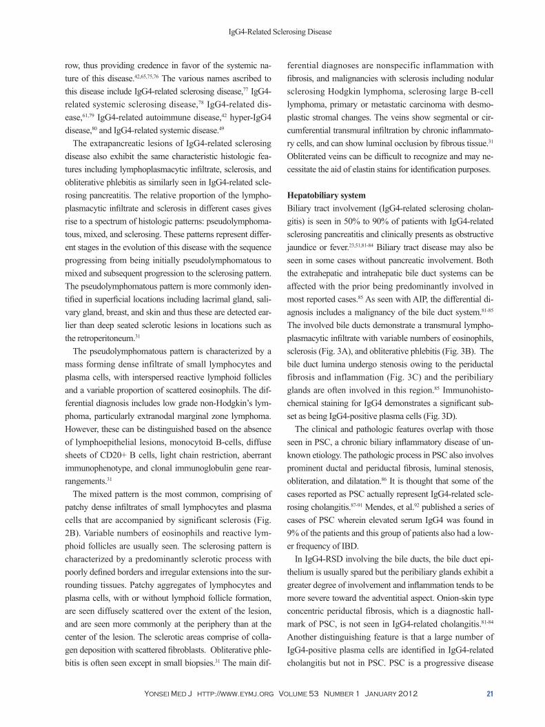
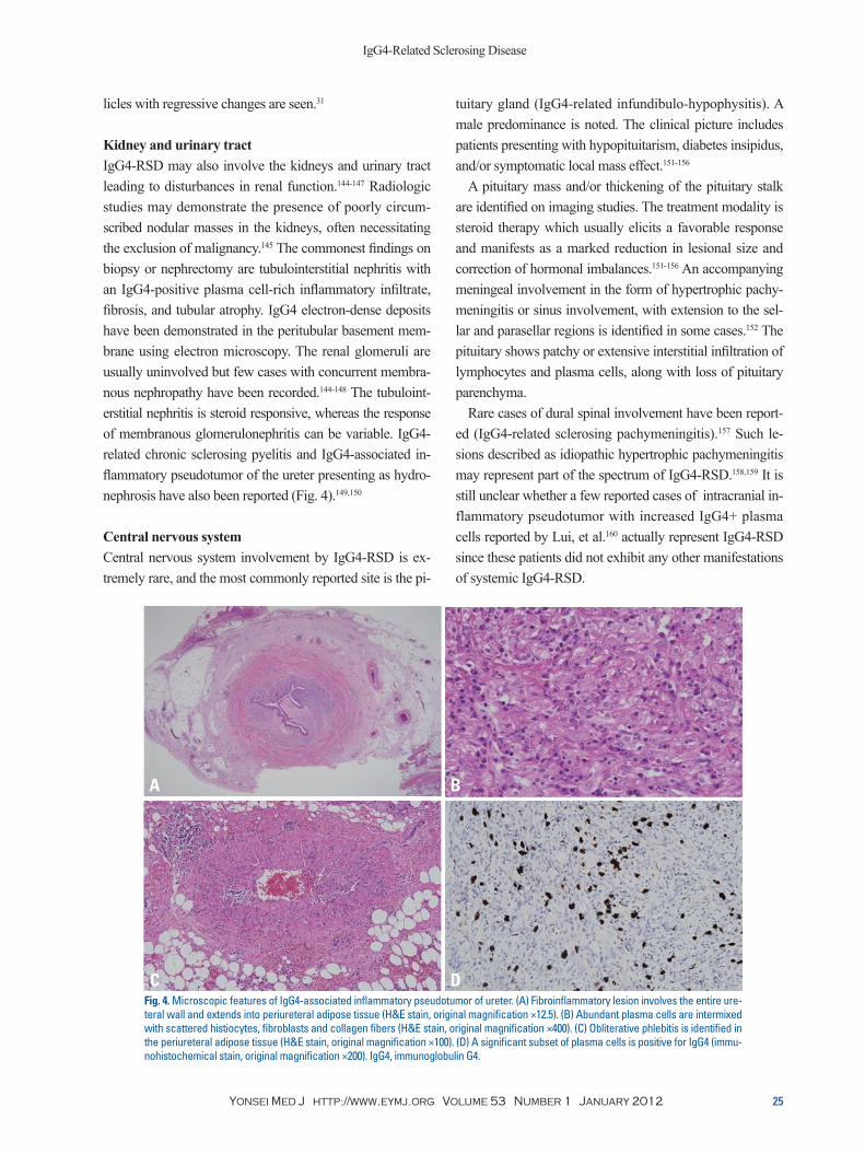
Kidney and urinary tractIgG4-RSD may also involve the kidneys and urinary tract leading to disturbances in renal function.144-147 Radiologic studies may demonstrate the presence of poorly circum-scribed nodular masses in the kidneys, often necessitating the exclusion of malignancy.145 The commonest findings on biopsy or nephrectomy are tubulointerstitial nephritis with an IgG4-positive plasma cell-rich inflammatory infiltrate, fibrosis, and tubular atrophy. IgG4 electron-dense deposits have been demonstrated in the peritubular basement mem-brane using electron microscopy. The renal glomeruli are usually uninvolved but few cases with concurrent membra-nous nephropathy have been recorded.144-148 The tubuloint-erstitial nephritis is steroid responsive, whereas the response of membranous glomerulonephritis can be variable. IgG4-related chronic sclerosing pyelitis and IgG4-associated in-flammatory pseudotumor of the ureter presenting as hydro-nephrosis have also been reported (Fig. 4).149,150
Central nervous systemCentral nervous system involvement by IgG4-RSD is ex-tremely rare, and the most commonly reported site is the pi-
Fig. 4. Microscopic features of IgG4-associated inflammatory pseudotumor of ureter. (A) Fibroinflammatory lesion involves the entire ure-teral wall and extends into periureteral adipose tissue (H&E stain, original magnification ×12.5). (B) Abundant plasma cells are intermixed with scattered histiocytes, fibroblasts and collagen fibers (H&E stain, original magnification ×400). (C) Obliterative phlebitis is identified in the periureteral adipose tissue (H&E stain, original magnification ×100). (D) A significant subset of plasma cells is positive for IgG4 (immu-nohistochemical stain, original magnification ×200). IgG4, immunoglobulin G4.
A
C
B
D
Mukul Divatia, et al.
Yonsei Med J http://www.eymj.org Volume 53 Number 1 January 201226
be multifocal with the commonest nodes involved being mediastinal, abdominal, and axillary lymph nodes. Clinical manifestations vary greatly.66,161,172 Enlarged asymptomatic lymph nodes are identified in surgical resection specimens or identified on imaging studies of patients with known IgG4-RSD. Lymphadenopathy may also develop following an established diagnosis of extranodal IgG4-RSD or may be the initial presenting manifestation with subsequent discov-ery of systemic IgG4-RSD. The enlarged lymph nodes may be asymptomatic or may give rise to a mass effect in vari-ous body sites.
The differential diagnosis in these cases with lymphade-nopathy includes lymphoma, multicentric Castleman dis-ease, or metastatic malignant tumor. The lymph nodes are usually not exceedingly enlarged (≤2 cm) and not associat-ed with fever and weight loss. Serum lactate dehydrogenase level is normal or only slightly elevated. Other laboratory findings include raised serum IgG4, serum IgG, polyclonal hypergammaglobulinemia and elevated erythrocyte sedi-mentation rate (ESR).66,161
The morphologic features of IgG4-related lymphadenopa-thy are different from those of extranodal sites since there is usually no sclerosis or phlebitis. Five histologic patterns of IgG4-related lymphadenopathy have been described.66,79,161
Skin and other miscellaneous body sitesSkin involvement in IgG4-RSD may be either isolated or as part of systemic IgG4-RSD.68,161 The clinical presentation is in the form of cutaneous plaques or nodules tending to in-volve the head and neck. Microscopic examination reveals a dermal and subcutaneous nodular lymphoplasmacytic in-filtrate with formation of lymphoid follicles and dermal or subcutaneous sclerosis. A markedly increased number of IgG4+ plasma cells, small lymphocytes, and variable infil-trate of eosinophils is identified.31
Cases of IgG4-RSD have been reported in the pericardi-um,162 prostate,163 seminal vesicles,164 nose, paranasal sinus-es165,166 and uterine cervix (idiopathic cervical fibrosis).167 Questionable entities currently include autoimmune esoph-agitis and splenic sclerosing angiomatoid nodular transfor-mation, which exhibit some of the features overlapping with IgG4-RSD, including increased plasma cells and sclero-sis.168-171 However, there is no definite evidence to currently include these lesions under the spectrum of IgG4-RSD.
Lymph nodesLymphadenopathy is identified in up to 80% of patients with autoimmune (IgG4-related sclerosing) pancreatitis as evi-denced by imaging studies.62 Lymph node involvement may
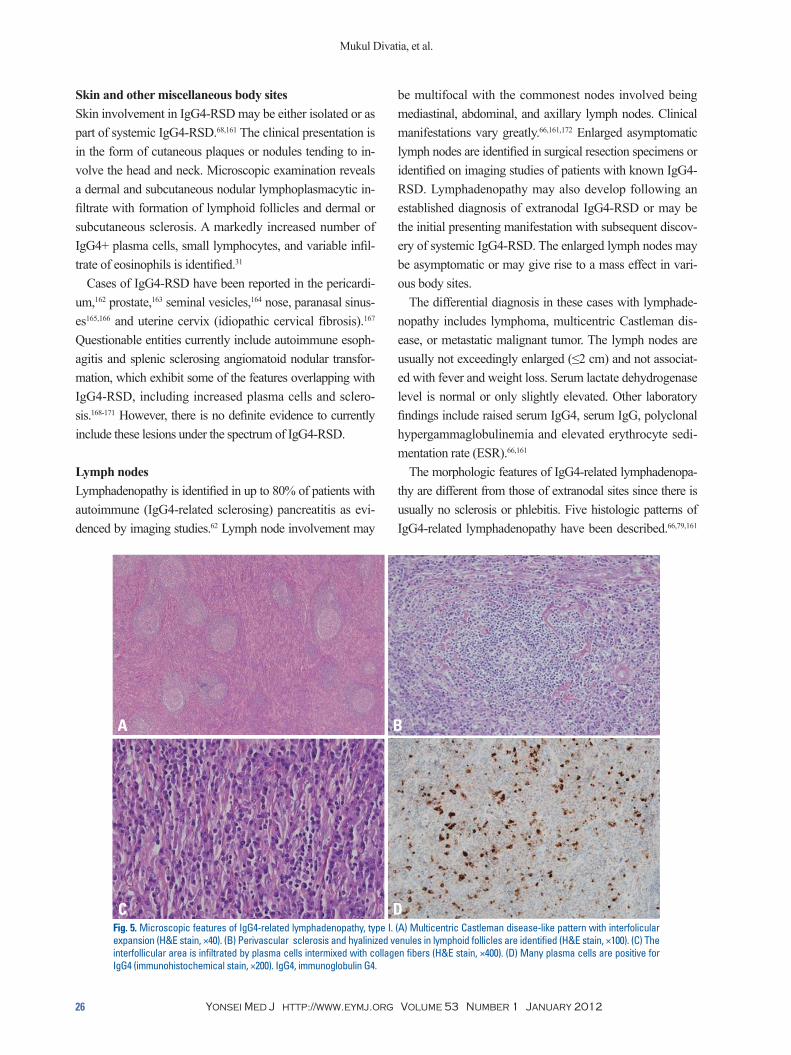
Fig. 5. Microscopic features of IgG4-related lymphadenopathy, type I. (A) Multicentric Castleman disease-like pattern with interfolicular expansion (H&E stain, ×40). (B) Perivascular sclerosis and hyalinized venules in lymphoid follicles are identified (H&E stain, ×100). (C) The interfollicular area is infiltrated by plasma cells intermixed with collagen fibers (H&E stain, ×400). (D) Many plasma cells are positive for IgG4 (immunohistochemical stain, ×200). IgG4, immunoglobulin G4.
A
C
B
D
IgG4-Related Sclerosing Disease
Yonsei Med J http://www.eymj.org Volume 53 Number 1 January 2012 27
mune disease.189 An association of IgG4-related AIP with gastric ulcer and Helicobacter pylori (H. pylori) infection has been proposed.190-192 It has been theorized by Okazaki, et al.193 that the development of the acronym ‘AIP’ involves a biphasic mechanism in which the initial response to self-anti-gens such as carbonic anhydrase, lactoferrin, pancreatic se-cretory trypsin inhibitor and ά-fodrin and molecular mimicry (H. pylori) are induced by decreased naive regulatory T cells, in conjunction with T-helper 1 cells releasing proinflammato-ry cytokines. Progression is mediated through increased memory regulatory T cell and T-helper 2 cell immune re-sponses resulting in chronic inflammation. The inflammatory infiltrate usually comprises of a mixed population of T and B cells, with increased CD4+, CD25+, FOXP3+ regulatory T cells.31,194,195 T-helper cell 2 cytokines (interleukin-4, interleu-kin-5, and interleukin-13) and regulatory cytokines (interleu-kin-10 and transforming growth factor) are upregulated in the involved tissues.194-196 Other proposed hypothesis regard-ing the pathogenesis include enhanced T-helper type 2 re-sponses to intestinal microflora197 and an immune-mediated pathogenesis based on the ultrastructural finding of electron dense immune complex deposits along the basement mem-branes of pancreatic acini and renal tubules.32
According to the results of a recent study conducted by screening of pooled IgG from patients with AIP, a specific reactivity with a peptide demonstrating homology with plasminogen-binding protein of H. pylori and with ubiqui-tin-protein ligase E3 component n-recognin 2 (an enzyme with high levels of expression in pancreatic acinar cells) was noted in the serum samples from 90% of patients with AIP and 10% of patients with pancreatic cancer. Healthy con-trols did not show any expression.198 It remains to be seen whether H. pylori truly plays a role in the development of AIP and IgG4-RSD.
DIAGNOSTIC CRITERIA FOR IgG4-RELATED SCLEROSING DISEASE
Diagnosis of IgG4-RSD requires not only an increase in the absolute number of IgG4+ cells but also an increased IgG4+/IgG+ ratio for the correct diagnosis. An absolute IgG4+ plas-ma cell count is not sufficient as a sole index as IgG4+ cells normally comprise approximately 5% of all IgG+ cells, and may be present in significant numbers in any inflammatory lesions with increased plasma cells even though the percent-age might remain in the acceptable range. A high IgG4+/
The various subtypes are as follows: multicentric Castle-man disease-like pattern (type I),172-178 follicular hyperplasia pattern (type II),66 interfollicular plasmacytosis and immu-noblastosis pattern (type III),66,79,161 progressive transforma-tion of germinal center-like pattern (type IV),79,179 and nodal inflammatory pseudotumor-like pattern (type V).79,179,180 Abundant IgG4+ polyclonal plasma cells are present in in-terfollicular and follicle center compartments in all of these patterns (Fig. 5).79
IgG4-RELATED SCLEROSING DISEASE AND ASSOCIATED
MALIGNANCIES
Sato, et al.79 have reported cases of ocular marginal zone lymphoma associated with ocular adnexal IgG4-related sclerosing disease. A few cases of localized non-Hodgkin’s lymphoma (marginal zone lymphoma and follicular lympho-ma) arising in a background of IgG4-related chronic scle-rosing dacryoadenitis have been recently reported.181 A rare example of composite marginal zone lymphoma and classi-cal Hodgkin lymphoma associated with IgG4-related scle-rosing disease of the cervical soft tissue has also been pub-lished.167 A recent study from the Mayo Clinic cites 3 cases of lymphoma (2 large B-cell lymphomas and 1 B-cell lym-phoma unspecified) discovered on follow-up of 111 patients with IgG4-related sclerosing disease, suggesting that the syndrome increases the risk of developing lymphoma in various body sites.182
Several cases of pancreatic ductal adenocarcinoma have been published in association with IgG4-RSD.183-186 Case reports of pulmonary adenocarcinoma, salivary duct carci-noma, urothelial carcinoma in situ and gastrointestinal clear cell sarcoma in association with IgG4-RSD have been re-ported in the lung, parotid gland, ureter and small intestine, respectively.140,150,187,188 Further studies are mandated in this field to determine if there is any causal relationship be-tween the malignancies and IgG4-RSD.
PROPOSED ETIOPATHOGENESIS OF IgG4-RELATED SCLEROSING DISEASE
The pathogenesis of IgG4-related sclerosing disease is still not clearly understood. The disease may represent a hyper-sensitive/allergic reaction as compared to being an autoim-
Mukul Divatia, et al.
Yonsei Med J http://www.eymj.org Volume 53 Number 1 January 201228
REFERENCES
1. Nirula A, Glaser SM, Kalled SL, Taylor FR. What is IgG4? A re-view of the biology of a unique immunoglobulin subtype. Curr Opin Rheumatol 2011;23:119-24.
2. Meulenbroek AJ, Zeijlemaker WP. Human IgG Subclasses: use-ful diagnostic markers for immunocompetence [online]. San-quin, Laboratory for Experimental and Clinical Immunology University of Amsterdam, the Netherlands; 1996 (http://www.xs4all.nl/_ednieuw/IgGsubclasses/subkl.htm).
3. Aucouturier P, Danon F, Daveau M, Guillou B, Sabbah A, Bes-son J, et al. Measurement of serum IgG4 levels by a competitive immunoenzymatic assay with monoclonal antibodies. J Immunol Methods 1984;74:151-62.
4. French MA. Serum IgG subclasses in normal adults. Monogr Al-lergy 1986;19:100-7.
5. Aalberse RC, Stapel SO, Schuurman J, Rispens T. Immunoglob-ulin G4: an odd antibody. Clin Exp Allergy 2009;39:469-77.
6. Tao MH, Smith RI, Morrison SL. Structural features of human immunoglobulin G that determine isotype-specific differences in complement activation. J Exp Med 1993;178:661-7.
7. Brekke OH, Michaelsen TE, Aase A, Sandin RH, Sandlie I. Hu-man IgG isotype-specific amino acid residues affecting comple-ment-mediated cell lysis and phagocytosis. Eur J Immunol 1994;24:2542-7.
8. Jeannin P, Lecoanet S, Delneste Y, Gauchat JF, Bonnefoy JY. IgE versus IgG4 production can be differentially regulated by IL-10. J Immunol 1998;160:3555-61.
9. Horner AA, Widhopf GF, Burger JA, Takabayashi K, Cinman N, Ronaghy A, et al. Immunostimulatory DNA inhibits IL-4-depen-dent IgE synthesis by human B cells. J Allergy Clin Immunol 2001;108:417-23.
10. Jones CC, Hamilton RG, Jordon RE. Subclass distribution of hu-man IgG autoantibodies in pemphigus. J Clin Immunol 1988;8: 43-9.
11. Futei Y, Amagai M, Ishii K, Kuroda-Kinoshita K, Ohya K, Ni-shikawa T. Predominant IgG4 subclass in autoantibodies of pem-phigus vulgaris and foliaceus. J Dermatol Sci 2001;26:55-61.
12. Hawrylowicz CM, O’Garra A. Potential role of interleukin-10-secreting regulatory T cells in allergy and asthma. Nat Rev Im-munol 2005;5:271-83.
13. Sarles H, Sarles JC, Muratore R, Guien C. Chronic inflammatory sclerosis of the pancreas--an autonomous pancreatic disease? Am J Dig Dis 1961;6:688-98.
14. Yoshida K, Toki F, Takeuchi T, Watanabe S, Shiratori K, Hayashi N. Chronic pancreatitis caused by an autoimmune abnormality. Proposal of the concept of autoimmune pancreatitis. Dig Dis Sci 1995;40:1561-8.
15. Okazaki K, Chiba T. Autoimmune related pancreatitis. Gut 2002;51:1-4.
16. Zamboni G, Lüttges J, Capelli P, Frulloni L, Cavallini G, Peder-zoli P, et al. Histopathological features of diagnostic and clinical relevance in autoimmune pancreatitis: a study on 53 resection specimens and 9 biopsy specimens. Virchows Arch 2004;445: 552-63.
17. Aparisi L, Farre A, Gomez-Cambronero L, Martinez J, De Las Heras G, Corts J, et al. Antibodies to carbonic anhydrase and IgG4 levels in idiopathic chronic pancreatitis: relevance for diag-nosis of autoimmune pancreatitis. Gut 2005;54:703-9.
IgG+ percentage alone is also not sufficient for a diagnosis because in cases with a few plasma cells, an erroneously high ratio may be calculated owing to the relative proportions of both IgG and IgG4+ plasma cells. We are in agreement with other investigators with regard to the cut-offs for the absolute number of IgG4+ cells >50/high-power fields (HPF) and ratio of IgG4+/IgG+ cells >40%.31 We also pro-pose selecting areas with the maximum density of IgG4+ plasma cells. At least three HPFs should be counted, and the average number of IgG4+ plasma cells per HPF should be calculated to give the results. In borderline cases, a de-scriptive diagnosis with a comment indicating “increased IgG4+ plasma cells” should be rendered along with a dis-claimer for the clinician requesting additional work-up or follow-up to determine if the lesion indeed represents IgG4-RSD.31 A raised serum IgG4 level is not mandatory for the diagnosis but may be of valuable assistance. Serum IgG4 titer often correlates with disease activity and the number of involved organs, but levels may also be normal.40 It is note-worthy that increased serum IgG4 has also been reported in a host of other diseases, including atopic dermatitis, parasit-ic infections, pemphigus vulgaris, pemphigus foliaceus, and pancreatic carcinoma.
Not every entity with increased IgG4+ plasma cells and a high IgG4/IgG ratio can be accepted as belonging to the spectrum of IgG4-RSD. Requirements such as concomitant morphologic features and an appropriate clinical context are mandatory for diagnosis. Autoimmune diseases include cases with an increased proportion of IgG4+ plasma cells but this does not qualify for inclusion as IgG4-RSD because the morphologic features and clinical findings do not corre-spond with the diagnostic criteria.
CONCLUSION In the past decade, significant progress has been made in our understanding of the genetic and immunologic triggers contributing to IgG4-RSD in addition to the discovery of the vast clinicopathologic spectrum of this entity. Our knowledge is currently evolving as newer developments come to light revealing a widely expanding histopathologic spectrum in this disease entity. Further studies are neces-sary to delineate the precise role played by IgG4 in the pathogenesis of this disease group in order to make mean-ingful contributions in the management of patients with this disease.
IgG4-Related Sclerosing Disease
Yonsei Med J http://www.eymj.org Volume 53 Number 1 January 2012 29
36. Otsuki M, Chung JB, Okazaki K, Kim MH, Kamisawa T, Kawa S, et al. Asian diagnostic criteria for autoimmune pancreatitis: consensus of the Japan-Korea Symposium on Autoimmune Pan-creatitis. J Gastroenterol 2008;43:403-8.
37. Chari ST, Smyrk TC, Levy MJ, Topazian MD, Takahashi N, Zhang L, et al. Diagnosis of autoimmune pancreatitis: the Mayo Clinic experience. Clin Gastroenterol Hepatol 2006;4:1010-6.
38. Frulloni L, Scattolini C, Falconi M, Zamboni G, Capelli P, Man-fredi R, et al. Autoimmune pancreatitis: differences between the focal and diffuse forms in 87 patients. Am J Gastroenterol 2009;104:2288-94.
39. Sugumar A, Klöppel G, Chari ST. Autoimmune pancreatitis: pathologic subtypes and their implications for its diagnosis. Am J Gastroenterol 2009;104:2308-10.
40. Hamano H, Kawa S, Horiuchi A, Unno H, Furuya N, Akamatsu T, et al. High serum IgG4 concentrations in patients with scleros-ing pancreatitis. N Engl J Med 2001;344:732-8.
41. Kamisawa T, Funata N, Hayashi Y, Tsuruta K, Okamoto A, Amemiya K, et al. Close relationship between autoimmune pan-creatitis and multifocal fibrosclerosis. Gut 2003;52:683-7.
42. Kamisawa T, Funata N, Hayashi Y, Eishi Y, Koike M, Tsuruta K, et al. A new clinicopathological entity of IgG4-related autoim-mune disease. J Gastroenterol 2003;38:982-4.
43. Sepehr A, Mino-Kenudson M, Ogawa F, Brugge WR, Deshpande V, Lauwers GY. IgG4+ to IgG+ plasma cells ratio of ampulla can help differentiate autoimmune pancreatitis from other “mass forming” pancreatic lesions. Am J Surg Pathol 2008;32:1770-9.
44. Kamisawa T, Yoshiike M, Egawa N, Nakajima H, Tsuruta K, Okamoto A, et al. Chronic pancreatitis in the elderly in Japan. Pancreatology 2004;4:223-7.
45. Kim KP, Kim MH, Song MH, Lee SS, Seo DW, Lee SK. Autoim-mune chronic pancreatitis. Am J Gastroenterol 2004;99:1605-16.
46. Nishimori I, Tamakoshi A, Otsuki M; Research Committee on Intractable Diseases of the Pancreas, Ministry of Health, Labour, and Welfare of Japan. Prevalence of autoimmune pancreatitis in Japan from a nationwide survey in 2002. J Gastroenterol 2007; 42:6-8.
47. Hardacre JM, Iacobuzio-Donahue CA, Sohn TA, Abraham SC, Yeo CJ, Lillemoe KD. Results of pancreaticoduodenectomy for lymphoplasmacytic sclerosing pancreatitis. Ann Surg 2003;237: 853-8.
48. Abraham SC, Wilentz RE, Yeo CJ, Sohn TA, Cameron JL, Boit-nott JK, et al. Pancreaticoduodenectomy (Whipple resections) in patients without malignancy: are they all ‘chronic pancreatitis’? Am J Surg Pathol 2003;27:110-20.
49. Khosroshahi A, Bloch DB, Deshpande V, Stone JH. Rituximab therapy leads to rapid decline of serum IgG4 levels and prompt clinical improvement in IgG4-related systemic disease. Arthritis Rheum 2010;62:1755-62.
50. Suda K, Nishimori I, Takase M, Oi I, Ogawa M. Autoimmune pancreatitis can be classified into early and advanced stages. Pancreas 2006;33:345-50.
51. Zen Y, Harada K, Sasaki M, Sato Y, Tsuneyama K, Haratake J, et al. IgG4-related sclerosing cholangitis with and without hepatic inflammatory pseudotumor, and sclerosing pancreatitis-associat-ed sclerosing cholangitis: do they belong to a spectrum of scle-rosing pancreatitis? Am J Surg Pathol 2004;28:1193-203.
52. Ito T, Nishimori I, Inoue N, Kawabe K, Gibo J, Arita Y, et al. Treatment for autoimmune pancreatitis: consensus on the treat-ment for patients with autoimmune pancreatitis in Japan. J Gas-
18. Okazaki K, Uchida K, Ohana M, Nakase H, Uose S, Inai M, et al. Autoimmune-related pancreatitis is associated with autoanti-bodies and a Th1/Th2-type cellular immune response. Gastroen-terology 2000;118:573-81.
19. Deshpande V, Mino-Kenudson M, Brugge W, Lauwers GY. Au-toimmune pancreatitis: more than just a pancreatic disease? A contemporary review of its pathology. Arch Pathol Lab Med 2005;129:1148-54.
20. Kamisawa T, Okamoto A. Autoimmune pancreatitis: proposal of IgG4-related sclerosing disease. J Gastroenterol 2006;41:613-25.
21. Kawa S, Ota M, Yoshizawa K, Horiuchi A, Hamano H, Ochi Y, et al. HLA DRB10405-DQB10401 haplotype is associated with autoimmune pancreatitis in the Japanese population. Gastroen-terology 2002;122:1264-9.
22. Sood S, Fossard DP, Shorrock K. Chronic sclerosing pancreatitis in Sjögren’s syndrome: a case report. Pancreas 1995;10:419-21.
23. Kawaguchi K, Koike M, Tsuruta K, Okamoto A, Tabata I, Fujita N. Lymphoplasmacytic sclerosing pancreatitis with cholangitis: a variant of primary sclerosing cholangitis extensively involving pancreas. Hum Pathol 1991;22:387-95.
24. Ectors N, Maillet B, Aerts R, Geboes K, Donner A, Borchard F, et al. Non-alcoholic duct destructive chronic pancreatitis. Gut 1997;41:263-8.
25. Yadav D, Notahara K, Smyrk TC, Clain JE, Pearson RK, Farnell MB, et al. Idiopathic tumefactive chronic pancreatitis: clinical profile, histology, and natural history after resection. Clin Gastro-enterol Hepatol 2003;1:129-35.
26. Wakabayashi T, Kawaura Y, Satomura Y, Watanabe H, Motoo Y, Sawabu N. Long-term prognosis of duct-narrowing chronic pan-creatitis: strategy for steroid treatment. Pancreas 2005;30:31-9.
27. Notohara K, Burgart LJ, Yadav D, Chari S, Smyrk TC. Idiopath-ic chronic pancreatitis with periductal lymphoplasmacytic infil-tration: clinicopathologic features of 35 cases. Am J Surg Pathol 2003;27:1119-27.
28. Sah RP, Chari ST, Pannala R, Sugumar A, Clain JE, Levy MJ, et al. Differences in clinical profile and relapse rate of type 1 versus type 2 autoimmune pancreatitis. Gastroenterology 2010; 139:140-8.
29. Kamisawa T, Funata N, Hayashi Y. Lymphoplasmacytic scleros-ing pancreatitis is a pancreatic lesion of IgG4-related systemic disease. Am J Surg Pathol 2004;28:1114.
30. Zhang L, Notohara K, Levy MJ, Chari ST, Smyrk TC. IgG4-positive plasma cell infiltration in the diagnosis of autoimmune pancreatitis. Mod Pathol 2007;20:23-8.
31. Cheuk W, Chan JK. IgG4-related sclerosing disease: a critical appraisal of an evolving clinicopathologic entity. Adv Anat Pathol 2010;17:303-32.
32. Deshpande V, Chicano S, Finkelberg D, Selig MK, Mino-Ke-nudson M, Brugge WR, et al. Autoimmune pancreatitis: a sys-temic immune complex mediated disease. Am J Surg Pathol 2006;30:1537-45.
33. Mino-Kenudson M, Smyrk TC, Deshpande V, Fujisawa M, Shi-mizu M, Uehara T, et al. Autoimmune pancreatitis: West vs. East. Mod Pathol 2008;21(Suppl 1):312A.
34. Okazaki K, Kawa S, Kamisawa T, Naruse S, Tanaka S, Nishi-mori I, et al. Clinical diagnostic criteria of autoimmune pancre-atitis: revised proposal. J Gastroenterol 2006;41:626-31.
35. Kim KP, Kim MH, Kim JC, Lee SS, Seo DW, Lee SK. Diagnos-tic criteria for autoimmune chronic pancreatitis revisited. World J Gastroenterol 2006;12:2487-96.
Mukul Divatia, et al.
Yonsei Med J http://www.eymj.org Volume 53 Number 1 January 201230
suda S, et al. IgG4-positive plasma cells in inflammatory pseu-dotumor (plasma cell granuloma) of the lung. Hum Pathol 2005;36:710-7.
70. Cheuk W, Chan AC, Lam WL, Chow SM, Crowley P, Lloydd R, et al. IgG4-related sclerosing mastitis: description of a new mem-ber of the IgG4-related sclerosing diseases. Am J Surg Pathol 2009;33:1058-64.
71. Kasashima S, Zen Y, Kawashima A, Konishi K, Sasaki H, Endo M, et al. Inflammatory abdominal aortic aneurysm: close rela-tionship to IgG4-related periaortitis. Am J Surg Pathol 2008;32: 197-204.
72. Kakudo K, Li Y, Hirokawa M, Ozaki T. Diagnosis of Hashimo-to’s thyroiditis and IgG4-related sclerosing disease. Pathol Int 2011;61:175-83.
73. Okazaki K, Uchida K, Chiba T. Recent concept of autoimmune-related pancreatitis. J Gastroenterol 2001;36:293-302.
74. Lara LP, Chari ST. Autoimmune pancreatitis. Curr Gastroenterol Rep 2005;7:101-6.
75. Saeki T, Saito A, Hiura T, Yamazaki H, Emura I, Ueno M, et al. Lymphoplasmacytic infiltration of multiple organs with immu-noreactivity for IgG4: IgG4-related systemic disease. Intern Med 2006;45:163-7.
76. Shinji A, Sano K, Hamano H, Unno H, Fukushima M, Nakamu-ra N, et al. Autoimmune pancreatitis is closely associated with gastric ulcer presenting with abundant IgG4-bearing plasma cell infiltration. Gastrointest Endosc 2004;59:506-11.
77. Kamisawa T. IgG4-related sclerosing disease. Intern Med 2006;45:125-6.
78. Bateman AC, Deheragoda MG. IgG4-related systemic sclerosing disease - an emerging and under-diagnosed condition. Histopa-thology 2009;55:373-83.
79. Sato Y, Notohara K, Kojima M, Takata K, Masaki Y, Yoshino T. IgG4-related disease: historical overview and pathology of he-matological disorders. Pathol Int 2010;60:247-58.
80. Neild GH, Rodriguez-Justo M, Wall C, Connolly JO. Hyper-IgG4 disease: report and characterisation of a new disease. BMC Med 2006;4:23.
81. Deshpande V, Sainani NI, Chung RT, Pratt DS, Mentha G, Rub-bia-Brandt L, et al. IgG4-associated cholangitis: a comparative histological and immunophenotypic study with primary scleros-ing cholangitis on liver biopsy material. Mod Pathol 2009;22: 1287-95.
82. Ghazale A, Chari ST, Zhang L, Smyrk TC, Takahashi N, Levy MJ, et al. Immunoglobulin G4-associated cholangitis: clinical profile and response to therapy. Gastroenterology 2008;134:706-15.
83. Kamisawa T, Nakajima H, Egawa N, Funata N, Tsuruta K, Oka-moto A. IgG4-related sclerosing disease incorporating sclerosing pancreatitis, cholangitis, sialadenitis and retroperitoneal fibrosis with lymphadenopathy. Pancreatology 2006;6:132-7.
84. Nakazawa T, Ohara H, Sano H, Ando T, Aoki S, Kobayashi S, et al. Clinical differences between primary sclerosing cholangitis and sclerosing cholangitis with autoimmune pancreatitis. Pancre-as 2005;30:20-5.
85. Nakanuma Y, Zen Y. Pathology and immunopathology of immu-noglobulin G4-related sclerosing cholangitis: the latest addition to the sclerosing cholangitis family. Hepatol Res 2007;37:S478-86.
86. Nakanuma Y, Harada K, Katayanagi K, Tsuneyama K, Sasaki M. Definition and pathology of primary sclerosing cholangitis. J Hepatobiliary Pancreat Surg 1999;6:333-42.
troenterol 2007;42:50-8. 53. Dhall D, Suriawinata AA, Tang LH, Shia J, Klimstra DS. Use of
immunohistochemistry for IgG4 in the distinction of autoim-mune pancreatitis from peritumoral pancreatitis. Hum Pathol 2010;41:643-52.
54. Kamisawa T, Okamoto A, Funata N. Clinicopathological fea-tures of autoimmune pancreatitis in relation to elevation of serum IgG4. Pancreas 2005;31:28-31.
55. Raina A, Krasinskas AM, Greer JB, Lamb J, Fink E, Moser AJ, et al. Serum immunoglobulin G fraction 4 levels in pancreatic cancer: elevations not associated with autoimmune pancreatitis. Arch Pathol Lab Med 2008;132:48-53.
56. Ghazale A, Chari ST, Smyrk TC, Levy MJ, Topazian MD, Taka-hashi N, et al. Value of serum IgG4 in the diagnosis of autoim-mune pancreatitis and in distinguishing it from pancreatic cancer. Am J Gastroenterol 2007;102:1646-53.
57. Kitagawa S, Zen Y, Harada K, Sasaki M, Sato Y, Minato H, et al. Abundant IgG4-positive plasma cell infiltration characterizes chronic sclerosing sialadenitis (Küttner’s tumor). Am J Surg Pathol 2005;29:783-91.
58. Wang WL, Farris AB, Lauwers GY, Deshpande V. Autoimmune pancreatitis-related cholecystitis: a morphologically and immu-nologically distinctive form of lymphoplasmacytic sclerosing cholecystitis. Histopathology 2009;54:829-36.
59. Chandan VS, Iacobuzio-Donahue C, Abraham SC. Patchy distri-bution of pathologic abnormalities in autoimmune pancreatitis: implications for preoperative diagnosis. Am J Surg Pathol 2008;32:1762-9.
60. Moon SH, Kim MH, Park do H, Song TJ, Eum J, Lee SS, et al. IgG4 immunostaining of duodenal papillary biopsy specimens may be useful for supporting a diagnosis of autoimmune pancre-atitis. Gastrointest Endosc 2010;71:960-6.
61. Geyer JT, Ferry JA, Harris NL, Stone JH, Zukerberg LR, Lauw-ers GY, et al. Chronic sclerosing sialadenitis (Küttner tumor) is an IgG4-associated disease. Am J Surg Pathol 2010;34:202-10.
62. Hamano H, Arakura N, Muraki T, Ozaki Y, Kiyosawa K, Kawa S. Prevalence and distribution of extrapancreatic lesions compli-cating autoimmune pancreatitis. J Gastroenterol 2006;41:1197-205.
63. Kitagawa S, Zen Y, Harada K, Sasaki M, Sato Y, Minato H, et al. Abundant IgG4-positive plasma cell infiltration characterizes chronic sclerosing sialadenitis (Küttner’s tumor). Am J Surg Pathol 2005;29:783-91.
64. Zen Y, Nakanuma Y. IgG4-related disease: a cross-sectional study of 114 cases. Am J Surg Pathol 2010;34:1812-9.
65. Cheuk W, Yuen HK, Chan JK. Chronic sclerosing dacryoadeni-tis: part of the spectrum of IgG4-related Sclerosing disease? Am J Surg Pathol 2007;31:643-5.
66. Cheuk W, Yuen HK, Chu SY, Chiu EK, Lam LK, Chan JK. Lymphadenopathy of IgG4-related sclerosing disease. Am J Surg Pathol 2008;32:671-81.
67. Zen Y, Sawazaki A, Miyayama S, Notsumata K, Tanaka N, Na-kanuma Y. A case of retroperitoneal and mediastinal fibrosis ex-hibiting elevated levels of IgG4 in the absence of sclerosing pan-creatitis (autoimmune pancreatitis). Hum Pathol 2006;37:239-43.
68. Cheuk W, Lee KC, Chong LY, Yuen ST, Chan JK. IgG4-related Sclerosing disease: a potential new etiology of cutaneous pseu-dolymphoma. Am J Surg Pathol 2009;33:1713-9.
69. Zen Y, Kitagawa S, Minato H, Kurumaya H, Katayanagi K, Ma-
IgG4-Related Sclerosing Disease
Yonsei Med J http://www.eymj.org Volume 53 Number 1 January 2012 31
dibular gland is mainly due to a T lymphocyte immune reaction. Mod Pathol 2002;15:845-52.
105. Cheuk W, Chan JK. Kuttner tumor of the submandibular gland: fine-needle aspiration cytologic findings of seven cases. Am J Clin Pathol 2002;117:103-8.
106. Kamisawa T, Nakajima H, Hishima T. Close correlation between chronic sclerosing sialadenitis and immunoglobulin G4. Intern Med J 2006;36:527-9.
107. Chan JK. Kuttner tumor (chronic sclerosing sialadenitis) of the submandibular gland: an underrecognized entity. Adv Anat Pathol 1998;5:239-51.
108. Yamamoto M, Takahashi H, Sugai S, Imai K. Clinical and patho-logical characteristics of Mikulicz’s disease (IgG4-related plas-macytic exocrinopathy). Autoimmun Rev 2005;4:195-200.
109. Masaki Y, Dong L, Kurose N, Kitagawa K, Morikawa Y, Yama-moto M, et al. Proposal for a new clinical entity, IgG4-positive multiorgan lymphoproliferative syndrome: analysis of 64 cases of IgG4-related disorders. Ann Rheum Dis 2009;68:1310-5.
110. Yamamoto M, Takahashi H, Ohara M, Suzuki C, Naishiro Y, Yamamoto H, et al. A new conceptualization for Mikulicz’s dis-ease as an IgG4-related plasmacytic disease. Mod Rheumatol 2006;16:335-40.
111. Yamamoto H, Yamaguchi H, Aishima S, Oda Y, Kohashi K, Os-hiro Y, et al. Inflammatory myofibroblastic tumor versus IgG4-related sclerosing disease and inflammatory pseudotumor: a comparative clinicopathologic study. Am J Surg Pathol 2009;33: 1330-40.
112. Comings DE, Skubi KB, Van Eyes J, Motulsky AG. Familial multifocal fibrosclerosis. Findings suggesting that retroperitoneal fibrosis, mediastinal fibrosis, sclerosing cholangitis, Riedel’s thy-roiditis, and pseudotumor of the orbit may be different manifes-tations of a single disease. Ann Intern Med 1967;66:884-92.
113. Klisnick A, Fourcade J, Ruivard M, Baud O, Souweine B, Boyer L, et al. Combined idiopathic retroperitoneal and mediastinal fi-brosis with pericardial involvement. Clin Nephrol 1999;52:51-5.
114. Graal MB, Lustermans FA. A patient with combined mediastinal, mesenteric and retroperitoneal fibrosis. Neth J Med 1994;44:214-9.
115. Owen K, Lane H, Jones MK. Multifocal fibrosclerosis: a case of thyroiditis and bilateral lacrimal gland involvement. Thyroid 2001;11:1187-90.
116. Morris WR, Haik BG, Osborn D, Fleming JC. Intraocular in-volvement in multifocal fibrosclerosis. Ophthalmology 2000; 107:962-6.
117. Levey JM, Mathai J. Diffuse pancreatic fibrosis: an uncommon feature of multifocal idiopathic fibrosclerosis. Am J Gastroenter-ol 1998;93:640-2.
118. Fukuda W, Kimura M, Akaogi T, Sako M, Ohiwa K, Yamamoto Y, et al. Multifocal fibrosclerosis: retroperitoneal fibrosis associ-ated with a suprasellar tumor and pachymeningitis. Intern Med 2003;42:1006-10.
119. Pacini D, Leone O, Turci S, Camurri N, Giunchi F, Martinelli GN, et al. Incidence, etiology, histologic findings, and course of thoracic inflammatory aortopathies. Ann Thorac Surg 2008;86: 1518-23.
120. Miller DV, Isotalo PA, Weyand CM, Edwards WD, Aubry MC, Tazelaar HD. Surgical pathology of noninfectious ascending aor-titis: a study of 45 cases with emphasis on an isolated variant. Am J Surg Pathol 2006;30:1150-8.
121. Burke AP, Tavora F, Narula N, Tomaszewski JE, Virmani R. Aortitis and ascending aortic aneurysm: description of 52 cases
87. Oseini AM, Chaiteerakij R, Shire AM, Ghazale A, Kaiya J, Mos-er CD, et al. Utility of serum immunoglobulin G4 in distinguish-ing immunoglobulin G4-associated cholangitis from cholangio-carcinoma. Hepatology 2011 Jun 14. doi: 10.1002/hep.24487. [Epub ahead of print]
88. Erkelens GW, Vleggaar FP, Lesterhuis W, van Buuren HR, van der Werf SD. Sclerosing pancreato-cholangitis responsive to ste-roid therapy. Lancet 1999;354:43-4.
89. Nakazawa T, Ohara H, Yamada T, Ando H, Sano H, Kajino S, et al. Atypical primary sclerosing cholangitis cases associated with unusual pancreatitis. Hepatogastroenterology 2001;48:625-30.
90. Kojima E, Kimura K, Noda Y, Kobayashi G, Itoh K, Fujita N. Autoimmune pancreatitis and multiple bile duct strictures treated effectively with steroid. J Gastroenterol 2003;38:603-7.
91. Kuroiwa T, Suda T, Takahashi T, Hirono H, Natsui M, Motoya-ma H, et al. Bile duct involvement in a case of autoimmune pan-creatitis successfully treated with an oral steroid. Dig Dis Sci 2002;47:1810-6.
92. Mendes FD, Jorgensen R, Keach J, Katzmann JA, Smyrk T, Donlinger J, et al. Elevated serum IgG4 concentration in patients with primary sclerosing cholangitis. Am J Gastroenterol 2006; 101:2070-5.
93. Webster GJ, Pereira SP, Chapman RW. Autoimmune pancreatitis/IgG4-associated cholangitis and primary sclerosing cholangitis--overlapping or separate diseases? J Hepatol 2009;51:398-402.
94. Abraham SC, Cruz-Correa M, Argani P, Furth EE, Hruban RH, Boitnott JK. Lymphoplasmacytic chronic cholecystitis and bili-ary tract disease in patients with lymphoplasmacytic sclerosing pancreatitis. Am J Surg Pathol 2003;27:441-51.
95. Jessurun J, Bolio-Solis A, Manivel JC. Diffuse lymphoplasma-cytic acalculous cholecystitis: a distinctive form of chronic cho-lecystitis associated with primary sclerosing cholangitis. Hum Pathol 1998;29:512-7.
96. Zen Y, Fujii T, Sato Y, Masuda S, Nakanuma Y. Pathological classification of hepatic inflammatory pseudotumor with respect to IgG4-related disease. Mod Pathol 2007;20:884-94.
97. Kanno A, Satoh K, Kimura K, Masamune A, Asakura T, Unno M, et al. Autoimmune pancreatitis with hepatic inflammatory pseudotumor. Pancreas 2005;31:420-3.
98. Travis WD, Elisabeth B, Muller-Hermelink HK, Harris CC, edi-tors. Pathology and Genetics of Tumours of the Lung, Pleural, Thymus and Heart. Lyon: IARC Press; 2004.
99. Cheuk W, Chan JK, Shek TW, Chang JH, Tsou MH, Yuen NW, et al. Inflammatory pseudotumor-like follicular dendritic cell tu-mor: a distinctive low-grade malignant intra-abdominal neo-plasm with consistent Epstein-Barr virus association. Am J Surg Pathol 2001;25:721-31.
100. Umemura T, Zen Y, Hamano H, Kawa S, Nakanuma Y, Kiyosa-wa K. Immunoglobin G4-hepatopathy: association of immuno-globin G4-bearing plasma cells in liver with autoimmune pan-creatitis. Hepatology 2007;46:463-71.
101. Umemura T, Zen Y, Hamano H, Ichijo T, Kawa S, Nakanuma Y, et al. IgG4 associated autoimmune hepatitis: a differential diag-nosis for classical autoimmune hepatitis. Gut 2007;56:1471-2.
102. Kuttner H. Uber entzundiche Tumoren der Submaaxillar-. speicheldruse. Bruns Beitr Klin Chir 1896;15:815-34.
103. Pilch BZ. Head and Neck Surgical Pathology. Philadelphia: Lip-pincott Williams and Wilkin; 2001.
104. Tiemann M, Teymoortash A, Schrader C, Werner JA, Parwaresch R, Seifert G, et al. Chronic sclerosing sialadenitis of the subman-
Mukul Divatia, et al.
Yonsei Med J http://www.eymj.org Volume 53 Number 1 January 201232
Smyrk TC, et al. Distinctive pulmonary histopathology with in-creased IgG4-positive plasma cells in patients with autoimmune pancreatitis: report of 6 and 12 cases with similar histopathology. Am J Surg Pathol 2009;33:1450-62.
140. Zen Y, Inoue D, Kitao A, Onodera M, Abo H, Miyayama S, et al. IgG4-related lung and pleural disease: a clinicopathologic study of 21 cases. Am J Surg Pathol 2009;33:1886-93.
141. Yamashita K, Haga H, Kobashi Y, Miyagawa-Hayashino A, Yo-shizawa A, Manabe T. Lung involvement in IgG4-related lym-phoplasmacytic vasculitis and interstitial fibrosis: report of 3 cases and review of the literature. Am J Surg Pathol 2008;32:1620-6.
142. Kobayashi H, Shimokawaji T, Kanoh S, Motoyoshi K, Aida S. IgG4-positive pulmonary disease. J Thorac Imaging 2007;22: 360-2.
143. Zen Y, Kasahara Y, Horita K, Miyayama S, Miura S, Kitagawa S, et al. Inflammatory pseudotumor of the breast in a patient with a high serum IgG4 level: histologic similarity to sclerosing pancre-atitis. Am J Surg Pathol 2005;29:275-8.
144. Watson SJ, Jenkins DA, Bellamy CO. Nephropathy in IgG4-re-lated systemic disease. Am J Surg Pathol 2006;30:1472-7.
145. Cornell LD, Chicano SL, Deshpande V, Collins AB, Selig MK, Lauwers GY, et al. Pseudotumors due to IgG4 immune-complex tubulointerstitial nephritis associated with autoimmune pancre-atocentric disease. Am J Surg Pathol 2007;31:1586-97.
146. Dhobale S, Bedetti C, Killian P, Ilyas M, Liput J, Jasnosz K, et al. IgG4 related sclerosing disease with multiple organ involve-ments and response to corticosteroid treatment. J Clin Rheumatol 2009;15:354-7.
147. Takeda S, Haratake J, Kasai T, Takaeda C, Takazakura E. IgG4-associated idiopathic tubulointerstitial nephritis complicating au-toimmune pancreatitis. Nephrol Dial Transplant 2004;19:474-6.
148. Saeki T, Nishi S, Ito T, Yamazaki H, Miyamura S, Emura I, et al. Renal lesions in IgG4-related systemic disease. Intern Med 2007;46:1365-71.
149. Kuroda N, Nakamura S, Miyazaki K, Inoue K, Ohara M, Mizuno K, et al. Chronic sclerosing pyelitis with an increased number of IgG4-positive plasma cells. Med Mol Morphol 2009;42:236-8.
150. Kim SA, Lee SR, Huh J, Shen SS, Ro JY. IgG4-associated in-flammatory pseudotumor of ureter: clinicopathologic and immu-nohistochemical study of 3 cases. Hum Pathol 2011;42:1178-84.
151. Hori M, Makita N, Andoh T, Takiyama H, Yajima Y, Sakatani T, et al. Long-term clinical course of IgG4-related systemic disease accompanied by hypophysitis. Endocr J 2010;57:485-92.
152. Shimatsu A, Oki Y, Fujisawa I, Sano T. Pituitary and stalk lesions (infundibulo-hypophysitis) associated with immunoglobulin G4-related systemic disease: an emerging clinical entity. Endocr J 2009;56:1033-41.
153. Wong S, Lam WY, Wong WK, Lee KC. Hypophysitis presented as inflammatory pseudotumor in immunoglobulin G4-related systemic disease. Hum Pathol 2007;38:1720-3.
154. Osawa S, Ogawa Y, Watanabe M, Tominaga T. Hypophysitis presenting with atypical rapid deterioration: with special refer-ence to immunoglobulin G4-related disease-case report-. Neurol Med Chir (Tokyo) 2009;49:622-5.
155. Tanabe T, Tsushima K, Yasuo M, Urushihata K, Hanaoka M, Koizumi T, et al. IgG4-associated multifocal systemic fibrosis complicating sclerosing sialadenitis, hypophysitis, and retroperi-toneal fibrosis, but lacking pancreatic involvement. Intern Med 2006;45:1243-7.
156. Yamamoto M, Takahashi H, Ohara M, Suzuki C, Naishiro Y,
and proposal of a histologic classification. Hum Pathol 2008;39: 514-26.
122. Dehner LP, Coffin CM. Idiopathic fibrosclerotic disorders and other inflammatory pseudotumors. Semin Diagn Pathol 1998; 15:161-73.
123. Clark A, Zeman RK, Choyke PL, White EM, Burrell MI, Grant EG, et al. Pancreatic pseudotumors associated with multifocal idiopathic fibrosclerosis. Gastrointest Radiol 1988;13:30-2.
124. Hamano H, Kawa S, Ochi Y, Unno H, Shiba N, Wajiki M, et al. Hydronephrosis associated with retroperitoneal fibrosis and scle-rosing pancreatitis. Lancet 2002;359:1403-4.
125. Zen Y, Onodera M, Inoue D, Kitao A, Matsui O, Nohara T, et al. Retroperitoneal fibrosis: a clinicopathologic study with respect to immunoglobulin G4. Am J Surg Pathol 2009;33:1833-9.
126. Chen TS, Montgomery EA. Are tumefactive lesions classified as sclerosing mesenteritis a subset of IgG4-related sclerosing disor-ders? J Clin Pathol 2008;61:1093-7.
127. Matsumoto Y, Kasashima S, Kawashima A, Sasaki H, Endo M, Kawakami K, et al. A case of multiple immunoglobulin G4-relat-ed periarteritis: a tumorous lesion of the coronary artery and ab-dominal aortic aneurysm. Hum Pathol 2008;39:975-80.
128. Sakata N, Tashiro T, Uesugi N, Kawara T, Furuya K, Hirata Y, et al. IgG4-positive plasma cells in inflammatory abdominal aortic aneurysm: the possibility of an aortic manifestation of IgG4-re-lated sclerosing disease. Am J Surg Pathol 2008;32:553-9.
129. Qian Q, Kashani KB, Miller DV. Ruptured abdominal aortic an-eurysm related to IgG4 periaortitis. N Engl J Med 2009;361: 1121-3.
130. Ito H, Kaizaki Y, Noda Y, Fujii S, Yamamoto S. IgG4-related in-flammatory abdominal aortic aneurysm associated with autoim-mune pancreatitis. Pathol Int 2008;58:421-6.
131. Ishida M, Hotta M, Kushima R, Asai T, Okabe H. IgG4-related inflammatory aneurysm of the aortic arch. Pathol Int 2009;59: 269-73.
132. Stone JH, Khosroshahi A, Deshpande V, Stone JR. IgG4-related systemic disease accounts for a significant proportion of thoracic lymphoplasmacytic aortitis cases. Arthritis Care Res (Hoboken) 2010;62:316-22.
133. Komatsu K, Hamano H, Ochi Y, Takayama M, Muraki T, Yo-shizawa K, et al. High prevalence of hypothyroidism in patients with autoimmune pancreatitis. Dig Dis Sci 2005;50:1052-7.
134. Li Y, Bai Y, Liu Z, Ozaki T, Taniguchi E, Mori I, et al. Immuno-histochemistry of IgG4 can help subclassify Hashimoto’s auto-immune thyroiditis. Pathol Int 2009;59:636-41.
135. Li Y, Nishihara E, Hirokawa M, Taniguchi E, Miyauchi A, Kakudo K. Distinct clinical, serological, and sonographic char-acteristics of hashimoto’s thyroiditis based with and without IgG4-positive plasma cells. J Clin Endocrinol Metab 2010;95: 1309-17.
136. Drieskens O, Blockmans D, Van den Bruel A, Mortelmans L. Riedel’s thyroiditis and retroperitoneal fibrosis in multifocal fi-brosclerosis: positron emission tomographic findings. Clin Nucl Med 2002;27:413-5.
137. Dahlgren M, Khosroshahi A, Nielsen GP, Deshpande V, Stone JH. Riedel’s thyroiditis and multifocal fibrosclerosis are part of the IgG4-related systemic disease spectrum. Arthritis Care Res (Hoboken) 2010;62:1312-8.
138. Zhang L, Smyrk TC. Autoimmune pancreatitis and IgG4-related systemic diseases. Int J Clin Exp Pathol 2010;3:491-504.
139. Shrestha B, Sekiguchi H, Colby TV, Graziano P, Aubry MC,
IgG4-Related Sclerosing Disease
Yonsei Med J http://www.eymj.org Volume 53 Number 1 January 2012 33
Gouilleux-Gruart V, et al. Polyclonal IgG4 hypergammaglobu-linemia associated with plasmacytic lymphadenopathy, anemia and nephropathy. Ann Hematol 2006;85:833-40.
174. Kojima M, Nakamura N, Tsukamoto N, Otuski Y, Shimizu K, Itoh H, et al. Clinical implications of idiopathic multicentric cas-tleman disease among Japanese: a report of 28 cases. Int J Surg Pathol 2008;16:391-8.
175. Nishimoto N, Kanakura Y, Aozasa K, Johkoh T, Nakamura M, Nakano S, et al. Humanized anti-interleukin-6 receptor antibody treatment of multicentric Castleman disease. Blood 2005;106: 2627-32.
176. Kojima M, Hosomura Y, Itoh H, Johshita T, Yoshida K, Naka-mura S, et al. Reactive proliferative lesions in lymph nodes from rheumatoid arthritis patients. A clinicopathological and immuno-histological study. Acta Pathol Jpn 1990;40:249-54.
177. Kojima M, Nakamura S, Morishita Y, Itoh H, Yoshida K, Ohno Y, et al. Reactive follicular hyperplasia in the lymph node lesions from systemic lupus erythematosus patients: a clinicopathologi-cal and immunohistological study of 21 cases. Pathol Int 2000; 50:304-12.
178. Koo CH, Nathwani BN, Winberg CD, Hill LR, Rappaport H. Atypical lymphoplasmacytic and immunoblastic proliferation in lymph nodes of patients with autoimmune disease (autoimmune-disease-associated lymphadenopathy). Medicine (Baltimore) 1984;63:274-90.
179. Ioachim HL, Medeiros LJ. Ioachim’s Lymph Node Pathology, 4th ed. Philadelphia, PA: Lippincott, Williams & Wilkins; 2009. p.186-9.
180. Moran CA, Suster S, Abbondanzo SL. Inflammatory pseudotu-mor of lymph nodes: a study of 25 cases with emphasis on mor-phological heterogeneity. Hum Pathol 1997;28:332-8.
181. Cheuk W, Yuen HK, Chan AC, Shih LY, Kuo TT, Ma MW, et al. Ocular adnexal lymphoma associated with IgG4+ chronic scle-rosing dacryoadenitis: a previously undescribed complication of IgG4-related sclerosing disease. Am J Surg Pathol 2008;32:1159-67.
182. Takahashi N, Ghazale AH, Smyrk TC, Mandrekar JN, Chari ST. Possible association between IgG4-associated systemic disease with or without autoimmune pancreatitis and non-Hodgkin lym-phoma. Pancreas 2009;38:523-6.
183. Motosugi U, Ichikawa T, Yamaguchi H, Nakazawa T, Katoh R, Itakura J, et al. Small invasive ductal adenocarcinoma of the pan-creas associated with lymphoplasmacytic sclerosing pancreatitis. Pathol Int 2009;59:744-7.
184. Witkiewicz AK, Kennedy EP, Kennyon L, Yeo CJ, Hruban RH. Synchronous autoimmune pancreatitis and infiltrating pancreatic ductal adenocarcinoma: case report and review of the literature. Hum Pathol 2008;39:1548-51.
185. Kamisawa T, Chen PY, Tu Y, Nakajima H, Egawa N, Tsuruta K, et al. Pancreatic cancer with a high serum IgG4 concentration. World J Gastroenterol 2006;12:6225-8.
186. Inoue H, Miyatani H, Sawada Y, Yoshida Y. A case of pancreas cancer with autoimmune pancreatitis. Pancreas 2006;33:208-9.
187. Joo M, Chang SH, Kim H, Gardner JM, Ro JY. Primary gastro-intestinal clear cell sarcoma: report of 2 cases, one case associat-ed with IgG4-related sclerosing disease, and review of literature. Ann Diagn Pathol 2009;13:30-5.
188. Gill J, Angelo N, Yeong ML, McIvor N. Salivary duct carcinoma arising in IgG4-related autoimmune disease of the parotid gland. Hum Pathol 2009;40:881-6.
Yamamoto H, et al. A case of Mikulicz’s disease (IgG4-related plasmacytic disease) complicated by autoimmune hypophysitis. Scand J Rheumatol 2006;35:410-1.
157. Chan SK, Cheuk W, Chan KT, Chan JK. IgG4-related sclerosing pachymeningitis: a previously unrecognized form of central ner-vous system involvement in IgG4-related sclerosing disease. Am J Surg Pathol 2009;33:1249-52.
158. Kupersmith MJ, Martin V, Heller G, Shah A, Mitnick HJ. Idio-pathic hypertrophic pachymeningitis. Neurology 2004;62:686-94.
159. Riku S, Kato S. Idiopathic hypertrophic pachymeningitis. Neu-ropathology 2003;23:335-44.
160. Lui PC, Fan YS, Wong SS, Chan AN, Wong G, Chau TK, et al. Inflammatory pseudotumors of the central nervous system. Hum Pathol 2009;40:1611-7.
161. Sato Y, Kojima M, Takata K, Morito T, Asaoku H, Takeuchi T, et al. Systemic IgG4-related lymphadenopathy: a clinical and pathologic comparison to multicentric Castleman’s disease. Mod Pathol 2009;22:589-99.
162. Sugimoto T, Morita Y, Isshiki K, Yamamoto T, Uzu T, Kashiwagi A, et al. Constrictive pericarditis as an emerging manifestation of hyper-IgG4 disease. Int J Cardiol 2008;130:e100-1.
163. Uehara T, Hamano H, Kawakami M, Koyama M, Kawa S, Sano K, et al. Autoimmune pancreatitis-associated prostatitis: distinct clinicopathological entity. Pathol Int 2008;58:118-25.
164. Taniguchi T, Kobayashi H, Fukui S, Ogura K, Saiga T, Okamoto M. A case of multifocal fibrosclerosis involving posterior medi-astinal fibrosis, retroperitoneal fibrosis, and a left seminal vesicle with elevated serum IgG4. Hum Pathol 2006;37:1237-9; author reply 1239.
165. Ikeda R, Awataguchi T, Shoji F, Oshima T. A case of paranasal sinus lesions in IgG4-related sclerosing disease. Otolaryngol Head Neck Surg 2010;142:458-9.
166. Ishida M, Hotta M, Kushima R, Shibayama M, Shimizu T, Ok-abe H. Multiple IgG4-related sclerosing lesions in the maxillary sinus, parotid gland and nasal septum. Pathol Int 2009;59:670-5.
167. Cheuk W, Tam FK, Chan AN, Luk IS, Yuen AP, Chan WK, et al. Idiopathic cervical fibrosis--a new member of IgG4-related scle-rosing diseases: report of 4 cases, 1 complicated by composite lymphoma. Am J Surg Pathol 2010;34:1678-85.
168. Lopes J, Hochwald SN, Lancia N, Dixon LR, Ben-David K. Au-toimmune esophagitis: IgG4-related tumors of the esophagus. J Gastrointest Surg 2010;14:1031-4.
169. Martel M, Cheuk W, Lombardi L, Lifschitz-Mercer B, Chan JK, Rosai J. Sclerosing angiomatoid nodular transformation (SANT): report of 25 cases of a distinctive benign splenic lesion. Am J Surg Pathol 2004;28:1268-79.
170. Nagai Y, Hayama N, Kishimoto T, Furuya M, Takahashi Y, Ot-suka M, et al. Predominance of IgG4+ plasma cells and CD68 positivity in sclerosing angiomatoid nodular transformation (SANT). Histopathology 2008;53:495-8.
171. Kashiwagi S, Kumasaka T, Bunsei N, Fukumura Y, Yamasaki S, Abe K, et al. Detection of Epstein-Barr virus-encoded small RNA-expressed myofibroblasts and IgG4-producing plasma cells in sclerosing angiomatoid nodular transformation of the spleen. Virchows Arch 2008;453:275-82.
172. Kojima M, Miyawaki S, Takada S, Kashiwabara K, Igarashi T, Nakamura S. Lymphoplasmacytic infiltrate of regional lymph nodes in Kuttner’s tumor (chronic sclerosing sialadenitis): a re-port of 3 cases. Int J Surg Pathol 2008;16:263-8.
173. Boulanger E, Fuentes V, Meignin V, Mougenot B, Labaume S,
Mukul Divatia, et al.
Yonsei Med J http://www.eymj.org Volume 53 Number 1 January 201234
tributes to IgG4 production and the initiation of Mikulicz's dis-ease. Arthritis Rheum 2011 Sep 6. doi: 10.1002/art.33320. [Epub ahead of print]
195. Koyabu M, Uchida K, Miyoshi H, Sakaguchi Y, Fukui T, Ikeda H, et al. Analysis of regulatory T cells and IgG4-positive plasma cells among patients of IgG4-related sclerosing cholangitis and autoimmune liver diseases. J Gastroenterol 2010;45:732-41.
196. Nakashima H, Miyake K, Moriyama M, Tanaka A, Watanabe M, Abe Y, et al. An amplification of IL-10 and TGF-beta in patients with IgG4-related tubulointerstitial nephritis. Clin Nephrol 2010;73:385-91.
197. Akitake R, Watanabe T, Zaima C, Uza N, Ida H, Tada S, et al. Possible involvement of T helper type 2 responses to Toll-like receptor ligands in IgG4-related sclerosing disease. Gut 2010; 59:542-5.
198. Frulloni L, Lunardi C, Simone R, Dolcino M, Scattolini C, Fal-coni M, et al. Identification of a novel antibody associated with autoimmune pancreatitis. N Engl J Med 2009;361:2135-42.
189. Zen Y, Fujii T, Harada K, Kawano M, Yamada K, Takahira M, et al. Th2 and regulatory immune reactions are increased in immu-noglobin G4-related sclerosing pancreatitis and cholangitis. Hep-atology 2007;45:1538-46.
190. Chang MC, Chang YT, Wei SC, Kuo CH, Liang PC, Wong JM. Autoimmune pancreatitis associated with high prevalence of gastric ulcer independent of Helicobacter pylori infection status. Pancreas 2009;38:442-6.
191. Kountouras J, Zavos C, Chatzopoulos D. A concept on the role of Helicobacter pylori infection in autoimmune pancreatitis. J Cell Mol Med 2005;9:196-207.
192. Guarneri F, Guarneri C, Benvenga S. Helicobacter pylori and au-toimmune pancreatitis: role of carbonic anhydrase via molecular mimicry? J Cell Mol Med 2005;9:741-4.
193. Okazaki K, Uchida K, Fukui T. Recent advances in autoimmune pancreatitis: concept, diagnosis, and pathogenesis. J Gastroenter-ol 2008;43:409-18.
194. Tanaka A, Moriyama M, Nakashima H, Miyake K, Hayashida JN, Maehara T, et al. Th2 and regulatory immune reactions con-