Embed Size (px)
Citation preview
IgG4-Related Mastoiditis, Hypertrophic Pachymeningitis andInflammatory Pseudotumor: A Case Report and Review of the LiteratureXiao-Li Li1, Xiu-Hua Wang2, Rui-Sheng Duan1, Zhan-Kui Wang2, Bing Yang1, Zhao-Xu Zhang3, Bin Liu1, Heng Li1 and Yan-Bin Li1*
1Department of Neurology, Shandong Provincial Qianfoshan Hospital, Shandong University, Jinan, 250014, PR China2Department of Rheumatology, Shandong Provincial Qianfoshan Hospital, Shandong University, Jinan, 250014, PR China3Department of Neurology, Peking University peoples hospital, Beijing, 100000, PR China*Corresponding author: Yan-Bin Li, Department of Neurology, Shandong Provincial Qianfoshan Hospital, Shandong University, Jinan, 250014, PR China, Tel: +8653189269311; E-mail: [email protected]
Received date: September 05, 2018; Accepted date: September 24, 2018; Published date: September 28, 2018
Copyright: ©2018 Li Li X, et al. This is an open-access article distributed under the terms of the Creative Commons Attribution License, which permits unrestricted use,distribution, and reproduction in any medium, provided the original author and source are credited.
Abstract
Background: We present a case of IgG4-related mastoiditis hypertrophic pachymeningitis and intracranialinflammatory pseudotumor.
Case presentation: We report a case of a patient presenting with left ear hearing loss and headaches.Computed tomography (CT) imaging revealed a mass in the left mastoid associated with mastoiditis. She was notresponsive to antibiotics and non-systemic steroids. She subsequently underwent mastoidectomy. However, thepatient developed central nervous system involvement with frequent headaches. Magnetic resonance imaging (MRI)revealed a mass in the brain associated with hypertrophic pachymeningitis and intracranial inflammatorypseudotumor. Tissue pathology revealed extensive lymphocyte cell infiltration. Immunohistochemistry revealedpositive signals for CD38 and IgG4. Importantly, the number of IgG4-positive plasma cells was >50 per high-powerfield. The patient was diagnosed with IgG4-related disease and responded well to steroid treatment.
Conclusion: This case highlights a unique IgG4-related disease characterized by mastoiditis, hypertrophicpachymeningitis and intracranial inflammatory pseudotumor that was successfully treated with steroids.
Keywords: IgG4-related disease; Mastoiditis; Hypertrophicpachymeningitis; Intracranial inflammatory pseudotumor
IntroductionIgG4-related disease is a systemic disease that mainly involves the
pancreas but can affect other organs, such as salivary glands, lacrimalglands, hepatobiliary tract, orbit, lymph node, retroperitoneum, aorta,mediastinum, soft tissue, skin, breast, kidney, prostate, upperaerodigestive tract, and lung [1]. However, IgG4-related diseaseinvolving the central nervous system is rarely reported. These casesmainly involve the pituitary [2] and spinal cord [3,4]. Reports of IgG4-related disease involvement of the meninges and intracranialorganization have recently been published [5,6]. However, the exactpathogenesis of IgG4-related disease remains unclear.
The diagnosis of IgG4-related disease is mainly based on tissuepathological changes of lymphoplasmacytic infiltration, a storiformpattern of fibrosis, and obliterative phlebitis. Importantly, the disease isassociated with increased numbers of IgG4 plasma cells. Thediagnostic criteria of IgG4-related disease is based on IgG4 serumconcentrations and the number of IgG4-positive plasma cells at highmagnification (>50 per high-power field, with an IgG4/IgG ratio>40%) [1]. IgG4-related disease responds well to steroid therapy. It isimportant to distinguish IgG4-related disease from malignant tumorsand other similar diseases of the affected organ to apply appropriatetherapy, which can avoid unnecessary surgery and reduce the pain andeconomic burden of patients. In this paper, we report a patient withIgG4-related mastoiditis, hypertrophic pachymeningitis and
intracranial inflammatory pseudotumor. The symptoms and lesionswere significantly alleviated after steroid treatment.
Case PresentationA 59-year-old woman presented with left ear hearing loss and
headaches. Computed tomography (CT) imaging revealed a mass inthe left mastoid (Figure 1). She was diagnosed with mastoiditis andtreated with a combination of antibiotics and steroids erratically. Threemonths later, the patient developed central nervous systeminvolvement with frequent headaches. She was evaluated byotolaryngology services. Mastoiditis surgery was performed withbiopsy. The specimens were stained with hematoxylin-eosin (HE) andvarious immunohistochemical stains for immune cells. The resultsdemonstrated that ck-pan was only expressed on residual epithelium(Figure 2A), which eliminated the possibility of cancer. TheKappa:Lambda ratio of 1.5:1 excluded the possibility of a lymphocyticproliferative tumor (Figures 2B and 2C). Pathology revealedconsiderable lymphocyte infiltration accompanied by fibrosis (Figure2D) (original magnification 100 ×). Plasma cells (CD38-positive cells)constituted most of the immune cell population (Figure 2E). Shereceived oral steroid treatment daily for a short period of time.However, she also presented with frequent headaches. Brain magneticresonance imaging (MRI) revealed a hyperintense lesion in the lefttemporal lobe (Figure 3). The patient was admitted to ShandongProvincial Qianfoshan Hospital. Physical examination revealed mildhearing loss of the left ear. Full blood count and biochemistry testswere within normal limits with the exception of a mild increase in
Jour
nal o
f Clin
ical & Cellular Imm
unology
ISSN: 2155-9899
Journal of Clinical & CellularImmunology Li Li et al., J Clin Cell Immunol 2018, 9:5
DOI: 10.4172/2155-9899.1000561
Case Report Open Access
J Clin Cell Immunol, an open access journalISSN:2155-9899
Volume 9 • Issue 5 • 1000561
the percentage of neutrophils (77.3 %). Serum autoimmune markers,including erythrocyte sedimentation rate, C3, C4, antineutrophiliccytoplasmic antibody, and antiextractable nuclear antigen, were allwithin the normal range with the exception of an increased C-reactiveprotein level (6.80 mg/L; normal range 0–3.48 mg/L), elevatedrheumatoid factor (339.00 IU/ml; normal range 0–15 IU/ml),antinuclear antibodies (1:100; types of antinuclear antibodies:homogeneous + particle type) and weak positivity for anti-SSAantibody. In addition, the results of the liver and kidney enzyme levelsand the tumor marker test results were negative. Cerebral spinal fluid(CSF) markers, including cell count, protein, and glucose, were withinnormal limits except for increased IgG levels of 48.70 mg/L. CSFcytology did not reveal malignant cells. Her serum IgG4 levels were0.35 g/L (range 0.03–2.01 g/L). MRI of the brain revealed hyperintenselesions in the left temporal lobe in the T2 image and a homogenouslyenhanced mass in the left temporal lobe and near the dura mater.Furthermore, histologic examination of mastoiditis tissues revealed alarge amount of IgG4-positive plasma cells (>50 per high-power field)(Figure 2F). IgG4-related mastoiditis, hypertrophic pachymeningitisand intracranial inflammatory pseudotumor of the central nervoussystem were diagnosed.
For treatment, 40 mg prednisone per day was started for 10 dayswith a gradual reduction to 5 mg for 10 weeks followed bymaintenance of 2.5 mg to date. After 4 months and at the 2 y follow-upvisit, her headaches had disappeared, and the lesion was significantlyreduced on MRI (Figure 4). Full blood count and biochemistry testswere within normal limits. C-reactive protein was normal. Rheumatoidfactor levels were reduced to 77.2 IU/ml and 74 IU/ml at the 4 monand 2 y follow-up visits, respectively. Following successful induction,the patient responded to the steady maintenance dose of prednisonewithout any side effects.
Discussion and ConclusionsIgG4-related disease is a recently recognized inflammatory and
fibrosing disease that can affect almost any organ, including the centralnervous system. The clinical symptoms of IgG4-related disease dependon the pattern of organ involvement and the severity of disease activity.The diagnostic criteria of IgG4-related disease are roughly based on thefollowing features: typical radiological findings; increased serum IgG4levels; abundant infiltration of IgG4-positive plasma cells andlymphocytes, fibrosis, and obliterative phlebitis; association with otherIgG4-related diseases; and response to steroids [7]. In general, systemicglucocorticoids are the first-line approach for IgG4-related disease.
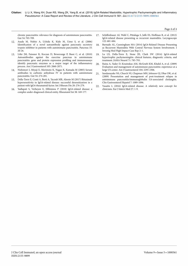
Figure 1: CT reveals confluent areas of the left mastoid.
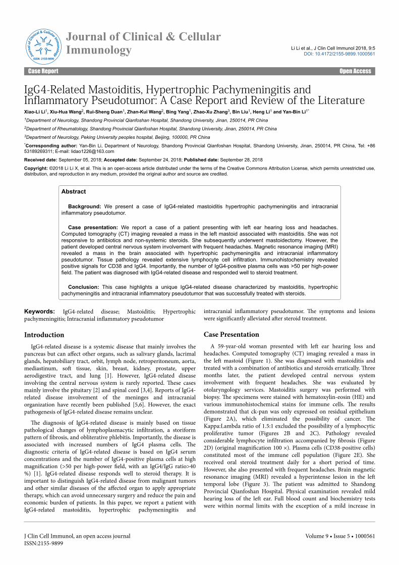
Figure 2: (A) Ck-pan is only expressed on residual epithelium(original magnification 100 ×). (B, C) Lymphocytic proliferativetumor was excluded based on a Kappa:Lambda ratio of 1.5:1. (D)Histological examination of the mastoid tissues revealed that theinterstitial infiltration is composed of a large number oflymphoplasmacytic cells and a few eosinophils infiltrated withfibrosis (original magnification 100 ×). (E) Animmunohistochemical study revealed a large number of plasmacells (CD38+) in the infiltrate, which constituted most of theimmune cell population (original magnification 200 ×). (F)Immunohistochemistry of mastoid tissues revealed markedlyincreased numbers of IgG4-positive plasma cells (>50 per high-power field) (original magnification 200 ×).
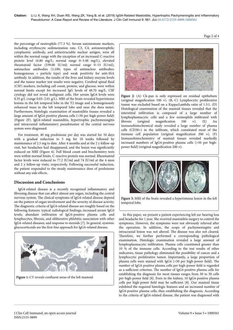
Figure 3: MRI of the brain revealed a hyperintense lesion in the lefttemporal lobe.
In this paper, we present a patient experiencing left ear hearing lossand headache for 1 year. She received mastoiditis surgery to control thesymptoms. However, the symptoms were not obviously relieved afterthe operation. In addition, the scope of pachymeningitis andintracranial lesion was not altered. The disease was also not cleared.Therefore, we further performed a corresponding pathologicalexamination. Histologic examination revealed a large amount oflymphoplasmacytic infiltration. Plasma cells constituted greater than10 % of the immune cells. According to the test results of otherindicators, tissue pathology eliminated the possibility of cancer and alymphocytic proliferative tumor. Importantly, a large proportion ofplasma cells were stained with IgG4 (>50 per high-power field). Thenumber of IgG4-positive plasma cells per high-power field is regardedas a sufficient criterion. The number of IgG4-positive plasma cells forestablishing the diagnosis for most tissues ranges from 30 to 50 cellsper high-power field [8]. Even in the kidney, 10 IgG4-positive plasmacells per high-power field may be sufficient [8]. Our mastoid tissueexhibited the required histologic features and an increased number ofIgG4-positive plasma cells, thus establishing the diagnosis. Accordingto the criteria of IgG4-related disease, the patient was diagnosed with
Citation: Li Li X, Wang XH, Duan RS, Wang ZK, Yang B, et al. (2018) IgG4-Related Mastoiditis, Hypertrophic Pachymeningitis and InflammatoryPseudotumor: A Case Report and Review of the Literature. J Clin Cell Immunol 9: 561. doi:10.4172/2155-9899.1000561
Page 2 of 4
J Clin Cell Immunol, an open access journalISSN:2155-9899
Volume 9 • Issue 5 • 1000561
IgG4-related mastoiditis, hypertrophic pachymeningitis andintracranial inflammatory pseudotumor.
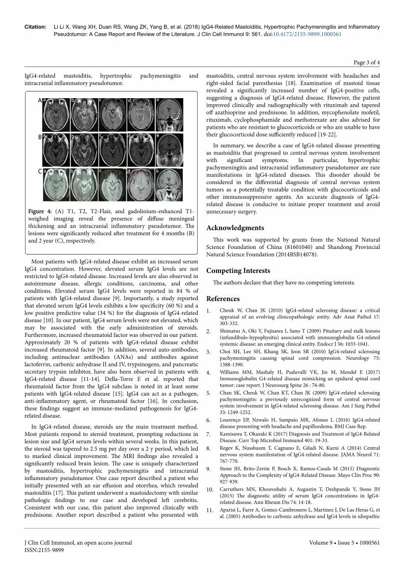
Figure 4: (A) T1, T2, T2-Flair, and gadolinium-enhanced T1-weighed imaging reveal the presence of diffuse meningealthickening and an intracranial inflammatory pseudotumor. Thelesions were significantly reduced after treatment for 4 months (B)and 2 year (C), respectively.
Most patients with IgG4-related disease exhibit an increased serumIgG4 concentration. However, elevated serum IgG4 levels are notrestricted to IgG4-related disease. Increased levels are also observed inautoimmune disease, allergic conditions, carcinoma, and otherconditions. Elevated serum IgG4 levels were reported in 84 % ofpatients with IgG4-related disease [9]. Importantly, a study reportedthat elevated serum IgG4 levels exhibits a low specificity (60 %) and alow positive predictive value (34 %) for the diagnosis of IgG4-relateddisease [10]. In our patient, IgG4 serum levels were not elevated, whichmay be associated with the early administration of steroids.Furthermore, increased rheumatoid factor was observed in our patient.Approximately 20 % of patients with IgG4-related disease exhibitincreased rheumatoid factor [9]. In addition, several auto-antibodies,including antinuclear antibodies (ANAs) and antibodies againstlactoferrin, carbonic anhydrase II and IV, trypsinogens, and pancreaticsecretary trypsin inhibitor, have also been observed in patients withIgG4-related disease [11-14]. Della-Torre E et al. reported thatrheumatoid factor from the IgG4 subclass is noted in at least somepatients with IgG4-related disease [15]. IgG4 can act as a pathogen,anti-inflammatory agent, or rheumatoid factor [16]. In conclusion,these findings suggest an immune-mediated pathogenesis for IgG4-related disease.
In IgG4-related disease, steroids are the main treatment method.Most patients respond to steroid treatment, prompting reductions inlesion size and IgG4 serum levels within several weeks. In this patient,the steroid was tapered to 2.5 mg per day over a 2 y period, which ledto marked clinical improvement. The MRI findings also revealed asignificantly reduced brain lesion. The case is uniquely characterizedby mastoiditis, hypertrophic pachymeningitis and intracranialinflammatory pseudotumor. One case report described a patient whoinitially presented with an ear effusion and otorrhea, which revealedmastoiditis [17]. This patient underwent a mastoidectomy with similarpathologic findings to our case and developed left cerebritis.Consistent with our case, this patient also improved clinically withprednisone. Another report described a patient who presented with
mastoiditis, central nervous system involvement with headaches andright-sided facial paresthesias [18]. Examination of mastoid tissuerevealed a significantly increased number of IgG4-positive cells,suggesting a diagnosis of IgG4-related disease. However, the patientimproved clinically and radiographically with rituximab and taperedoff azathioprine and prednisone. In addition, mycophenolate mofetil,rituximab, cyclophosphamide and methotrexate are also advised forpatients who are resistant to glucocorticoids or who are unable to havetheir glucocorticoid dose sufficiently reduced [19-22].
In summary, we describe a case of IgG4-related disease presentingas mastoiditis that progressed to central nervous system involvementwith significant symptoms. In particular, hypertrophicpachymeningitis and intracranial inflammatory pseudotumor are raremanifestations in IgG4-related diseases. This disorder should beconsidered in the differential diagnosis of central nervous systemtumors as a potentially treatable condition with glucocorticoids andother immunosuppressive agents. An accurate diagnosis of IgG4-related disease is conducive to initiate proper treatment and avoidunnecessary surgery.
AcknowledgmentsThis work was supported by grants from the National Natural
Science Foundation of China (81601040) and Shandong ProvincialNatural Science Foundation (2014BSB14078).
Competing InterestsThe authors declare that they have no competing interests.
References1. Cheuk W, Chan JK (2010) IgG4-related sclerosing disease: a critical
appraisal of an evolving clinicopathologic entity. Adv Anat Pathol 17:303-332.
2. Shimatsu A, Oki Y, Fujisawa I, Sano T (2009) Pituitary and stalk lesions(infundibulo-hypophysitis) associated with immunoglobulin G4-relatedsystemic disease: an emerging clinical entity. Endocr J 56: 1033-1041.
3. Choi SH, Lee SH, Khang SK, Jeon SR (2010) IgG4-related sclerosingpachymeningitis causing spinal cord compression. Neurology 75:1388-1390.
4. Williams MM, Mashaly H, Puduvalli VK, Jin M, Mendel E (2017)Immunoglobulin G4-related disease mimicking an epidural spinal cordtumor: case report. J Neurosurg Spine 26 : 76-80.
5. Chan SK, Cheuk W, Chan KT, Chan JK (2009) IgG4-related sclerosingpachymeningitis: a previously unrecognized form of central nervoussystem involvement in IgG4-related sclerosing disease. Am J Surg Pathol33: 1249-1252.
6. Lourenço EP, Nzwalo H, Sampaio MR, Afonso L (2016) IgG4-relateddisease presenting with headache and papilloedema. BMJ Case Rep.
7. Kamisawa T, Okazaki K (2017) Diagnosis and Treatment of IgG4-RelatedDisease. Curr Top Microbiol Immunol 401: 19-33.
8. Regev K, Nussbaum T, Cagnano E, Giladi N, Karni A (2014) Centralnervous system manifestation of IgG4-related disease. JAMA Neurol 71:767-770.
9. Stone JH, Brito-Zerón P, Bosch X, Ramos-Casals M (2015) DiagnosticApproach to the Complexity of IgG4-Related Disease. Mayo Clin Proc 90:927-939.
10. Carruthers MN, Khosroshahi A, Augustin T, Deshpande V, Stone JH(2015) The diagnostic utility of serum IgG4 concentrations in IgG4-related disease. Ann Rheum Dis 74: 14-18.
11. Aparisi L, Farre A, Gomez-Cambronero L, Martinez J, De Las Heras G, etal. (2005) Antibodies to carbonic anhydrase and IgG4 levels in idiopathic
Citation: Li Li X, Wang XH, Duan RS, Wang ZK, Yang B, et al. (2018) IgG4-Related Mastoiditis, Hypertrophic Pachymeningitis and InflammatoryPseudotumor: A Case Report and Review of the Literature. J Clin Cell Immunol 9: 561. doi:10.4172/2155-9899.1000561
Page 3 of 4
J Clin Cell Immunol, an open access journalISSN:2155-9899
Volume 9 • Issue 5 • 1000561
chronic pancreatitis: relevance for diagnosis of autoimmune pancreatitis.Gut 54: 703-709.
12. Asada M, Nishio A, Uchida K, Kido M, Ueno S, et al. (2006)Identification of a novel autoantibody against pancreatic secretorytrypsin inhibitor in patients with autoimmune pancreatitis. Pancreas 33:20-26.
13. Löhr JM, Faissner R, Koczan D, Bewerunge P, Bassi C, et al. (2010)Autoantibodies against the exocrine pancreas in autoimmunepancreatitis: gene and protein expression profiling and immunoassaysidentify pancreatic enzymes as a major target of the inflammatoryprocess. Am J Gastroenterol 105: 2060-2071.
14. Nishimori I, Miyaji E, Morimoto K, Nagao K, Kamada M (2005) Serumantibodies to carbonic anhydrase IV in patients with autoimmunepancreatitis. Gut 54: 274-281.
15. Della-Torre E, Conti A, Berti A, Yacoub MR, Alessio M (2017) Rituximabhypersensitivity in IgG4-related disease: successful desensitization in apatient with IgG4 rheumatoid factor. Int J Rheum Dis 20: 276-279.
16. Yadlapati S, Verheyen E, Efthimiou P (2018) IgG4-related disease: acomplex under-diagnosed clinical entity. Rheumatol Int 38: 169-177.
17. Schiffenbauer AI, Wahl C, Pittaluga S, Jaffe ES, Hoffman R, et al. (2012)IgG4-related disease presenting as recurrent mastoiditis. Laryngoscope122: 681-684.
18. Barnado AL, Cunningham MA (2014) IgG4-Related Disease Presentingas Recurrent Mastoiditis With Central Nervous System Involvement. JInvestig Med High Impact Case Rep 2: 1.
19. Lu LX, Della-Torre E, Stone JH, Clark SW (2014) IgG4-relatedhypertrophic pachymeningitis: clinical features, diagnostic criteria, andtreatment. JAMA Neurol 71: 785-793.
20. Raina A, Yadav D, Krasinskas AM, McGrath KM, Khalid A, et al. (2009)Evaluation and management of autoimmune pancreatitis: experience at alarge US center. Am J Gastroenterol 104: 2295-2306.
21. Sandanayake NS, Church NI, Chapman MH, Johnson GJ, Dhar DK, et al.(2009) Presentation and management of post-treatment relapse inautoimmune pancreatitis/immunoglobulin G4-associated cholangitis.Clin Gastroenterol Hepatol 7: 1089-1096.
22. Vasaitis L (2016) IgG4-related disease: A relatively new concept forclinicians. Eur J Intern Med 27: 1-9.
Citation: Li Li X, Wang XH, Duan RS, Wang ZK, Yang B, et al. (2018) IgG4-Related Mastoiditis, Hypertrophic Pachymeningitis and InflammatoryPseudotumor: A Case Report and Review of the Literature. J Clin Cell Immunol 9: 561. doi:10.4172/2155-9899.1000561
Page 4 of 4
J Clin Cell Immunol, an open access journalISSN:2155-9899
Volume 9 • Issue 5 • 1000561