Embed Size (px)
Citation preview
IgG4-Related Disease: Puzzles and Pitfalls
Roberto Novoa, MD
Section of Dermatopathology
Departments of Pathology and Dermatology
Stanford University Medical Center
Stanford, CA
Disclosures
• I have no relevant disclosures
• Consultant: Zebra Medical Technologies
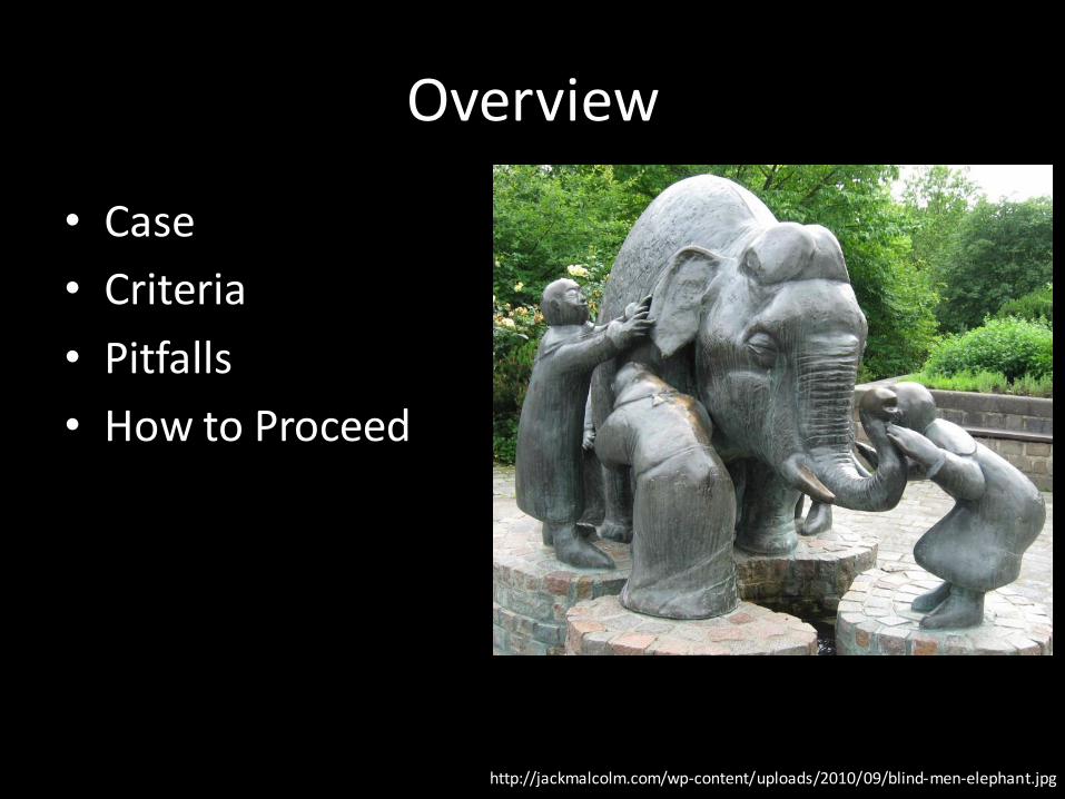
Overview
• Case
• Criteria
• Pitfalls
• How to Proceed
http://jackmalcolm.com/wp-content/uploads/2010/09/blind-men-elephant.jpg

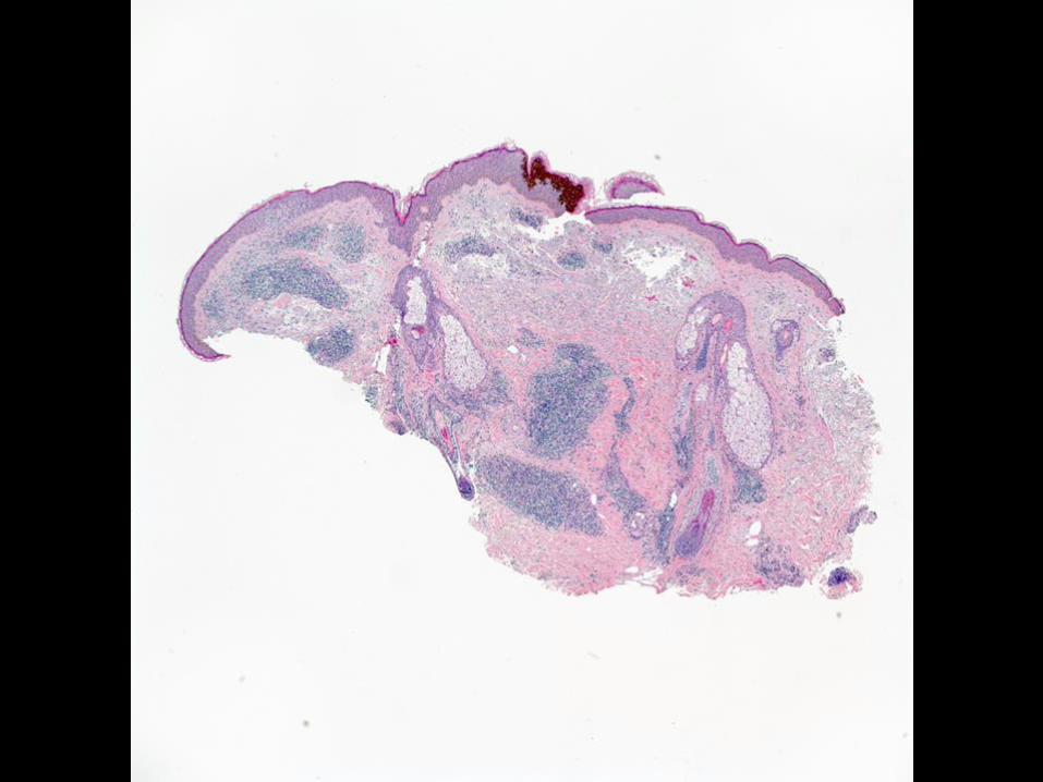
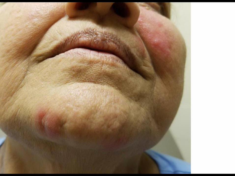
Case
• 72 year-old woman with a 3-year history of pruritic rash on cheeks, neck, chest, and upper arms
• Treated with doxycycline, topical steroids without relief
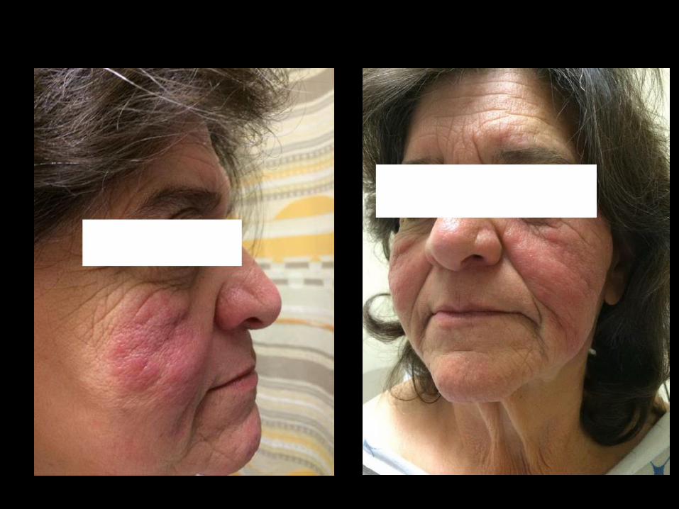

Slide consult requested by clinician

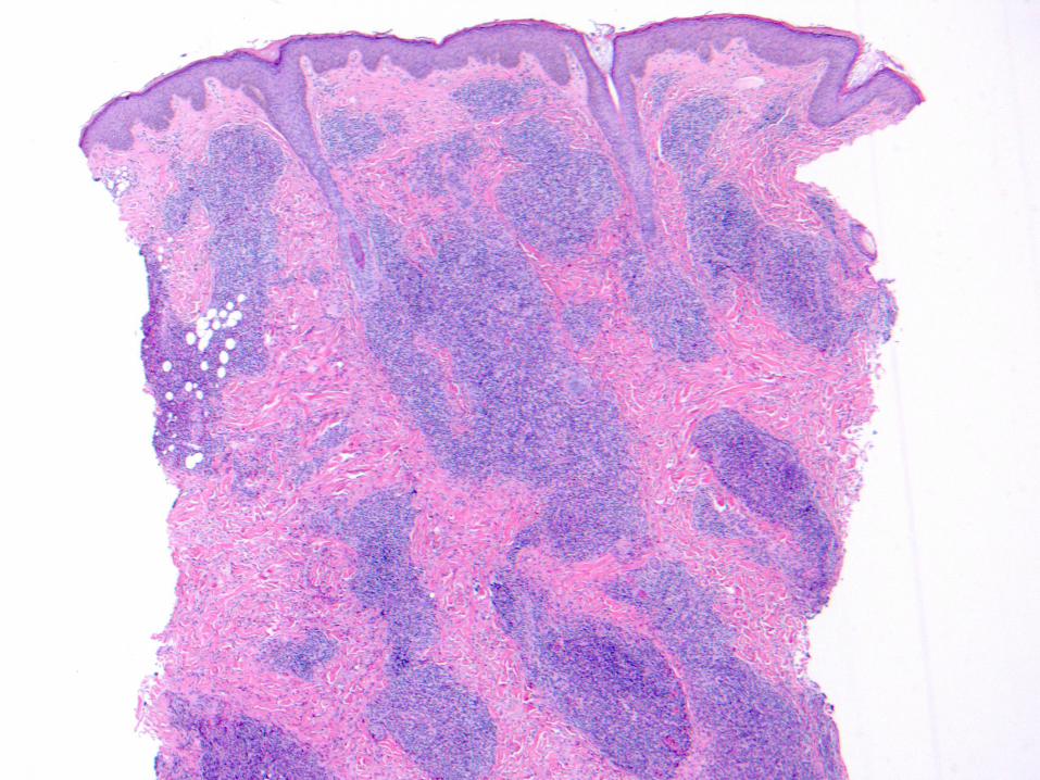
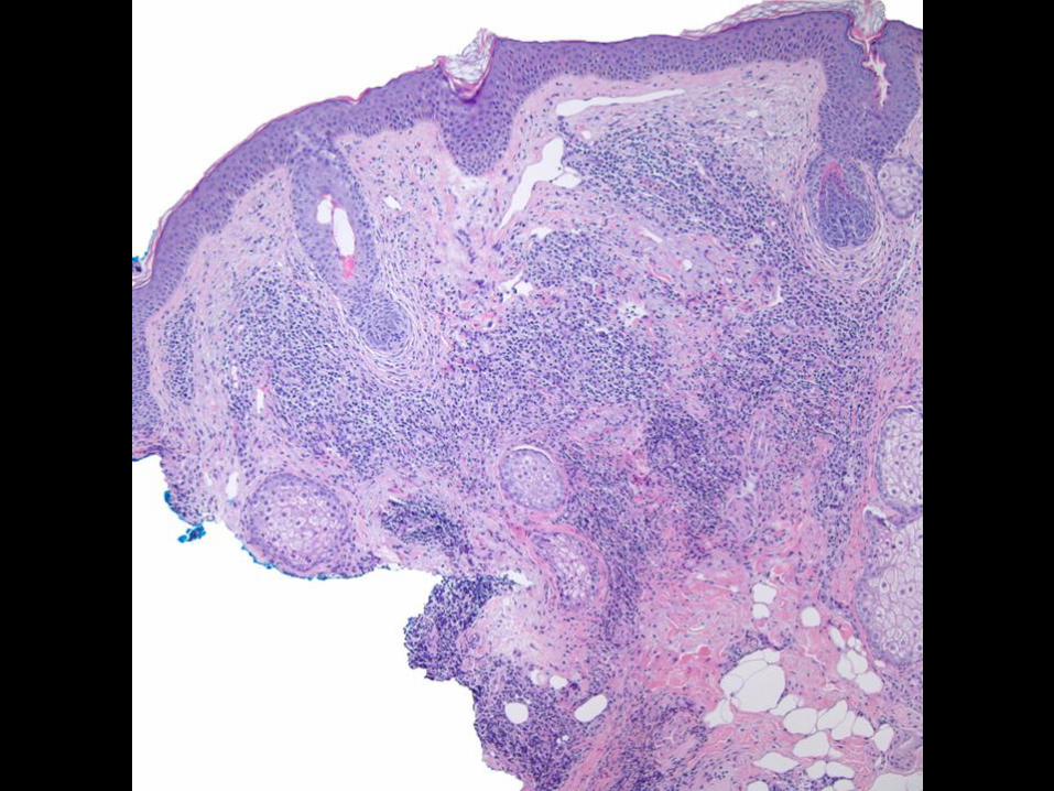
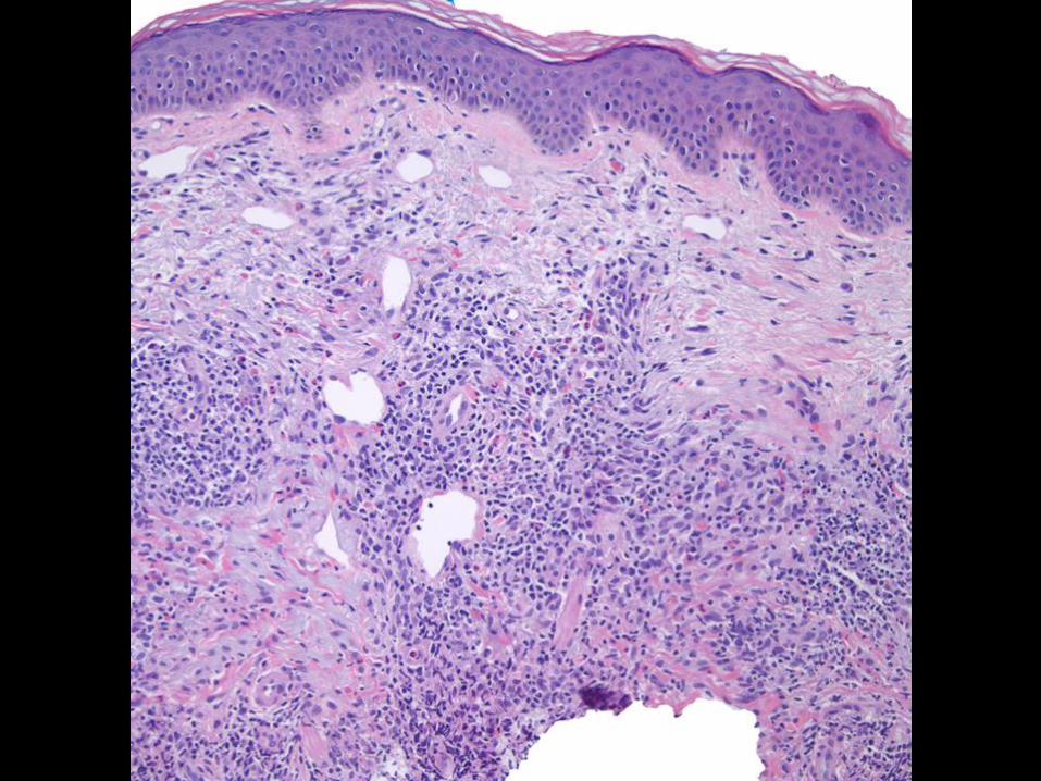
Case
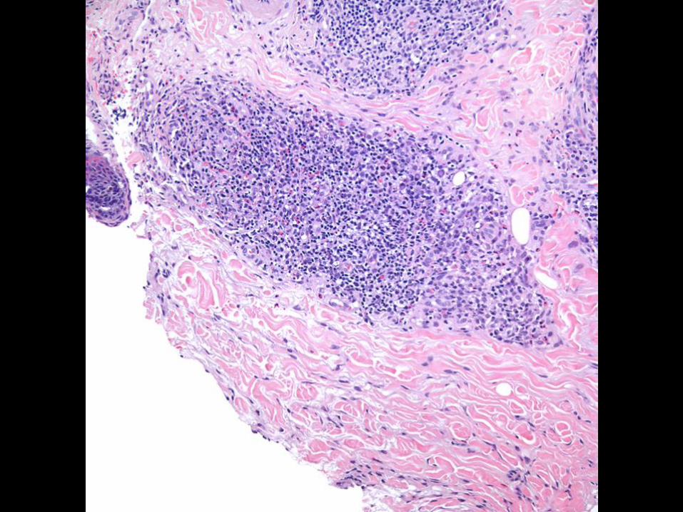
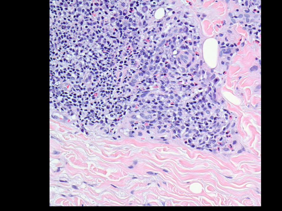
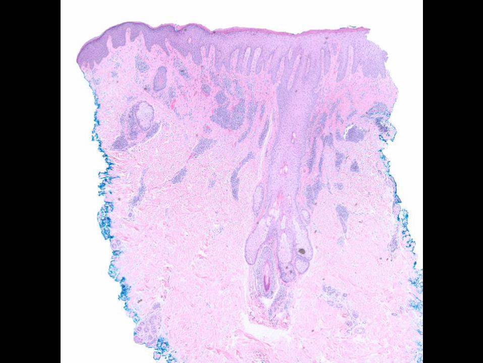
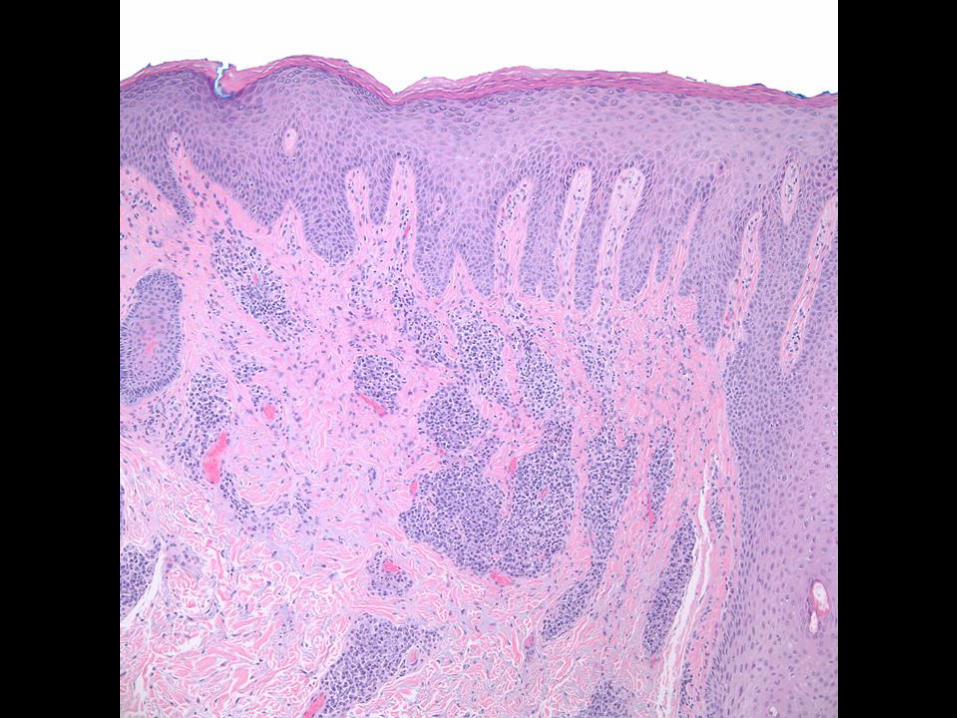
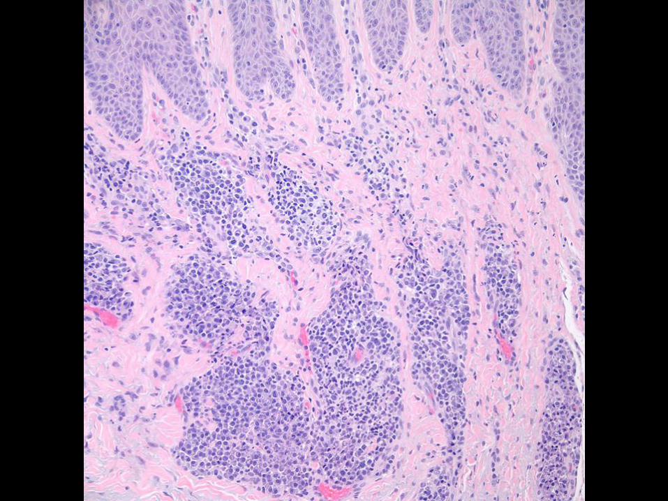
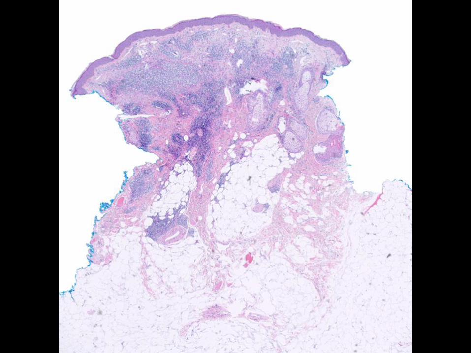
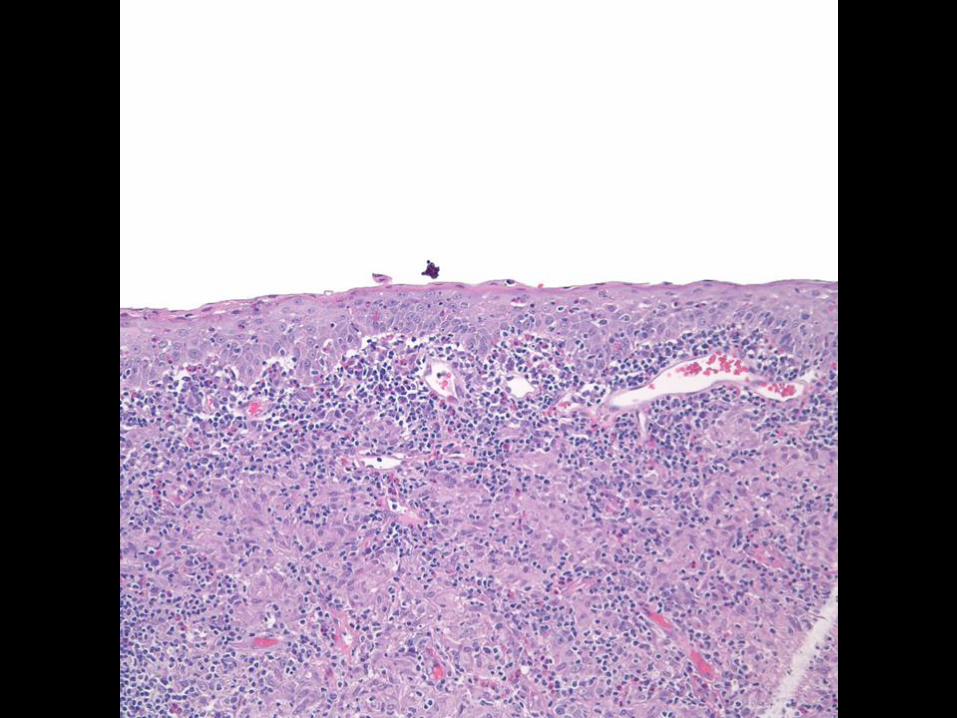
• My signout:
SUPERFICIAL AND DEEP PERIVASCULAR AND PERI-ADNEXAL LYMPHOPLASMACYTIC INFILTRATES WITH EOSINOPHILS (SEE COMMENT)
Case
Clinical Ddx
• Rosacea
• Photodrug
• Dermal hypersensitivity
• Granuloma faciale
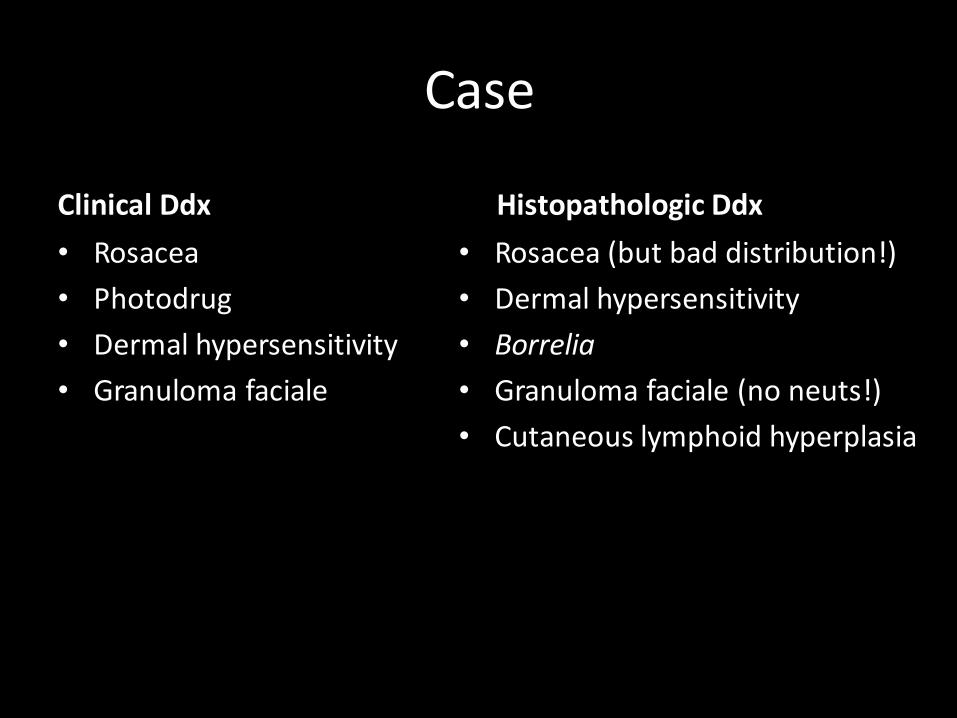
Case
Clinical Ddx
• Rosacea
• Photodrug
• Dermal hypersensitivity
• Granuloma faciale
Histopathologic Ddx
• Rosacea (but bad distribution!)
• Dermal hypersensitivity
• Borrelia
• Granuloma faciale (no neuts!)
• Cutaneous lymphoid hyperplasia
• PMH:
– Thyroiditis, unspecified
• ROS: 60 lb weight loss
https://www.reddit.com/r/marvelstudios/comments/4j5wha/mild_spoilers_didnt_pick_up_on_this_the_first_time/
• PMH:
– Thyroiditis, unspecified
• ROS: 60 lb weight loss
https://www.reddit.com/r/marvelstudios/comments/4j5wha/mild_spoilers_didnt_pick_up_on_this_the_first_time/

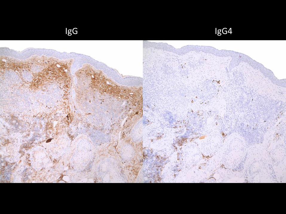
Case
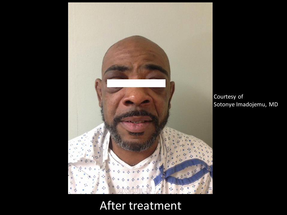
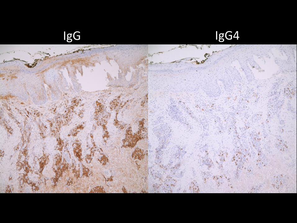
• Challenging fellowship case of patient with proptosis, weight loss, and similar-appearing biopsy specimens
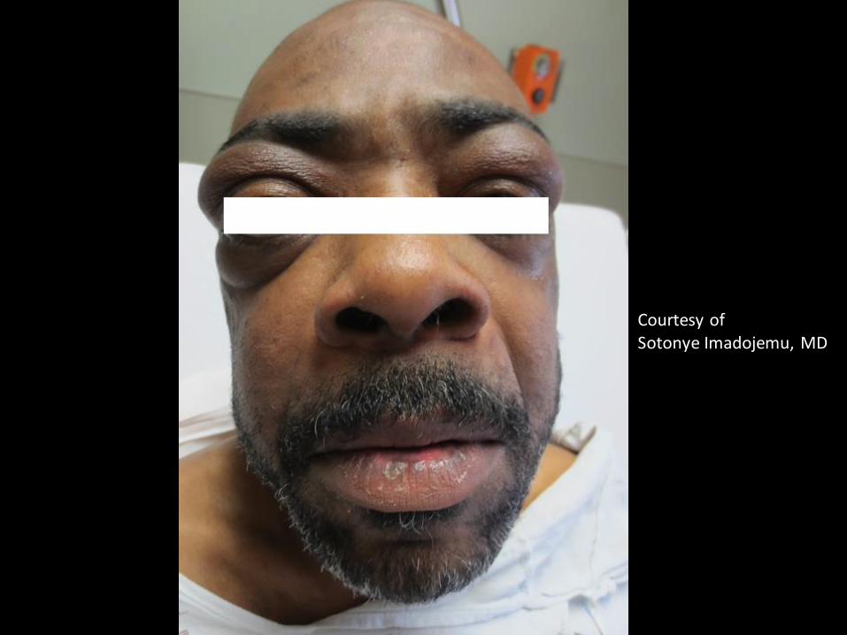
Courtesy of Sotonye Imadojemu, MD
IgG IgG4
After treatment
Courtesy of Sotonye Imadojemu, MD

Back to our case
• Called the clinician and had a conversation
• Clinician thought it worth investigating
• Ordered outside block
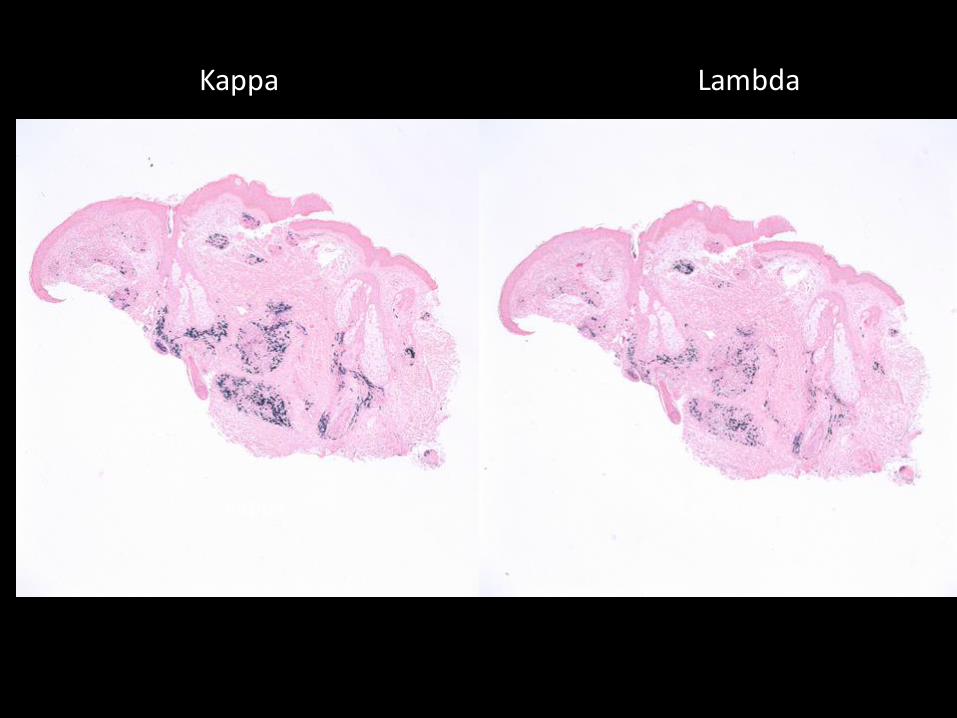
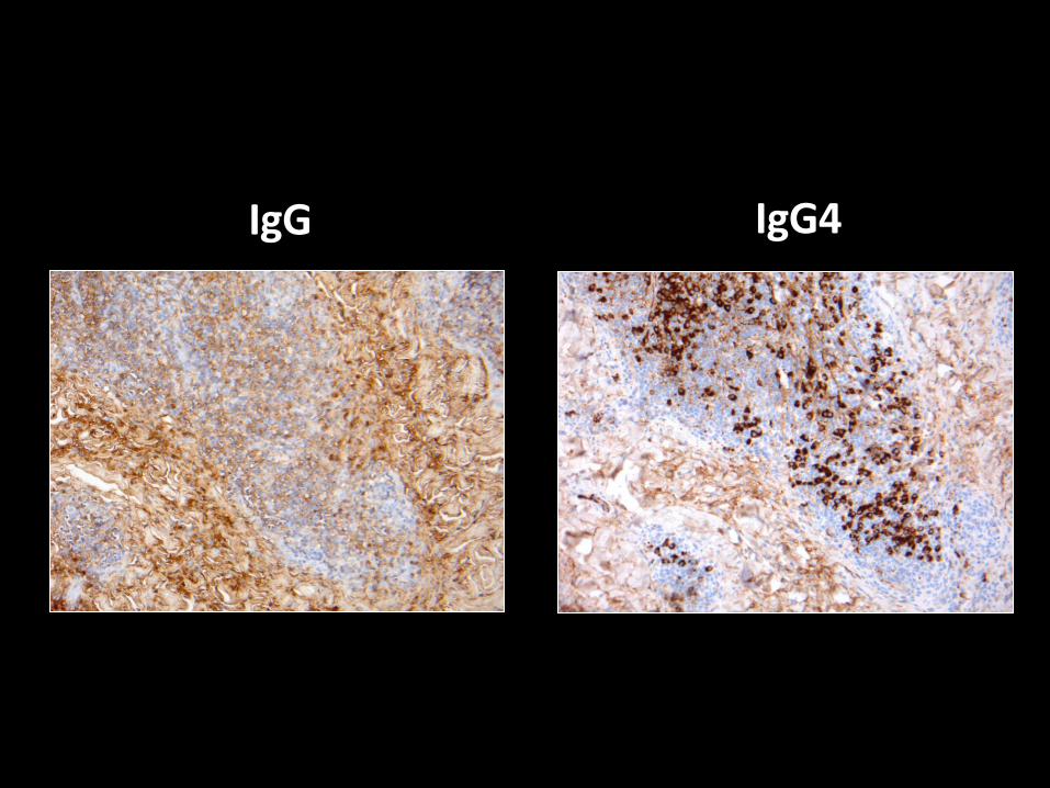
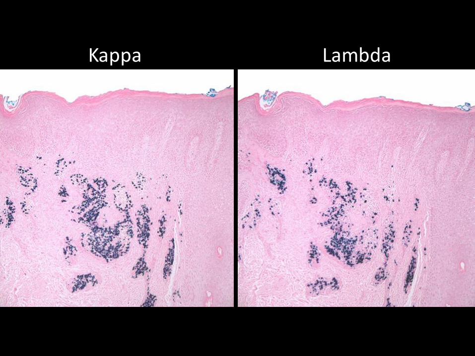
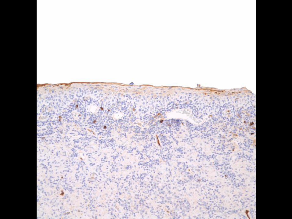
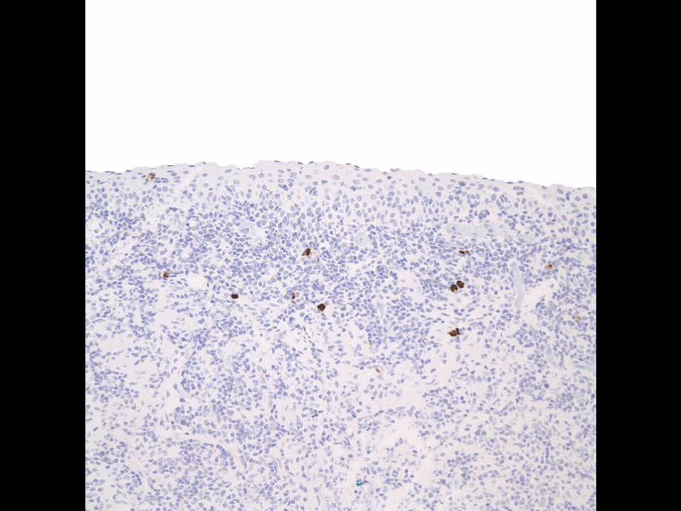
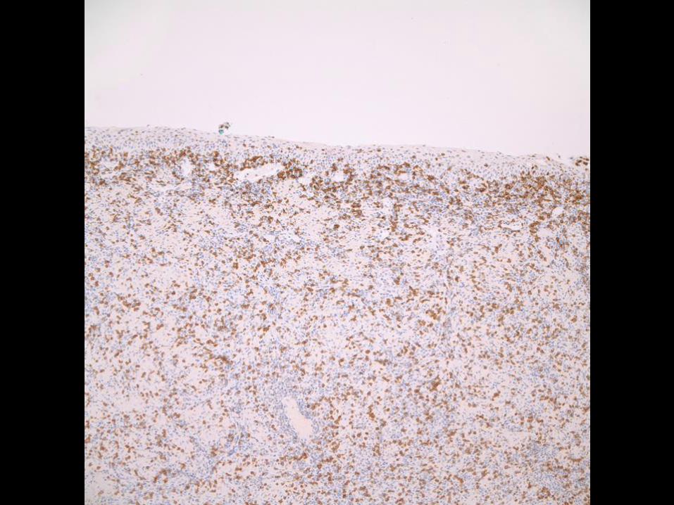
Kappa Lambda
Kappa Lambda
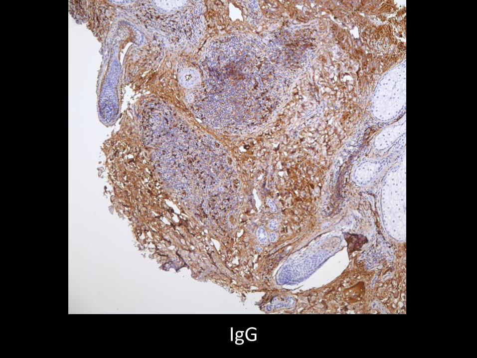
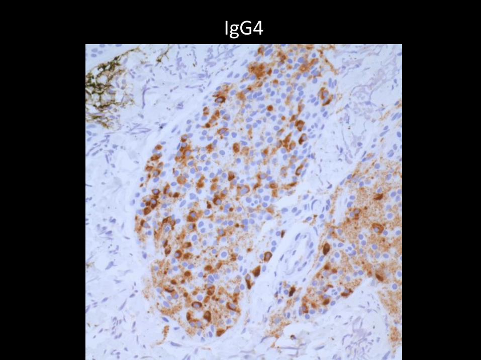
IgG
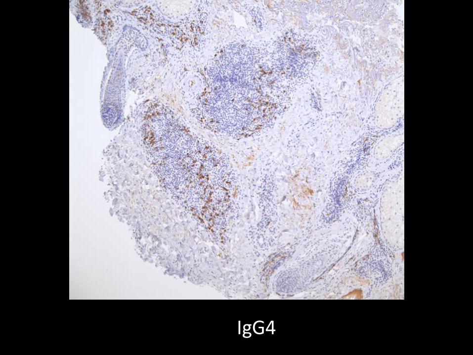
IgG4
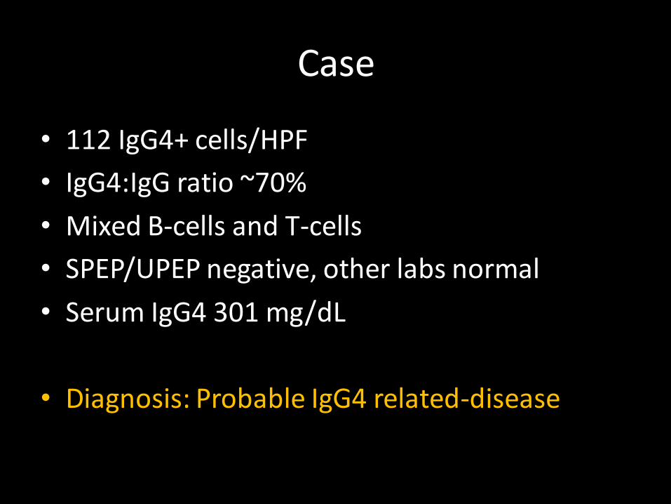
Case
• 112 IgG4+ cells/HPF
• IgG4:IgG ratio ~70%
• Mixed B-cells and T-cells
• SPEP/UPEP negative, other labs normal
• Serum IgG4 301 mg/dL
• Diagnosis: Probable IgG4 related-disease
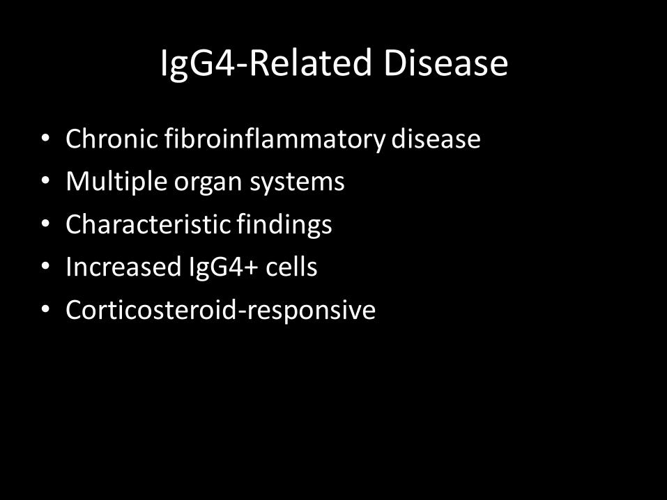
IgG4-Related Disease
• Chronic fibroinflammatory disease
• Multiple organ systems
• Characteristic findings
• Increased IgG4+ cells
• Corticosteroid-responsive
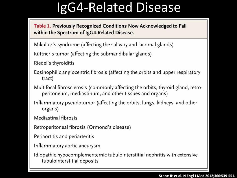
IgG4-Related Disease
Stone JH et al. N Engl J Med 2012;366:539-551.
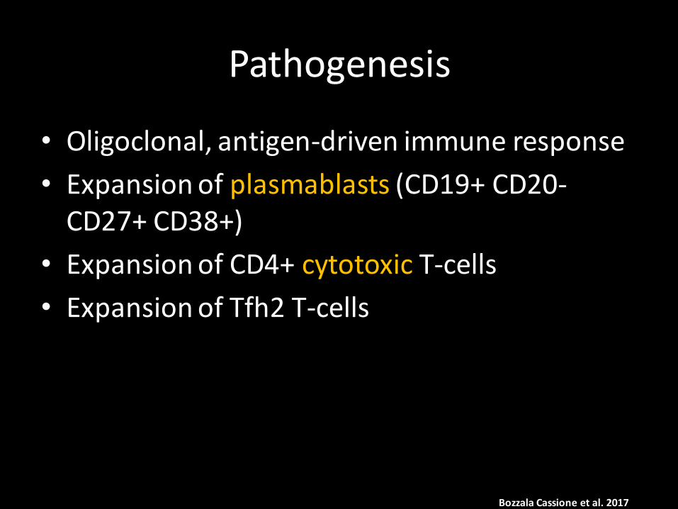
Pathogenesis
• Oligoclonal, antigen-driven immune response
• Expansion of plasmablasts (CD19+ CD20-CD27+ CD38+)
• Expansion of CD4+ cytotoxic T-cells
• Expansion of Tfh2 T-cells
Bozzala Cassione et al. 2017
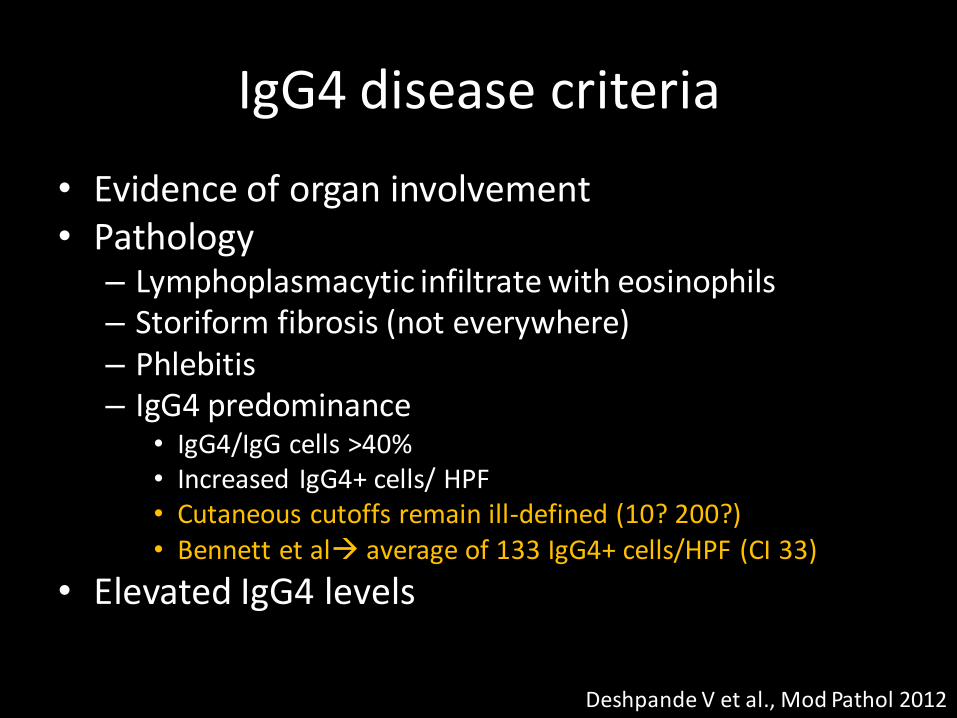
IgG4 disease criteria
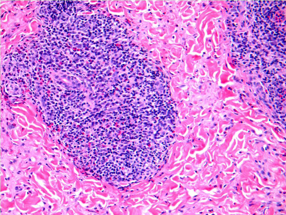
• Evidence of organ involvement• Pathology
– Lymphoplasmacytic infiltrate with eosinophils– Storiform fibrosis (not everywhere)– Phlebitis– IgG4 predominance
• IgG4/IgG cells >40%• Increased IgG4+ cells/ HPF• Cutaneous cutoffs remain ill-defined (10? 200?)• Bennett et al average of 133 IgG4+ cells/HPF (CI 33)
• Elevated IgG4 levels
Deshpande V et al., Mod Pathol 2012
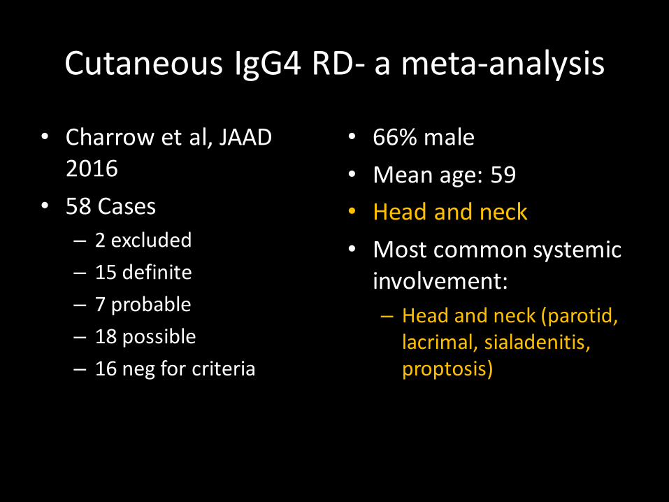
Cutaneous IgG4 RD- a meta-analysis
• Charrow et al, JAAD2016
• 58 Cases
– 2 excluded
– 15 definite
– 7 probable
– 18 possible
– 16 neg for criteria
• 66% male
• Mean age: 59
• Head and neck
• Most common systemic involvement:
– Head and neck (parotid, lacrimal, sialadenitis, proptosis)
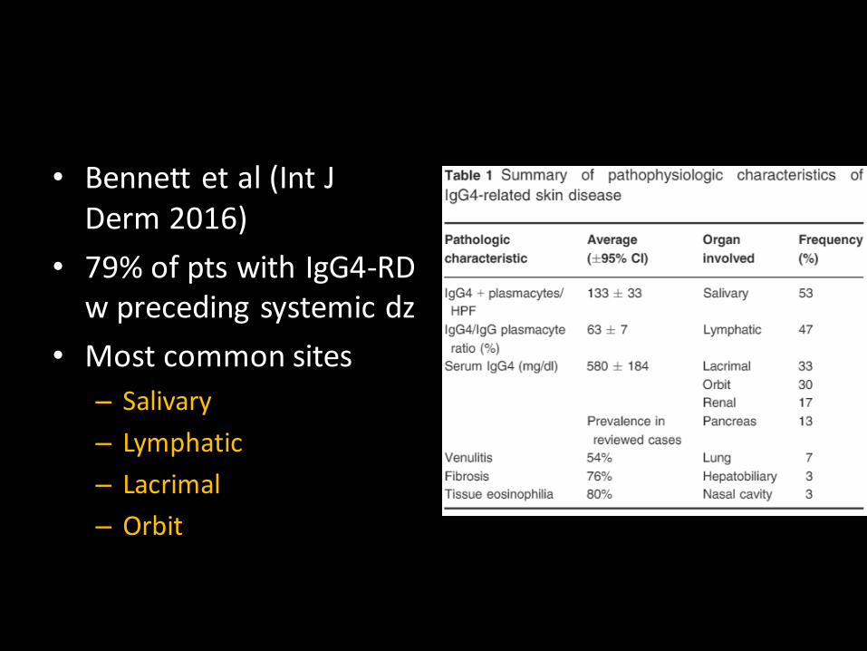
• Bennett et al (Int J Derm 2016)
• 79% of pts with IgG4-RD w preceding systemic dz
• Most common sites
– Salivary
– Lymphatic
– Lacrimal
– Orbit
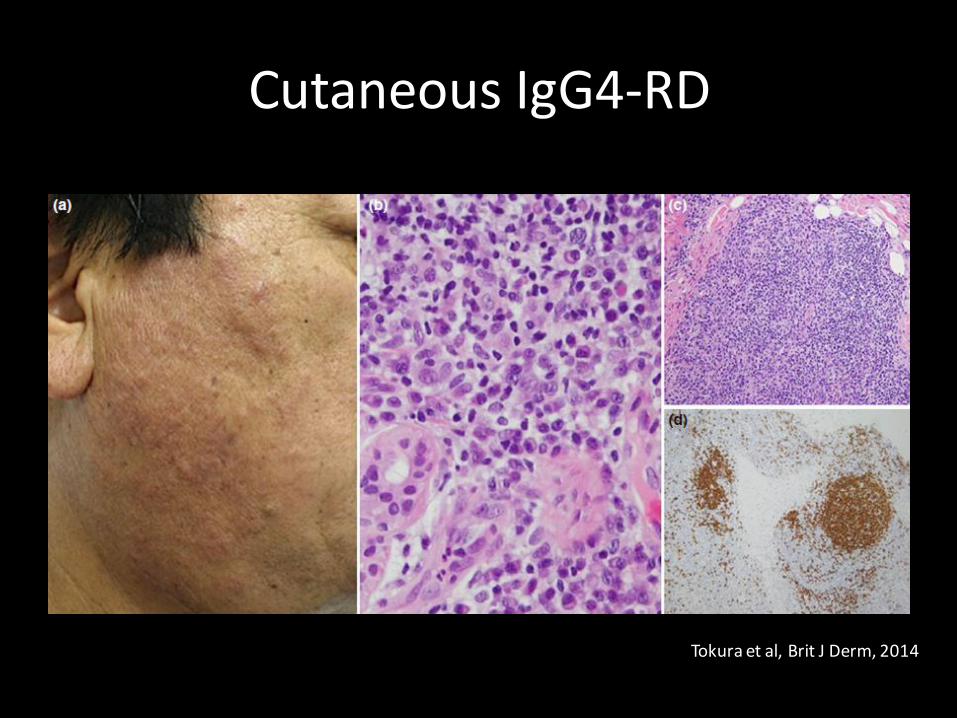
Cutaneous IgG4-RD
Tokura et al, Brit J Derm, 2014
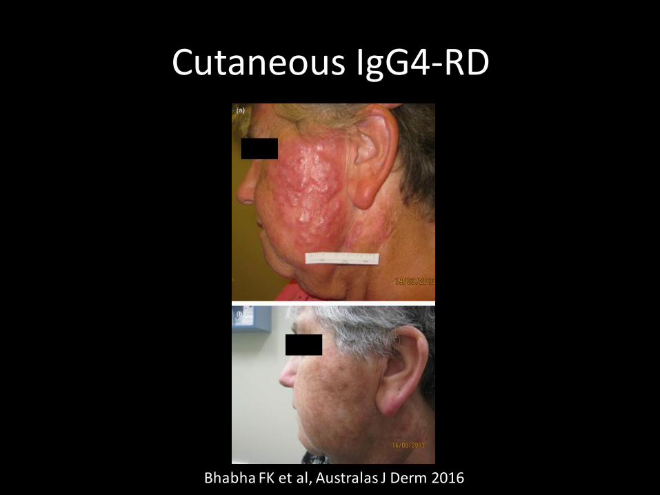
Cutaneous IgG4-RD
Bhabha FK et al, Australas J Derm 2016
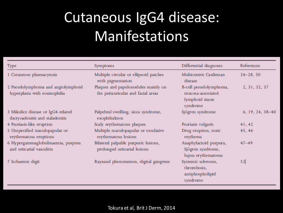
Cutaneous IgG4 disease: Manifestations
Tokura et al, Brit J Derm, 2014
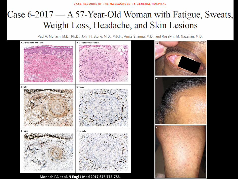
Monach PA et al. N Engl J Med 2017;376:775-786.
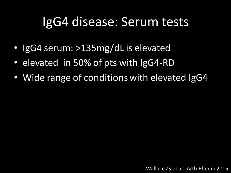
IgG4 disease: Serum tests
• IgG4 serum: >135mg/dL is elevated
• elevated in 50% of pts with IgG4-RD
• Wide range of conditions with elevated IgG4
Wallace ZS et al. Arth Rheum 2015
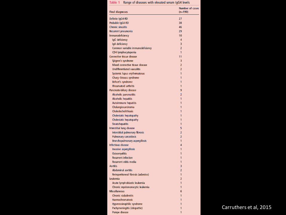
Carruthers et al, 2015
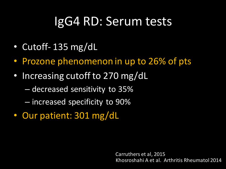
IgG4 RD: Serum tests
• Cutoff- 135 mg/dL
• Prozone phenomenon in up to 26% of pts
• Increasing cutoff to 270 mg/dL
– decreased sensitivity to 35%
– increased specificity to 90%
• Our patient: 301 mg/dL
Khosroshahi A et al. Arthritis Rheumatol 2014 Carruthers et al, 2015
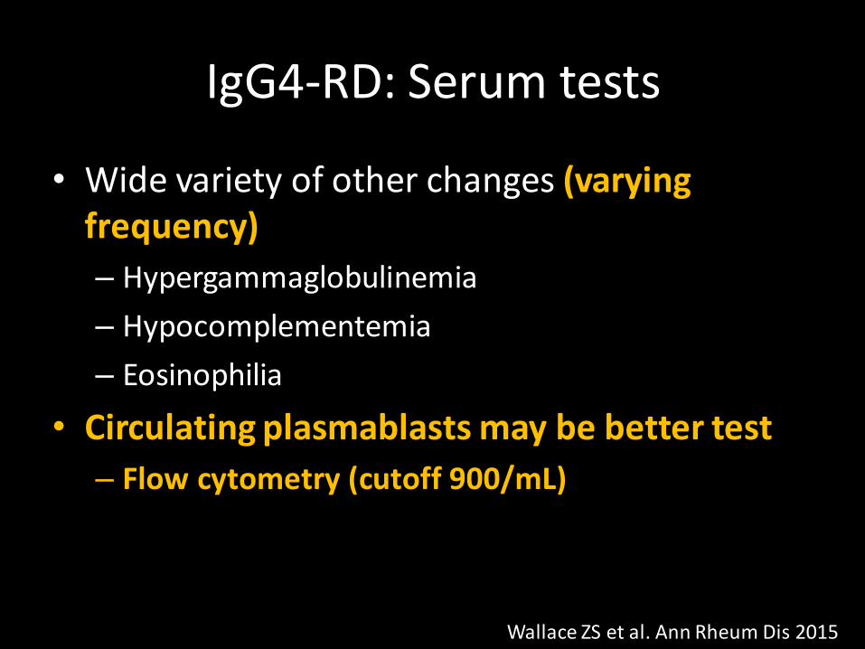
IgG4-RD: Serum tests
• Wide variety of other changes (varying frequency)
– Hypergammaglobulinemia
– Hypocomplementemia
– Eosinophilia
• Circulating plasmablasts may be better test
– Flow cytometry (cutoff 900/mL)
Wallace ZS et al. Ann Rheum Dis 2015
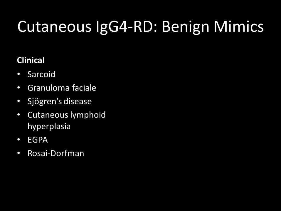
Cutaneous IgG4-RD: Benign Mimics
Clinical
• Sarcoid
• Granuloma faciale
• Sjögren’s disease
• Cutaneous lymphoid hyperplasia
• EGPA
• Rosai-Dorfman
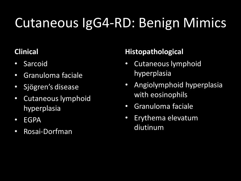
Cutaneous IgG4-RD: Benign Mimics
Clinical
• Sarcoid
• Granuloma faciale
• Sjögren’s disease
• Cutaneous lymphoid hyperplasia
• EGPA
• Rosai-Dorfman
Histopathological
• Cutaneous lymphoid hyperplasia
• Angiolymphoid hyperplasia with eosinophils
• Granuloma faciale
• Erythema elevatumdiutinum
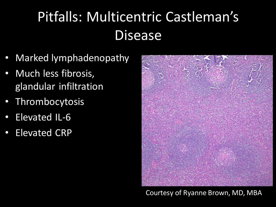
Pitfalls: Multicentric Castleman’sDisease
• Marked lymphadenopathy
• Much less fibrosis, glandular infiltration
• Thrombocytosis
• Elevated IL-6
• Elevated CRP
Courtesy of Ryanne Brown, MD, MBA
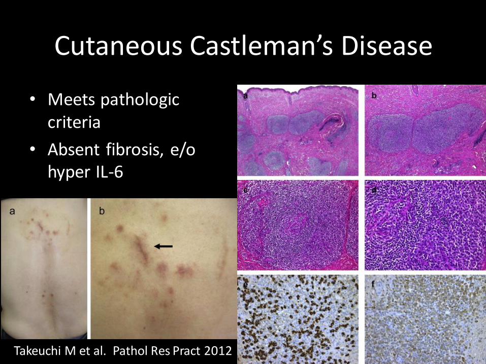
Cutaneous Castleman’s Disease
• Meets pathologic criteria
• Absent fibrosis, e/o hyper IL-6
Takeuchi M et al. Pathol Res Pract 2012
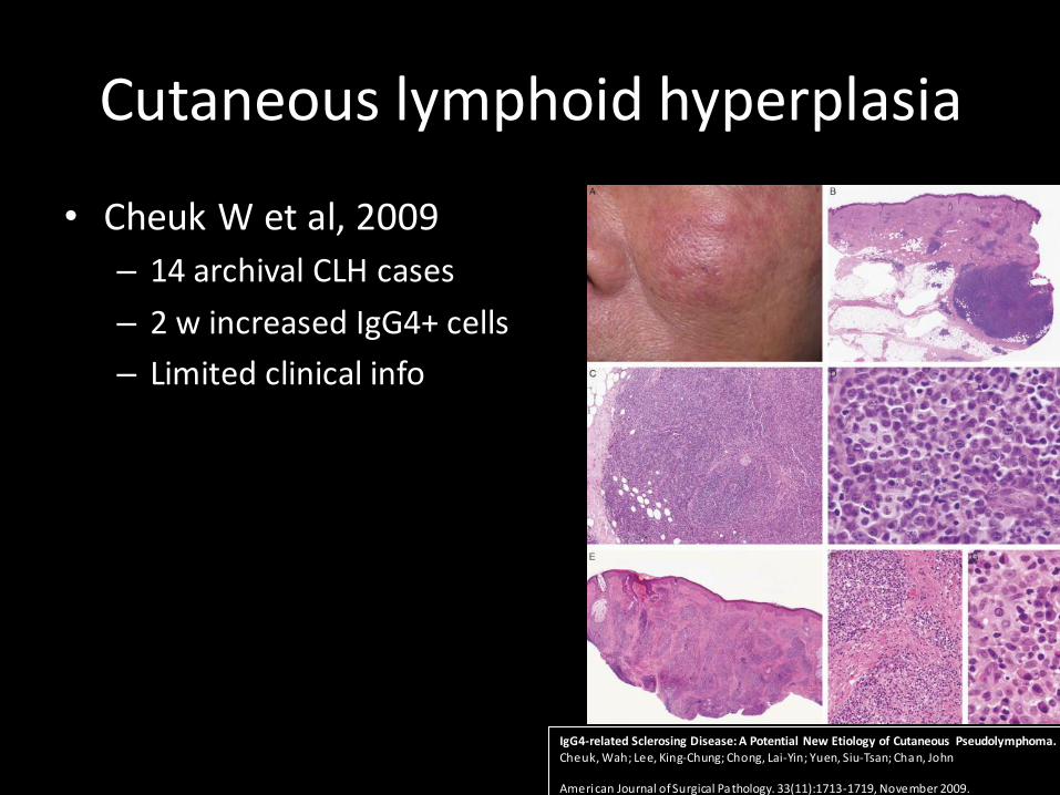
Cutaneous lymphoid hyperplasia
• Cheuk W et al, 2009
– 14 archival CLH cases
– 2 w increased IgG4+ cells
– Limited clinical info
IgG4-related Sclerosing Disease: A Potential New Etiology of Cutaneous Pseudolymphoma.Cheuk, Wah; Lee, King-Chung; Chong, Lai-Yin; Yuen, Siu-Tsan; Chan, John
American Journal of Surgical Pathology. 33(11):1713-1719, November 2009.
Pitfalls: Malignant Mimics
Clinical
• MALT lymphoma
• Angioimmunoblastic T-cell lymphoma
• CTCL variants
Histopathological
• IgG4-restricted myeloma
• IgG4-restricted MZL
Lowe GC et al. Int J Derm 2015
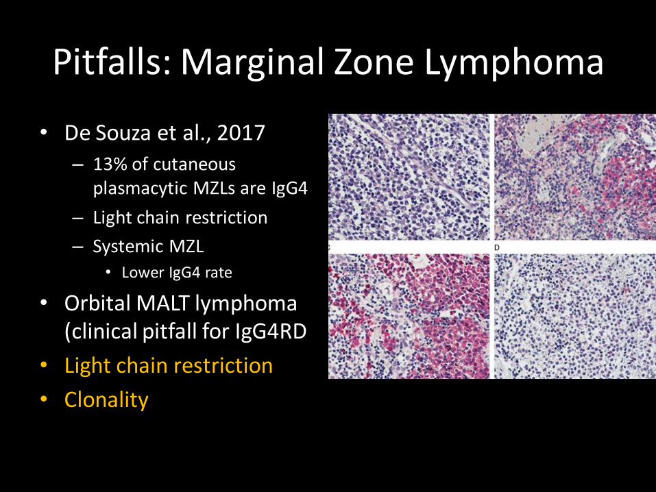
Pitfalls: Marginal Zone Lymphoma
• De Souza et al., 2017
– 13% of cutaneous plasmacytic MZLs are IgG4
– Light chain restriction
– Systemic MZL
• Lower IgG4 rate
• Orbital MALT lymphoma (clinical pitfall for IgG4RD
• Light chain restriction
• Clonality
IgG4-RD and Malignancy
• Malignancy 2.5x more common in IgG4-RD pts
– 19% lymphoma
• Slightly different cohort

Challenging patients
http://discounicornss.tumblr.com/post/57403826324
Challenging patients
IgG4!!
http://discounicornss.tumblr.com/post/57403826324
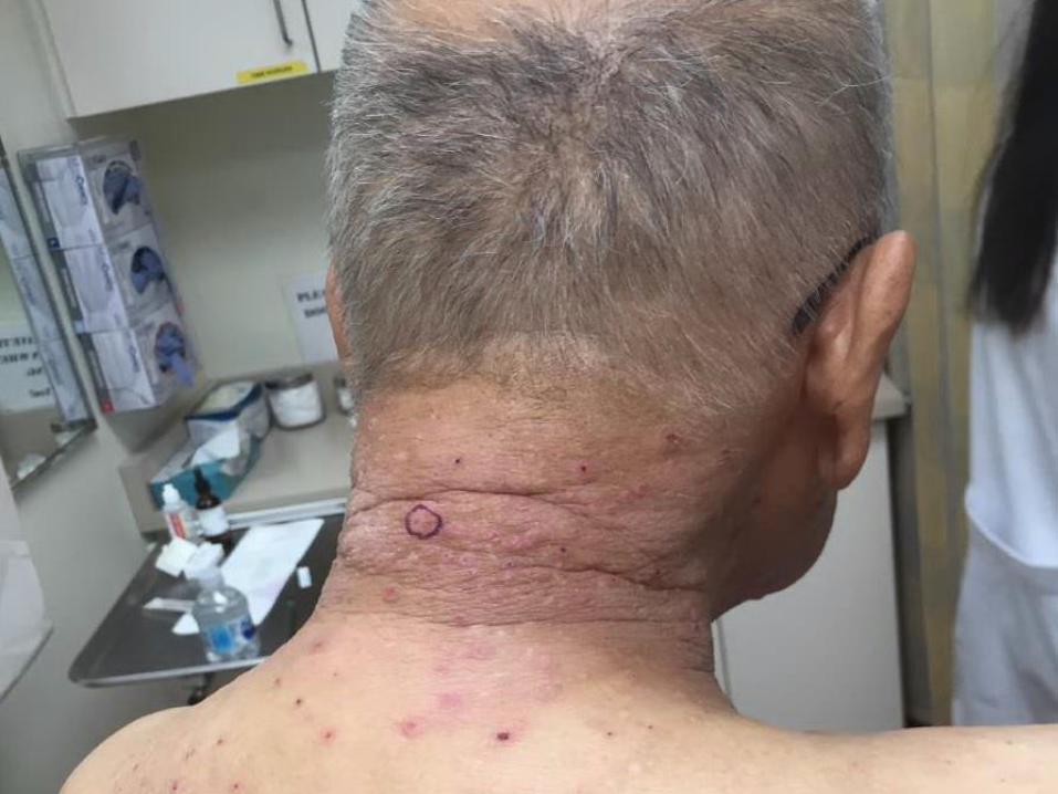
Kappa Lambda
IgG IgG4
IgG4

Diagnosis: Chronic actinic dermatitis
IgG IgG4

Diagnosis: Rosacea…for now
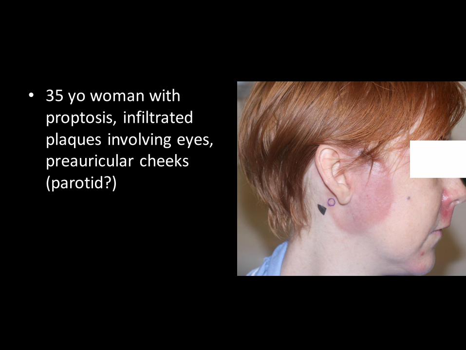
• 35 yo woman with proptosis, infiltrated plaques involving eyes, preauricular cheeks (parotid?)
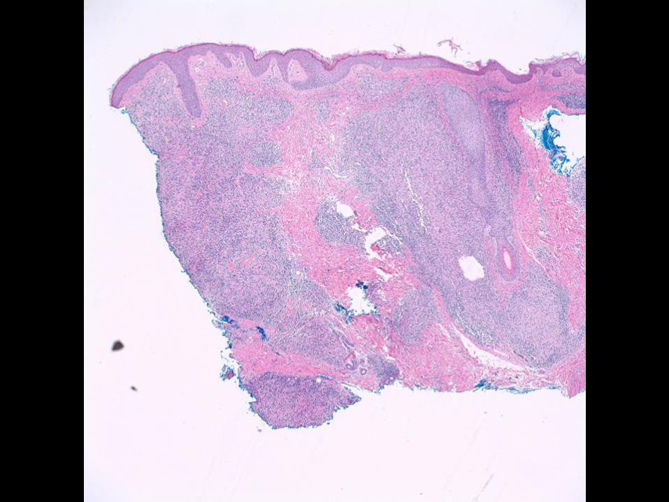
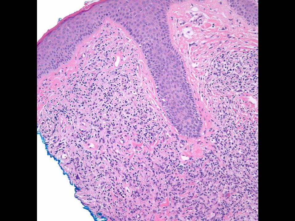
Diagnosis: T-cell lymphoma
• Work in progress
Conclusions
1. IgG4 disease– varied morphologies, but esp think in clinical scenario with dermal nodules on face, neck, upper body
2. If thinking about CLH and pt has systemic sx, consider IgG4
3. Criteria are still evolving, but application of strict criteria probably best at present time (>100 IgG4+ cells/HPF, Ig4/IgG ratio >40%)
4. Serum IgG4 can be useful, but circulating plasmablasts may be better test
5. Thorough evaluation for malignancy
6. Beware of pitfalls!
References
1. Stone JH, Zen Y, Deshpande V. IgG4-related disease. N Engl J Med. 2012 Feb 9;366(6):539-51. doi: 10.1056/NEJMra1104650.2. Tokura Y, Yagi H, Yanaguchi H, Majima Y, Kasuya A, Ito T, Maekawa M, Hashizume H. IgG4-related skin disease. Br J Dermatol. 2014
Nov;171(5):959-67. doi: 10.1111/bjd.13296. Epub 2014 Oct 20.3. Deshpande V, Zen Y, Chan JK, Yi EE, Sato Y, Yoshino T, Klöppel G, Heathcote JG, Khosroshahi A, Ferry JA, Aalberse RC, Bloch DB, Brugge WR,
Bateman AC, Carruthers MN, Chari ST, Cheuk W, Cornell LD, Fernandez-Del Castil lo C, Forcione DG, Hamilos DL, Kamisawa T, Kasashima S, Kawa S, Kawano M, Lauwers GY, Masaki Y, Nakanuma Y, Notohara K, Okazaki K, Ryu JK, Saeki T, Sahani DV, Smyrk TC, Stone JR, Takahira M, Webster GJ, Yamamoto M, Zamboni G, Umehara H, Stone JH. Consensus statement on the pathology of IgG4-related disease. Mod Pathol. 2012 Sep;25(9):1181-92.
4. Bennett AE, Fenske NA, Rodriguez-Waitkus P, Messina JL. IgG4-related skin disease may have distinct systemic manifestations: a systematic review. Int J Dermatol. 2016 Nov;55(11):1184-1195.
5. Bhabha FK, Turner JJ, Bleasel K, Sladden MJ. Immunoglobulin Type Gamma 4-Related Disease Presenting with Cutaneous Plaques. Australas J Dermatol. 2016 Feb;57(1):74.
6. Monach PA, Stone JH, Sharma A, Nazarian RM. Case 6-2017. A 57-year-old woman with fatigue, sweats, weight loss, headache, and skin lesions. N Engl J Med. 2017 Feb 23;376(8):775-786
7. Wallace ZS, Deshpande V, Mattoo H, Mahajan VS, Kulikova M, Pil lai S, Stone JH. IgG4-Related Disease: Clinical and Laboratory Features in One Hundred Twenty-Five Patients. Arthritis Rheumatol. 2015 Sep;67(9):2466-75.
8. Takeuchi M, Sato Y, Takata K, Kobayashi K, Ohno K, Iwaki N, Orita Y, Yoshino T. Cutaneous multicentric Castleman's disease mimicking IgG4-related disease. Pathol Res Pract. 2012 Dec 15;208(12):746-9. doi: 10.1016/j.prp.2012.09.006. Epub 2012 Oct 25.
9. Cheuk, Wah; Lee, King-Chung; Chong, Lai-Yin; Yuen, Siu-Tsan; Chan, John. IgG4-related Sclerosing Disease: A Potential New Etiology of Cutaneous Pseudolymphoma. American Journal of Surgical Pathology. 33(11):1713-1719, November 2009.
10. Lowe GC, Bogner RR, el-Azhary RA, Gonzalez-Santiago TM, Kindle SA, Lehman JS, Gibson LE. Cutaneous manifestations of immunoglobulin G4-related disease: what dermatologists need to know. Int J Dermatol. 2015 Apr;54(4):377-82. doi: 10.1111/ijd.12953.
11. De Souza A, Ferry JA, Burghart DR, Tinguely M, Goyal A, Duncan LM, Kutzner H, Kempf W. IgG4 Expression in Primary Cutaneous Marginal Zone Lymphoma: A Multicenter Study.Appl Immunohistochem Mol Morphol. 2017 Feb 1.