Embed Size (px)
Citation preview
1
IgG4 detection of Echinococcus granulosus paramyosin: a useful diagnostic test for human 1
hydatidosis 2
3
Running title: E. granulosus paramyosin for diagnosis of hydatidosis 4
5
Zohreh Kazemi Moghadam Kakhki,a Fatemeh Ghaffarifar,b Akbar Khalilpour,a Farhanah Abdul 6
Aziz,a Geita Saadatnia, a ,c and Rahmah Noordina# 7
8
a Institute for Research in Molecular Medicine (INFORMM), Universiti Sains Malaysia, Penang, 9
Malaysia 10
b Department of Parasitology, School of Medical Sciences, Tarbiat Modarres University, Tehran, 11
Iran 12
c Department of Biotechnology, Iranian Research Organization for Science and Technology 13
(IROST), Tehran, Iran 14
15
# Corresponding author. Tel.: +60 4 653 4802. Fax: +60 4 653 4803 16
Email address: [email protected] 17
18
19
20
Copyright © 2013, American Society for Microbiology. All Rights Reserved.Clin. Vaccine Immunol. doi:10.1128/CVI.00019-13 CVI Accepts, published online ahead of print on 30 January 2013
on June 4, 2018 by guesthttp://cvi.asm
.org/D
ownloaded from
2
ABSTRACT 21
Hydatidosis is a public health problem in many parts of the world, and improvement in diagnosis 22
of the disease is still being pursued. Protoscoleces of Echinococcus granulosus were isolated 23
from hydatid cysts collected from naturally infected sheep slaughtered in abattoirs in Iran. 24
Sonicated extract of protoscolex was subjected to two-dimensional gel electrophoresis and 25
western blot analysis. Primary antibodies were from serum samples from 130 hydatidosis 26
patients, 38 individuals infected with other parasitic infections and 30 healthy people; while 27
peroxidase (HRP)-conjugated anti-human IgG and IgG4 were used as secondary antibodies. The 28
recombinant form of the identified protein was produced and tested for its sensitivity and 29
specificity for the detection of human hydatidosis. An antigenic band of ~ 60 kDa was found to 30
be sensitive (82%) and specific (100%) for the detection of hydatidosis when probed with anti-31
human IgG4-HRP, while the sensitivity and specificity were 33% and 100%, respectively, with 32
anti-human IgG-HRP. By mass-spectrometry, the band was identified as protoscolex tegument 33
paramyosin. The sensitivity and specificity of full-length paramyosin-recombinant protein in 34
IgG4 blots were found to be 86% and 98%, respectively. In conclusion, IgG4 detection of 35
Echinococcus granulosus paramyosin was found to be useful for the diagnosis of human 36
hydatidosis. 37
38
39
40
on June 4, 2018 by guesthttp://cvi.asm
.org/D
ownloaded from
3
INTRODUCTION 41
Hydatid cyst, or hydatidosis, is caused by the larval stage (metacestode) of the tapeworm 42
Echinococcus granulosus in human and livestock. As the intermediate host, humans acquire the 43
disease by accidental ingestion of vegetables or water contaminated with the ova of adult worms 44
that live in the small intestine of canids, the definitive host. After exposure to gastrointestinal 45
enzymes, the infective ovum hatches into an oncosphere, which is able to reach organs such as 46
the liver and lungs through the vascular and lymphatic systems. The larva then develops into a 47
hydatid cyst, which is gradually filled with fluid and protoscoleces. Hydatid cysts not only cause 48
severe illness, but also cause economic losses due to costs related to diagnosis and surgery (3). 49
This disease is scattered throughout the world, with an emerging or re-emerging status in several 50
countries (11) and is also highly prevalent in Iran (26). It is estimated that 2-3 million people are 51
infected with this neglected disease worldwide (10). Early diagnosis is based on clinical signs, 52
which is followed by imaging of suspected organs. Clinical signs in humans are not specific, and 53
the imaging methods cannot differentiate between hydatid cysts, tumors and other lesions (1, 14). 54
Therefore, immunodiagnosis remains an important tool in the diagnosis of the disease. Chordi 55
and Kagan were the first to use immunoelectrophoresis to identify the antigenic components of 56
sheep hydatid cyst fluid, and subsequently determined which antigenic components were active 57
in detecting antibodies in the sera of patients with hydatid cysts (8). A successful 58
immunodiagnostic test depends on the use of highly specific and sensitive antigens, as well as 59
the detection of the appropriate antibody class or subclass (18, 27). Detection of circulating 60
antigens in serum was reported to be less sensitive than detection of Echinococcus-specific 61
antibodies (31). In this study, two-dimensional electrophoresis (2-DE) and western blots using 62
IgG and IgG4 as secondary antibodies were performed to identify antigen of E. granulosus 63
on June 4, 2018 by guesthttp://cvi.asm
.org/D
ownloaded from
4
protoscolex with good diagnostic potential. 64
65
MATERIALS AND METHODS 66
Serum samples. Group I serum samples were collected from 81 patients with cysts in their liver 67
or lungs who were diagnosed based on clinical symptoms and serodiagnosis and/or with 68
magnetic resonance imaging (MRI). The samples were obtained from sera banks of pathology 69
laboratories in Tehran, Iran. Nine pools of sera, containing 9 samples per pool, were created. 70
Group II serum samples (obtained prior to surgery) were individual samples from 49 confirmed 71
hydatidosis patients from Imam Hospital, Tehran, Iran who had cysts in their liver or lungs, and 72
who underwent surgical removal of the cysts. Consent was obtained from the patients for 73
collection of the serum samples, and the Tarbiat Modarres University research ethics committee 74
approved the study. Group III control samples (68) were from sera from healthy people (n=30) 75
and patients infected with other parasites (n=38) [ Ascaris lumbricoides (5), Fasciola hepatica 76
(2), Taenia saginata (1), Toxocara canis (2), Toxoplasma gondii (4), Trichuris trichiura (5), 77
Strongyloides stercoralis (2), Brugia malayi (11), Gnathostoma spinigerum (1), Taenia solium 78
(cystircercosis) (1), Necator americanus (2) and Ancylostoma duodenale (2)]. 79
80
Preparation of the protoscolex antigens. Protoscoleces were aseptically isolated from hydatid 81
cysts collected from infected sheep slaughtered in abattoirs in Iran. The protoscoleces were 82
washed three times with sterile phosphate-buffered saline (PBS, pH 7.2) by centrifugation at 83
3000 ×g for 10 min. The final pellet was suspended in an equal volume of sterile PBS containing 84
a cocktail of protease inhibitors (Roche Diagnostics, Germany) at 40 µl⁄ml. Three cycles of 85
freeze–thaw (water bath and liquid nitrogen) were performed, followed by sonication on ice 86
on June 4, 2018 by guesthttp://cvi.asm
.org/D
ownloaded from
5
(Liquid Pressor: Model XL2020, USA). The homogenate was centrifuged (10,000 ×g, 1 hour), 87
and the pellet was discarded. The supernatant was washed twice with 10 mM Tris and 88
concentrated by centrifugation at 3,000 ×g for 30 min using a spin filter with a 5 kDa MW cut-89
off (VivaspinTM, Sartorius, Germany). The protein concentration of the supernatant was 90
determined using a Bio-Rad RCDC protein assay kit (Bio Rad, USA), and then the supernatant 91
was stored in aliquots at -80 ˚C. 92
93
One-dimensional electrophoresis. One-dimensional electrophoresis was performed using an 94
OFFGEL Fractionator (Agilent, USA) to separate the protein according to isoelectric point. 95
Ready-made 13 cm IPG dry strip gels with a pH range of 3-10 were used. In total, 1000 µg of 96
desalted protein was fractionated into 12 wells. 97
98
SDS-PAGE (Sodium dodecyl sulfate-polyacrylamide gel electrophoresis). The second 99
dimension of electrophoresis was performed on a 12% SDS-PAGE gel Mini-Protean 3 cell 100
apparatus (Bio-Rad, USA). The amount of protein loaded was optimized at 20 µg per well. 101
Standard molecular weight markers were loaded in the first lane. Samples were run at 100 V for 102
110 min. 103
104
Western blot analysis of native antigens. Protein was electro-transferred from gel to a 105
nitrocellulose paper (NCP, 0.45) using a Trans-Blot Cell (Bio-Rad, USA) at 12 A for 30 min. 106
The NCP was stained with Ponceau S solution (Sigma, USA) and washed with TBS-T (Tris-HCl 107
buffered saline-0.05% Tween 20) washing buffer. It was blocked with blocking buffer (Roche 108
Diagnostics, Germany) for one hour at room temperature. The NCP was then washed four times 109
on June 4, 2018 by guesthttp://cvi.asm
.org/D
ownloaded from
6
at 1, 5, 10 and 15-min intervals consecutively and cut into 3 mm strips. The strips were then 110
incubated with human serum diluted 1:100 in blocking buffer for 2 hours at room temperature 111
and then overnight at 4 °C. The next day, after a washing step, anti-human IgG4 or IgG 112
conjugated to horseradish peroxidase at dilutions of 1:2000 or 1:1000, respectively, (Invitrogen, 113
USA) were incubated with the strips for 1 hour at room temperature. The strips were then 114
washed and developed with chemiluminescence substrate (Roche Diagnostics, Germany). 115
Initially, the potential diagnostic band was identified from results of western blots performed 116
with each of the nine pools from the Group I sera and pooled healthy sera, using anti-human 117
IgG4-HRP as the secondary antibody. Subsequently, individual sera from Group II and Group III 118
samples were tested to determine the sensitivity and specificity of the identified band, using both 119
HRP-conjugated anti-human IgG and IgG4 as secondary antibodies. 120
121
Staining of gels. The gels were stained with Coomassie blue or with MS-compatible silver stain. 122
The latter was performed according to the method described by Shevchenko (29). The identified 123
band for MS-MS analysis was manually excised and transferred to a microfuge tube. 124
125
In-gel digestion of a silver stained gel. Fifty microliters of acetonitrile was added into each 126
microfuge tube containing the sliced band and incubated for 15 min at room temperature. The 127
supernatant was removed and 25 µl of ammonium bicarbonate (25 mM) was added and 128
incubated for 10 min. The supernatant was again removed and the gel pieces were air dried for 129
one hour. Resuspension solution containing 10 ng/µl trypsin was added and the gel pieces were 130
incubated on ice for 5 min. Subsequently, the gel pieces were immersed in 25 µl of ammonium 131
bicarbonate (25 mM) and incubated at 30 °C overnight. Two volumes of acetonitrile were added 132
on June 4, 2018 by guesthttp://cvi.asm
.org/D
ownloaded from
7
for digestion, and then the samples were vortexed and incubated for 20 min at room temperature. 133
Finally, the supernatant was transferred into a fresh 1.5 ml microfuge tube. Sample cleanup for 134
MALDI –TOF mass spectrometry analysis was performed using Zip-Tip pipette tips (Zip-Tip U-135
C18, Millipore, USA). 136
137
Mass spectrometry analysis. In-gel digested protein from a silver-stained gel was sent for 138
MS/MS analysis using a MALDI TOF-TOF 4800 (ABSCIEX, USA) at the Protein and 139
Proteomics Centre, National University of Singapore. Meanwhile, a Coomassie blue-stained, 140
sliced gel band was sent for processing and analysis using a MALDI TOF-TOF 5800 141
(ABSCIEX, USA) at Proteomics International, Australia. 142
143
Preparation of recombinant antigens. The gene sequence that corresponded to the MS/MS-144
identified protein was custom-cloned into a pET28 expression vector by Epoch Life Science 145
(USA). The recombinant plasmid received from the company was transformed into competent E. 146
coli BL21 (DES) expression host cells. A single colony containing the recombinant plasmid was 147
inoculated in 100 ml terrific broth containing 100 μg/ml of kanamycin and incubated overnight at 148
37 °C with agitation at 180 rpm. Fifty microliters of the overnight culture was inoculated into 149
500 ml terrific broth with kanamycin and incubated at 37 °C with agitation at 200 rpm until the 150
OD at 600 nm reached 0.4-0.5. Protein expression was induced by incubation in 1 mM isopropyl 151
β-D-1- thiogalactopyranoside (IPTG) for 5 hours at 37 °C. The cells were harvested by 152
centrifugation at 10,000 ×g, 4 °C for 10 min. The cell pellet was resuspended in 1.5 v/w lysis 153
buffer containing protease inhibitors and 0.5 mg/ml lysozyme and incubated on ice for 30 min. 154
The cell suspension was then lysed using a sonicator, followed by centrifugation at 10,000 ×g, 4 155
on June 4, 2018 by guesthttp://cvi.asm
.org/D
ownloaded from
8
°C for 30 min. The supernatant was transferred to a new tube, DNase (2500 ug/ml) was added 156
and incubated on ice for 10-15 min, followed by centrifugation of the tube at 10,000 ×g, 4 °C for 157
30 min. The resultant supernatant was filtered using a syringe filter (0.45 µm). The filtered 158
supernatant was mixed with Ni-NTA resin (Qiagen, USA), incubated at 4 °C (rotating) for 30 159
min, and then the mixture was loaded into a chromatography column. Gradient washings of the 160
column were performed using 10 ml each of four kinds of washing buffers containing 500 mM 161
NaCl (10mM, 20mM, 30mM and 40mM imidazole respectively). The protein fractions were 162
eluted using elution buffer containing 250 mM imidazole. 163
164
Western blot of recombinant antigens. Protein-containing fractions from the affinity column 165
were pooled and buffer exchanged into PBS with 1 M urea (pH=7). The fractions were 166
concentrated by centrifugation at 3000 ×g for 30 min with a spin filter as described above. After 167
the protein was transferred onto the NCP, it was blocked with 5% alkali-soluble casein 168
(Novagen, USA) for 1 hour at room temperature. After blocking, the NCP was washed with 169
TBS-T washing buffer and incubated with His-tag antibody-HRP at a 1:1000 dilution for 1 hour 170
at room temperature. Western blotting was also performed using human sera as described above. 171
The same set of serum samples was used, but due to the limited volumes of some samples a 172
reduced number of control sera were used. 173
174
RESULTS 175
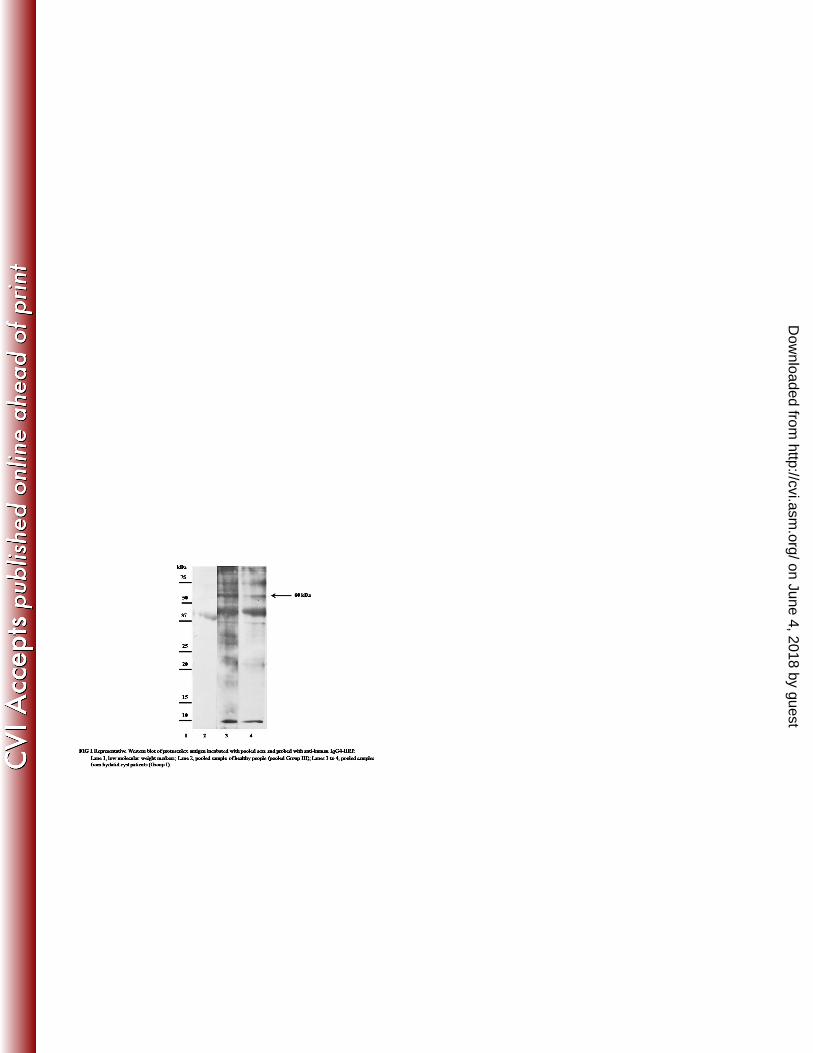
In the initial set of experiments using pooled serum samples, a band of ~ 60 kDa was observed in 176
IgG4 blots using Group I sera from patients with hydatidosis. This band was not observed with 177
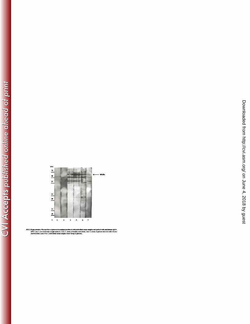
pooled healthy serum (Figure 1). Figure 2 shows representative IgG4 blots with individual serum 178
on June 4, 2018 by guesthttp://cvi.asm
.org/D
ownloaded from
9
samples; the ~ 60 kDa band was found to be present when blotted with sera of hydatidosis 179
patients but not when blotted with control sera. 180
181
As shown in Table 1, the 60 kDa band in IgG4 blots of the native protoscolex antigen showed 182
82% sensitivity (40/49) with Group II serum samples. Out of the 40 serum samples which 183
reacted with the 60 kDa band, 18 were from patients with liver cysts, 19 with lung cysts and 3 184
with mixed liver and lung cysts. Out of the 9 false negative results, 5 were from patients with 185
liver cysts and 4 from patients with lung cysts. A100% specificity (67/67) was seen with Group 186
III sera. In IgG blots of the native protoscolex antigen, the 60 kDa band showed 33% sensitivity 187
of (10/30) and 100% specificity with the same set of sera. 188
189
Database search was performed using Platyhelminthes database from UniProtKB. Both 190
proteomic service centers identified the band as Echinococcus tegument paramyosin (accession 191
number gi|547974) with protein scores (> 200) and peptide scores well above the cut-off values. 192
No protein other than paramyosin was identified. Two tryptic peptides were reported which 193
covered 3% of the paramyosin protein sequence. It showed 100% homology to E. granulosus 194
and 97% to Taenia solium. 195
196
Custom cloning into the pET28 expression vector was made based on the coding sequence (cds) 197
of the previously submitted GenBank sequence, accession Z21787.1. The recombinant 198
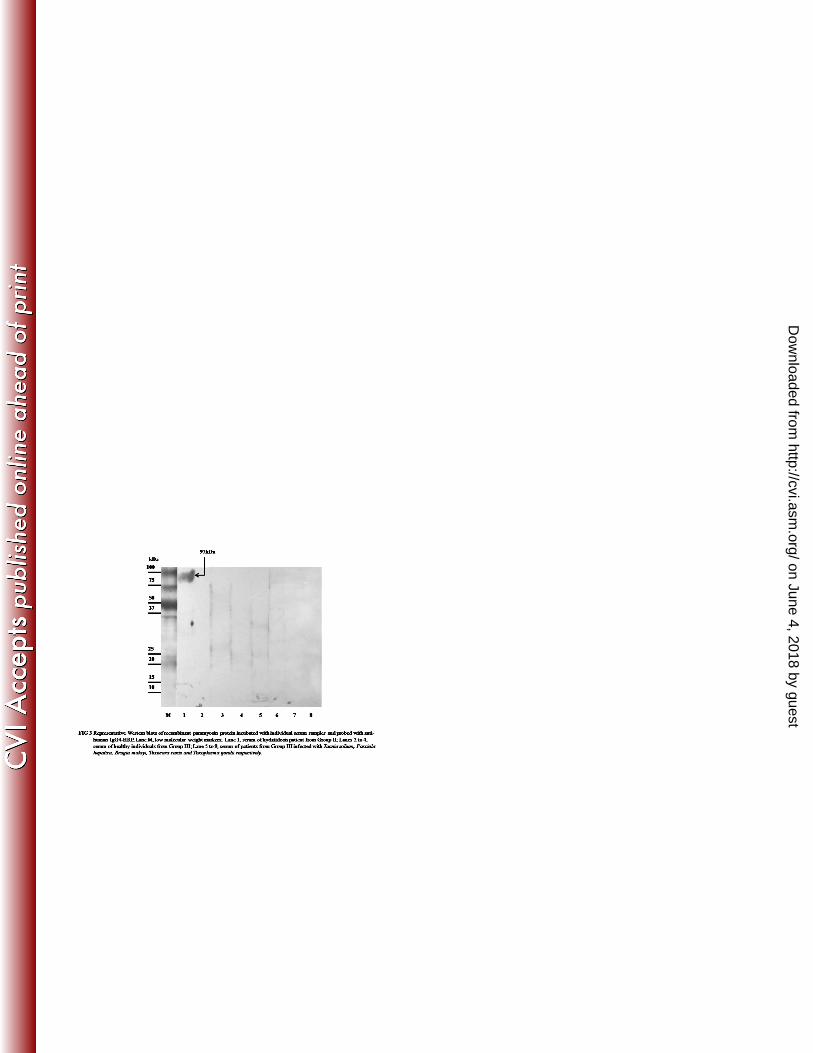
paramyosin showed 86% sensitivity (42/49) with Group II serum samples from hydatid cyst 199
on June 4, 2018 by guesthttp://cvi.asm
.org/D
ownloaded from
10
patients and 98% specificity (58/59) with Group III control sera from 24 healthy people and 35 200
patients with other parasitic infections. Figure 3 shows a representative western blot performed 201
with the recombinant paramyosin. 202
203
204
DISCUSSION 205
Clinical signs of human hydatid cysts are variable and non-specific. Primary diagnosis is based 206
on imaging methods such as ultrasound, computed tomography and magnetic resonance imaging 207
(17). To complement the radiological findings, immunological tests such as enzyme-linked 208
immunosorbent assay (ELISA), indirect immunofluorescence, immunoelectrophoresis (IEP) and 209
immunoblotting are used in diagnostic laboratories. However, false negative (up to 25%) and 210
false positive results remain a problem with the available immunodiagnostic assays (21). To date 211
there is no standardized, highly sensitive and specific test available for immunodiagnosis of 212
human cystic echinococcosis (16). Therefore, during the last three decades, extensive efforts 213
have been made to characterize antigenic components of protoscolex, germinal layer and hydatid 214
cyst fluid of E. granulosus for use in serodiagnostic assays. Hydatid cyst fluid (HCF) is one of 215
the main antigen sources for serodiagnosis of hydatid disease. Two major components of HCF, 216
thermostable antigen B (AgB) and thermolabile antigen 5 (Ag5), are the most widely used 217
antigens in reported assays for the disease (4). However, there are difficulties related to their lack 218
of sensitivity and to their cross-reactivity with antigens from other parasites, notably other 219
taeniid cestodes, as well as problems with the standardization of the antigens. Thus far, the use of 220
these antigens is predominantly restricted to scientific applications and neither is available for 221
general use (1). 222
on June 4, 2018 by guesthttp://cvi.asm
.org/D
ownloaded from
11
The number of reports on the use of protoscolex as an antigen for detecting antibodies in human 223
hydatidosis is limited (5, 6, 22, 2). Chemale et al. (2003) described the analysis of E. granulosus 224
metacestode protein extract by 2-DE and the identification of prominent proteins by peptide mass 225
fingerprinting (PMF). A total of 100 prominent protein spots from three 2-DE gels were analyzed 226
by MALDI-TOF-MS; 15 of them were identified by their PMFs, which include protoscoleces 227
tegument paramyosin (7). Monteiro et al. (2010) performed a proteomic analysis of E. 228
granulosus metacestodes in a bovine host. They used two complementary proteomic approaches, 229
2-DE/MALDI-TOF-MS/MS and LC-MS/MS, to analyze proteins expressed in E. granulosus 230
protoscoleces. The 2-DE IgG immunoblots using a pool of six sera from hydatid disease patients 231
showed 14 protoscolex proteins recognized by hydatid cyst patients’ sera. These proteins were 232
reported to contribute to immunoregulatory events at the host-parasite interface during infection. 233
According to the investigators, some of them, such as paramyosin and tetraspanin, may be 234
potentially useful for vaccine development (19). 235
236
In this study, protoscoleces isolated from hydatid cysts from the livers of sheep were analyzed by 237
2-DE, using OFFGEL for the first dimension of separation followed by mini SDS-PAGE. This 238
approach has the advantage of using less volume of the serum samples in the subsequent western 239
blots (25). The clear band on the western blots allowed the band on the corresponding SDS-240
PAGE gels to be readily identified and excised for mass-spectrometry analysis. The high protein 241
and peptide scores obtained in MS-MS analysis of the in-gel digested protein provided a 242
confident identification of the protein as paramyosin. 243
244
Paramyosin is a 97 kDa muscle protein present in the tegument of protoscoleces. It is an α-245
on June 4, 2018 by guesthttp://cvi.asm
.org/D
ownloaded from
12
helical protein and was first noted in invertebrate muscle as a structural component (9). The 246
report of paramyosin-based vaccination against schistosomes led to the characterization of the 247
protein in other organisms (12). In the platyhelminths Taenia, Echinococcus and Schistosoma, an 248
extra muscular localization of paramyosin has been reported (28). Muhlschlegel et al. (1993) 249
described the cloning of a paramyosin-homologous protein of E. granulosus; and by 250
immunofluorescence they demonstrated its presence in the tegument, subtegument of the body 251
wall and muscles of the four oral suckers of the E. granulosus larvae (20). Paramyosin has also 252
been identified as an immunogenic protein in several parasitic infections, such as Schistosoma 253
japonicum, Schistosoma mansoni, Taenia solium and Taenia saginata (12, 15). 254
255
In this study, the identified E. granulosus paramyosin amino acid sequence was 97% 256
homologous to the paramyosin sequence of T. solium. Thus it is not surprising that the 257
recombinant paramyosin protein showed cross-reactivity with serum from cysticercosis patient. 258
Interestingly the same serum was not cross-reactive with the 60kDa protein of the native 259
protoscolex antigen. One possible reason for this difference is that the recombinant paramyosin 260
comprised the full length paramyosin (~97 kDa), while the native paramyosin band on SDS-261
PAGE was not the full-length protein since its molecular weight was ~60 kD. The paramyosin 262
antigenic epitope may reside in the non-homologous part of the sequence. However, before 263
performing further studies to identify the epitope fragment, several more serum samples from 264
cysticercosis patients should be tested to confirm the western blot results. 265
266
on June 4, 2018 by guesthttp://cvi.asm
.org/D
ownloaded from
13
The levels of serum IgG4 antibodies were reported to have increased and remained at high levels 267
in patients with active cysts or in patients with relapsing disease (23, 24). Patients with relapsing 268
disease were reported to maintain high IgG4 titers in ELISA, whereas the levels of IgG4 269
decreased in patients with infiltration or calcified hydatid cysts and became negative in patients 270
after removal of cyst(s) by surgery or pharmacological treatment (13, 31). These results suggest 271
that the IgG4 subclass is a good marker for follow-up of hydatidosis cases. Previous studies 272
related to paramyosin used IgG in the seroanalysis; however, our study showed that the use of 273
IgG4 as secondary antibody led to much higher sensitivity and similar specificity compared with 274
the use of IgG. Both native and recombinant antigens demonstrated similar sensitivities and 275
specificities; however, the latter would be more useful for patient diagnosis because it can be 276
produced as a standardized and reproducible diagnostic reagent (30). 277
278
One limitation of this study is the lack of samples from patients post-treatment; thus, further 279
studies should be performed on this type of samples. In addition, a lateral flow format of the 280
IgG4 test using the paramyosin recombinant antigen would be useful in facilitating a multicenter 281
evaluation of the test. In conclusion, this study showed that sensitive and specific diagnosis of 282
human hydatidosis could be achieved by performing an IgG4 assay using either the native or 283
recombinant form of E. granulosus paramyosin. 284
285
ACKNOWLEDGEMENTS 286
This study was funded by USM Research University grant No: 1001/CIPPM/8130132 and USM 287
Postgraduate Student grant No. 1001 ⁄CIPPM⁄ 844079. The first author received financial support 288
from a USM fellowship program. We would like to thank Tan Sin Yee for her technical 289
on June 4, 2018 by guesthttp://cvi.asm
.org/D
ownloaded from
14
assistance. 290
291
REFERENCES 292
1. Babba H, Messedi A, Masmoudi S, Zribi M, Grillot R, Ambriose-Thomas P, 293
Beyrouti I, Sahnoun Y. 1994. Diagnosis of Human Hydatidosis-Comparison Between 294
Imagery and 6 Serologic Techniques. Am J Trop Med Hyg. 50:64-68. 295
2. Ben Nouir N, Nunez S, Gianinazzi C, Gorcii M, Muller N, Nouri A, Babba H, 296
Gottstein B. 2008. Assessment of Echinococcus granulosus somatic protoscoleces 297
antigens for Serological follow-up of young patients surgically treated for cystic 298
echinococcosis. J Clin Microbiol. 46:1631-1640. 299
3. Budke CM, Deplazes P, Torgerson PR. 2006. Global socioeconomic impact of Cystic 300
echinococcosis. Emerg Infect Dis. 12:296–303. 301
4. Carmena D, Benito A, Eraso E. 2006. Antigens for the immunodiagnosis of 302
Echinococcus granulosus infection: An update. Acta Trop. 98:74-86. 303
5. Carmena D, Martinez J, Benito A, Guisantes JA. 2005. Shared and non-shared 304
Antigens from three different extracts of the metacestode of Echinococcus Granulosus. 305
Mem Inst Oswaldo Cruz. 100:861-367. 306
6. Carmena D, Martinez J, Benito A, Guisantes JA. 2004. Characterization of Excretory-307
secretory products from protoscoleces of Echinococcus granulosus and Evaluation of 308
their potential for immunodiagnosis of human cystic echinococcosis. Parasitology. 309
129:371–378. 310
on June 4, 2018 by guesthttp://cvi.asm
.org/D
ownloaded from
15
7. Chemale G, van Rossum AJ, Jefferies JR, Barrett J, Brophy PM. 2003. Proteomic 311
analysis of the larval stage of the parasite Echinococcus granulosus: Causative agent of 312
cystic hydatid disease. Proteomics. 3:1633-1636. 313
8. Chordi A, Kagan I G. 1965. Identification and characterization of antigenic components 314
of sheep hydatid cyst fluid by immunoelectrophoresis. J Parasitol. 51:63–71. 315
9. Cohen C. 1982. Matching molecules in the catch mechanism. Proc Natl Acad Sci USA. 316
79:3176-3182. 317
10. Craig PS, Rogar MT, Allan CC. 1996. Detection, creening and community 318
Epidemology of taeniid cestode zoonoses: cystic echinococcosis, alveolar 319
Echinococcocsis and neurocysticercosis. Advan Parasitol. 36:169-250. 320
11. Eckert J, Conraths FJ, Tackmann K. 2000. Echinococcosis: an emerging or re-321
emerging zoonosis?. Int J Parasitol. 30:1283-94 322
12. Gobert GN, McManus DP. 2005. Update on paramyosin in parasitic worms. Parasitol 323
Int. 54:101-107. 324
13. Guerri ML, Davila M, Rodriguez M, Nieto FJ, Ladron de Guevara C. 2000. Utility 325
of IgG subclasses in the diagnosis and follow up of hydatidosis. Enferm Infecc Microbiol 326
Clin. 18:262–2666. 327
14. Hira PR, Shweiki HM, Francis I. 1993. Cystic Hydatid-Disease-Pitfalls in Diagnosis In 328
the Middle-East Endemic Area. Trap Med Hyg. 96:363-369. 329
15. Lanar DE, Pearce EJ, James SL, Sher A. 1986. Identification of paramyosin as 330
Schistosome antigen recognized by intradermally vaccinated mice. Science. 234:593-596. 331
16. Li J, Zhang W-B, Wilson M, Ito A, McManus DP. 2003. A novel recombinant Antigen 332
for immunodiagnosis of human cystic echinococcosis. J Infect Dis. 188:1951–1960. 333
on June 4, 2018 by guesthttp://cvi.asm
.org/D
ownloaded from
16
17. Lightowlers MW, Gottstein B. 1995. Echinococcosis/hydatidosis: antigens, 334
Immunological and molecular diagnosis 355-410. In R. C. A. Thompson and A. J. 335
Lymbery (ed), the biology of Echinococcus and hydatid disease. CAB International, 336
Wallingford, United Kingdom. 337
18. Matossian RM, Anami SY, Salti I, Araj GF. 1976. Serum immunoglobulin levels in 338
Human hydatidosis. Int J Parasitol. 6:367–371. 339
19. Monteiro KM, de Carvalho MO, Zaha A, Ferreira HB. 2010. Proteomic analysis Of 340
the Echinococcus granulosus metacestode during infection of its intermediate Host. 341
Proteomics. 10:1985-1999. 342
20. Muhlschlegel F, Sygulla L, Frosch P, Massetti P, Frosch M. 1993. Paramyosin of 343
Echinococcus granulosus: cDNA sequence and characterization of a tegumental Antigen. 344
Parasitol Res. 79:660-666. 345
21. Ortona E, Rigano R, Buttari B, Delunardo F, Ioppolo S, Margutti P, Profumo E, 346
Teggi A, Vaccari S, Siracusano A. 2003. An update on Immunodiagnosis of cystic 347
echinococcosis. Acta Trop. 85:165-171. 348
22. Rafiei A, Craig PS. 2002. The immunodiagnostic potential of protoscoleces Antigens in 349
human cystic echinococcosis and the possible influence of parasite Strain. Ann Trop Med 350
Parasitol. 96:383–389. 351
23. Rigano R, Profumo E, Teggi A, Siracusano A. 1996. Production of IL-5 and IL-6 By 352
peripheral blood mononuclear cells (PBMC) from patients with Echinococcus 353
Granulosus infection Clin Exp Immunol. 105:456–459. 354
on June 4, 2018 by guesthttp://cvi.asm
.org/D
ownloaded from
17
24. Rigano R, Profumo E, Ioppolo S, Notargiacomo S, Ortona E, Teggi A, Siracusano A. 355
1995. Immunological markers indicating the effectiveness of pharmacological treatment 356
in Human hydatid disease. Clin Exp Immunol. 102:281–285. 357
25. Saadatnia G, Mohamed Z, Ghaffarifar F, Osman E, Moghadam ZK, Noordin R. 358
2012. Toxoplasma gondii excretory secretory antigenic proteins of diagnostic potential. 359
APMIS. 120:47-55. 360
26. Sadjjadi SM, Ardehali S, Noman-Pour B, Kumar V, Izadpanah A. 2001. Diagnosis 361
Of cystic echinococcosis: Ultrasound imaging or counter current Immunoelectrophoresis? 362
East Mediterr Health J. 7:907-911. 363
27. Sbihi Y, Janssen D, Osuna A. 1997. Specific Recognition of Hydatid Cyst Antigens By 364
Serum IgG, IgE, and IgA Using Western Blot. J Clin Lab Anal. 11:154–157. 365
28. Schmidt J, Bodor O, Gohr L, Kunz W. 1996. Paramyosin isoforms of Schistosoma 366
mansoni are phosphorylated and localized in a large variety of muscle Types. 367
Parasitology. 112 (Pt 5): 459-467. 368
29. Shevchenko A, Wilm M, Vorm O, Mann M. 1996. Mass spectrometric sequencing of 369
proteins silver-stained polyacrylamide gels. Anal Chem. 68:850-858. 370
30. Siracusano A, Teggi A, Ortona E. 2009. Human Cystic Echinococcosis: Old Problems 371
and New Perspectives. Interdisciplinary Perspectives on Infectious Diseases. 372
2009:474368. 373
31. Zhang W, Li J, McManus DP. 2003. Concepts in Immunology and Diagnosis of 374
Hydatid Disease. Clin Microbiol Rev. 16:18-36. 375
376
377
on June 4, 2018 by guesthttp://cvi.asm
.org/D
ownloaded from
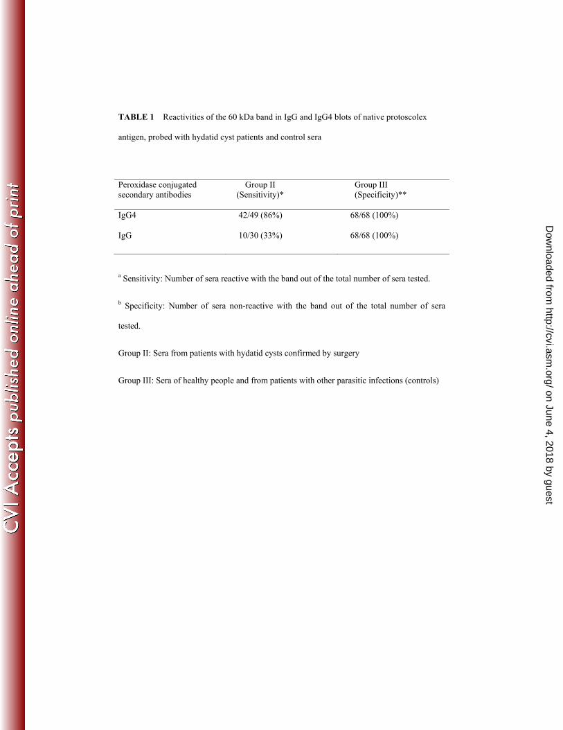
TABLE 1 Reactivities of the 60 kDa band in IgG and IgG4 blots of native protoscolex
antigen, probed with hydatid cyst patients and control sera
Peroxidase conjugated secondary antibodies
Group II (Sensitivity)*
Group III (Specificity)**
IgG4
42/49 (86%) 68/68 (100%)
IgG 10/30 (33%) 68/68 (100%)
a Sensitivity: Number of sera reactive with the band out of the total number of sera tested.
b Specificity: Number of sera non-reactive with the band out of the total number of sera
tested.
Group II: Sera from patients with hydatid cysts confirmed by surgery
Group III: Sera of healthy people and from patients with other parasitic infections (controls)
on June 4, 2018 by guesthttp://cvi.asm
.org/D
ownloaded from


![Echinococcus granulosus [Modo de compatibilidad].pdf](https://img.pdfslide.us/doc/110x75/577cc4d81a28aba7119aa462/echinococcus-granulosus-modo-de-compatibilidadpdf.jpg)