Embed Size (px)
Citation preview
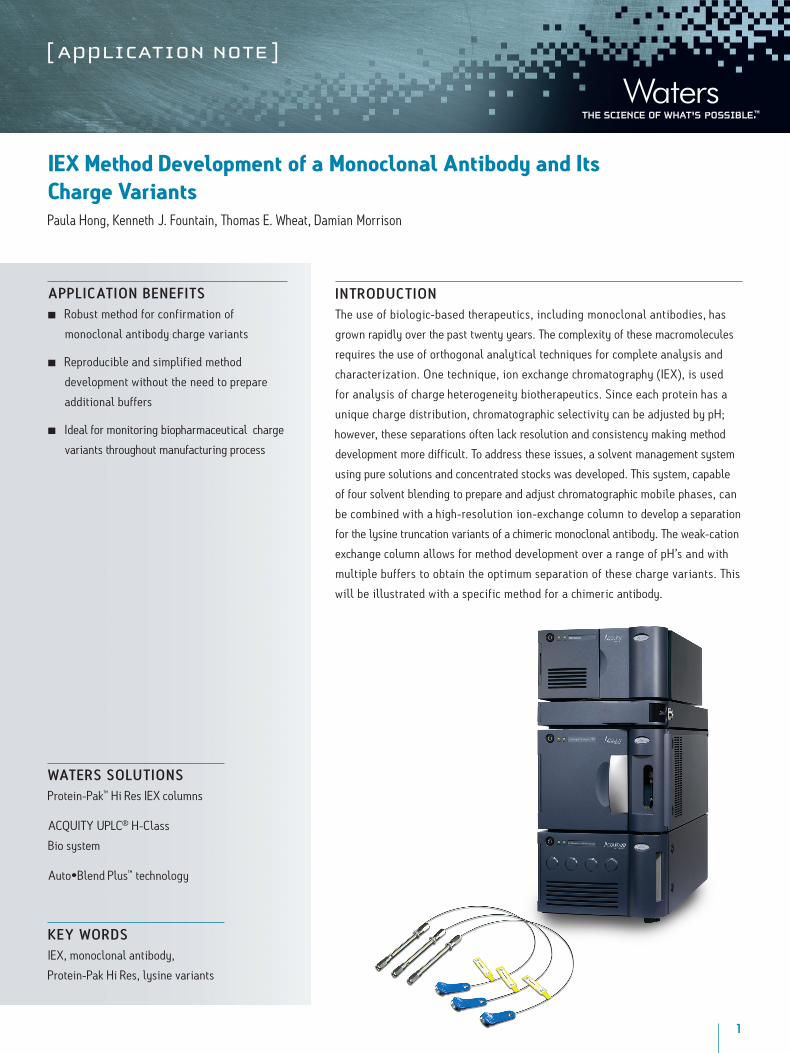
1
Wat ers solut ionsProtein-Pak™ Hi Res IEX columns
ACQUITY UPLC® H-Class
Bio system
Auto•Blend Plus™ technology
k ey WordsIEX, monoclonal antibody,
Protein-Pak Hi Res, lysine variants
aP PliCat ion BeneFitsn Robust method for confirmation of
monoclonal antibody charge variants
n Reproducible and simplified method
development without the need to prepare
additional buffers
n Ideal for monitoring biopharmaceutical charge
variants throughout manufacturing process
IEX Method Development of a Monoclonal Antibody and Its Charge VariantsPaula Hong, Kenneth J. Fountain, Thomas E. Wheat, Damian Morrison
int roduCt ionThe use of biologic-based therapeutics, including monoclonal antibodies, has
grown rapidly over the past twenty years. The complexity of these macromolecules
requires the use of orthogonal analytical techniques for complete analysis and
characterization. One technique, ion exchange chromatography (IEX), is used
for analysis of charge heterogeneity biotherapeutics. Since each protein has a
unique charge distribution, chromatographic selectivity can be adjusted by pH;
however, these separations often lack resolution and consistency making method
development more difficult. To address these issues, a solvent management system
using pure solutions and concentrated stocks was developed. This system, capable
of four solvent blending to prepare and adjust chromatographic mobile phases, can
be combined with a high-resolution ion-exchange column to develop a separation
for the lysine truncation variants of a chimeric monoclonal antibody. The weak-cation
exchange column allows for method development over a range of pH’s and with
multiple buffers to obtain the optimum separation of these charge variants. This
will be illustrated with a specific method for a chimeric antibody.

IEX Method Development of a Monoclonal Antibody and its Charge VariantsCharge Variants2
results and disCussionCharge heterogeneity of a monoclonal antibody may be caused by several structural
changes including C-terminal lysine processing.1 When present in biopharmaceuticals,
these charge variants are often monitored throughout manufacturing to ensure
control of the process. In the following study, an IEX method was developed to
confirm and quantify the presence of C-terminal lysine truncation variants in
a chimeric monoclonal antibody therapeutic. Method development was performed
on a weak-cation exchange column (Protein-Pak Hi Res CM, 4.6 x 100 mm, 7 µm)
by manipulating pH and ionic strength. A four solution blending system was used
to make pH buffer adjustments by using a weak acid (line A) and the cognate base
(line B). Sodium chloride (NaCl, line C) and water (line D) were used to adjust the
ionic strength of the buffer. These adjustments were performed using Auto•Blend
Plus Technology, which allowed the gradient to be expressed directly in terms of pH
and ionic strength. The operating software automatically calculates the percentage
of acid and base required for the specified pH from the known or measured pKa of
the selected buffer system.
des-lys di-lys mono-lys
pH 6.0
pH 6.4
pH 6.6
pH 7.0
pH 6.8
0.002
0.000
0.002
0.004
0.000
0.002
0.004
0.000
0.002
0.004
0.000
0.005
0.000
0 42 6 8 10 12 14 16 18 20 22 24 26 28 30 32 34 min
AUAU
AUAU
AU
Figure 1. Analysis of a chimeric antibody and its truncated C-terminal lysine variants on a Protein-Pak Hi Res CM column with sodium phosphate buffer. Separations were performed over a pH range of 6.0-7.0 using Auto•Blend Plus Technology.
0.002
0.004
0.000
0 108 12 14 16 18 20 22 24 26 28 30 32 34 36 38 40 42 min
AUAU
AUAU
AU
pH 6.0
pH 6.4
pH 6.6
pH 7.0
pH 6.8
des-lys di-lys
mono-lys
0.002
0.004
0.000
0.002
0.004
0.000
0.002
0.004
0.000
0.002
0.004
0.000
Figure 2. Analysis of a chimeric antibody and its truncated C-terminal lysine variants on a Protein-Pak Hi Res CM column with MES buffer. Separations were performed over a pH range of 6.0-7.0 using Auto•Blend Plus Technology.
eX PeriMentalsaMPle desCriPtion: A chimeric monoclonal antibody sample containing C-terminal lysine truncation variants was prepared at 1.25 mg/mL in 20 mM MES buffer, pH 6.
C-terminal lysine cleavage was performed using Carboxypeptidase B (CpB) (Worthington Biochemical Corp., p/n LS005304) prepared at 1 mg/mL. The monoclonal antibody (1000 µL, 1.25 mg/mL) and CpB (12.2 µL, 1 mg/mL) were combined. At predetermined time intervals (t= 0, 1, 2.5, 5, 7.5, 10, 12.5, 15, and 20 min), a 100 µL aliquot of the mixture was removed and combined with glacial acetic acid (1.7 µL) to halt the reaction.
lC conditions LC system: ACQUITY UPLC H-Class Bio System with Auto•Blend Plus Technology
Detector: PDA Detection with Titanium Flow Cell
Wavelength: 280 nm
Sampling rate: 20 pts/sec
Filter time constant: Normal
Column: Protein-Pak Hi Res IEX CM, 4.6 x 100 mm, 7 µm (P/N 186004929)
Column temp.: 30 °C
Sample temp.: 4 °C
Injection volume: 10 µL
Flow rate: 0.5 mL/min
Mobile phase A: 100 mM Sodium Phosphate, monobasic, or 100 mM MES monohydrate
Mobile phase B: 100 mM Sodium Phosphate, dibasic, or 100 mM MES sodium salt
Mobile phase C: 1000 mM Sodium Chloride (NaCl)
Mobile phase D: Water
Purge and wash solvents: 20mM Sodium Phosphate, pH 6.0 or 20mM MES, pH 6.0
Gradient: 0-10% C in 60 min,
(pH specified in figures)
data management Software: Empower™ 2 with Auto•Blend
Plus Technology
IEX Method Development of a Monoclonal Antibody and its Charge VariantsCharge Variants3
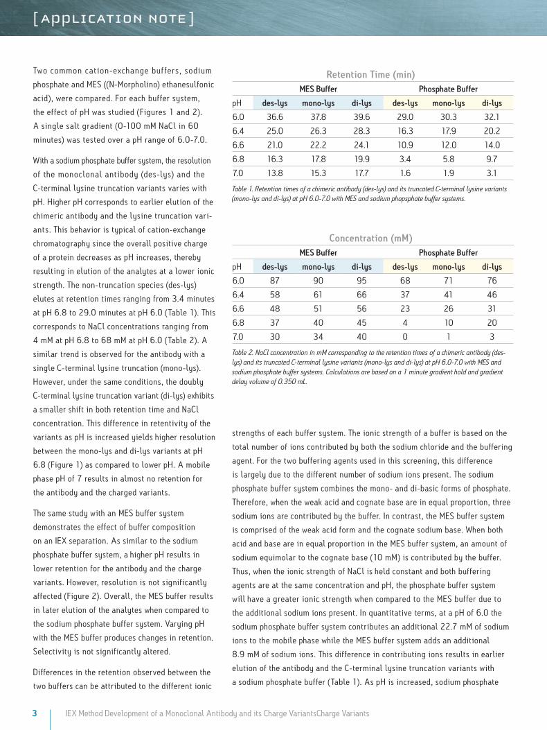
Table 1. Retention times of a chimeric antibody (des-lys) and its truncated C-terminal lysine variants (mono-lys and di-lys) at pH 6.0-7.0 with MES and sodium phopsphate buffer systems.
retention time (min)Mes Buffer Phosphate Buffer
pH des-lys mono-lys di-lys des-lys mono-lys di-lys
6.0 36.6 37.8 39.6 29.0 30.3 32.1
6.4 25.0 26.3 28.3 16.3 17.9 20.2
6.6 21.0 22.2 24.1 10.9 12.0 14.0
6.8 16.3 17.8 19.9 3.4 5.8 9.7
7.0 13.8 15.3 17.7 1.6 1.9 3.1
Two common cation-exc hange buffers, sodium
phosphate and MES ((N-Morpholino) ethanesulfonic
acid), were compared. For each buffer system,
the effect of pH was studied (Figures 1 and 2).
A single salt gradient (0-100 mM NaCl in 60
minutes) was tested over a pH range of 6.0-7.0.
With a sodium phosphate buffer system, the resolution
of the monoclonal antibody (des-lys) and the
C-terminal lysine truncation variants varies with
pH. Higher pH corresponds to earlier elution of the
chimeric antibody and the lysine truncation vari-
ants. This behavior is typical of cation-exchange
chromatography since the overall positive charge
of a protein decreases as pH increases, thereby
resulting in elution of the analytes at a lower ionic
strength. The non-truncation species (des-lys)
elutes at retention times ranging from 3.4 minutes
at pH 6.8 to 29.0 minutes at pH 6.0 (Table 1). This
corresponds to NaCl concentrations ranging from
4 mM at pH 6.8 to 68 mM at pH 6.0 (Table 2). A
similar trend is observed for the antibody with a
single C-terminal lysine truncation (mono-lys).
However, under the same conditions, the doubly
C-terminal lysine truncation variant (di-lys) exhibits
a smaller shift in both retention time and NaCl
concentration. This difference in retentivity of the
variants as pH is increased yields higher resolution
between the mono-lys and di-lys variants at pH
6.8 (Figure 1) as compared to lower pH. A mobile
phase pH of 7 results in almost no retention for
the antibody and the charged variants.
The same study with an MES buffer system
demonstrates the effect of buffer composition
on an IEX separation. As similar to the sodium
phosphate buffer system, a higher pH results in
lower retention for the antibody and the charge
variants. However, resolution is not significantly
affected (Figure 2). Overall, the MES buffer results
in later elution of the analytes when compared to
the sodium phosphate buffer system. Varying pH
with the MES buffer produces changes in retention.
Selectivity is not significantly altered.
Differences in the retention observed between the
two buffers can be attributed to the different ionic
Table 2. NaCl concentration in mM corresponding to the retention times of a chimeric antibody (des-lys) and its truncated C-terminal lysine variants (mono-lys and di-lys) at pH 6.0-7.0 with MES and sodium phosphate buffer systems. Calculations are based on a 1 minute gradient hold and gradient delay volume of 0.350 mL.
Concentration (mM)Mes Buffer Phosphate Buffer
pH des-lys mono-lys di-lys des-lys mono-lys di-lys
6.0 87 90 95 68 71 76
6.4 58 61 66 37 41 46
6.6 48 51 56 23 26 31
6.8 37 40 45 4 10 20
7.0 30 34 40 0 1 3
strengths of each buffer system. The ionic strength of a buffer is based on the
total number of ions contributed by both the sodium chloride and the buffering
agent. For the two buffering agents used in this screening, this difference
is largely due to the different number of sodium ions present. The sodium
phosphate buffer system combines the mono- and di-basic forms of phosphate.
Therefore, when the weak acid and cognate base are in equal proportion, three
sodium ions are contributed by the buffer. In contrast, the MES buffer system
is comprised of the weak acid form and the cognate sodium base. When both
acid and base are in equal proportion in the MES buffer system, an amount of
sodium equimolar to the cognate base (10 mM) is contributed by the buffer.
Thus, when the ionic strength of NaCl is held constant and both buffering
agents are at the same concentration and pH, the phosphate buffer system
will have a greater ionic strength when compared to the MES buffer due to
the additional sodium ions present. In quantitative terms, at a pH of 6.0 the
sodium phosphate buffer system contributes an additional 22.7 mM of sodium
ions to the mobile phase while the MES buffer system adds an additional
8.9 mM of sodium ions. This difference in contributing ions results in earlier
elution of the antibody and the C-terminal lysine truncation variants with
a sodium phosphate buffer (Table 1). As pH is increased, sodium phosphate
4 IEX Method Development of a Monoclonal Antibody and its Charge VariantsCharge Variants
contributes even more sodium ions in the form of the base (32.3 mM at pH 7) as compared to MES buffer
system (17.8 mM sodium ions), resulting in a greater ionic strength at a constant NaCl concentration, and
thus a greater retention time shift with pH as compared to MES buffer system (Table 1).
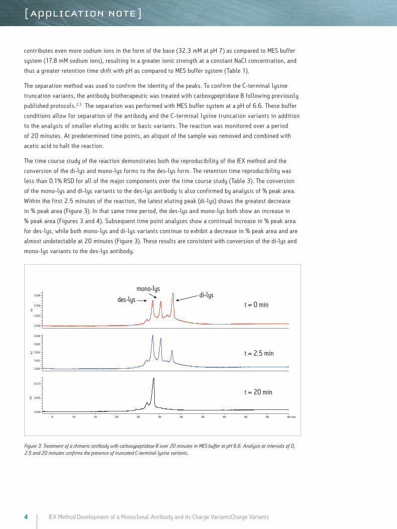
The separation method was used to confirm the identity of the peaks. To confirm the C-terminal lysine
truncation variants, the antibody biotherapeutic was treated with carboxypeptidase B following previously
published protocols.2,3 The separation was performed with MES buffer system at a pH of 6.6. These buffer
conditions allow for separation of the antibody and the C-terminal lysine truncation variants in addition
to the analysis of smaller eluting acidic or basic variants. The reaction was monitored over a period
of 20 minutes. At predetermined time points, an aliquot of the sample was removed and combined with
acetic acid to halt the reaction.
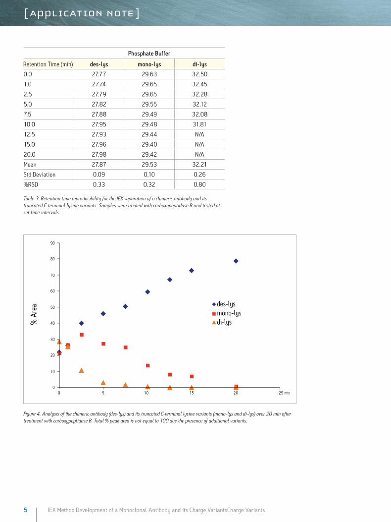
The time course study of the reaction demonstrates both the reproducibility of the IEX method and the
conversion of the di-lys and mono-lys forms to the des-lys form. The retention time reproducibility was
less than 0.1% RSD for all of the major components over the time course study (Table 3). The conversion
of the mono-lys and di-lys variants to the des-lys antibody is also confirmed by analysis of % peak area.
Within the first 2.5 minutes of the reaction, the latest eluting peak (di-lys) shows the greatest decrease
in % peak area (Figure 3). In that same time period, the des-lys and mono-lys both show an increase in
% peak area (Figures 3 and 4). Subsequent time point analyses show a continual increase in % peak area
for des-lys, while both mono-lys and di-lys variants continue to exhibit a decrease in % peak area and are
almost undetectable at 20 minutes (Figure 3). These results are consistent with conversion of the di-lys and
mono-lys variants to the des-lys antibody.
t = 0 min
t = 2.5 min
t = 20 min
des-lys di-lys
AU
0.000
0.002
0.004
0.006
AU
0.000
0.005
0.010
5 10 15 20 25 30 35 40 45 50 55 60 min
mono-lys
AU
0.000
0.002
0.004
0.006
0.008
Figure 3. Treatment of a chimeric antibody with carboxypeptidase B over 20 minutes in MES buffer at pH 6.6. Analysis at intervals of 0, 2.5 and 20 minutes confirms the presence of truncated C-terminal lysine variants..
5 IEX Method Development of a Monoclonal Antibody and its Charge VariantsCharge Variants
Figure 4. Analysis of the chimeric antibody (des-lys) and its truncated C-terminal lysine variants (mono-lys and di-lys) over 20 min after treatment with carboxypeptidase B. Total % peak area is not equal to 100 due the presence of additional variants.
% A
rea
30
20
10
00 5 10 15 20 25 min
70
60
90
80
50
40
des-lysmono-lysdi-lys
Table 3. Retention time reproducibility for the IEX separation of a chimeric antibody and its truncated C-terminal lysine variants. Samples were treated with carboxypeptidase B and tested at set time intervals.
Phosphate Buffer
Retention Time (min) des-lys mono-lys di-lys
0.0 27.77 29.63 32.50
1.0 27.74 29.65 32.45
2.5 27.79 29.65 32.28
5.0 27.82 29.55 32.12
7.5 27.88 29.49 32.08
10.0 27.95 29.48 31.81
12.5 27.93 29.44 N/A
15.0 27.96 29.40 N/A
20.0 27.98 29.42 N/A
Mean 27.87 29.53 32.21
Std Deviation 0.09 0.10 0.26
%RSD 0.33 0.32 0.80
Waters Corporation 34 Maple Street Milford, MA 01757 U.S.A. T: 1 508 478 2000 F: 1 508 872 1990 www.waters.com
Waters, and ACQUITY UPLC are registered trademarks of Waters Corporation. Protein-Pak, Auto-Blend Plus, Empower and The Science of What’s Possible are trademarks of Waters Corporation.All other trademarks are the property of their respective owners.
©2011 Waters Corporation. Produced in the U.S.A.March 2011 720003836EN KK-PDF
ConClusionsThe heterogeneity of a biopharmaceutical monoclonal antibody from C-terminal
lysine truncation is typically monitored throughout manufacturing to ensure
process stability and insure quality control. For these charge variants, the
Protein-Pak Hi Res CM column provides a tool for the analysis and confirmation
of a c himeric antibody and its C-terminal lysine truncation variants. T he
column, in combination with the ACQUITY H-Class Bio System and Auto•Blend
Plus Technology, allows for simplified pH screening and evaluation of multiple
buffer systems. Method development studies for the chimeric antibody
demonstrate the dramatic affect of pH with a sodium phosphate buffer, which is
partially due to the ionic strength of the buffering agent. In contrast, minimal
resolution effects are observed with varying pH in a MES buffer system. All of
the screening studies performed are simplified with the use of a four-solvent
blending system and Auto•Blend Plus Technology. The resulting separation
provides a robust method for analysis and confirmation of a monoclonal
antibody and its C-terminal lysine truncation variants.
references
1. Vlasak,J and Ionescu, “Hereterogeneity of Monoclonal Anibodies Revelealed by Charge-Sensitive Methods. ’’ Curr. Pham. BioTech. , 2008, 9. 468-481
2. Walker JM and Winder J S‚ “C Terminal Sequence Analysis woith Carboxypeptidase Y.” from Protein Protocols Handbook, Ed Walker, JM. Huamna Press Ince, Totowa, NJ.
3. Chen N, Nguyen M, Jacobsen F, Ouyang, J. “CEX- HPLC and Imaged cIEF of Antibodies with Engineered and Upaired Cysteines: from Multiple Main Peaks to One Peak.” APPS, Nov 2008
solutions in Practice
Auto•Blend Plus Tutorial, IEX Technical Brief, Literature Reference 720003601en.