Embed Size (px)
Citation preview

IEEE TRANSACTIONS ON IMAGE PROCESSING, VOL. 13, NO. 6, JUNE 2004 739
Efficient Iris Recognition by CharacterizingKey Local Variations
Li Ma, Tieniu Tan, Fellow, IEEE, Yunhong Wang, Member, IEEE, and Dexin Zhang
Abstract—Unlike other biometrics such as fingerprints and face,the distinct aspect of iris comes from randomly distributed fea-tures. This leads to its high reliability for personal identification,and at the same time, the difficulty in effectively representing suchdetails in an image. This paper describes an efficient algorithm foriris recognition by characterizing key local variations. The basicidea is that local sharp variation points, denoting the appearing orvanishing of an important image structure, are utilized to repre-sent the characteristics of the iris. The whole procedure of featureextraction includes two steps: 1) a set of one-dimensional intensitysignals is constructed to effectively characterize the most impor-tant information of the original two-dimensional image; 2) usinga particular class of wavelets, a position sequence of local sharpvariation points in such signals is recorded as features. We alsopresent a fast matching scheme based on exclusive OR operation tocompute the similarity between a pair of position sequences. Ex-perimental results on 2 255 iris images show that the performanceof the proposed method is encouraging and comparable to the bestiris recognition algorithm found in the current literature.
Index Terms—Biometrics, iris recognition, local sharp varia-tions, personal identification, transient signal analysis, wavelettransform.
I. INTRODUCTION
WITH AN increasing emphasis on security, automatedpersonal identification based on biometrics has been
receiving extensive attention over the past decade. Biometrics[1], [2] aims to accurately identify each individual usingvarious physiological or behavioral characteristics, such as fin-gerprints, face, iris, retina, gait, palm-prints and hand geometryetc. Recently, iris recognition is becoming an active topic inbiometrics due to its high reliability for personal identification[1]–[3], [9], [10], [12]. The human iris, an annular part betweenthe pupil (generally appearing black in an image) and thewhite sclera as shown in Fig. 1, has an extraordinary structureand provides many interlacing minute characteristics such asfreckles, coronas, stripes, furrows, crypts and so on. Thesevisible characteristics, generally called the texture of the iris,are unique to each subject [5]–[12], [34]–[36]. The uniqueness
Manuscript received January 1, 2003; revised October 1, 2003. This work wassupported by the National Science Foundation of China (NSFC) under Grants69825105, 60121302, 60332010, and 60275003, and by the Chinese Academyof Sciences. The associate editor coordinating the review of this manuscript andapproving it for publication was Dr. Philippe Salembier.
L. Ma is with the National Laboratory of Pattern Recognition, Institute ofAutomation, Chinese Academy of Sciences, Beijing 100080, China (e-mail:[email protected]).
T. Tan, Y. Wang, and D. Zhang are with the National Laboratory of PatternRecognition, Institute of Automation, Chinese Academy of Sciences, Beijing100080, China (e-mail: [email protected]; [email protected]; [email protected]).
Digital Object Identifier 10.1109/TIP.2004.827237
Fig. 1. Samples of iris images.
of the iris pattern is the direct result of the individual differencesthat exist in the development of the anatomical structures in thebody. Some research work [12], [34]–[36] has also stated thatthe iris is essentially stable over a person’s life. Furthermore,since the iris is an internal organ as well as externally visible,iris-based personal identification systems can be noninvasiveto their users [9]–[12], [35], [36], which is of great importancefor practical applications. All these desirable properties (i.e.,uniqueness, stability, and noninvasiveness) make iris recogni-tion a particularly promising solution to security.
A. Related Work
Flom and Safir first proposed the concept of automated irisrecognition in 1987 [34]. Since then, some researchers workedon iris representation and matching and have achieved greatprogress [7]–[23], [35], [36]. Daugman [8]–[10] made use ofmultiscale Gabor filters to demodulate texture phase structureinformation of the iris. Filtering an iris image with a family offilters resulted in 1024 complex-valued phasors which denotethe phase structure of the iris at different scales. Each phasorwas then quantized to one of the four quadrants in the com-plex plane. The resulting 2048-component iriscode was usedto describe an iris. The difference between a pair of iriscodeswas measured by their Hamming distance. Sanchez-Reillo etal. [16] provided a partial implementation of the algorithm byDaugman. Wildes et al. [11] represented the iris texture witha Laplacian pyramid constructed with four different resolu-tion levels and used the normalized correlation to determinewhether the input image and the model image are from thesame class. Boles and Boashash [13] calculated a zero-crossingrepresentation of one-dimensional (1-D) wavelet transform atvarious resolution levels of a concentric circle on an iris image
1057-7149/04$20.00 © 2004 IEEE

740 IEEE TRANSACTIONS ON IMAGE PROCESSING, VOL. 13, NO. 6, JUNE 2004
to characterize the texture of the iris. Iris matching was basedon two dissimilarity functions. In [19], Sanchez-Avila et al.further developed the iris representation method by Boles etal. [13]. They made an attempt to use different similaritymeasures for matching, such as Euclidean distance and Ham-ming distance. Lim et al. [15] decomposed an iris image intofour levels using 2-D Haar wavelet transform and quantizedthe fourth-level high-frequency information to form an 87-bitcode. A modified competitive learning neural network (LVQ)was adopted for classification. Tisse et al. [20] analyzed theiris characteristics using the analytic image constructed by theoriginal image and its Hilbert transform. Emergent frequencyfunctions for feature extraction were in essence samples ofthe phase gradient fields of the analytic image’s dominantcomponents [25], [26]. Similar to the matching scheme ofDaugman, they sampled binary emergent frequency functionsto form a feature vector and used Hamming distance formatching. Park et al. [21] used a directional filter bank todecompose an iris image into eight directional subband out-puts and extracted the normalized directional energy as fea-tures. Iris matching was performed by computing Euclideandistance between the input and the template feature vectors.Kumar et al. [22] utilized correlation filters to measure theconsistency of iris images from the same eye. The correlationfilter of each class was designed using the two-dimensional(2-D) Fourier transforms of training images. If the correla-tion output (the inverse Fourier transform of the product ofthe input image’s Fourier transform and the correlation filter)exhibited a sharp peak, the input image was determined tobe from an authorized subject, otherwise an imposter. Bae etal. [23] projected the iris signals onto a bank of basis vectorsderived by independent component analysis and quantized theresulting projection coefficients as features.
Our earlier attempts to iris recognition developed the textureanalysis-based methods [14], [17], [18] and a local intensityvariation analysis-based method [37]. In [17], the global tex-ture features of the iris were extracted by means of well-knownGabor filters at different scales and orientations. Based on theexperimental results and analysis obtained in [17], we furtherconstructed a bank of spatial filters [18], whose kernels are suit-able for iris recognition, to represent the local texture featuresof the iris and thus achieved much better results. Different fromthe above two methods, we also developed a Gaussian–Her-mite moments-based method [37]. This method is our prelimi-nary work using local intensity variations of the iris as features.Gaussian–Hermite moments [24] which use Gaussian–Hermitepolynomial functions as transform kernels belong to a class oforthogonal moments. This means that they produce minimalinformation redundancy. Gaussian–Hermite moments can wellcharacterize local details of a signal since they construct orthog-onal features from the signal’s derivatives of different orders[24]. We decomposed an iris image into a set of 1-D intensitysignals (see Section IV-A for more details) and represented localvariations of the intensity signals using Gaussian–Hermite mo-ments (order 1 to 4). To reduce computational cost and improveclassification accuracy, we adopted Fisher linear discriminantto reduce the dimensionality of original features and the nearestcenter classifier for matching.
It should be noted that all these algorithms are based on grayimages, and color information is not used. The main reason isthat the most important information for recognition (i.e., texturevariations of the iris) is the same in both gray and color images.From the methods described above, we can conclude that thereare four main approaches to iris representation: phase-basedmethods [8]–[10], zero-crossing representation [13], [19], tex-ture analysis [11], [14], [15], [17], [18], [21], and intensity vari-ation analysis [23], [37]. However, the question of which ap-proach is most suitable for extracting iris features has never beenanswered.
B. Outline
In this paper, we first present our intuitive observations aboutthe characteristics of the iris based on the appearance of nu-merous iris images, and then introduce a new algorithm for irisrecognition inspired by such observations. Finally, we perform aseries of experiments to evaluate the proposed algorithm. More-over, in order to answer the question of which approach is mostsuitable for extracting iris features, we carry out extensive quan-titative comparison among some existing methods and providedetailed discussions on the overall experimental results. To thebest of our knowledge, this is the first attempt in comparing theexisting algorithms on a reasonably sized database.
The remainder of this paper is organized as follows. Section IIprovides an overview of our method based on an intuitive under-standing for iris features. Detailed descriptions of image prepro-cessing, feature extraction and matching are given in Section IIIand Section IV respectively. Experimental results and discus-sions are reported in Section V. Section VI concludes this paper.
II. OVERVIEW OF OUR APPROACH
Six iris samples captured by our home-made digital opticalsensor are shown in Fig. 1. We can see from these images thatthe iris consists of many irregular small blocks, such as freckles,coronas, stripes, furrows, crypts, and so on. Furthermore, thedistribution of these blocks in the iris is also random. Such ran-domly distributed and irregular blocks constitute the most dis-tinguishing characteristics of the iris.
Intuitively, if we can precisely locate each of these blocks inthe image and recognize the corresponding shape as well, thenwe will obtain a high performance algorithm. But it is almost im-possible to realize such an idea. Unlike fingerprint verification,where feature extraction can rely on ridge following, it is diffi-cult to well segment and locate such small blocks in gray im-ages. Moreover, classifying and recognizing the shape of suchblocks is unpractical due to their great irregularity. From theviewpoint of signal processing, however, we can regard theseirregular blocks as a kind of transient signals. Therefore, irisrecognition can be solved using some approaches to transientsignal analysis. As we know, local sharp variations denote themost important properties of a signal. In our framework, we thusrecord the position of local sharp variation points as features in-stead of locating and recognizing those small blocks. Fig. 2 il-lustrates the main steps of our method.
First, the background in the iris image is removed by local-izing the iris. In order to achieve invariance to translation andscale, the annular iris region is normalized to a rectangular block

MA et al.: EFFICIENT IRIS RECOGNITION BY CHARACTERIZING KEY LOCAL VARIATIONS 741
Fig. 2. Diagram of our approach.
of a fixed size. After lighting correction and image enhance-ment, we construct a set of 1-D intensity signals containing themain intensity variations of the original iris for subsequent fea-ture extraction. Using wavelet analysis, we record the positionof local sharp variation points in each intensity signal as fea-tures. Directly matching a pair of position sequences is also verytime-consuming. Here, we adopt a fast matching scheme basedon the exclusive OR operation to solve this problem. The pro-posed method is detailed in the following sections.
III. IRIS IMAGE PREPROCESSING
Our preprocessing operates in three steps. First, the iris is lo-calized and the irrelevant parts (e.g. eyelid, pupil etc.) are re-moved from the original image. Then, the localized iris is un-wrapped to a rectangular block of a fixed size in order to reducethe deformation caused by variations of the pupil and obtain ap-proximate scale invariance. Finally, lighting correction and con-trast improvement are applied to compensate for differences ofimaging conditions.
A. Localization
The iris is an annular portion between the pupil (innerboundary) and the sclera (outer boundary). Both the innerboundary and the outer boundary of a typical iris can approxi-mately be taken as circles. However, the two circles are usuallynot concentric [8]. We first roughly determine the iris regionin the original image, and then use edge detection and Houghtransform to exactly compute the parameters of the two circlesin the determined region. The detailed steps are as follows.
1) Project the image in the vertical and horizontal direc-tion to approximately estimate the center coordinates
of the pupil. Since the pupil is generally darkerthan its surroundings, the coordinates corresponding tothe minima of the two projection profiles are consideredas the center coordinates of the pupil
(1)
Fig. 3. Iris image preprocessing: (a) original image; (b) localized image;(c) normalized image; (d) estimated local average intensity; and (e) enhancedimage.
where and denote the center coordinates of thepupil in the original image .
2) Compute a more accurate estimate of the center coordi-nates of the pupil. We binarize a 120 120 region cen-tered at the point by adaptively selecting a rea-sonable threshold using the gray level histogram of thisregion. The centroid of the resulting binary region is con-sidered as a new estimate of the pupil coordinates. Notethat one can improve accuracy for estimating the centercoordinates of the pupil by repeating this step since thecoordinates estimated by image projection de-scribed in the first step are sometimes slightly far fromthe real center coordinates of the pupil.
3) Calculate the exact parameters of these two circles usingedge detection (Canny operator [27] in our experiments)and Hough transform [28] in a certain region determinedby the center of the pupil .
In the experiments, we perform the second step twice fora reasonably accurate estimate. Compared with the localiza-tion method by Wildes et al. [11] where the combination ofedge detection and Hough transform is also adopted, our methodapproximates the iris region before edge detection and Houghtransform. This will reduce the region for edge detection andthe search space of Hough transform, and thus result in lowercomputational cost. An example of iris localization is shown inFig. 3(b).
B. Normalization
Irises from different people may be captured in different size,and even for irises from the same eye, the size may change dueto illumination variations and changes of the camera-to-eye dis-

742 IEEE TRANSACTIONS ON IMAGE PROCESSING, VOL. 13, NO. 6, JUNE 2004
tance. Such elastic deformation in iris texture will affect thematching results. For the purpose of achieving more accuraterecognition results, it is necessary to compensate for such de-formation. Wildes et al. [11] solved this problem by registeringthe input image with the model image. Daugman [8]–[10] rep-resented the iris using a fixed parameter interval in a doubly di-mensionless pseudo polar coordinate system, whereas our pre-vious method [14], [17], [18] normalized the iris into an imageof a fixed size. The normalization schemes described in [15],[16], [20], [21], and [23] are similar to our approach. These ex-isting methods are essentially the same except the method byWildes et al. [11]. In experiments, we counter-clockwise un-wrap the annular iris to a rectangular texture block with a fixedsize. The normalization not only reduces to a certain extent thedistortion of the iris caused by pupil movement but also simpli-fies subsequent processing.
C. Enhancement
The normalized iris image has low contrast and may havenonuniform brightness caused by the position of light sources.All these may affect the subsequent processing in feature ex-traction and matching. In order to obtain a more well-distributedtexture image, we first approximate intensity variations acrossthe whole image. The mean of each 16 16 small block con-stitutes a coarse estimate of the background illumination. Thisestimate is further expanded to the same size as the normalizedimage by bicubic interpolation. The estimated background illu-mination as shown in Fig. 3(d) is subtracted from the normal-ized image to compensate for a variety of lighting conditions.Then we enhance the lighting corrected image by means of his-togram equalization in each 32 32 region. Such processingcompensates for the nonuniform illumination, as well as im-proves the contrast of the image. Fig. 3(e) shows the prepro-cessing result of an iris image, from which we can see that finertexture characteristics of the iris become clearer than those inFig. 3(c).
IV. FEATURE EXTRACTION AND MATCHING
As mentioned earlier, the characteristics of the iris can be con-sidered as a sort of transient signals. Local sharp variations aregenerally used to characterize the important structures of tran-sient signals. We thus construct a set of 1-D intensity signalswhich are capable of retaining most sharp variations in the orig-inal iris image. Wavelet transform is a particularly popular ap-proach to signal analysis and has been widely used in image pro-cessing [29]–[33]. In this paper, a special class of 1-D wavelets(the wavelet function is a quadratic spline of a finite support)is adopted to represent the resulting 1-D intensity signals. Theposition of local sharp variation points is recorded as features.
A. Generation of 1-D Intensity Signals
Local details of the iris generally spread along the radial di-rection in the original image corresponding to the vertical direc-tion in the normalized image [see Fig. 3(e)]. Therefore, infor-mation density in the angular direction corresponding to the hor-izontal direction in the normalized image is much higher thanthat in other directions [9], [18]; i.e., it may suffice only to cap-
ture local sharp variations along the horizontal direction in thenormalized image to characterize an iris. In addition, since ourbasic idea is to represent the randomly distributed blocks of theiris by characterizing local sharp variations of the iris, it is un-necessary to capture local sharp variation points in every lineof the iris image for recognition. Bearing these two aspects inmind, we decompose the 2-D normalized image into a set of1-D intensity signals according to the following equation:
...
...
(2)
where is the normalized image of (64 512 in our ex-periments), denotes gray values of the th row in the image ,
is the total number of rows used to form a signal , is thetotal number of 1-D signals. In essence, each intensity signal isa combination of successive horizontal scan lines which re-flect local variations of an object along the horizontal direction.A set of such signals contains the majority of the local sharpvariations of the iris. This is confirmed by the experimental re-sults reported in Section V. Moreover, such processing reducesthe computational cost required for subsequent feature repre-sentation. In experiments, we find that the iris regions close tothe sclera contain few texture characteristics and are easy to beoccluded by eyelids and eyelashes. Therefore, we extract fea-tures only in the top-most 78% section (corresponding to the re-gions closer to the pupil) of the normalized image. The relationbetween the total row number of the normalized image, thetotal number of 1-D signals and the number of rows usedto form a 1-D signal is denoted as . Sincethe total row number of the normalized image is fixed, theproduct of the total number of 1-D signals and the number
of rows used to form a 1-D signal is a constant in experi-ments. The recognition rate of the proposed algorithm can beregulated by changing the parameter . A small leads to alarge set of signals which results in characterizing the iris de-tails more completely, and thus increases recognition accuracy.A large , however, implies a lower recognition rate with ahigher computational efficiency. This way, we can trade off be-tween speed and accuracy. In experiments, we chooseand .
B. Feature Vector
As a well-known multiresolution analysis approach, thedyadic wavelet transform has been widely used in variousapplications, such as texture analysis, edge detection, imageenhancement and data compression [29]–[33]. It can decom-pose a signal into detail components appearing at differentscales. The scale parameter of the dyadic wavelets varies onlyalong the dyadic sequence . Here, our purpose is toprecisely locate the position of local sharp variations whichgenerally indicate the appearing or vanishing of an important

MA et al.: EFFICIENT IRIS RECOGNITION BY CHARACTERIZING KEY LOCAL VARIATIONS 743
image structure. The dyadic wavelets satisfy such requirementsas well as incur lower computational cost, and are thus adoptedin our experiments. The dyadic wavelet transform of a signal
at scale is defined as follows (in convolution form):
(3)
where is the wavelet function at scale . In our algo-rithm, the function is a quadratic spline which has a com-pact support and one vanishing moment [29], [31], [33]. Thismeans that local extremum points of the wavelet transform cor-respond to sharp variation points of the original signal. There-fore, using such a transform, we can easily locate the iris sharpvariation points by local extremum detection. Mallat [33] hasproved that the dyadic wavelet transform based on the abovewavelet function could be calculated with a fast filter bank al-gorithm. The detailed implementation may be referred to [33].As (3) shows, the wavelet transform of a signal includes a familyof signals providing detail components at different scales. Thereis an underlying relationship between information at consecu-tive scales, and the signals at finer scales are easily contami-nated by noise. Considering these two points (information re-dundancy at consecutive scales and the effect of noise on sig-nals at finer scales), we only use two scales to characterize dif-ferences among 1-D intensity signals. As we know, a local ex-tremum is either a local minimum or a local maximum. Irisimages shown in Fig. 3 illustrate that the irregular blocks ofthe iris are slightly darker than their surroundings. Therefore,it is reasonable to consider that a local minimum of the wavelettransform described above denotes the appearing of an irreg-ular block and a local maximum denotes the vanishing of anirregular block. A pair of adjacent local extremum points (aminimum point and a maximum point) indicates that a smallblock may exist between them. However, there are a few adja-cent local extremum points between which the amplitude differ-ence is very small. Such local extremum points may correspondto relatively faint characteristics in the iris image (i.e., local slowvariations in the 1-D intensity signals) and are less stable andreliable for recognition. A threshold-based scheme is used tosuppress them. If the amplitude difference between a pair of ad-jacent local extrema is less than a predetermined threshold, suchtwo local extremum points are considered from faint iris char-acteristics and not used as discriminating features. That is, weonly utilize distinct iris characteristics (hence local sharp varia-tions) for accurate recognition. For each intensity signal , theposition sequences at two scales are concatenated to form thecorresponding features:
(4)where the first components are from the first scale, the next
components from the other scale, denotes the position ofa local sharp variation point in the intensity signal, and ,respectively, represent the property of the first local sharp vari-ation point at two scales. If the first local sharp variation point
(or ) is a local minimum of the wavelet transform,(or ) is set to 1, otherwise 1. Features from different 1-D in-
Fig. 4. Illustration of feature transform.
tensity signals are concatenated to constitute an ordered featurevector
(5)
where denotes the features from the th intensity signal, andis the total number of 1-D intensity signals. Note that since
the number of local sharp variation points is distinct for differentirises, the dimensionality of the feature vector is not a con-stant. In our experiments, the feature vector generally consistsof about 660 components. In addition, at each scale, we replaceeach feature component denoting the actual position of localsharp variation points by its difference with the previous com-ponent. This will save memory since the difference (i.e., theinterval between two consecutive local sharp variation points)is less than 256 and can be represented in a byte.
C. Matching
We determine whether two irises are from the same class bycomparing the similarity between their corresponding featurevectors. Directly computing the distance between a pair of posi-tion sequences is inconvenient. Inspired by the matching schemeof Daugman [8]–[10], a two-step approach is proposed to solvethis problem.
1) The original feature vector is expanded into a binary fea-ture vector (called feature transform, in our algorithm).
2) The similarity between a pair of expanded feature vectorsis calculated using the exclusive OR operation.
Fig. 4 illustrates how to transform original features derived fromone intensity signal at a scale into a sequence of 0’s and 1’s(hereinafter, called a binary sequence). The length of the bi-nary sequence at a scale is the same as the length of the 1-Dintensity signal defined in (2). At each position denoted by theoriginal feature components, the binary sequence changes from1 to 0 or from 0 to 1. In the algorithm, we utilize the component
of the original features to set the values of the firstelements of the binary sequence. We define that the values ofthe first elements of the binary sequence are set to 1if is equal to 1 (i.e., is a local maximum point of thewavelet transform), otherwise 0. By such processing, a positionsequence as shown in Fig. 4 can be expanded into a binary se-quence. Using the same scheme, the original features of an irisdefined in (5) can be written as
(6)
where and are the binary sequences from theth 1-D intensity signal at the first and the second scale, re-
744 IEEE TRANSACTIONS ON IMAGE PROCESSING, VOL. 13, NO. 6, JUNE 2004
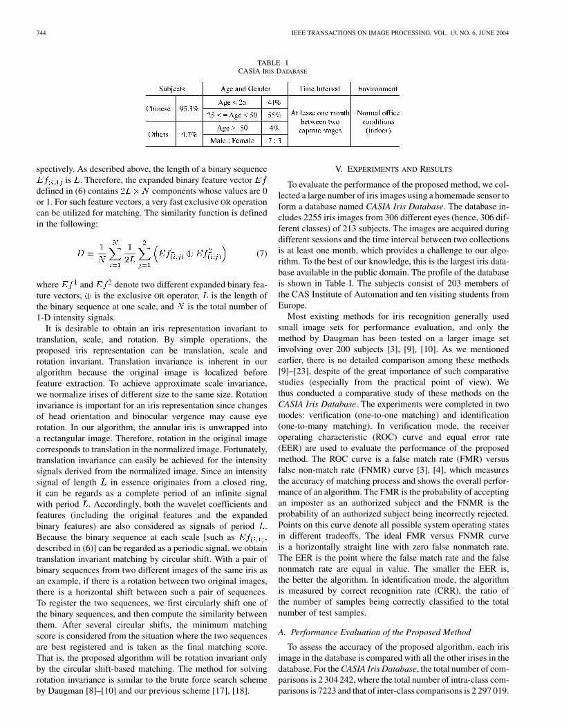
TABLE ICASIA IRIS DATABASE
spectively. As described above, the length of a binary sequenceis . Therefore, the expanded binary feature vector
defined in (6) contains components whose values are 0or 1. For such feature vectors, a very fast exclusive OR operationcan be utilized for matching. The similarity function is definedin the following:
(7)
where and denote two different expanded binary fea-ture vectors, is the exclusive OR operator, is the length ofthe binary sequence at one scale, and is the total number of1-D intensity signals.
It is desirable to obtain an iris representation invariant totranslation, scale, and rotation. By simple operations, theproposed iris representation can be translation, scale androtation invariant. Translation invariance is inherent in ouralgorithm because the original image is localized beforefeature extraction. To achieve approximate scale invariance,we normalize irises of different size to the same size. Rotationinvariance is important for an iris representation since changesof head orientation and binocular vergence may cause eyerotation. In our algorithm, the annular iris is unwrapped intoa rectangular image. Therefore, rotation in the original imagecorresponds to translation in the normalized image. Fortunately,translation invariance can easily be achieved for the intensitysignals derived from the normalized image. Since an intensitysignal of length in essence originates from a closed ring,it can be regards as a complete period of an infinite signalwith period . Accordingly, both the wavelet coefficients andfeatures (including the original features and the expandedbinary features) are also considered as signals of period .Because the binary sequence at each scale [such as ,described in (6)] can be regarded as a periodic signal, we obtaintranslation invariant matching by circular shift. With a pair ofbinary sequences from two different images of the same iris asan example, if there is a rotation between two original images,there is a horizontal shift between such a pair of sequences.To register the two sequences, we first circularly shift one ofthe binary sequences, and then compute the similarity betweenthem. After several circular shifts, the minimum matchingscore is considered from the situation where the two sequencesare best registered and is taken as the final matching score.That is, the proposed algorithm will be rotation invariant onlyby the circular shift-based matching. The method for solvingrotation invariance is similar to the brute force search schemeby Daugman [8]–[10] and our previous scheme [17], [18].
V. EXPERIMENTS AND RESULTS
To evaluate the performance of the proposed method, we col-lected a large number of iris images using a homemade sensor toform a database named CASIA Iris Database. The database in-cludes 2255 iris images from 306 different eyes (hence, 306 dif-ferent classes) of 213 subjects. The images are acquired duringdifferent sessions and the time interval between two collectionsis at least one month, which provides a challenge to our algo-rithm. To the best of our knowledge, this is the largest iris data-base available in the public domain. The profile of the databaseis shown in Table I. The subjects consist of 203 members ofthe CAS Institute of Automation and ten visiting students fromEurope.
Most existing methods for iris recognition generally usedsmall image sets for performance evaluation, and only themethod by Daugman has been tested on a larger image setinvolving over 200 subjects [3], [9], [10]. As we mentionedearlier, there is no detailed comparison among these methods[9]–[23], despite of the great importance of such comparativestudies (especially from the practical point of view). Wethus conducted a comparative study of these methods on theCASIA Iris Database. The experiments were completed in twomodes: verification (one-to-one matching) and identification(one-to-many matching). In verification mode, the receiveroperating characteristic (ROC) curve and equal error rate(EER) are used to evaluate the performance of the proposedmethod. The ROC curve is a false match rate (FMR) versusfalse non-match rate (FNMR) curve [3], [4], which measuresthe accuracy of matching process and shows the overall perfor-mance of an algorithm. The FMR is the probability of acceptingan imposter as an authorized subject and the FNMR is theprobability of an authorized subject being incorrectly rejected.Points on this curve denote all possible system operating statesin different tradeoffs. The ideal FMR versus FNMR curveis a horizontally straight line with zero false nonmatch rate.The EER is the point where the false match rate and the falsenonmatch rate are equal in value. The smaller the EER is,the better the algorithm. In identification mode, the algorithmis measured by correct recognition rate (CRR), the ratio ofthe number of samples being correctly classified to the totalnumber of test samples.
A. Performance Evaluation of the Proposed Method
To assess the accuracy of the proposed algorithm, each irisimage in the database is compared with all the other irises in thedatabase. For the CASIA Iris Database, the total number of com-parisons is 2 304 242, where the total number of intra-class com-parisons is 7223 and that of inter-class comparisons is 2 297 019.

MA et al.: EFFICIENT IRIS RECOGNITION BY CHARACTERIZING KEY LOCAL VARIATIONS 745
Fig. 5. Distributions of intra-class and inter-class distances. (a) The results from comparing images taken at the same session. (b) The results from comparingimages taken at different sessions.
As we know, the time lag between the date when images usedfor building the templates are captured and the date when thetest images are taken has an effect on intra-class matching dis-tances since there may be great variations between images ofthe same iris taken at different time. To explore the impact ofthe time lag on the proposed method, we respectively analyzethe experimental results based on comparisons between imagestaken at the same session and those based on comparisons be-tween images taken at different sessions. In our experiments,the time lag between different capture sessions is at least onemonth. For the results from comparing images taken at the samesession, the distribution of the intra-class matching distance isestimated with 3512 comparisons and the inter-class distribu-tion is estimated with 1 165 164 comparisons. For the resultsfrom comparing images taken at different sessions, the distribu-tion of the intra-class matching distance is estimated with 3711comparisons and the inter-class distribution is estimated with1 131 855 comparisons. Fig. 5 shows distributions of intra-classand inter-class matching distances in the two cases.
For a satisfying biometrics algorithm, intra-class distancesshould hardly vary with time. From the results shown in Fig. 5,we can see that the intra-class distance distribution derived fromcomparing images of the same iris taken at the same sessionand that derived from comparing images of the same iris takenat different sessions are very close. This demonstrates the highstability of the proposed iris features. It should be noted that thesame iris sensor is used to capture images at different sessions.If we make use of different iris sensors at different image cap-ture sessions, the differences between the above two intra-classdistance distributions may increase. However, this needs to befurther investigated. Fig. 5 also reveals that the distance be-tween the intra-class and the inter-class distribution is large, in-dicating the good discriminability of the extracted features. Thisis verified by the following verification results. Fig. 6 shows theROC curves of the proposed method, from which two observa-
tions can be made. First, the ROC curve based on different ses-sion comparisons interlaces with that based on the same sessioncomparisons. That is, the performance change caused by thetime-lag is extremely small for the proposed method. Second,the performance of our algorithm is very high and the EER isonly 0.09% for different session comparisons. In particular, ifone and only one false match occurs in 1 000 000 trails, falsenonmatch rate is less than 1.60%. The above experimental re-sults are highly encouraging. This also demonstrates that our irisrepresentation and matching schemes are very effective and the1-D intensity signals defined in (2) well capture the most dis-criminating information of the iris.
Experiments were carried out to investigate the cause of afew large intra-class distances. Such two pairs of iris imagesare listed in Figs. 7 and 8, from which two main reasons can beidentified.
1) Eyelids and eyelashes may occlude the effective regionsof the iris for feature extraction, and the failure of iris lo-calization (i.e., large localization errors) may cause falsenonmatching. Such an example is shown in Fig. 7. Inour experiments, we found that 57.7% false nonmatchesare incurred by the occlusion of eyelids and eyelashesand 21.4% false nonmatches come from the inaccuratelocalization. In addition, the inaccurate localization usu-ally occurs in the occluded images since the eyelids andeyelashes bring about some edge noises and decrease lo-calization accuracy. In order to reduce such false non-matches, we are working on detecting eyelids and eye-lashes so that feature extraction is only performed in theregions of no occlusion as well as localizing the iris moreexactly.
2) As shown in Fig. 8, the difference of the pupil size be-tween these two original images of the same eye is verysignificant. One has a pupil of normal size, and the othera considerably dilated pupil. Currently, we can recover

746 IEEE TRANSACTIONS ON IMAGE PROCESSING, VOL. 13, NO. 6, JUNE 2004
Fig. 6. Verification results.
Fig. 7. An example of false nonmatch due to eyelid/eyelash occlusion.
Fig. 8. An example of false nonmatch due to excessive pupil dilation.
the deformed iris caused by the dilation or constriction ofthe pupil to the normal iris by normalization. However,under the extreme conditions (namely the iris texture isexcessively compressed by the pupil), the iris after nor-malization still has many differences with its normal state
(i.e., the iris has a pupil of normal size). Therefore, thematching distance between such a pair of iris images isvery large. In our experiments, 10.7% false nonmatchesresult from the pupil changes. This is a common problemin all iris recognition methods. Iris normalization is thusan important research issue in the future.
Besides the above reasons, other factors that can result in falsenonmatching include the specular reflection from the cornea oreyeglasses, poorly focused images and motion-blurred images.Since these factors are relevant to noninvasive iris imaging, itis difficult to completely avoid the corresponding false non-matches. However, with the improvement of iris imaging, suchcases can be reduced.
B. Comparison With Existing Methods
The methods proposed by Daugman [8]–[10], Wildes et al.[11], Boles and Boashash [13] are the best known among ex-isting schemes for iris recognition. Furthermore, they charac-terize local details of the iris based on phase, texture analysisand zero-crossing representation. Therefore, we choose to com-pare our algorithm with theirs. For the purpose of comparison,we implemented the three methods according to the relatedpapers [8]–[13], [35], [36] (in our current implementations ofthe methods by Daugman [9] and Wildes et al. [11], we didnot carry out their schemes for eyelid and eyelash detection.Nevertheless, this should not invalidate our comparison exper-iments and the resulting conclusions. This is because if weperformed eyelid and eyelash detection with all these methods,their respective performance will be slightly improved.). Themethod by Wildes et al. only works in verification mode [11],so we do not test its performance in identification mode. Somemethods such as our previous method based on local intensityvariation analysis [37] and those presented in [11], [18] needmore than one sample for each class for training. Therefore, for

MA et al.: EFFICIENT IRIS RECOGNITION BY CHARACTERIZING KEY LOCAL VARIATIONS 747
TABLE IICOMPARISON OF CRRS AND EERS
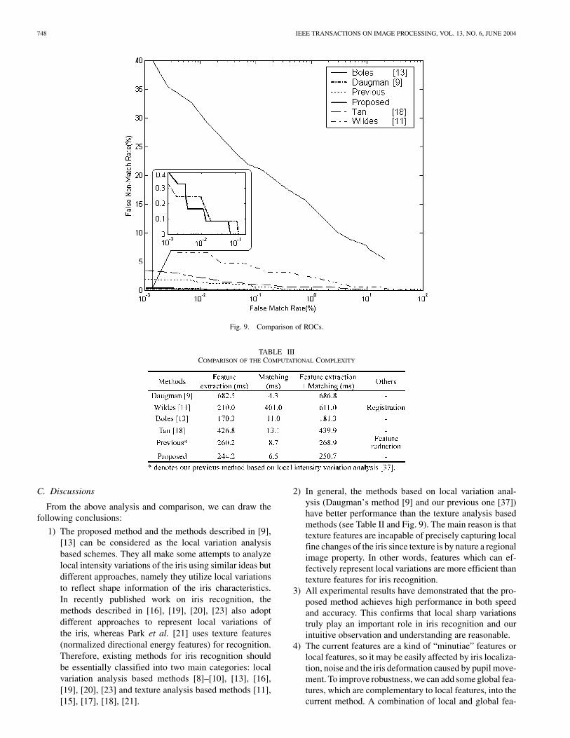
each iris class, we choose three samples from images taken atthe first session for training and all samples captured at othersessions serve as test samples. This is also consistent with thewidely accepted standard for biometrics algorithm testing [3],[4] (training images and testing images should be respectivelycaptured at different sessions). For the CASIA Iris Database,there are 918 images for training and 1237 images for testing.(To satisfy the above requirement, 100 images taken at the firstsession are not used in the experiments.) When matching theinput feature vector with the three templates of a class, the av-erage of the three scores is taken as the final matching distance.Table II and Fig. 9 describe the experimental results conductedon the CASIA Iris Database in two modes (verification andidentification), where denotes our previous method based onlocal intensity variation analysis [37].
From the results shown in Table II and Fig. 9, we can findthat Daugman’s method and the proposed method have the bestperformance, followed by our previous method based on localintensity variation analysis [37], our previous method [18], themethods by Wildes et al. [11] and Boles et al. [13]. The pro-posed method has an encouraging performance and its EERis only 0.07%. Wildes et al. [11] decomposed the iris textureinto four different frequency bands and used the normalizedcorrelation for verification. Image registration, which generallybrings about high computational cost, is an important step inthis method. Although combining the block correlation valuesby the median operation leads certain robustness against mis-registration, misregistration is still a main reason for false non-matching. That is, misregistration may affect the verificationaccuracy of this method. Our previous method [18] adopted abank of spatial filters to represent local texture information ofthe iris. The disadvantage of this method is that it cannot ex-actly capture the fine spatial changes of the iris. The theoret-ical basis of the method [13] comes from the signal reconstruc-tion theory based on the wavelet transform [30], [33]. However,good reconstruction does not necessarily mean accurate recog-nition. This is because that information used for reconstructionincludes some “individual” features of an image which do notexist in all samples from the same class and may reduce recog-nition accuracy. Furthermore, this method only employed ex-tremely little information along a concentric circle on the iristo represent the whole iris. These factors result in a relativelylow accuracy as shown in Fig. 9. More recently, Sanchez-Avilaand Sanchez-Reillo [19] further developed the method of Boleset al. by using different similarity measures for matching. Whenthe similarity measure was Hamming distance, this method cap-tured a small amount of local variations of the iris and thus
achieved 97.9% correct recognition rate on a data set of 200images from 10 subjects. However, the proposed method con-structs a set of intensity signals to contain the most importantdetails of the iris and makes use of stable and reliable local vari-ations of the intensity signals as features. This leads to the highperformance of the proposed method. Another method from ourgroup was based on local intensity variation analysis [37]. Sim-ilar to the proposed method, it characterized local variationsof a set of 1-D intensity signals using Gaussian–Hermite mo-ments and obtained satisfying results. But averaging the ad-jacent feature components for dimensionality reduction over-looks the effect of the most discriminating features on recog-nition accuracy. Both the proposed algorithm and the methodby Daugman achieve the best results. Daugman projected eachsmall local region onto a bank of Gabor filters, and then quan-tized the resulting phasor denoted by a complex-valued coef-ficient to one of the four quadrants in the complex plane. Inessence, this scheme analyzed local variations of the iris bycomparing and quantizing the similarity between Gabor filtersand the local regions. To achieve high accuracy, the local re-gion for feature extraction must be small enough. This resultsin a high dimensional feature vector (2048 components). Com-pared with Daugman’s method (which is the most exploitedcommercially), our current method contains about 660 compo-nents. This is because that our method only records the positionof local sharp variations as features and contains less redundantinformation. Daugman skillfully applied Gabor filters to rep-resent local shape of the iris, while we used quadratic splinewavelets to characterize local sharp variations of the iris. Thatis, the two methods aim to capture local variations of the iris asdiscriminating features. Therefore, they achieve quite close per-formance. Since the proposed iris representation is based on 1-Dfiltering and feature matching adopts the exclusive OR operation,our method is very efficient. Table III illustrates the computa-tional cost of the methods described in [9], [11], [13], [18] andthe current algorithm, including the CPU time for feature ex-traction (from an input image to a feature vector) and matching(from a pair of feature vectors to the matching result).
The above experiments used 200 different iris images andwere performed in Matlab 6.0 on a 500-MHz PC with 128 MRAM. Table III shows that the current method, our previousmethod [37] and the method of Boles et al. [13] consume lesstime than others [9], [18] for feature extraction. The reason isthat they are based on 1-D signal analysis and the other methodsinvolve 2-D mathematical operation. The method by Wildes etal. [11] only takes about 210 ms to build a four-level Laplacianpyramid representation of an image, whereas the piecewise cor-relation based matching generally needs high computational ex-pense as shown in Table III. Since Daugman’s method and ourmethod can compute the distance between a pair of feature vec-tors by the exclusive OR operation, they implement matchingfaster than others. If the exclusive OR operation is carried outusing some optimization methods in C/C++, the running timefor matching may be further reduced (as Daugman reported in[9], the matching speed of his method can be only 0.01 ms). Notethat the method by Wildes et al. [11] and our previous method[37] require extra cost for image registration and feature reduc-tion, respectively.
748 IEEE TRANSACTIONS ON IMAGE PROCESSING, VOL. 13, NO. 6, JUNE 2004
Fig. 9. Comparison of ROCs.
TABLE IIICOMPARISON OF THE COMPUTATIONAL COMPLEXITY
C. Discussions
From the above analysis and comparison, we can draw thefollowing conclusions:
1) The proposed method and the methods described in [9],[13] can be considered as the local variation analysisbased schemes. They all make some attempts to analyzelocal intensity variations of the iris using similar ideas butdifferent approaches, namely they utilize local variationsto reflect shape information of the iris characteristics.In recently published work on iris recognition, themethods described in [16], [19], [20], [23] also adoptdifferent approaches to represent local variations ofthe iris, whereas Park et al. [21] uses texture features(normalized directional energy features) for recognition.Therefore, existing methods for iris recognition shouldbe essentially classified into two main categories: localvariation analysis based methods [8]–[10], [13], [16],[19], [20], [23] and texture analysis based methods [11],[15], [17], [18], [21].
2) In general, the methods based on local variation anal-ysis (Daugman’s method [9] and our previous one [37])have better performance than the texture analysis basedmethods (see Table II and Fig. 9). The main reason is thattexture features are incapable of precisely capturing localfine changes of the iris since texture is by nature a regionalimage property. In other words, features which can ef-fectively represent local variations are more efficient thantexture features for iris recognition.
3) All experimental results have demonstrated that the pro-posed method achieves high performance in both speedand accuracy. This confirms that local sharp variationstruly play an important role in iris recognition and ourintuitive observation and understanding are reasonable.
4) The current features are a kind of “minutiae” features orlocal features, so it may be easily affected by iris localiza-tion, noise and the iris deformation caused by pupil move-ment. To improve robustness, we can add some global fea-tures, which are complementary to local features, into thecurrent method. A combination of local and global fea-

MA et al.: EFFICIENT IRIS RECOGNITION BY CHARACTERIZING KEY LOCAL VARIATIONS 749
tures is expected to further improve the performance ofour method. Texture features are well-known global in-formation. In the next stage, we will associate the currentfeatures with global texture information for more efficientand robust iris recognition.
VI. CONCLUSIONS
In this paper, we have presented an efficient algorithm for irisrecognition which is invariant to translation, scale and rotation.This method regards the texture of the iris as a kind of transientsignals and uses the wavelet transform to process such signals.The local sharp variation points, good indicators of importantimage structures, are extracted from a set of intensity signalsto form discriminating features. Experimental results have il-lustrated the encouraging performance of the current method inboth accuracy and speed. In particular, a comparative study ofexisting methods for iris recognition has been conducted. Suchperformance evaluation and comparison not only verify the va-lidity of our observation and understanding for the characteris-tics of the iris but also will provide help for further research.
ACKNOWLEDGMENT
The authors would like to thank Dr. J. Daugman (CambridgeUniversity, U.K.) and Dr. R. Wildes (York University, Canada)for their helpful comments and discussions. They also thank theanonymous referees for their thorough reviews and constructivecomments.
REFERENCES
[1] A. Jain, R. Bolle, and S. Pankanti, Eds., Biometrics: Personal Identifi-cation in a Networked Society. Norwell, MA: Kluwer, 1999.
[2] D. Zhang, Automated Biometrics: Technologies and Systems. Norwell,MA: Kluwer, 2000.
[3] T. Mansfield, G. Kelly, D. Chandler, and J. Kane, “Biometric producttesting final report,” Nat. Physical Lab., Middlesex, U.K., 2001.
[4] A. Mansfield and J. Wayman, “Best practice standards for testing andreporting on biometric device performance,” Nat. Physical Lab., Mid-dlesex, U.K., 2002.
[5] F. Adler, Physiology of the Eye: Clinical Application, 4th ed. London,U.K.: C.V. Mosby Co., 1965.
[6] H. Davision, The Eye. London, U.K.: Academic, 1962.[7] R. Johnson, “Can iris patterns be used to identify people?,” Chemical
and Laser Sciences Division LA-12 331-PR, Los Alamos Nat. Lab., LosAlamos, CA, 1991.
[8] J. Daugman, “High confidence visual recognition of persons by a test ofstatistical independence,” IEEE Trans. Pattern Analy. Machine Intell.,vol. 15, pp. 1148–1161, Nov. 1993.
[9] , “Statistical richness of visual phase information: update on recog-nizing persons by iris patterns,” Int. J. Comput. Vis., vol. 45, no. 1, pp.25–38, 2001.
[10] , “Demodulation by complex-valued wavelets for stochastic patternrecognition,” Int. J. Wavelets, Multi-Res. and Info. Processing, vol. 1, no.1, pp. 1–17, 2003.
[11] R. Wildes, J. Asmuth, G. Green, S. Hsu, R. Kolczynski, J. Matey, andS. McBride, “A machine-vision system for iris recognition,” Mach. Vis.Applic., vol. 9, pp. 1–8, 1996.
[12] R. Wildes, “Iris recognition: an emerging biometric technology,” Proc.IEEE, vol. 85, pp. 1348–1363, Sept. 1997.
[13] W. Boles and B. Boashash, “A human identification technique using im-ages of the iris and wavelet transform,” IEEE Trans. Signal Processing,vol. 46, pp. 1185–1188, Apr. 1998.
[14] Y. Zhu, T. Tan, and Y. Wang, “Biometric personal identification basedon iris patterns,” in Proc. Int. Conf. Pattern Recognition, vol. II, 2000,pp. 805–808.
[15] S. Lim, K. Lee, O. Byeon, and T. Kim, “Efficient iris recognition throughimprovement of feature vector and classifier,” ETRI J., vol. 23, no. 2, pp.1–70, 2001.
[16] R. Sanchez-Reillo and C. Sanchez-Avila, “Iris recognition with low tem-plate size,” in Proc. Int. Conf. Audio and Video-Based Biometric PersonAuthentication, 2001, pp. 324–329.
[17] L. Ma, Y. Wang, and T. Tan, “Iris recognition based on multichannelGabor filtering,” in Proc. 5th Asian Conf. Computer Vision, vol. I, 2002,pp. 279–283.
[18] , “Iris recognition using circular symmetric filters,” in Proc. 16thInt. Conf. Pattern Recognition, vol. II, 2002, pp. 414–417.
[19] C. Sanchez-Avila and R. Sanchez-Reillo, “Iris-based biometric recog-nition using dyadic wavelet transform,” IEEE Aerosp. Electron. Syst.Mag., vol. 17, pp. 3–6, Oct. 2002.
[20] C. Tisse, L. Martin, L. Torres, and M. Robert, “Person identificationtechnique using human iris recognition,” in Proc. Vision Interface, 2002,pp. 294–299.
[21] C. Park, J. Lee, M. Smith, and K. Park, “Iris-based personal authenti-cation using a normalized directional energy feature,” in Proc. 4th Int.Conf. Audio- and Video-Based Biometric Person Authentication, 2003,pp. 224–232.
[22] B. Kumar, C. Xie, and J. Thornton, “Iris verification using correlationfilters,” in Proc. 4th Int. Conf. Audio- and Video-Based Biometric PersonAuthentication, 2003, pp. 697–705.
[23] K. Bae, S. Noh, and J. Kim, “Iris feature extraction using independentcomponent analysis,” in Proc. 4th Int. Conf. Audio- and Video-BasedBiometric Person Authentication, 2003, pp. 838–844.
[24] J. Shen, W. Shen, and D. Shen, “On geometric and orthogonal mo-ments,” Int. J. Pattern Recognit. Artific. Intell., vol. 14, no. 7, pp.875–894, 2000.
[25] T. Tangsukson and J. Havlicek, “AM-FM image segmentation,” in Proc.EEE Int. Conf. Image Processing, 2000, pp. 104–107.
[26] J. Havlicek, D. Harding, and A. Bovik, “The mutli-component AM-FMimage representation,” IEEE Trans. Image Processing, vol. 5, pp.1094–1100, June 1996.
[27] J. Canny, “A computational approach to edge detection,” IEEE Trans.Pattern Anal. Machine Intell., vol. PAMI-8, pp. 679–698, Nov. 1986.
[28] D. Ballard, “Generalized Hough transform to detect arbitrary patterns,”IEEE Trans. Pattern Anal. Machine Intell., vol. PAMI-13, pp. 111–122,1981.
[29] S. Mallat and W. Hwang, “Singularity detection and processing withwavelets,” IEEE Trans. Inform. Theory, vol. 38, pp. 617–643, Mar. 1992.
[30] S. Mallat, “Zero-crossings of a wavelet transform,” IEEE Trans. on In-form. Theory, vol. 37, pp. 1019–1033, July 1992.
[31] S. Mallat and S. Zhong, “Characterization of signals from multiscaleedges,” IEEE Trans. Pattern Anal. Machine Intell., vol. 14, pp. 710–732,July 1992.
[32] Q. Tieng and W. Boles, “Recognition of 2D object contours using thewavelet transform zero-crossing representation,” IEEE Trans. PatternAnal. Machine Intell., vol. 19, pp. 910–916, Aug. 1997.
[33] S. Mallat, A Wavelet Tour of Signal Processing. New York: Academic,1999.
[34] L. Flom and A. Safir, “Iris Recognition system,” U.S. Patent 4 641 394,1987.
[35] J. Daugman, “Biometric personal identification system based on irisanalysis,” U.S. Patent 5 291 560, 1994.
[36] R. Wildes, J. Asmuth, S. Hsu, R. Kolczynski, J. Matey, and S. Mcbride,“Automated, noninvasive iris recognition system and method,” U.S.Patent 5 572 596, 1996.
[37] L. Ma, “Personal identification based on iris recognition,” Ph.D disser-tation, Inst. Automation, Chinese Academy of Sciences, Beijing, China,June 2003.
Li Ma received the B.Sc. and M.Sc. degrees inautomation engineering from Southeast University,Nanjing, China, in 1997 and 2000, respectively,and the Ph.D. degree in computer science from theNational Laboratory of Pattern Recognition, ChineseAcademy of Sciences, Beijing, China, in 2003.
Currently, he is a Research Member of the IBMChina Research Laboratory, Beijing, China. Hisresearch interests include image processing, patternrecognition, biometrics, Web mining, and knowledgemanagement.

750 IEEE TRANSACTIONS ON IMAGE PROCESSING, VOL. 13, NO. 6, JUNE 2004
Tieniu Tan (M’92–SM’97–F’04) received the B.Sc.degree in electronic engineering from Xi’an JiaotongUniversity, Xi’an, China, in 1984 and the M.Sc., DIC,and Ph.D. degrees in electronic engineering from Im-perial College of Science, Technology and Medicine,London, U.K., in 1986, 1986, and 1989, respectively.
He joined the Computational Vision Group at theDepartment of Computer Science, The University ofReading, Reading, U.K., in October 1989, where heworked as Research Fellow, Senior Research Fellow,and Lecturer. In January 1998, he returned to China
to join the National Laboratory of Pattern Recognition, Institute of Automation,Chinese Academy of Sciences, Beijing, China. He is currently Professor andDirector of the National Laboratory of Pattern Recognition, as well as Presidentof the Institute of Automation. He has published widely on image processing,computer vision, and pattern recognition. His current research interests includespeech and image processing, machine and computer vision, pattern recogni-tion, multimedia, and robotics.
Dr. Tan serves as referee for many major national and international journalsand conferences. He is an Associate Editor of Pattern Recognition and IEEETRANSACTIONS ON PATTERN ANALYSIS AND MACHINE INTELLIGENCE, and theAsia Editor of Image and Vision Computing. He was an elected member of theExecutive Committee of the British Machine Vision Association and Societyfor Pattern Recognition (1996–1997) and is a Founding Co-Chair of the IEEEInternational Workshop on Visual Surveillance.
Yunhong Wang (M’03) received the B.Sc. degreein electronic engineering from Northwestern Poly-technical University, Xi’an, China, and the M.S. andPh.D. degrees in electronic engineering from Nan-jing University of Science and Technology, Nanjing,China, in 1995 and 1998, respectively.
She joined the National Laboratory of PatternRecognition, Institute of Automation, ChineseAcademy of Sciences, Beijing, China, in 1998,where she has been an Associate Professor since2000. Her research interests include biometrics,
pattern recognition, and image processing.
Dexin Zhang received the B.Sc. degree in automa-tion engineering from Tsinghua University, Beijing,China, in 2000. He is currently pursuing his Master’sdegree at the National Laboratory of Pattern Recog-nition, Chinese Academy of Sciences, Beijing.
His research interests include biometrics, imageprocessing, and pattern recognition.