Embed Size (px)
DESCRIPTION
ID_Journal_2008_I
Citation preview

Cranio-maxillofacial
Implant Directions®
Vol.3 No.1 March 2008
Published by IF Publishing, Germany
EvidEncE REpoRt »Acid EtchEd vs. MAchinE
suRfAcE iMplAnts
litERAtuRE AnAlysis »sMoking And dEntAl iMplAnts
cRiticAl AppRAisAl »EnhAncEd BonE Apposition to A
chEMicAlly ModslA titAniuM suRfAcE
coRREctivE intERvEntion »iMMEdiAtE REstoRAtion AftER fAiluRE And
REplAcEMEnt of BAsAl iMplAnts
REsEARch in contExt »RAndoM AssignMEnt: lEt chAncE BE youR fRiEnd.
shoRt coMMunicAtion »A novEl ostEoinductivE
BonE gRAfting suBstitutE?
full lEnght ARticlE »Boi® iMplAnts As thE thERApy of choicE in dEntAl officEs
clinicAl REsEARch »outcoMEs of iMMEdiAtEly loAdEd full ARch
REconstRuctions on BAsAl iMplAnts And tEEth in thE MAndiBlE
cAsE REpoRt »iMMEdiAtE loAding of A MAxillARy full-ARch
REhABilitAtion suppoRtEd By BAsAl And cREstAl iMplAnts
ISS
N 1
86
4-1
19
9 /
e-IS
SN
18
64
-12
37

2
Editorial board
Editor-in-chief Dr. Werner Mander, [email protected]
Managing editor Dr. Sigmar Kopp, [email protected]
Coordinating editorN. N., Switzerland
Editorial board (in alphabetic order)Prof. Dr. Volker Bienengräber, GermanyHenri Diederich med.dent, LuxemburgDr. Yassen Dimitrov, BulgariaZa. Stephan Haas, GermanyProf. Dr. Vitomir S. Konstantinovic, SerbiaCarlos Mendez, SpainDr. Richard Musicer, USADr. Gerald Schillig, GermanyDr. Katrin Tost, Greece
Evidence reports and Critical AppraisalsIF Research & Evidence Dept. Single Issue Price Euro 30 Annual SubscriptionEuro 120
Copyright Copyright ©2008 byInternational Implant FoundationDE- 80802 Munich / Germanywww.implantfoundation.org
CMF.Impl.dir.ISSN 1864-1199e-ISSN 1864-1237
Disclaimer
HazardsGreat care has been taken to maintain the accuracy of the informa-tion contained in this publication. However, the publisher and/or the distributer and/or the editors and/or the authors cannot be held re-sponsible for errors or any consequences arising from the use of the information contained in this publication. The statements or opinions contained in editorials and articles in this publication are solely those of the authors thereof and not of the publisher, and/or the distributer, and/or the IIF.The products, procedures and therapies described in this work are hazardous and are therefore only to be applied by certified and trained medical professionals in environment specially designed for such pro-cedures. No suggested test or procedure should be carried out un-less, in the user‘s professional judgment, its risk is justified. Whoever applies products, procedures and therapies shown or described in this publication will do this at their own risk. Because of rapid advances in the medical sience, IF recommends that independent verification of diagnosis, therapies, drugs, dosages and operation methods should be made before any action is taken. Although all advertising material which may be inserted into the work is expected to conform to ethical (medical) standards, inclusion in this publication does not constitute a guarantee or endorsement by the publisher regarding quality or value of such product or of the claims made of it by its manufacturer.
Legal restrictionsThis work was produced by IF Publishing, Munich, Germany. All rights reserved by IF Publishing. This publication including all parts thereof, is legally protected by copyright. Any use, exploitation or commercializa-tion outside the narrow limits set forth by copyright legislation and the restrictions on use laid out below, without the publisher‘s consent, is illegal and liable to prosecution. This applies in particular to photostat reproduction, copying, scanning or duplication of any kind, translation, preparation of microfilms, electronic data processing, and storage such as making this publication available on Intranet or Internet. Some of the products, names, instruments, treatments, logos, desi-gns, etc. reffered to in this publication are also protected by patents and trademarks or by other intellectual property protection laws« (eg. «IF«, «IIF« and the IF-Logo) are registered trademarks even though specific reference to this fact is not always made in the text. Therefore, the appearance of a name, instrument, etc. without desi-gnation as proprietary is not to be construed as a representation by publisher that it is in the public domain.Institutions‘ subscriptions allow to reproduce tables of content or pre-pare lists of Articles including abstracts for internal circulation within the institutions concerned. Permission of the publisher is required for all other derivative works, including compilations and translations. Per-mission of the publisher is required to store or use electronically any material contained in this journal, including any article or part of an article. For inquiries contact the publisher at the adress indicated.

CMF.Impl.Dir. Vol 1-2008 3
Typical contents in ID
Evidence Reports summarize the latest «Hot Topics» from relevant journals putting similar studies «side-by-side». This unique presentation of studies allows you to compare and contrast the patient populations, the treatment interventions, and the quality of the scientific methods. The «evidence-based bottom line» is presented with an overall summary statement at the beginning. Clinical notes by implantologists with special expertise on the topic complete the Evidence Report by providing their expert clinical opinion. ID is an implantology publication that provides attention to detail in balancing science with clinical opinion in such a clear, concise, and visually-friendly presentation.
Literature Analyses provide you with an in-depth look at the research on a given topic. A «Literature Analysis» is a critical review of the literature on the epidemiology, treatment methods, and prognosis for implant-related topics or conditions. Literature Analyses are broader than «Evidence Reports» and are written to serve as a reference tool for implantologists to help them make decisions regarding how to manage patients, to assist them in evaluating needs for future research, and to use the material for future presentations.
Critical Appraisals summarize the findings from important papers used for clinical decision making or marketing by implant companies. In addition to the summary, the study‘s methods and clinical conclusions are critically reviewed in an effort to challenge the implantology community into not accepting everything that is published, while fostering alternative explanations and ideas.
Case reports give implantologists the opportunity to publish on unique patients using innovative or alternative methods for treating challenging patient conditions.
Research in Context is a helpful «what is» section to consult if you’ve ever read a study and asked «what is a p-value» or any other research method question. It assists clinicians with the critical evaluation of the literature by briefly describing relevant aspects of research methods and statistical analysis that may bias results and lead to erroneous conclusions.
•
•
•
•
•

4
Evidence Report
Effect of machined-surfaced versus chemical-ly conditioned surfaced dental implants upon implant survival and complications
Evidence Report Purpose
Increased surface roughness may enhance mechanical interlocking between the implant surface and the bone, which may result in in-creased resistance to compression, tension, and shear stress. Surface roughness may also stimulate faster and stronger osseointegra-tion because host tissue biomolecules adapt more firmly to the implant surface. By chemi-cally altering the surface morphology of dental implants, bone-to-implant contact may be en-hanced, thereby influencing the rate and extent of osseointegration of titanium implants.
Objective
To critically summarize the recently published literature examining implant survival and other outcomes in studies comparing machined-sur-faced with chemically conditioned surfaced (i.e. dual acid-etched, titanium-oxide, anodized sur-face) dental implants.
Summary
Cumulative survival rates were similar com-paring machined-surfaced to chemically condi-tioned surfaced dental implants in all studies. One study found lower success rates in ma-chined-surfaced compared to chemically condi-
tioned implants. There are conflicting findings with respect to peri-implant bone resorption comparing the two groups. Further, there were no statistically significant differences for implant stability or peri-implant bone resorption between machined-surfaced and chemically conditioned surfaced implants. Additional meth-odologically rigorous comparative studies, and studies evaluating other implants, are needed to better evaluate advantages and disadvantag-es of implant surface conditioning.
Sampling
A MEDLINE search was performed to iden-tify recent studies published between January 2000 and September 2007 examining the ef-fect of machined-surfaced versus chemically conditioned surfaced dental implants upon treatment outcomes. Twelve articles evaluated the treatment comparison of interest. Five ar-ticles which included outcomes on implant sur-vival met our criteria and are included in this report.
Common Outcome MeasuresImplant survivalImplant successPeri-implant bone resorptionImplant stabilitySoft-tissue parameters
•••••
CMF.Impl.Dir. Vol 1-2008 5
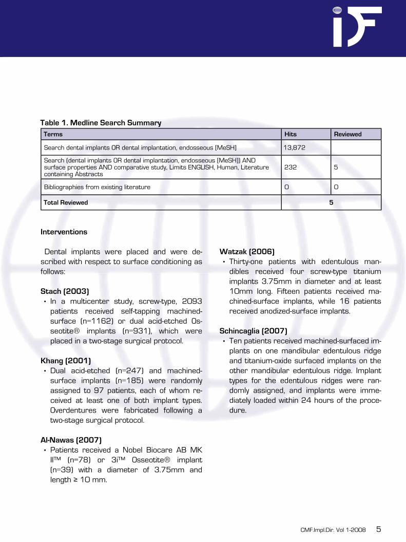
Table 1. Medline Search SummaryTerms Hits Reviewed
Search dental implants OR dental implantation, endosseous [MeSH] 13,872
Search (dental implants OR dental implantation, endosseous [MeSH]) AND surface properties AND comparative study, Limits ENGLISH, Human, Literature containing Abstracts
232 5
Bibliographies from existing literature 0 0
Total Reviewed 5
Interventions
Dental implants were placed and were de-scribed with respect to surface conditioning as follows:
Stach (2003)In a multicenter study, screw-type, 2093 patients received self-tapping machined-surface (n=1162) or dual acid-etched Os-seotite® implants (n=931), which were placed in a two-stage surgical protocol.
Khang (2001)Dual acid-etched (n=247) and machined-surface implants (n=185) were randomly assigned to 97 patients, each of whom re-ceived at least one of both implant types. Overdentures were fabricated following a two-stage surgical protocol.
Al-Nawas (2007)Patients received a Nobel Biocare AB MK II™ (n=78) or 3i™ Osseotite® implant (n=39) with a diameter of 3.75mm and length ≥ 10 mm.
•
•
•
Watzak (2006)Thirty-one patients with edentulous man-dibles received four screw-type titanium implants 3.75mm in diameter and at least 10mm long. Fifteen patients received ma-chined-surface implants, while 16 patients received anodized-surface implants.
Schincaglia (2007)Ten patients received machined-surfaced im-plants on one mandibular edentulous ridge and titanium-oxide surfaced implants on the other mandibular edentulous ridge. Implant types for the edentulous ridges were ran-domly assigned, and implants were imme-diately loaded within 24 hours of the proce-dure.
•
•

6
Table 2. Comparative studies evaluating machined-surfaced versus chemically conditioned sur-faced dental implants.
Treatment
Author(year)
Study Design
Population Diagnostic Characteristics
Machined-Surfaced(Group A)
Chemically Conditioned Surfaced(Group B)
Follow-up (%)
LoE†
Stach (2003)
Randomized controlled trial
N = 2093female: 58%age: MS=51.6±11.1 years; DAE=54.3±8.3 years
Indication for dental implant placement
N=1162; Ni=2585
N=931; Ni=2236
5 years: 98%
High
Khang(2001)
Randomized controlled trial
N = 97; Ni=432female: 62% age: 60±12 years
Indication for placement of >1 dental implant in the same restoration
N=NR; Ni=185
N=NR; Ni=247
3 years: NR*
High
Al-Nawas (2007)
Retrospective cohort
N = 118; Ni=264female: 60%age: 51±17 yrs
Indication for dental implant placement
N=78 N=39 43-51 months: 74%
Moderate
Watzak (2006)
Retrospective cohort
N = 31female: 58%age: 67.6 (52-86) years
Edentulous mandible, treated with implant-supported fixed bridges
N=15 N=16 30-48 months: 62%
Moderate
Schincaglia (2007)
Randomized controlled trial
N=10; Ni=42female: 40%age: 61.3 (37-74) years
Bilateral edentulous posterior mandible requiring a fixed partial denture of at least 2 teeth per side, same type of opposing occlusion bilaterally
N=10; Ni=20
N=10; Ni=22
12 months: 100%
Moderate
N=number of subjects; Ni=number of implants; MS=machined-surfaced; DAE=dual acid-etched*NR (not reported) = for follow-up rate either not reported or precise follow-up rate could not be determined since the initial number of eligible patients or number lost to follow-up were not provided.†Level of Evidence (LoE) is based on study design and methods (Very high, High, Moderate, and Poor)

CMF.Impl.Dir. Vol 1-2008 7
Table 3. Evaluation of articles comparing machined-surfaced versus chemically conditioned sur-faced dental implantsStudy design and methods Stach
(2003)Khang (2001)
Al-Nawas (2007)
Watzak (2006)
Schincaglia (2007)
1. What type of study design? RCT RCT RetrospectiveCohort
RetrospectiveCohort
RCT
2. Statement of concealed allocation?* NO NO N/A N/A YES
3. Intention to treat?* NO NO N/A N/A NO
4. Independent or blind assessment? NO NO NO NO NO
5. Complete follow-up of >85%? YES NO NO NO NO
6. Adequate sample size? YES YES YES NO YES
7. Controlling for possible confounding? NO YES YES YES YES
LEVEL OF EVIDENCE High High Moderate Moderate High
* Applies to randomized controlled trials only
ResultsImplant survival (Figure 1)
There are conflicting findings when comparing survival rates in machined-surfaced and chemi-cally conditioned surfaced implants; however, differences were not statistically significant:
At 5 years, there were no statically signifi-cant differences in survival rates between machined-surface and anodized-surface im-plants (92.7% vs. 98.3%, p>.05) [Stach].At a mean functional loading time of 33 months, there were no statistically signifi-cant differences in survival rates between machined-surface and anodized-surface im-plants (100% vs. 98.4%, p>.05) [Watzak].At one year, there was a higher survival rate for machined-surfaced compared to tita-nium-oxide surfaced implants, but the differ-ence was not statistically significant (100% vs. 90.5%, p>.05) [Schincaglia].
•
•
•
Implant successOverall success was defined as pocket probing
depth ≤ 5mm, negative bleeding on probing, and bone loss < 0.2mm annually.
Significantly lower implant success rates were found for machined-surface implants compared to dual acid-etched implants at 36 months (86.7% and 95.0%, respectively; p<.01). In good quality bone, the cumulative success rates at 48 months for machined-surface and dual acid-etched implants were 87.8% and 93.8%, respectively; the 48-month cumulative success rates in poor quality bone were 84.8% and 96.8%, re-spectively (p<.01) [Khang].Mean implant success rates did not re-veal any statistically significant differences between machined-surface and dual acid-etched implants (49 vs. 46 months, respec-tively; HR=0.7, 95% CI 0.3-1.5) [Al-Nawas].
•
•

8
Peri-implant bone resorption (Figure 2)There is a trend towards increased peri-implant
bone resorption associated with machined-sur-faced implants compared to chemically condi-tioned surfaced implants.
One study reported a significantly greater mean marginal bone loss around machined-surface implants compared to anodized surface implants (-1.4 ± 0.1mm vs. -1.2 ± 0.1mm, p=.03) [Watzak].Another study found no statistically signifi-cant differences for radiographic bone loss between machined-surfaced and titanium-oxide surfaced implants at one year (-1.1 ± 0.6mm vs. -0.9 ± 0.7mm, p=.224). Although these differences are not statistically signifi-cant, these findings may be due to the small number of subjects in this study. [Schinca-glia]
Implant stability
No statistically significant differences were found for Periotest® or resonance frequen-cy analysis values between machined-sur-faced and dual acid-etched dental implants at 2 years (p>0.05) [Al-Nawas].No statistically significant differences were found for resonance frequency analysis val-ues between machined-surfaced and titani-um-oxide surfaced dental implants at 1 year (p>0.05) [Schincaglia].
•
•
•
•
Soft tissue parameters
No statistically significant differences were found for peri-implant soft-tissue param-eters (probing depths, bleeding on probing) between machined-surface and dual acid-etched dental implants at 2 years (p>0.05) [Al-Nawas].No statistically significant differences were found for peri-implant soft-tissue parame-ters (marginal plaque index, probing depths, bleeding on probing) between machined-sur-face and anodized surface dental implants at a mean functional loading time of 33 months (p>0.05) [Watzak].
Methodological considerations
All studies reviewed were randomized con-trolled trials with a rating of high (low quality RCT) or cohort studies with a rating of mod-erate (low quality cohort) level of evidence. No very high quality randomized controlled trials or high quality cohort studies were identified in the literature. One of the studies [Schincaglia] had a sam-ple size that was likely inadequate to show a difference between the study groups.Since multiple implants in the same subject are not statistically independent, either one implant should be chosen per patient or sta-tistical analysis should account for multiple implants per patient. None of the studies reviewed accounted for multiple implants in the same subject.Only two of the studies reported a follow-up rate or provided data adequate enough to
•
•
•
•
•
•
CMF.Impl.Dir. Vol 1-2008 9
calculate the follow-up rate. A follow-up rate of ≥85% is necessary to ensure valid study results.
References
Studies Study 1Stach RM and Kohles SS (2003)A meta-analysis examining the clinical survivabil-ity of machined-surfaced and osseotite implants in poor-quality boneImplant Dentistry 12(1):87-93.
Study 2Khang W, Feldman S, Hawley CE, Gunsolley JA multi-center study comparing dual acid-etched and machined-surfaced implants in various bone qualitiesJ Periodontol 72(10):1384-90.
Study 3Al-Nawas B, Hangen U, Duschner H, Krum-menauer F, Wagner W (2007) Turned, machined versus double-etched dental implants in vivoClin Implant Dent Relat Res 9(2):71-78.
Study 4Watzak G, Zechner W, Busenlechner D, Arn-hart C, Gruber R, Watzak G (2006)Radiological and clinical follow-up of machined- and anodized-surface implants after mean func-tional loading for 33 monthsClin Oral Impl Res 17:651-7.
Study 5Schincaglia GP, Marzola R, Scapoli C, Scotti R (2007)Immediate loading of dental implants supporting fixed partial dentures in the posterior mandible: a randomized controlled split-mouth study—mach-inged versus titanium oxide implant surfaceInt J Oral Maxillofac Implants 22:35-46.

10
0 %
100 %
80 %
60 %
40 %
20 %
92,7%
100,0%
p> 0.05
Machined-Surfaced
Conditioned Surfaced
Statistical significance noted on graphs if provided by author
Cu
mu
lati
ve
Su
rviv
al
Ra
te (
%)
98,3% 98,4%100,0%
90,5%
p> 0.05
p> 0.05
60 months, n=2093 (Stach)
12 months, n=10 (Schincaglia)
33 months, n=31 (Watzak)
Figure 1. Cumulative survival rates for machined-surfaced vs. chemically conditioned surfaced dental implants*

CMF.Impl.Dir. Vol 1-2008 11
Figure 2. Mean peri-implant bone loss for machined-surfaced vs. chemically conditioned surfaced dental implants*
-5
0
-1
-2
-3
-4
-1,4
p> 0.05
Machined-Surfaced
Conditioned-Surfaced
Statistical significance noted on graphs if provided by author
-5
-5
33 months, n=31 (Stach) 12 months, n=10 (Schincaglia)
-1,2
-0,9
-1,1
p> 0.05
Me
an
Bo
ne
Lo
ss
(m
m)

12
Literature Analysis
Smoking and Dental ImplantsAre patients who smoke at greater risk of implant failure?
A “Literature Analysis” is a critical review of the literature on the epidemiology, treatment meth-ods, and prognosis for implant-related topics or conditions. Literature Analyses are broader than “Evidence Reports” (also published in each issue of Implant Directions) which focus on one specific treatment intervention by comparing and contrasting only 3 to 5 high quality articles in greater depth.
Literature Analyses are written to serve as a reference tool for implantologists:
To help them make decisions regarding how to manage patients;To assist them in evaluating needs for future research;To use the material for future presentations.
Purpose
The purpose of this Literature Analysis was to systematically search the literature to identify key articles in an effort to better understand the risk of implant failure in patients who are smok-ers. We were interested in how smoking may contribute to implant failure and other compli-cations. Moreover, we wanted to compare sur-vival rates of dental implants in smokers versus nonsmokers. This literature analysis will address the following objectives:
•
•
•
Report definition(s) of implant failureSummarize how smoking may contribute to implant failureExamine the effect of smoking upon implant failureExamine the effect of smoking upon implant complicationsSummarize survival rates of dental implants in smokers versus nonsmokersCompare survival rates of one and two-stage procedures in smoker and nonsmokersReport upon BOI as a potential alternative to dental implantation in smokers
Data Sources and Search Strategy
MEDLINE was searched to identify studies reporting data on smoking as a risk factor for dental implant failure (Table 1). There was no restriction on year published. An attempt was made to identify studies of high methodologi-cal quality (systematic reviews, RCT and cohort studies) comparing dental implant failure in smokers and nonsmokers. Studies evaluating a series of patients (i.e. case-series) and studies of < 10 subjects were excluded from the pri-mary review but may have been used to sup-port some of the background information. The following strategies were employed to identify literature to meet the objectives:
First strategy: Identify systematic review ar-ticles evaluating smoking as a risk factor for dental implant failure. Topics such as criteria for implant failure, dental implant survival rates, and risk factors for failure were included.
1.2.
3.
4.
5.
6.
7.

CMF.Impl.Dir. Vol 1-2008 13
Second strategy: Identify comparative studies evaluating smoking as a risk factor for dental implant failure.
Third strategy: Identify comparative studies evaluating smoking as a risk factor for dental implant complications.
The following are results of the various search strategies:
First strategy: We identified three systematic reviews on risk factors contributing to dental implant failure. One of these reviews did not evaluate smoking as a risk factor due to poor data, and two of these reviews highlighted stud-ies which were of poor quality, so they were ex-cluded.
Second strategy: We identified six comparative studies which evaluated evaluating smoking as a risk factor for dental implant failure.
Table 1. Medline Search SummaryTerms Hits Reviewed
Search („dental implantation, endosseous“ [MeSH] OR “dental implants” [MeSH]) AND “smoking” [MeSH] 160 8
Search („dental implantation, endosseous“ [MeSH] OR “dental implants” [MeSH]) AND “smoking” [MeSH] AND systematic review NOT case report, Limits ENGLISH, Literature containing Abstracts
22 0
Bibliographies from existing literature 2
Total Reviewed 10
Third strategy:: We identified four comparative studies which evaluated evaluating smoking as a risk factor for dental implant complications.
Background
Dental Implant Failure
Definition: Implant failures can be categorized into biologi-
cal failure, mechanical failure, iatrogenic failure, and inadequate patient adaptation1. A radio-graphic finding of implant failure is progressive bone loss. Clinically, implant failures may pres-ent with mobility, pain and/or infection.
14
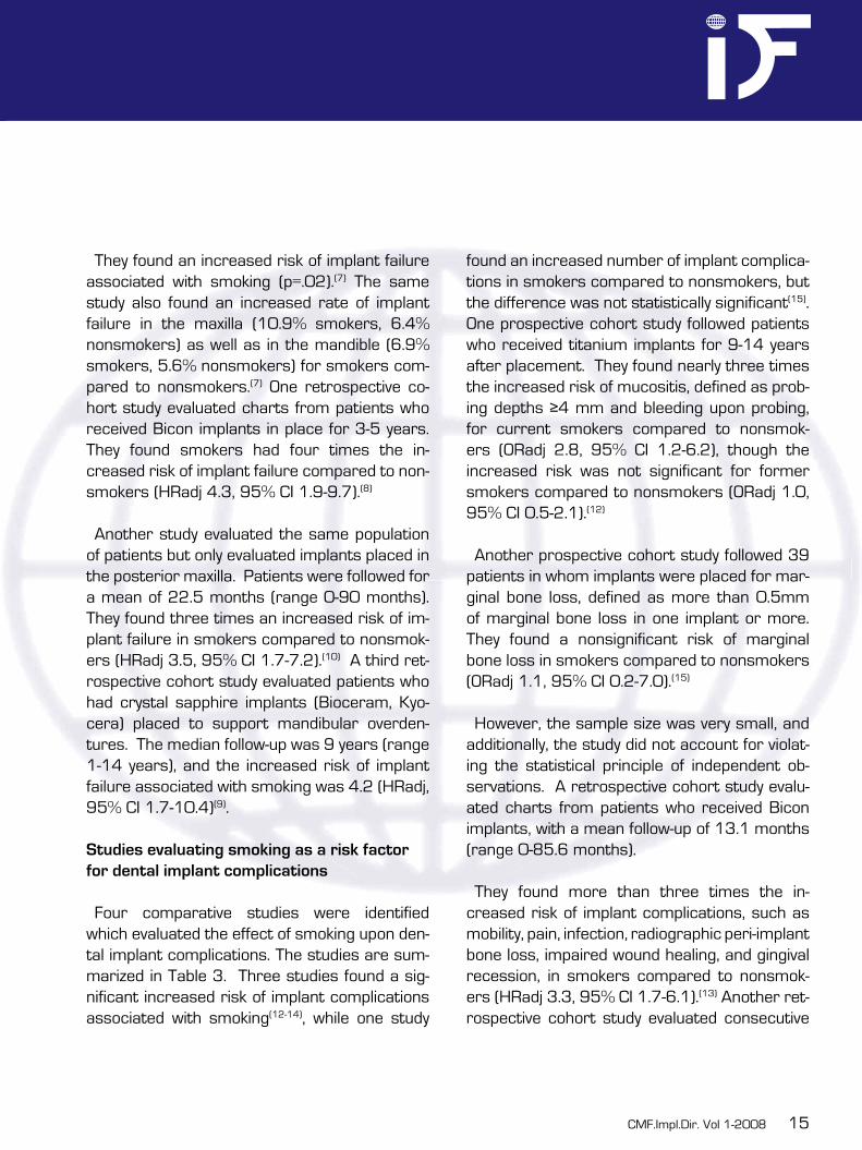
Smoking as an Etiology for Dental Implant Failure
Mechanism for smoking causing implant fail-ure:The negative effect of tobacco use on im-plant success may be related to the deleterious effect of smoking on wound healing, including a diminished proliferation of red blood cells, fibro-blasts, and macrophages.(2) Nicotine has also been associated with in-
creased platelet adhesiveness, which causes microclots and decreases microperfusion(3) and may lead to tissue ischemia. In addition, nicotine causes vasoconstriction, resulting from the release of adrenal and peripheral cat-echolamines.(4) Studies have shown that cate-cholamines released in this fashion undermine wound healing by retarding and decreasing the rate of epithelialization.(5) Furthermore, the in-take of carbon monoxide decreases the levels of oxygen available for tissue perfusion and leads to cellular hypoxia and diminished wound heal-ing.
Methodological Definitions
The relative risk (RR) is a relative comparison of outcomes between two groups that have dif-ferent exposures; it is the proportion of patients with the outcome in the treatment group (A) di-vided by the proportion of patients with the out-come in the control group (B). In survival analy-sis, the hazard ratio (HR) is reported and can be interpreted similarly. Statistical significance is reached if the 95% confidence intervals do not cross the value of one. The cumulative haz-ard is the probability of the endpoint of interest
(e.g. dental implant failure), taking into account the effect of several risk factors upon this prob-ability. The odds ratio (OR) is an estimate of the strength of the association between the risk factor and the disease outcome. The adjusted odds ratio is an odds ratio that takes into ac-count the effect of several risk factors upon the association.
Studies evaluating smoking as a risk factor for dental implant failure
Six comparative studies were identified which evaluated the effect of smoking upon dental im-plant failure. The studies are summarized in Table 2. Five studies found a significant increased risk of implant failure associated with smoking(6-10), while one study found an increased number of implant failures in smokers compared to non-smokers, but the difference was not statistically significant.(11)
One prospective cohort study followed patients who received implants for 2 years. They found the increased risk of implant failure associated with smoking was 14.4 (p<.0001)(6). Another prospective cohort study followed patients for 10 years after having received hollow screw im-plants of the ITI Dental Implant System. 35.7% of smokers experienced implant failure, while 22.6% of nonsmokers had failed implants, though this difference was not statistically sig-nificant in a multivariate logistic regression(11). A third prospective cohort study evaluated 3 - 5 year follow-up data of patients who received im-plants in a multicenter clinical study.
CMF.Impl.Dir. Vol 1-2008 15
They found an increased risk of implant failure associated with smoking (p=.02).(7) The same study also found an increased rate of implant failure in the maxilla (10.9% smokers, 6.4% nonsmokers) as well as in the mandible (6.9% smokers, 5.6% nonsmokers) for smokers com-pared to nonsmokers.(7) One retrospective co-hort study evaluated charts from patients who received Bicon implants in place for 3-5 years. They found smokers had four times the in-creased risk of implant failure compared to non-smokers (HRadj 4.3, 95% CI 1.9-9.7).(8)
Another study evaluated the same population of patients but only evaluated implants placed in the posterior maxilla. Patients were followed for a mean of 22.5 months (range 0-90 months). They found three times an increased risk of im-plant failure in smokers compared to nonsmok-ers (HRadj 3.5, 95% CI 1.7-7.2).(10) A third ret-rospective cohort study evaluated patients who had crystal sapphire implants (Bioceram, Kyo-cera) placed to support mandibular overden-tures. The median follow-up was 9 years (range 1-14 years), and the increased risk of implant failure associated with smoking was 4.2 (HRadj, 95% CI 1.7-10.4)(9).
Studies evaluating smoking as a risk factor for dental implant complications
Four comparative studies were identified which evaluated the effect of smoking upon den-tal implant complications. The studies are sum-marized in Table 3. Three studies found a sig-nificant increased risk of implant complications associated with smoking(12-14), while one study
found an increased number of implant complica-tions in smokers compared to nonsmokers, but the difference was not statistically significant(15).One prospective cohort study followed patients who received titanium implants for 9-14 years after placement. They found nearly three times the increased risk of mucositis, defined as prob-ing depths ≥4 mm and bleeding upon probing, for current smokers compared to nonsmok-ers (ORadj 2.8, 95% CI 1.2-6.2), though the increased risk was not significant for former smokers compared to nonsmokers (ORadj 1.0, 95% CI 0.5-2.1).(12)
Another prospective cohort study followed 39 patients in whom implants were placed for mar-ginal bone loss, defined as more than 0.5mm of marginal bone loss in one implant or more. They found a nonsignificant risk of marginal bone loss in smokers compared to nonsmokers (ORadj 1.1, 95% CI 0.2-7.0).(15)
However, the sample size was very small, and additionally, the study did not account for violat-ing the statistical principle of independent ob-servations. A retrospective cohort study evalu-ated charts from patients who received Bicon implants, with a mean follow-up of 13.1 months (range 0-85.6 months).
They found more than three times the in-creased risk of implant complications, such as mobility, pain, infection, radiographic peri-implant bone loss, impaired wound healing, and gingival recession, in smokers compared to nonsmok-ers (HRadj 3.3, 95% CI 1.7-6.1).(13) Another ret-rospective cohort study evaluated consecutive

16
patients who underwent placement of implants plus guided bone regeneration. They found an adjusted odds ratio of exposed surface area of the implant (bone fill <87%) to be 2.9 (95% CI 1.0-7.9).(14) This study did not account for violat-ing the statistical principle of independent ob-servations.
Survival rates of one and two-stage procedu-res in smokers and nonsmokers
One study estimated implant survival rates while taking into account potential confound-ers which may contribute to implant failure. In a retrospective cohort study(8), 553 patients who received Bicon implants and in whom smoking status was known were followed for 3-5 years.
Implant survival rates were estimated using a multivariate model. The highest survival rates were associated with nonsmokers who had im-plants placed in 2 stages. The 1- and 5- year survival rates, estimated from the multivariate model, were 97.1% (95% CI: 95.4-96.7) and 92.9% (95% CI: 88.9-97.1), respectively. Sur-vival rates for nonsmokers who had 1- stage implants were 91.1% (95% CI: 85.4-97.2) 1-year survival and 79.5% (95% CI: 65.0-97.3) 5-year survival. Smokers who have implants placed in 2-stages had a 1- year survival rate of 88.2% (95% CI: 80.6-96.5) and a 5-year sur-vival rate of 73.5% (95% CI: 57.4-94.0). The poorest outcome was expected from smokers who underwent a single-stage implant place-ment procedure. For this group, the estimated 1- and 5- year survival rates were 67.6% (95% CI: 47.6-96.0) and 38.3% (95% CI: 13.5-100),
respectively. BOI® as a potential alternative to conventio-nal implants among smokers
The following findings from this literature over-view make BOI® a potential alternative to bone augmentation procedures:
There appears to be an increased risk of implant failure in smokers compared to non-smokers.A proposed mechanism for implant failure is poor wound healing following the invasive procedure of implant placement.Since BOI® can be an effective implant in poor bone by distributing the loads laterally and capitalizing on cortical support, it may be superior dental implant in smokers when compared to root-form implants.
Future research recommendations
MEDLINE was searched to identify studies reporting data on smoking as a risk factor for dental implant failure. An attempt was made to identify studies of high methodological qual-ity (systematic reviews, RCT and cohort stud-ies) comparing dental implant failure in smokers and nonsmokers.
Although there is substantial literature on smoking as a risk factor for dental implant failure, the majority of these studies have significant methodological flaws. Few studies took into account any possible confounders which may also contribute to implant failure and/or obtaining poor follow-up of subjects.
1.
2.
3.
•
•

CMF.Impl.Dir. Vol 1-2008 17
Nevertheless, the included trials did provide limited but useful clinical information on the association between smoking and dental im-plant failure.Future studies on this topic should be clini-cal trials, concentrating research efforts on few important clinical questions, increasing the sample size, and decreasing the number of treatment variables in the trials. Collabor-ative efforts among various research groups are also encouraged.Clinical trials comparing BOI® to other im-plant procedures in smokers compared to nonsmokers would be an addition to the cur-rent literature. Longitudinal studies are needed to deter-mine long-term survival rates of dental im-plants in smokers compared to nonsmok-ers.
Executive Summary
We identified three systematic reviews on risk factors contributing to dental implant failure, though they were eliminated be-cause they did not address smoking as a risk factor or highlighted studies which were of poor quality. Ten additional comparative studies were identified, six of which evalu-ated the association between smoking and dental implant failure, and four studies ana-lyzed the association between smoking and dental implant complications.Five studies found a significant increased risk of implant failure associated with smoking(6-10), while one study found an increased number
•
•
•
•
•
•
of implant failures in smokers compared to nonsmokers, but the difference was not sta-tistically significant.(11)
Three studies found a significant increased risk of implant complications associated with smoking(12-14), while one study found an in-creased number of implant complications in smokers compared to nonsmokers, but the difference was not statistically significant.(15)
Implant survival rates were highest in non-smokers who had implants placed in two stages. The 1- and 5-year survival rates, es-timated from the multivariate model, were 97.1% (95% CI: 95.4-96.7) and 92.9% (95% CI: 88.9-97.1), respectively. The poor-est outcome occurred smokers who un-derwent a single-stage implant placement procedure. For this group, the estimated 1- and 5-year survival rates were 67.6% (95% CI: 47.6-96.0) and 38.3% (95% CI: 13.5-100), respectively.In the literature, it has been recommended that smokers refrain from smoking 1 week prior to and 8 week after implant place-ment.(16)
Since BOI® can be an effective implant in poor bone by distributing the loads laterally and capitalizing on cortical support, it may be superior dental implant in smokers when compared to root-form implants.
•
•
•
•
18
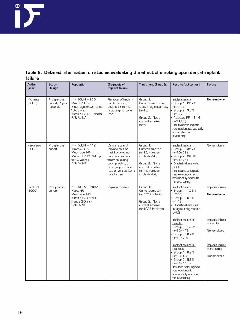
Table 2. Detailed information on studies evaluating the effect of smoking upon dental implant failure
Author(year)
Study Design
Population Diagnosis of Implant failure
Treatment Group (s) Results (outcomes) Favors
Moheng (2005)
Prospective cohort, 2 year follow-up
N = 93, Ni = 266;Male: 61.3%;Mean age: 60.3, range 18-85 yrs;Median F/U*: 2 years;F/U %: NA
Removal of implant due to probing depths ≥3 mm or radiographic bone loss
Group 1:Current smoker, at least 1 cigarette/day (n=15)
Group 2: Not a current smoker (n=78)
Implant failure• Group 1: 26.7% (n=4/15)
• Group 2: 3.8% (n=3/78)
• Adjusted RR = 14.4 (p<.0001) [multivariate logistic regression, statistically accounted for clustering]
Nonsmokers
Karoussis (2003)
Prospective cohort
N = 53, Ni = 112; Male: 40.2%;Mean age: NR;Median F/U*: NR (up to 10 years);F/U %: NR
Clinical signs of implant pain or mobility, probing depths >5mm or
=5mm+bleeding upon probing, or radiographic bone loss or vertical bone loss >2mm
Group 1:Current smoker (n=12, number implants=28)
Group 2: Not a current smoker (n=41, number implants=84)
Implant failure• Group 1: 35.7% (n=10/28)
• Group 2: 22.6% (n=65/84)
• Statistical analysis: p>.05[multivariate logistic regression, did not statistically account for clustering]
Nonsmokers
Lambert (2000)
Prospective cohort
N = NR, Ni = 2887; Male: NR;Mean age: NR;Median F/U*: NR (range 3-5 yrs);F/U %: 90
Implant removal Group 1:Current smoker (n=959 implants)
Group 2: Not a current smoker (n=1928 implants)
Implant failure• Group 1: 10.8% (±0.68)
• Group 2: 6.9% (±1.48)
• Statistical analysis: In logistic regression, p=.02
Implant failure in maxilla
• Group 1: 10.9% (n=52/478)
• Group 2: 6.4% (n=51/793)
Implant failure in mandible
• Group 1: 6.9% (n=33/481)
• Group 2: 5.6% (n=64/1135) [multivariate logistic regression, did statistically account for clustering]
Implant failure
Nonsmokers
Implant failure in maxilla
Nonsmokers
Implant failure in mandible
Nonsmokers

CMF.Impl.Dir. Vol 1-2008 19
Author(year)
Study Design
Population Diagnosis of Implant failure
Treatment Group (s)
Results (outcomes) Favors
Vehemente (2005)
Retrospective cohort
N = 553, Ni = 553; Male: 49.9%;Mean age: 53.3 ± 13.9 yrs;Median F/U*: NR (range 3-5 yrs);F/U %: 96.4
Implant removal Group 1:Current smoker (n=57)
Group 2: Not a current smoker (n=496)
Implant failure• Adjusted HR = 4.3 (1.9-9.7) [1 implant randomly selected from each patient]
Nonsmokers
Berge (2000)
Retrospective cohort, sapphire implants placed to support mandibular overdentures
N = 30, Ni = 116; Male: 30.0%; Mean age: 63.8 yrs (range 47-77 yrs); Median F/U*: 9 yrs (range 1-14 yrs);F/U %: 50
Clinical signs of implant infection, pain, mobility or radiographic marginal bone loss >4mm
Group 1:Current smoker (n=16, n=61 implants)
Group 2: Not a current smoker (n=14, n=55 implants)
Implant failure• Group 1: Mean implant survival = 10.19 yrs (8.85-11.54) • Group 2: Mean implant survival = 11.74 yrs (10.89-12.59)• Crude HR = 3.26 (1.38-7.72)• Adjusted HR = 4.21 (1.71-10.43) [multivariate logistic regression, did not statistically account for clustering]
Nonsmokers
McDermott (2006)
Retrospective cohort, implants placed in posterior maxilla
N = 269;Male: 50%; Mean age: 56.1±12.4; Mean F/U*: 22.5 mo (range 0-90.0 mos);F/U %: NR
Implant renoval, secondary to mobility
Group 1:Current smoker (n=28)
Group 2: Not a current smoker (n=241)
Implant failure• Unadjusted HR: 3.9 (2.1-7.5)• Adjusted HR: 3.5 (1.7-7.2) [multivariate logistic regression, did statistically account for clustering]
Nonsmokers
Bolded findings are statistically significant, p <0.05, while those that are not bolded are NOT statistically significant but tended to favor one treatmentPatient characteristics include sample size (N), number of implants (Ni), proportion male, and mean age or range or standard deviation (SD), and mean follow-up (F/U) and range if available; NR = Not reported

20
Author(year)
Study Design
Population Diagnosis of Implant complications
Treatment Group (s)
Results (outcomes) Favors
Roos-Jansaker (2006)
Prospective cohort
N=218, Ni = 999;Male: 50.9%;Mean age: NR;Median F/U*: NR (range 9-14 yrs);F/U %: 74.1
Mucositis: Probing depth ≥4mm and bleeding upon probing
Group 1:Current smoker (n=307)
Group 2: Former smoker (n=382)
Group 3: Never smoker (n=309)
Mucositis• Group 1: 59.0% (n=181/307)• Group 2: 42.9% (n=164/382)• Group 3: 42.7% (n=132/309)• Crude OR, former = 1.1 (0.6-2.3) • Crude OR, current = 2.9 (1.4-6.0) • Adj OR, former = 1.0 (0.5-2.1) • Adj OR, current = 2.8 (1.2-6.2) [multivariate logistic regression, statistically account for clustering]
Nonsmokers
Shimpuku (2003)
Prospective cohort
N = 39, Ni = 251; Male: 38.5%;Mean age: 55.1±9.4 yrs (range 29-74 yrs);Median F/U*: NR;F/U %: NR
Marginal bone loss: More than 0.5mm of marginal bone loss in one implant or more
Group 1:Current smoker (n=14)
Group 2: Not a current smoker (n=25)
Marginal bone loss• Group 1: 35.7% (n=5/14)• Group 2: 48.0% (n=12/25)• Adjusted OR: 1.09 (0.17-6.97)[multivariate logistic regression, did not statistically account for clustering]
Nonsmokers
McDermott (2003)
Prospective cohort
N=553; Ni = 553;Male: 49.9%;Mean age: 53.5±13.9;Median F/U*: 13.1 mos (range 0-85.6 mos);F/U %: NR
Implant complication: mobiity, pain, infection, radiographic peri-implant bone loss, impaired wound healing, gingival recession
Group 1:Current smoker (n=57)
Group 2: Not a current smoker (n=496)
Implant failure• Adjusted HR = 3.26 (1.74-6.10) [1 implant randomly selected from each patient]
Nonsmokers
Zitzmann (1999)
Retrospective cohort, subjects underwent placement of implants + guided bone regeneration
N = 75, Ni = 112; Male: 25.3%;Mean age: NR;Median F/U*: NR;F/U %: NR
exposed surface area of implant at baseline compared to re-entry (4 months for mandible, 6 months for maxilla) < 87%
Group 1:Current smoker (n=22)
Group 2: Not a current smoker (n=53)
Increase in bony defect• Group 1: 18% of implants• Group 2: 11.9% of implants• Crude OR = 1.96 (0.78-4.76) • Adjusted OR = 2.86 (1.01-7.94) [multivariate logistic regression, did not statistically account for clustering]
Nonsmokers
Bolded findings are statistically significant, p <0.05, while those that are not bolded are NOT statistically significant but tended to favor one treatment
Table 3. Detailed information on studies evaluating the effect of smoking upon dental implant

CMF.Impl.Dir. Vol 1-2008 21
References
Esposito M, Hirsch JM, Lekholm U and Thomsen P: Biological factors contributing to failures of osseointegrated oral implants. (II). Etiopathogenesis. Eur J Oral Sci. 106: 721-64, 1998.Sherwin MA and Gastwirth CM: Detrimental effects of cigarette smoking on lower extremity wound healing. J Foot Surg. 29: 84-7, 1990.Mosely LH and Finseth F: Cigarette smoking: impairment of digital blood flow and wound healing in the hand. Hand. 9: 97-101, 1977.Nolan J, Jenkins RA, Kurihara K and Schultz RC: The acute effects of cigarette smoke exposure on experimental skin flaps. Plast Reconstr Surg. 75: 544-51, 1985.Mosely LH, Finseth F and Goody M: Nicotine and its effect on wound healing. Plast Reconstr Surg. 61: 570-5, 1978.Moheng P and Feryn JM: Clinical and biologic factors related to oral implant failure: a 2-year follow-up study. Implant Dent. 14: 281-8, 2005.Lambert PM, Morris HF and Ochi S: The influence of smoking on 3-year clinical success of osseointegrated dental implants. Ann Periodontol. 5: 79-89, 2000.Vehemente VA, Chuang SK, Daher S, Muftu A and Dodson TB: Risk factors affecting dental implant survival. J Oral Implantol. 28: 74-81, 2002.Berge TI and Gronningsaeter AG: Survival of single crystal sapphire implants supporting mandibular overdentures. Clin Oral Implants Res. 11: 154-62, 2000.McDermott NE, Chuang SK, Woo VV and Dodson TB: Maxillary sinus augmentation as a risk factor for implant failure. Int J Oral Maxillofac Implants. 21: 366-74, 2006.Karoussis IK, Muller S, Salvi GE, Heitz-Mayfield LJ, Bragger U and Lang NP: Association between periodontal and peri-implant conditions: a 10-year prospective study. Clin Oral Implants Res. 15: 1-7, 2004.Roos-Jansaker AM, Renvert H, Lindahl C and Renvert S: Nine- to fourteen-year follow-up of implant treatment. Part III: factors associated with peri-implant lesions. J Clin Periodontol. 33: 296-301, 2006.McDermott NE, Chuang SK, Woo VV and Dodson TB: Complications of dental implants: identification, frequency, and associated risk factors. Int J Oral Maxillofac Implants. 18: 848-55, 2003.Zitzmann NU, Scharer P and Marinello CP: Factors influencing the success of GBR. Smoking, timing of implant placement, im-plant location, bone quality and provisional restoration. J Clin Periodontol. 26: 673-82, 1999.Shimpuku H, Nosaka Y, Kawamura T, Tachi Y, Shinohara M and Ohura K: Genetic polymorphisms of the interleukin-1 gene and early marginal bone loss around endosseous dental implants. Clin Oral Implants Res. 14: 423-9, 2003.Bain CA: Smoking and implant failure--benefits of a smoking cessation protocol. Int J Oral Maxillofac Implants. 11: 756-9, 1996.
1.
2.
3.
4.
5.6.
7.
8.
9.
10.
11.
12.
13.
14.
15.
16.

22
Critical Appraisal
ReferenceBuser D., Broggini N., Wieland M., et al (2001)Enhanced Bone Apposition to a Chemically Modified SLA Titanium Surface.J Dent Res;83(7):529-533
Summary
This article concludes that the modified SLA surface promotes enhanced bone apposi-tion during early stages of bone regenera-tion. However, this animal study has many methodological weaknesses and significant bias cannot be ruled out. Furthermore, even if the results are valid, the authors ac-knowledge that these findings do not sug-gest superior bone anchoring at earlier time points. As a result, claims of this magnitude by manufacturers or other clinicians are not warranted.
Objectives/Aims
To examine bone apposition to a modified SLA (modSLA) surface in the maxillae of miniature pigs as compared with a standard SLA surface. The authors hypothesized that the mod SLA surface would promote a fast-er bone apposition in comparison with the standard SLA surface.
•
•
MethodsStudy Design
Prospective matched-cohort animal study.
SamplingSix adult miniature pigs.
Implant design and surface characterization
Test implants = modSLA surface rinsed un-der N2 protection and continuously stored in an isotonic NaCl solutionControl implants = Standard SLA surface All were cylindrical titanium with two circular bone chambers with a depth of 0.75 mm and a height of 1.8 mm (Institut Straumann AG, Waldenburg, Switzerland)Both implants underwent the same sand-blasting and acid-etching procedure
InterventionTwo surgical procedures per pig were per-formed:
First surgery = Anterior teeth in maxilla were removed by means of a flap elevation, careful osteotomy, and tooth separation. Af-ter wound closure, the sites were allowed to heal for at least 6 months.Second surgery = Titanium implants were inserted according to a low-trauma surgical technique. The implants were placed, with good primary stability provided by the press-fit of the implants with the bone walls of the prepared implant beds.Three or four implants were inserted on ei-
•
•
••
•
•
•
•
CMF.Impl.Dir. Vol 1-2008 23
ther side of the maxilla, in a split-mouth de-sign. Following irrigation, primary wound closure was achieved with interrupted sutures, and implants were left to heal in a submerged position.
Surface analysisFour different methods were employed:
Surface topography – 10 images examined under scanning electron microscopy (SEM).Quantitative 3-D topographical analysis – calculated dimensional roughness param-eters of 10 images under a white-light con-focal microscope.Surface wettability – Dynamic contact angle (DCA) measurements of 10 surfaces.Chemical composition – Six samples were examined for oxygen, titanium, and carbon by x-ray photoelectron spectroscopy (XPS)
Histological preparation and analysis
Two miniature pigs were killed after 2, 4, and 8 weeks of healing respectively. In each animal, two bone blocks were re-moved and immersed in a solution of form-aldehyde (4%) combined with CaCl2 (1%). The specimens were dehydrated and em-bedded in methylmethacrylate. ~500 µm thickness sections were prepared and stained superficially with toluidine blue fol-lowed by basic fuchsin.Assessment of bony ingrowthAssessment of bone density
•
•
•
•
•
•
•
•
••
Histomorphometric analysis
Bone to implant contact (BIC; %)
Timing of assessments
Surface analysis timing unknownHistological and histomorphometric analy-ses performed at 2, 4, and 8 weeks
Results Surface analysis
Surface topography – No qualitative differ-ences observed.Quantitative 3-D topographical analysis – No statistically significant differences in surface roughness parameters.Surface wettability – DCA measurements indicated that SLA was hydrophobic (DCA = 138.3o ± 4.2) and modSLA was hydrophilic (DCA = 0 o; p<0.05).Chemical composition – modSLA had in-creased oxygen and titanium concentrations (O, 55.0% ± 2.0; Ti, 26.5% ± 0.9) compared with SLA surface (O, 44.2% ± 1.9; Ti, 18.4% ± 1.6). Conversely, modSLA surface dem-onstrated reduced carbon concentration (C, 18.4% ± 1.6) compared with the standard SLA surface (C, 37.3% ± 3.4). No statistical tests reported.
•
••
•
•
•
•

24
Histological analysis For both implants:
At 2 weeks, bony ingrowth into the bone chambers and direct bone-to-implant con-tact were evident. A scaffold of woven bone formation was observed.At 4 weeks, bone density increased, as indi-cated by the reinforcement of woven bone trabeculae.At 8 weeks, bone density in the bone cham-bers had increased further and early signs of bone remodeling were apparent.
Histomorphometric analysis (Figure)
At 2 and 4 weeks, significant differences in percentage of BIC between test and control implants were observed.At 8 weeks, no significant differences were observed.
Methodological Principle
Randomized design NO
Independent or blind assessment NO
Adequate sample size YES
Controlling for possible confounding YES*
Appropriate measures
Histological analysis YES
Histomorphometric analysis YES
Biomechanical analysis NO
•
•
•
•
•
Reviewers Comment
What were the study’s methodological strengths?
Matched pair design – comparisons were made within the same pig and within the same region of the mouth.Some quantitative methods were used at dif-ferent time points.
What were the study’s methodological limi-tations?
Side of mouth was not randomized. We cannot be sure that all factors that might influence outcome were equal. One cannot prevent bias that may exist by pre-inspecting animals prior to placement of one implant or another.The most critical methodological principle violated was blinding of assessors. If the as-sessor was not blind (or at least indepen-dent) to the implant, we cannot be sure that knowledge of the implant did not have a di-rect or indirect effect on the interpretation of the analyses.There were no biomechanical tests per-formed. This makes the inference that in-creased bone apposition during the 2 and 4 week period leads to increased initial sta-bility rather tenuous. The authors acknowl-edge this weakness. Hence claims of this magnitude by manufacturers or other clini-cians are not warranted.
1.
•
•
1.2.
•
•
•

CMF.Impl.Dir. Vol 1-2008 25
100
20
40
60
80
0
SLA
mod
SLA
mod
SLA
mod
SLA
SLA SLA
2 weeks 8 weeks 4 weeks
T I M E
PE
RC
EN
T
Figure. Bone-to-implant contact (BIC) in two implant surfaces.
Clinical note
Was the preparation of the implant sites relevant to the clinical setting?
To improve the chances for the successful in-tegration of an implant, a direct bone-to-implant contact must be achieved by the surgeon. This means that there should be no gap between the drilled out cavity of bone and the implant. In this experiment, the authors show large cavities with no direct bone contact.
The soft tissue development inside these cavi-ties as well as the later ingrowth of secondary osteons is described. The tissue observed ini-tially is pre-bone converting to woven bone (i.e., a type of endosseous callus which requires a
blood clot and space to develop). Space, how-ever, is not present under normal placement conditions in crestal (i.e., screw) implantology.
The implants used do not appear to have threads in the pictures presented. Clinically, threads influence the load distribution of the im-planted bone and interfere with the direction of osteonal repair. If threads are not present, bony ingrowth into the cavities for this experiment are not under “normal conditions”.
The authors do not explain their “low trauma insertion technique”. If no flap was raised during the surgical procedure, this experiment will not be relevant for many dental implant cases. Rais-ing a full thickness flap will create a “regional acceleratory phenomenon” (RAP) (1), thus re-ducing the amount of spongious bone between
26
the cortical bone. This reduction of old (mature, mineralized) spongious bone may have a sig-nificant influence on the implant stability in the first 4-6 months and may be one of the reasons why crestal implants inserted after raising full thickness flaps in the upper jaw are more safely loaded after 4-6 months.
What is the clinical relevance of the chemical composition?
Increasing the amount of oxygen on the tita-nium surface of implants is not difficult to do. However, ion exchanges (in-diffusion of oxygen, out-diffusion of titanium) may lead to an overall increase of solubility of the outer area of the titanium body and to a decrease of the integrity thereby increasing the risk of fracture for the whole implant body.(2)
In particular, with 3.3 mm implants, the frac-ture resistance is critically reduced, because the thickness of the wall of the implants is thin. The standard implants described in this experiment were designed to be used as additional implants in non load bearing areas. The modified implants (modSLA) are potentially even weaker since ion exchanges of this magnitude do not only affect the actual surface, but create a considerable layer of defect areas in the depth of the surface .2 This may lead to cracks, which ultimately lead to the failure of the Titanium structure as the supporting bone retracts over time from the collar of the implant.
Fillies et al(3) evaluated SLA-surfaces and showed that the type and roughness of the
surface determines the behavior and develop-ment of cells with a potential to differentiate. On smooth and microstructured surfaces of pure titanium, bone forming cells are found predomi-nantly whereas the proportion of fibrous tissue cells is lower, whereas fibroblasts (instead of the desired osteoblasts) are increased on SLA-surfaces. This may have a negative influence on the integration of the implants. It may be considered that the changes in surface com-position from pure titanium to a titanium alloy (Ti55O18C) caused by the SLA-preparation are one of the major reasons for this observation.
Were all important assessments performed?
The degree of mineralization of the newly formed tissues was not determined in this ex-periment. This however should be a standard procedure for bone quality assessments.(4) If this experiment would have shown a consider-able amount of mineralization, statements about “increased bone apposition” would be justified. However, if no mineralization (or an increase in mineralization compared to SLA) was examined, we would have expected a statement about a histologically visible, blood-derived, granulation with later resorption and replacement by osteo-nal bone. One must be careful when describing this tissue as “bone”. Standard staining meth-ods, such as tetracycline labeling would have helped to enlighten the histological findings. Fur-thermore, the authors used the term “increased bone density” inappropriately. An increase in the volume of non-mineralized tissue was observed and should not be confused with bone density.
CMF.Impl.Dir. Vol 1-2008 27
Are there alternative explanations for the findings observed in this study?
The implants described (modSLA) are supplied in vials containing liquid; therefore, the implants are wet when they are inserted into the bone. Remnants of this wet storage might enhance the liquid quantity available on the surface of the implant after the implant is enveloped into the bone. In order to distinguish between the effects of this NaCl-coating, the modSLA should have been compared to pre-wetted SLA-Implants (us-ing sterile water). One must be careful in as-suming the surface made the difference when the wet supply condition may have contributed to an alternate behavior of the tissue. We can-not be sure without a comparable control.
How might the findings of this animal study be applied to patient care?
The average implantologist might consider us-ing modSLA implants for immediate or early
loading protocols. This should be considered with caution given the findings presented above. There does not appear to be “bone” available in the vicinity of the implant at this stage of heal-ing; hence, the load bearing capability of the peri-implant bone (being under heavy remodel-ing) is likely low.
Furthermore, without biomechanical testing, a statement of stability cannot be made. Howev-er, if prosthetic work pieces are to be inserted at this stage, abutments must be screwed in and tightened (e.g., with 25-30 Ncm) which may impose extremely high forces on the surfaces of bone. Especially in the maxilla, implants may show immediate loosening under these condi-tions. The bone in the anterior mandible is more resistant and less fragile and may tolerate this approach more successfully. Finally, the authors of this paper acknowledge that these findings do not suggest superior bone anchoring at ear-lier time points so careful consideration of early loading is highly recommended.
References
Yaffe A., Fine N., Binderman I. Regional accelerated phenomenon in the mandible following mucoperiostal flap surgery. J. Peri-odontol, 1994; 65(1): 79-83C. Leyens: Oxidationsverhalten und Oxidationsschutz von Titanlegierungen; In: Peter M. and Leyens C. Titan und Titanlegierungen, Wiley-VCH Publishers, 2002, p. 197-245 ; ISBN 3-527-30539-4Fillies at al.: Primäre Osteoblastenreaktionen auf SLA-und mikrostrukturierten Implantatoberflächen; Mund Kiefer GesichtsChir, 2005; 9:24-28.Currey JD (1981) In Covin SC (ed): Mechanical properties of bone; Am Soc. Of Mech,Engineers; New York, pp 13-26
1.
2.
3.
4.
28
Corrective Intervention
Immediate restoration after failure and re-placement of basal implants
AUTHOR:Ihde Stefan K.A., Dr.med.dent.* Lindenstr. 68CH-8738 Uetliburge-mail: [email protected]
ABSTRACT
Corrective interventions in basal implantology may be managed in one surgical intervention by qualified dentists. The failing implant is removed and the new implant is inserted. Given appro-priate amounts of bone and a suitable state of dentition in the opposite jaw, the treatment may be finished by an immediate load procedure.A corrective intervention using axial implants
to replace failed basal implants immediately is usually not the method of first choice when the initial treatment was performed because verti-cal bone was missing. However basal implants are the devices of first choice, when failed im-plants of any design have to be replaced. An appropriate surgical technique and tools are mandatory.
KEYWORDS:
Basal implants, corrective intervention, implant failure, immediate implant replacement
INTRODUCTION
Although dental implantology is assumed to be a relatively safe procedure, failures may occur. A large body of literature on complications in axial implants is available. Qualified reports and analyses about problematic treatments out-comes with basal implants are rare. Our clinic has reported earlier on a case of failed basal im-plants and the methods to solve the problem (7).
This article reports on the occurrence of a complete failure and the treatment steps until the case was recovered.
Case Report
A 47 year old woman was treated in 1997 in our office with basal implants (Diskimplant®, Victory SA, Nice, France). A total of 7 implants had been inserted: 5 single-disk-implants and two double-disk-implants. A circular bridge was cemented after 12 days on the screw-on abut-ments. After this, the patient did not appear for occlusal and masticatory adjustments until the middle of 2000. During this period, several of the implants had become mobility inside the bone and decementations had occured. This could be diagnosed clinically and with x-ray (Fig. 1). Due to her absence from the mandatory follow-up appointments, the masticatory condi-tions had slipped into a very unfavorable situa-tion, with heavy overloading having occurred in the distal mandible. We immediately corrected the bite situation by means of grinding and build-ing up and recommended the necessary follow-up interval of at least 6 months. The patient was

CMF.Impl.Dir. Vol 1-2008 29
informed that a problematic situation had de-veloped. She refused to undergo the proposed corrective surgical intervention, since she was able to function without any limitation and no pain at all.After this we had the chance to monitor the
gradual deterioration of the situation for anoth-er six years, because the patient appeard for follow-ups and x-rays, but refused any corrective intervention during this long time period.In 2006, the patient had a new full upper denture
fabricated alio loco. The dentist did not adjust the occlusion and mastication properly, but he cre-ated severe early contacts on the left distal side, inducing partial and punctual overloadings. This drastic change of resulting forces coupled with the unbalanced bite situation quickly led to severe deterioration in the implant-equipped opposite jaw (Fig. 2, 6/2006). Formerly separate defects in the lower left mandible became confluent and mobility severely increased. The bridge was only supported by two implants in area 43 and 42. The cementation on the implant in area 33 had been lost. Only when chewing became painful, the pa-tient agreed to a corrective surgical intervention. This intervention was performed in late 2006. One of the existing implants was still fixed (area 33), so the implant in area 33 was left in place while all others were removed. Immediately, three new basal implants were inserted in strategic positions 43, 47, 37, to create a basis for an “all on four” circular mandibular bridge (Fig. 3, 12/2006). The resoration was well balanced until the last follow up in July 2007 and the actual panoramic picture shows a complete recovery of the bony defects, formation of new cortical bone, the well integrated implants and the new bridge. (Fig.4, 7/2007).
Failure analysis
1. Implant design related problemsWhen the initial treatment was performed,
basal implants with round, rotation-symmetrical base-plates were all that were available. Achiev-ing primary fixation was not easy and the pos-sibility of initial basal implant rotation in the cav-ity was not hindered by implant design. As long as the fixation and splinting of the implants with the bridge is given, failures should not occur. As we understand today, the dual mechanism of integration involves callus formation in the void spaces of the cavity which forms and mineraliz-es quite fast. If the treatment protocol is delayed or infections occur, callus can not form and the integration gained from it will not be realized. In many cases, osteonal remodeling alone will be enough to secure integration.Further, at the time when initial treatment was
performed, no rotation-symmetrical abutments were available. The manufacturer had made only abutments with one flat vertical face but since the external connection of the implants was not designed to provide congruent design hinder-ing rotation, the abutments were not screwed tightly onto the threads, but “positioned” in the correct direction to fit the bridge. This way the bridge was more or less “swimming” on the im-plants and it was thus impossible to intention-ally distribute masticatory forces between all implants; In fact, the implants were not splinted at all due to this problem of implant and abut-ment design.In addition at the time of treatment, the sur-
faces of the disk-plates and the vertical shaft were roughened by sandblasting. The intention
30
of this surface treatment was to enable better bony integration. Roughened implants do pro-vide a better chance for the blood cloth to stabi-lize near/at the implant. On the other hand, the hose surfaces provide a lower chance for re-integration. They irritate the matrix of the bone during the movement. Modern basal implants are not sandblasted any more, their surface is machined & blanc.
2. Problems relating to the treatment protocol & the treatment itselfIt is understood today, that “immediate load-
ing” means loading within no more than 3 days postoperatively. At the time of the treatment this was not known as a general rule. Implan-tologists working in immediate load protocols tried to place prosthetics within 2-3 weeks, de-pending on the capacity and willingness of their dental laboratories (1). With today`s experience and knowledge, loading around day 12 must be considered to be of high failure risk. Implants should be loaded immediately or considerably later.We also have to face the fact, that especially
the distal implants in this case have been placed within the alveolar bone and not in the basal bone. As we know today, basal implants have to be placed in the resorption resistant basal bone (i.e. below the white linea oblique), a bone region which resists the masticatory forces better. At the time of the initial treatment, the term “basal implantology” had not been “invented” yet.
3. Problems stemming from missing follow-ups during the first post-treatment phase. When the patient reappeared in our of-fice three years post surgery the first time, several crowns had become unfixed in the abut-ments. This caused additional overload on the remaining fixed implants, resulting in increas-ing mobility in these implants. This envirunment may cause mobility to spread and reach addi-tional implants during functional time, until all implants became mobile. Since “dropping out” is not an easy option for implants at all, the situ-ation will deteriorate gradually, if no intervention takes place.
4. Tertiary problems during the last phase of usage.If basal implants are ailing, a recovery may be
attempted, as long as the interface with bone does not develop infections and stability can be guaranteed by any means, thus allowing the un-stable implant to re-integrate(2). Well trained and experienced basal implantolo-
gists manage early implant mobilities by means of prosthetic adjustments and the reduction of load by different means (6). However this has to be repeated regularly and early, as soon as mobility is discovered. Since we were able to evaluate and treat the patient after 2002 regularly, we adjusted the occlusal surface extremely carefully and managed to keep the situation more or less stable. The dentist, who inserted the new upper denture in 2006, likely did not have adequate experience and the insight into the necessity of precise adjustments. His careless intervention without any contact to our clinic quickly ruined the unfavorable, but balanced situation.

CMF.Impl.Dir. Vol 1-2008 31
Discussion
We are reporting about this case in such de-tail, because a number of basal implant specif-ics can be learned from this case.First of all, it is interesting that it is was pos-
sible to maintain the implants in situ for such a long time, despite in the year 2000, the nec-essary surgical revision was obvious. The indi-cation for removal of the implants in area 35, 36, 45, 46 was recommended as early as 2000(3), because sharp black zones of osteolytic were visible around the implants circularly.Recently in the german literature, two articles
were published (4, 5), stating that after the loss or (unqualified) removal of basal implants large bony defects are to be expected and that those defects can only be treated by means of ma-jor bone transplants (e.g. from the hip, parietal bone, etc) in order to allow the placement of another set of axial implants. The case shown here, clearly demonstrates, that this is not true. As a matter of fact, the authors of the above mentioned citations are maxillofacial surgeons who have at their disposal the ability to perform such autologous bone transplantations and a large financial incentive to do so. It would have been the duty of those surgeons
instead, to clearly inform the patient, that the maximally-invasive intervention is not necessary at all- that bone transplants are not necessary. Hospitalization is avoidable and no waiting time is required replacing the failed basal implants with new ones. Had they revealed this truth frankly to the pa-
tient, the patient would probably never have agreed to their ambiguous “treatment” plan. It
must be stated at this point that the treatments of Tetsch and Neukam were probably not based on a truly informed consent, which leads to a sit-uation where their “treatment” must be catego-rized as an intentional damage of the patient’s health. Both groups of authors can not excuse themselves, because they must have known de-tails of the existing scientific literature, namely the works of Scortecci (10-22) and Donsimoni et al. (23-28), Bocklage (8,9) (just to name a few).
Conclusion
Basal implants are the devices of first choice, when it comes to replacing implants. This is es-pecially true, when basal implants have to be re-placed. The patients have chosen this therapy for good reasons: they wanted an affordable, straight forward therapy and they wanted to avoid risky bone augmentations. For corrective interventions, there is no reason to change the therapy plan towards crestal implant designs and bone augmentations.

32
Fig.1 The first rediagraphic picture after the patient had been out of controll for more than 3 years after the placement of the prosthetical workpiece (2000)
Fig.2 A further radiological picture as taken in April 2001; the black zones around the imlpants present almost unchanged compared to Fig. 1
Fig.3 The radiological control in February 2002.
Fig.4 In 2005 confluent black zones in the left lower man-dible are visible. However the patient did not agree to a correc-tive intervention at that time.
CMF.Impl.Dir. Vol 1-2008 33
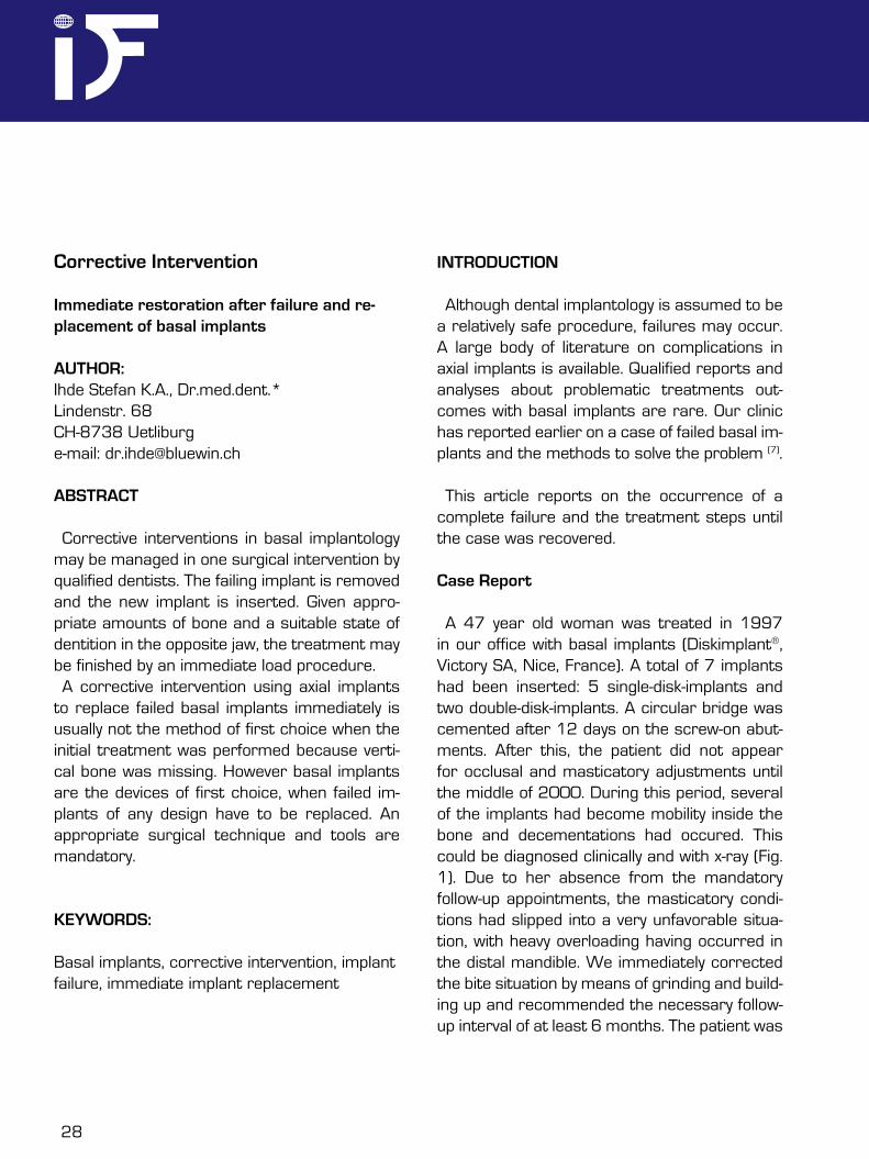
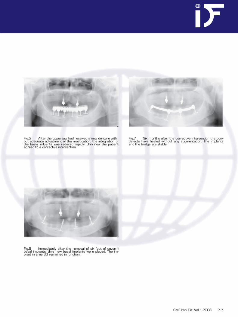
Fig.5 After the upper jaw had received a new denture with out adequate adjustment of the mastication, the integration of the basla imlpants was reduced rapidly. Only now the patient agreed to a corrective intervention.
Fig.6 Immediately after the removal of six (out of seven ) basal implants, thre new basal implants were placed. The im-plant in area 33 remained in function.
Fig.7 Six months after the corrective intervention the bony defects have healed without any augmentation. The implants and the bridge are stable.

34
References
(1) Ihde S.: Sanierung mit basal osseointegrierten Implantaten. Colleg Magazin, 1998; 5:138–146.(2) Ihde S. & Konstantinovic V. (3) Comparison and definition of the pathological phenomena occurring after a tooth replacement and the possible therapeutic stages implying basal and crestal implants(4) Implantodontie 14 (2005) 176-185(5) ICD (Implantoralclub Deutschland)/Besch K.-J.: Konsensus zu BOI. Schweiz Monatsschr Zahnmed,1999: 109:971–972(6) Tetsch J., P. Tetsch: Komplikationen und Folgeschäden nach Diskimplantationen; Z. Zahnärztl. Implant. 2006: 22(3) 118 - 123(7) Fenner M., Nkenke E., Holst S., Wichmann M., F.W. Neukam: Implantatprothetische Kaufunktionelle Rehabilitation nach Fraktur basal osseointegrierter Implantate (BOI); Z. Zahnärztl. Implantol. 2006: 22(2); 120 – 126(8) Ihde S. : Utilisation therapeutique de la toxin botulique dans le traitement de entretien en implantologie dentaire; Implant-odontie 14(2005): 56 – 61(9) Ihde S., Konstantinovic V., B. Cutilo: Der „kleine Reifenwechsel“ – Austausch eines BOI unter der vorhandenen festsitzenden Versorgung. Dent Implantol 2002: 6, 358 -361 (10) Bocklage R.: Advanced alveolar crest atrophy: an alternative treatment technique for maxilla and mandible. Implant Den-tistry 2001; 10 (1): 30-35(11) Bocklage R.: Rehabilitation of the edentulous maxilla and mandible with fixed imlpant-supported restorations applying im-mediate functional loading: A treatment concept.. Implant Dentistry 2002; 11 (2) (12) Scortecci G., Misch CE., Missika P.: Mise en charge fonctionnelle immédiate (MCI) chez l‘édenté partiel maxillaire. Apprt décisif de l‘implantologie basale. Implantodontie, n° 47, novembre 2002, 23-35(13) Scortecci G. Bourbon B.: Prothèse sur Diskimplant Sème partie : La Dent Unitaire. RFPD Actualités 1991;31 :21-29(14) Scortecci G., Bourbon B.: Prothèse sur Diskimplant. RFPD Actualités 1990 ; n° 13.31-48(15) Scortecci G., Bourbon B., Foesser P.: Dentures on Diskimplats 2. Complete denture: the antistress system REvFrProthes Dent 1990 Dec (22): 35-51(16) Scortecci G, Brenler P.: Insertion axiale et mise en charge immédiate : indications et limites. Implantodontie 2000, n° 37:73-75(17) Scortecci G., Doglioli P.: Behavior of human bone cells (maxilla and mandible) in contact with commercially pure titanium used for dental implants: in vitro study. Publication scientifique dans la Revue du Fourth World Biomaterials Congress, Berlin, avril 1992, 149; published in: Cytotechnology 7: 39-48, 1991(18) Scortecci G., Doglioli P.: Culture cellulaire issue de prélèvements effectués au niveau des maxillaires humains. Intérêt dans l‘évaluation in vitro de biomatériaux pour l‘implantologie dentaire. Les Publications de BIOMAT, 1989(19) Scortecci G., Doms P., Holsington W.: Osseointegrated System: Tissue-integrated prosthesis for small bone volumes. Pub-lication scientifique dans la Revue de l‘UCLA Symposium „Implants in the partially edentulous patient“, Palm Springs (CA), USA, avril 1990, n° 54(20) Scortecci G., Dôme P.: Biological anchorage in small bone volumes. Publication scientifique dans la Revue de la Première Rencontre Internationale de Rouen d‘Implantologie et des Biomatériaux, Mars 1991(21) Scortecci G., Doms P.: Racines artificielles bio-intégrées avec appui tricortical. Actualités Odonto-Stomatologiques 1987 ; n° 159 : 521-538(22) Scortecci G., Donsimoni JM., Spahn FP., Binderman I.: Ostéointégration et mise en fonction immédiate: sont-elles compati-bles? Alpha Oméga News, Décembre 1996, 4-5(23) Scortecci G., Donsimoni JM.: Mise en fonction immédiate des implants dentaires. La Lettre de Stomatologie, Juin 2000, n° 6:7-8(24) Scortecci G., Foesser P.: L‘implant ostéointégré dans les faibles volumes : au cabinet et au laboratoire. Intérêt de la mé-thode pour les os de type IV. Publication scientifique dans la Revue des VII Journées Inter Club, Antibes-Juan les Pins, avril 1992(25) Donsimoni JM., Dohan D.: Les implants maxillo-faciaux à plateaux d‘assise Concepts et technologies orthopédiques, réhabili-tations maxillo-mandibulaires, reconstructions maxillo-faciales, réhabilitations dentaires partielles, techniques de réintervention, méta-analyse. 1ère partie : concepts et technologies orthopédiques. Implantodontie 13, n°.1, 13-30, 2004
1.2.3.
4.5.6.
7.
8.
9.
10.
11.
12.
13.14.15.
16.
17.
18.
19.
20.
21.
22.
23.
24.
25.

CMF.Impl.Dir. Vol 1-2008 35
(26) Donsimoni JM. , Bermot P., Dohan D. : Les implants maxillo-faciaux à plateaux d‘assise Concepts et technologies ortho-pédiques, réhabilitations maxillo-mandibulaires, reconstructions maxillo-faciales, réhabilitations dentaires partielles, techniques de réintervention, méta-analyse. 2ème partie : rehabilitations maxillo-mandibulaires. Implantodontie 13 (2004) 31-41(27) Donsimoni JM. , Dohan A. ,Gabrieleff D. ,Dohan D. : Article original; Les implants maxillo-faciaux à plateaux d‘assise : troisième partie : reconstructions maxillo-faciales; Les implants maxillo-faciaux à plateaux d‘assise : concepts et technologies orthopédiques, réhabilitations maxillo-mandibulaires, reconstructions maxillo-faciales, réhabilitations dentaires partielles, tech-niques de réintervention, méta-analyse, Implantodontie 13, n°.2, 71-86 (2004)(28) Donsimoni JM., Gabrieleff, P. Bernot, D. Dohan: Les implants maxillo-faciaux à plateaux d`assise; Concepts et technologies orthopédiques, réhabilitations, maxillomandibulaires, reconstructions maxillo-faciales, réhabilitation dentaires partielles, tech-niques de réintervention, méta-analyse 4ème partie : réhabilitations dentaires partielles. Implantodontie 13 (2004) 139-150(29) Donsimoni JM., Gabrieleff, Bernot P. , Dohan D.: Les implants maxillo-faciaux à plateaux d`assise; Concepts et technologies orthopédiques, réhabilitations maxillo-mandibulaires, reconstructions maxillo-faciales, réhabilitations dentaires partielles, tech-niques de réintervention, méta-analyse. 5éme partie : techniques de réintervention. Implantodontie 13 (2004) 207-216(30) Donsimoni JM., Gabrieleff, Bernot P., Dohan D.: Les implants maxillo-faciaux à plateaux d`assise; Concepts et technologies orthopédiques, réhabilitations maxillo-mandibulaires, reconstructions maxillo-faciales, réhabilitations dentaires partielles, tech-niques de réintervention, méta-analyse. 6ème partie: une méta-analyse. Implantodontie 13 (2004) 217-228
26.
27.
28.
29.
30.
36
Short Communication
A novel osteoinductive bone grafting substitute?
AUTHOR:
V. Bienengräber*, S. Lenz, M. Kirchhoff Department of Oral and Plastic Maxillofacial Surgery, Rostock UniversityStrempelstr. 13D - 18057 Rostocke-mail: [email protected]
K.-O. HenkelDepartment of Oral and Maxillofacial Surgery, Federal Army Hospital Hamburg,Lesserstr. 180D-22049 Hamburg
ABSTRACT
Hydroxyapatite (HA) is the main component of bone. The bone grafting substitute described here consists of nanocrystalline HA embedded in a highly porous matrix of silica (SiO
2) gel. This
HA-silica-matrix is fully biodegradable and at the same time extremely osteoconductive. Ec-topic bone formation was induced even when implanting the HA silica matrix subcutaneously into porcine fatty tissue thus proving osteoin-ductive properties.
KEYWORDS:
Bone grafting substitute, osteoinductivity, hy-droxyapatite, silica, sol-gel-technique
Introduction
Hydroxyapatite (HA) - on the basis of alcox-ides - was distributed homogeneously in a SiO
2
sol (24% by weight), which formed a granular material during the transformation to a gel [1]. While drying the gel at a temperature of < 700 �C, the solvent evaporated and prompted a slight shrinking of the material. The production of the highly porous compound finally resulted in granules (density of 0.5 g/cm3). Evaporation also led to the formation of small pores (“micro-pores”, Ø 5-100µm), which allow the immigra-tion of osteoblasts and connective tissue fibrils. These are the starting points of bone formation within the HA silica matrix in multiple locations simultaneously. The loosely packed HA crystal-lites are held together by SiO
2 leading to nano-
pores (Ø 10-20 nm). The extremely enlarged outer and inner surfaces of the HA-silica-matrix (84 m2/g) allow the fast invasion of endoge-nous proteins, particularly the growth factors. The favourable biological behaviour of the new HA silica matrix is due to the natural nanocrys-talline structure of HA (Ø 90 nm) and the highly interconnecting porosity of the granules (61% of volume) [1]. In vivo studies demonstrate that the HA silica matrix is extremely osteoconduc-tive and at the same time fully biodegradable [2,
3].
Materials and methods
Osteoinductivity means that a material induc-es bone formation outside the skeleton. 0.3 ml of HA silica matrix granules (0.6 x 2 mm) were

CMF.Impl.Dir. Vol 1-2008 37
applied into the subcutaneous fatty tissue and the adjacent musculature in the neck region of 12 adult Goettingen minipigs. The surrounding tissue was excised 2.5 (group 1, 3 animals), 4 (group 2, 3 animals) and 8 months (group 3, 6 animals) postoperatively. The tissue samples were studied macroscopically, radiologically, his-tologically (cutting and grinding technique, Giem-sa - toluidine blue, Masson-Goldner and von Kos-sa stains) and histomorphometrically (ITEM®, Soft-Imaging-Systems, Münster, Germany).
Results and discussion
Biodegradation of the HA silica matrix was con-siderably slower in soft tissues than in bone [3]. Residues of the material were found, but could not be clearly differentiated from foci of ossi-fication macroscopically. In radiographs it was also not possible to differentiate safely between HA particles of the material and foci of ossifi-cation, since both were similarly radioopaque. Computer tomography (CT) produced better in-formation: e. g. in one animal two clearly sepa-rated foci of ossification were visible 8 months after implantation (Fig. A). These findings were confirmed histologically in five out of the six mini-pigs (group 3). Multiple foci were visible even macroscopically in three animals. The largest one measured 2.5 mm by 12.5 mm (Fig. B). Foci of ossification were found in all animals of groups 1 and 2: micro foci (Ø < 1mm) after 2.5 months and larger foci (Ø 1mm - 3.5 mm) after 4 months. The foci of ossification were located almost exclusively in the subcutaneous fatty tis-sue (Fig. C). No ossification took place in the muscular tissue probably due to the permanent
movement of the myofibrils. The close contact between partially biodegraded HA particles and newly formed bone spoke in favour of the osteo-inductive properties of this material (Fig. D)
Fig. A-D: Findings 8 months following implanta-tion of HA-silca-matrix into subcutaneously fatty tissue (minipig, neck region)..
Fig. A. High power computer tomography: two separate large calcified areas

38
Fig. B. Histological findings (cutting and grinding technique) Two large foci of ossification (same as in Fig. 1a), adjacent to muscula ture in subcutaneous fatty tissue (Giemsa-toluidine blue, original magnif. 4 x)
Fig. C. Histological findings (cutting and grinding technique) Ossified material (bright green) in subcutaneous tissue with subtotally biodegraded HA particles enclosed (dark green, Masson-Goldner, original magnif. 12,5 x)
Fig. D. Histological findings (cutting and grinding technique) Osteoid and of newly formed cancellous bone (dark brown/black) with subtotally biodegraded HA particles enclosed (arrows, von Kossa, original magnif. 40x)
CMF.Impl.Dir. Vol 1-2008 39
Conclusion
These studies led to the conclusion that dif-ferentiation of adipocytes (or other adult stem cells) into osteoblasts can be induced in vivo by highly porous bone grafting substitutes with nat-ural nanocrystalline structure. Further in vivo studies will be required to understand those processes.The HA- silica- matrix tested is available in the
form of granules for application in medicine (Nano Bone® / CE 0483, Artoss GmbH, Ros-tock / Germany). First, positive results in oral and maxillofacial surgery , i.e. in treating bone defects with the new HA-silica matrix, have been published [4]. At the same time the HA-silica ma-trix is a favourable carrier for osteoinductive material like bone morphogenic proteins, and stem cells. This is of particular clinical impor-tance in recipient tissues with poor regenera-tive properties.
References
1. Gerber T, Holzhüter G, Knoblich B, Dör fling P, Bienengräber V, Henkel K-O (2000) De-velopement of bioactive sol-gel material tem-plate for in vitro and in vivo synthesis of bone material. J Sol-Gel Sci Technol 19: 441-445
2. Gerber T, Trakova T, Henkel K-O, Bienen-graeber V, Witt M, Koewitz J (2003) Silica/cal-cium phosphate sol-gel derived bone grafting material and bone remodelling. An eight months in vivo study , Key Engin. Mat: 240-242, 411-414
3. Gerber T, Holzhüter G, Götz W, Bienen-gräber V, Henkel K-O, Rumpel E (2006) Nano-structuring of biomaterials – A pathway of bone grafting substitute. Eur. J. Trauma: 32, (132-140)4. Henkel K-O, Gerber T, Lenz J-H, Bienen-
gräber V (2005) Ein qualitativ neuartiges Kno-chenaufbaumaterial auf Hydroxylapatit-Xerogel-Basis. Zahnärztl. Welt 114: (416-418)
Acknowledgements
This research was funded by the Department of Education of the State of Mecklenburg-Vor-pommern (PTJ MGS/04 024 10). The authors thank Prof. Dr. Klaus U. Benner, Institute of Anatomy of the Munich University, for realizing the high power computer tomography (fig 1a).
40
Full length article
BOI® implants as the therapy of choice in dental offices
AUTHOR
Dr. Katrin TostPrivate Practice69100 KomotiniW. Georgiou 4GreeceContact: [email protected]
SUMMARY
BOI® implants allow cantilever situations in the mandible to be definitively restored in a prompt and cost-effective fashion. They can be used alone or in combination with natural tooth abut-ments and/or crestal implants. A number of case studies are presented to illustrate treat-ments of this type. They can be planned and con-ducted in any well-equipped dental office without deviating from routine treatment approaches. In her own dental office, the author relies on BOI® implants as the alternative of choice to remov-able restorations.
KEYWORDS
BOI® implants, cantilever situation in mandible, fast and cost-effective restoration
INTRODUCTION
Dental offices offer prosthetic restorations to replace any teeth that have been lost. Most pa-tients wish to have fixed restorations installed. Very often, this expectation can be met by de-livering bridges. However, if any gaps along the arch can no longer be reasonably closed with a bridge, removable partial dentures are indicated [1]. Given the very low acceptance of removable dentures, patients are today incre-asingly opting for implants. BOI® implants are capable of assuming the function of missing abutment teeth, allowing the masticatory func-tion to be restored in a timely fashion. With this strategy, functional problems can be avo-ided that would otherwise sooner or later af-fect the masticatory pattern in its entirety [2]. BOI® implants offer stable bicortical support due to their shape and transosseous insertion path. As a result, they can be immediately loa-ded. The amount of vertical bone is irrelevant with these designs, since the horizontal rather than the vertical structures of the jawbone are utilized for the transmission of forces. BOI® im-plants come in a large variety of shapes enab-ling dentists to select appropriate designs in accordance with individual bone conditions. Dif-ferent implant sizes should be kept in stock at all times, allowing clinicians to select the most suitable implant type even in the midst of the surgical procedure and to immediately place implants should a need for extractions arise unexpectedly. The available bone volume should be optimally utilized. Since the jawbone is safe from resorption in its basal segment, this area is typically the one that is most suitable[3].

CMF.Impl.Dir. Vol 1-2008 41
patient’s systemic health must be discussed as well. There is a need to define which teeth should be preserved and what steps should be taken for them to become healthy and stable (including measures such as caries treatment, root canal therapy, apical resection, periodontal treatment, or vitality testing).
Orthopantomographs
An orthopantomograph will normally suffice for prosthetic treatment planning. The image will reveal the state of the residual dentition, the vertical bone height, the position of the con-dyles, the bone structure, and the trabecular pattern. The location of the implant site can be determined as well. Both the maxillary sinus and the course of the mandibular nerve will be visible.
Clinical examination
The inspection of the oral cavity is followed by pal-pating the muscles, determining the jaw width, and exploring the mylohyoid line. Any systemic diseases need to be identified and, if necessary, controlled by the physician in charge or general practitioner. Examples would include diseases such as diabe-tes, HIV, cardiovascular disorders, hypertension, or curare patients.The author’s personal list of contraindications also includes excessive alcohol consumption, depressi-on, poor general hygiene, indecisiveness or lack of reliability, skeptical attitude toward the clinician or the treatment modality, and insistence on being in-formed about every tiny detail of treatment.Surgery is postponed if infections of the re-
First contact with patients
With patients who present at our office for the first time, our policy is to find out about their desires and treatment expectations. What should the outcome look like?Some patients may only ask to have a lost tooth restored because they were able to use it for mastication until recently, when the tooth was eventually lost.However, a new and broader perspective has emerged in modern dentistry, based on the no-tion that focusing on overall function is more productive. The stomatognathic system is com-prised of the maxilla and mandible including their dental arches and periodontal structures but also includes the temporomandibular joints, the masticatory muscles with adjoining musc-le system, the salivary glands, and the vessels and nerves supplying the various tissue struc-tures [4]. It must be made completely clear to the patient that appropriate dental treatment is mainly about restoring the functional unit as a whole. The objective is to ensure that the ma-sticatory organ can meet its primary tasks of breaking down food and ensuring mutual sta-bilization of both jaws during swallowing. Good esthetics with teeth of pleasant size, color and shape are of secondary importance – but ne-vertheless still very important. Patients must be willing to maintain their new teeth (e.g. by brushing them twice daily, rinsing, and comp-lying with scheduled recall visits).The first conversation with the patient inclu-des the medical history. Any problems that may have contributed to the present oral sta-tus need to be discussed and understood. The

42
is kept at hand to mitigate any panic attacks and to avoid hyperventilation.
Medication
Patients without systemic diseases are given lexotanil, xanax (Alprazolam) 0.25 mg as anxio-lytic medication if they become psychologically tense or experience fear. The patient should have had breakfast as usual. Men should sha-ve to avoid contamination of suture material by facial hair. Women susceptible to menstrual symptoms such as blood loss or psychological instability should have the day of surgery sche-duled accordingly. Our patients are advised to arrive at the office in company and to avoid dri-ving after the procedure. Lukewarm sweetened tea is always on hand for drinking with a straw. Communication with the patient (eye contact, conversation) is continuously maintained. Treat-ment is started with preparatory procedures of operative dentistry (abrasive instruments, ca-ries treatment, root-canal treatment, or root-tip resection). The patient’s oral cavity is disin-fected with betadine and the face with rubbing alcohol.Antibiotics are administered only to patients who required intraoperative coverage because of a concomitant disease. They are not routi-nely administered, however, since antibiotics do not per se improve treatment outcomes in any significant way [5]. Analgesics are offered in the form of preparations containing acetamino-phen or mefenamic acid 500 mg, administered 3 or 4 times daily. Patients are advised to use chlorhexidine mouthwash solutions.
spiratory tract, herpes sores, or aphthae are present. Establishing a harmonious relation-ship with the patient is essential for success. Implant therapy continues to be a service that involves special requirements. This treatment should only be performed by highly competent and responsible clinicians selecting only pati-ents with adequate levels of intelligence, de-mands on life, cooperation, compliance, and the ability to pay for the treatment. Implant treat-ment is elective in nature, to be conducted only on patients truly willing to receive them.
Treatment planning
First, a decision must be made which jaw to treat first. This should generally be the oppo-sing jaw or the jaw requiring fewer implants. The occlusal level is determined and the ver-tical dimension defined by bite registration. It must be checked whether suitable implant ty-pes and sizes are available and at hand. Occlu-sal splints may be helpful in raising the occlusal plane. It is important to follow Camper’s plane. All conservative treatment procedures need to be planned as well.
Preparation for surgery
We use Ringer solution as a cooling agent and store it in the refrigerator prior to surgery. The solution is applied by connecting special flasks to the dental unit. All instruments, the turbine including the angled handpiece and the surgical towels are autoclaved. Special caps are worn by the clinician, but also by the assistants and the patient, to cover their hair. An oxygen bottle

CMF.Impl.Dir. Vol 1-2008 43
Surgical approach
The clinician aims to create a congruent im-plant bed by raising and reflecting a mucope-riosteal flap using specialized burs and cutters. The BOI® implant is laterally advanced into the implant with the help of gentle knocking move-ments. Finally, the flap is repositioned and sutu-red.

44
Case report 1
A woman born in 1963 presented at my of-fice after a “cosmetic crown” restoration in her maxilla had fractured. After conducting a post-and-core buildup, the crown could be reinserted. However, it was pointed out to the patient that mastication was confined to her anterior teeth. The crown was likely to fracture again, and other teeth might be da-maged. The upper molars were significantly elongated because their lower antagonists had been missing for several years. A re-movable denture was refused by the patient out of hand. Two years later, debonding of the restoration on tooth 21 occurred. Rece-mentation was possible but did not address the underlying problem. The patient reque-sted that her lower anterior teeth should not be prepared to serve as abutments. After obtaining an orthopantomograph, the case was thoroughly examined and a treat-ment plan established. Four ceramic crowns were delivered in the maxilla to guide the oc-clusal plane. The molars were reduced in length and the teeth treated endodontically,
because encroachment on the pulp proved un-avoidable. In this way, the occlusal plane could be aligned with Camper’s plane. The new sha-pe of the dental arch is readily discernible in Figure 2. Rehabilitation of the mandible was planned as follows: One distal BOI® implant was selected for each posterior segment, and compression screws for both premolar sites. The canines were to be included in the struc-ture as well. Treatment began by preparing the canines under local anesthesia. The BOI® implants were inserted, followed by placement of the KOS® screws. The threaded pin of the left implant was too deep (or the number of threads too small). An elongated polished ab-utment was therefore selected to compensa-te for this deficit. The ceramic bridge could be inserted after 7 days on November 20, 2006. The patient continues to be happy with her new teeth after one year of clinical service.
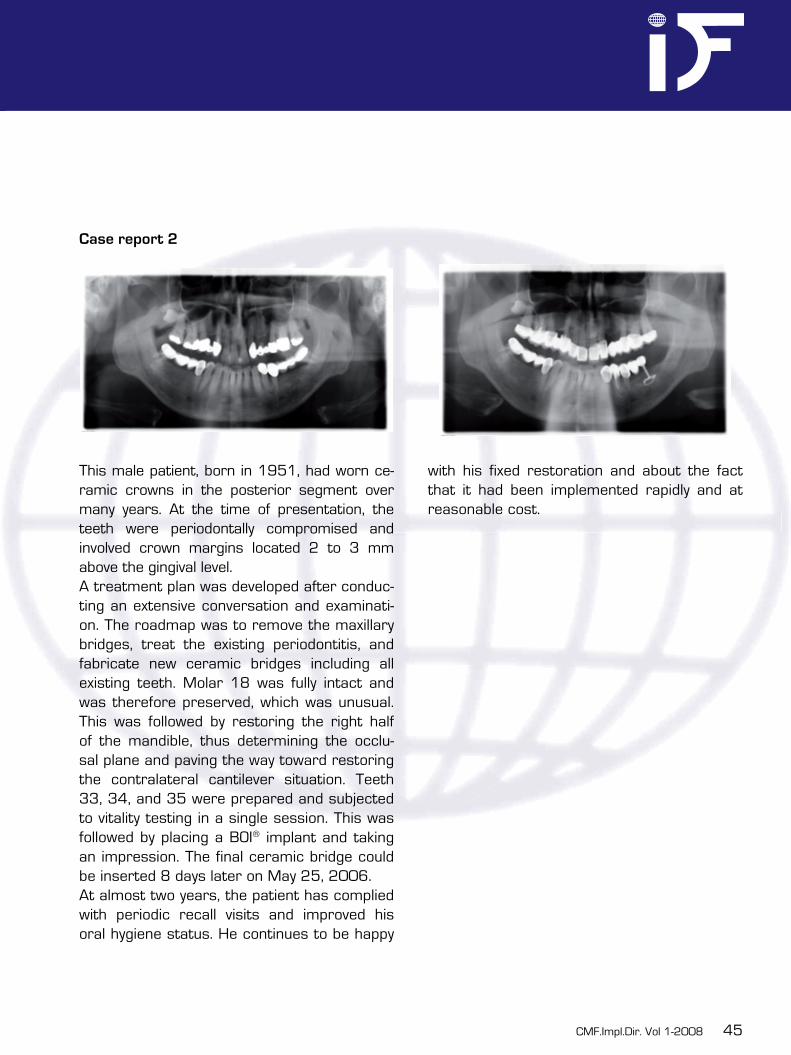
CMF.Impl.Dir. Vol 1-2008 45
Case report 2
This male patient, born in 1951, had worn ce-ramic crowns in the posterior segment over many years. At the time of presentation, the teeth were periodontally compromised and involved crown margins located 2 to 3 mm above the gingival level.A treatment plan was developed after conduc-ting an extensive conversation and examinati-on. The roadmap was to remove the maxillary bridges, treat the existing periodontitis, and fabricate new ceramic bridges including all existing teeth. Molar 18 was fully intact and was therefore preserved, which was unusual. This was followed by restoring the right half of the mandible, thus determining the occlu-sal plane and paving the way toward restoring the contralateral cantilever situation. Teeth 33, 34, and 35 were prepared and subjected to vitality testing in a single session. This was followed by placing a BOI® implant and taking an impression. The final ceramic bridge could be inserted 8 days later on May 25, 2006.At almost two years, the patient has complied with periodic recall visits and improved his oral hygiene status. He continues to be happy
with his fixed restoration and about the fact that it had been implemented rapidly and at reasonable cost.

46
Case report 3
This woman, born in 1958, was in good ge-neral health. Mastication was confined to the anterior segment. This situation was com-pounded by increasing mobility of the lower in-cisors. Dental treatment was therefore man-dated. A treatment plan was developed after duly completing the required discussions with the patient. Two bridges were planned in the upper anterior and lower right posterior seg-ments. Molars 27 and 28 were to be preser-ved. Considerable abrasive reductions were required because a considerable divergence was present between the teeth. All teeth were treated for periodontitis. Upon completion of these steps, the left posterior segment of the mandible was addressed. After taking an im-pression of the maxilla, teeth 34 and 35 were prepared with abrasive instruments and te-sted for vitality. Tooth 38 was hopeless and had to be extracted. In the same session, the jawbone was thoroughly cleared of granulation tissue, followed by inserting the BOII® implant infero-anteriorly to the socket of the extracted third molar. The load-transmitting segments of the implant were located in the linea obli-
qua region, which is safe from resorption. This jawbone region offers an enhanced rege-nerative tendency compared to other regions due to the local stresses involved[6]. The base-plate of the BOI® implant was located in the basal segment of the mandibular jawbone.This was followed by taking an impression of the mandible. The final ceramic bridge could be inserted 7 days later on February 5, 2008.
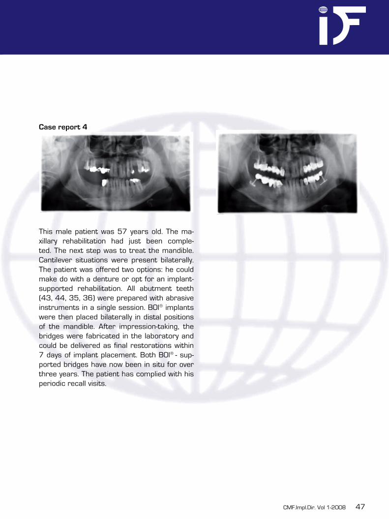
CMF.Impl.Dir. Vol 1-2008 47
Case report 4
This male patient was 57 years old. The ma-xillary rehabilitation had just been comple-ted. The next step was to treat the mandible. Cantilever situations were present bilaterally. The patient was offered two options: he could make do with a denture or opt for an implant-supported rehabilitation. All abutment teeth (43, 44, 35, 36) were prepared with abrasive instruments in a single session. BOI® implants were then placed bilaterally in distal positions of the mandible. After impression-taking, the bridges were fabricated in the laboratory and could be delivered as final restorations within 7 days of implant placement. Both BOI® - sup-ported bridges have now been in situ for over three years. The patient has complied with his periodic recall visits.
48
cal practice[6]. The blood supply is confined to the mandibular artery in treatment strategies of this type and is further restricted by the com-pact volume of these implants.Also, the surface area for transmucosal bacte-
rial colonization is increased due to the large di-ameter of these implants. BOI® implants feature shafts that are both very thin and highly polished. The surface for bacterial colonization is much smaller. Consequently, the condition of “peri-im-plantatis” commonly observed with crestal im-plants does not occur in basal implantology.
CONCLUSION
BOI® implants are a method of first choice whenever the function of the entire stomato-gnathic system needs to be restored by pro-sthetic rehabilitation of the mandible, even (and precisely) if little bone is available.
DISCUSSION
BOI implants are used to transfer masticatory forces to the jawbone instead of lost teeth. The force-transmitting disks are designed to offer bicortical support by transosseous position-ing. Cortical bone is dense and stable, offering an ideal level of load-bearing capacity. It is su-perior to cancellous bone (where conventional implants are inserted in the traditional way) in all respects. The basal jawbone area is safe from resorption. Achieving bicortical support in that region is essential to the success of im-plant therapy, whereas the amount of vertical bone present above the level of the basic disk becomes largely irrelevant. This bone-anchored disk connects to the prosthetic structure via the shaft of the BOI® implant. The length of this shaft may vary depending on the desired level of the occlusal plane along the bridge. We do not replace bone but rely entirely on available bone to anchor implants. This strategy eliminates the need for any adjunctive measures involving bone grafting or osseodistraction, reducing the over-all risk of treatment and avoiding waiting peri-ods. This makes the procedure easier to handle and more benign on patients.Bicortical support allows blood to be supplied
to implants that have been freshly inserted in posterior regions of the mandible through three different arteries: the mandibular artery inside the jawbone and the lingual/facial arteries in the periosteum on both sides of the mandible. This blood supply will improve wound healing and implant integration as well as implant function.Short crestal implants with large diameters
have not turned out to yield good results in clini-

CMF.Impl.Dir. Vol 1-2008 49
REFERENCES
(1) Breustedt, Lenz, Musil, Staegemann, Taege, Weiskopf: Prothetische Stomatologie, Leipzig 1987.(2) Pfanas P. La Rehabilitation Neuro-Occlusale, Masson, Paris 1982(3) Ihde S. Restoration of the atrophied mandibule using basal osseointegrated implants and fixed prosthetic superstructures. Implant Dent 2001(4) Fuhr, Reiber. Die Totalprothese, Urban u. Schwarzenberg 1993(5) Gynther G. H. et al. Dental implant installation without antibiotic prophylaxis. Oral Surg. Oral Med Oral Pathol 85, 509, 1998.(6) Ihde S., Mutter E. Versorgung von Freiendsituationen mit basal osseointegrierten Implantaten (BOI®) bei reduziertem vertikalen Knochenangebot. Deutsche Zahnärztliche Zeitschrift 58, 2003.

50
Clinical Research
Outcomes of immediately loaded full arch reconstructions on basal implants and teeth in the mandible: retrospective report on 115 consecutive cases during a period of up to 134 months.
AUTHOR:Ihde Stefan K.A., Dr.med.dent.* Lindenstr. 68CH-8738 Uetliburge-mail: [email protected]
ABSTRACT
We report on a retrospective review of out-comes after the insertion and usage of basal implants alone or in combination with natural abutments for treating patients with full arch bridges in the mandible. From June 1996 to August 2007, 115 consecutive patients see-king implant treatment in the lower jaw recei-ved 457 basal implants and a total of 130 bridges thereon. Only cases where basal im-plants were used alone or in combination with teeth were included. During the observation period no patient seeking treatment was tur-ned away for any reason. Teeth were included into the constructions whenever available and in an appropriate location and condition. The mean age at implant surgery was 60.7 years. Even in cases of severe bone atrophy, no aug-mentations were performed. All patients recei-ved fully loaded fixed bridges between 3 to 12
days postoperatively. Whenever implants had to be replaced after extractions, this was done in one surgical step without waiting or healing time. An overall success rate of 93.4 % was observed. Basal implants alone or in combina-tion with stable teeth can securely be placed and used in immediate load protocols to form a base for full arch bridges in the edentulous or partly dentate mandible.
KEYWORDS
Basal implants, BOI®, Diskos®, immediate load, immediate implant placement, full arch mandi-bular reconstructions.
INTRODUCTION
Placement of circular bridges on basal im-plants in immediate load conditions is a fre-quently performed procedure and there are va-rious reports on this procedure in the literarture1,
8, 11. Other than in axial (crestal) implantology, the restoration of basal implants is in most cases done by a fixed bridge: telescopes are contraindicated and bar-based restorations are rarely delivered. Due to the straight for-ward treatment protocol and the possibility to avoid intermediate prosthetical constructions, the overall treatment costs are low. All these aspects reflect the desire and expectations of the overwhelming majority of the patients 13. Atrophy is the result of a lack of bony stimu-lation and nutrition. Generalized diseases may
CMF.Impl.Dir. Vol 1-2008 51
vities, e.g. empty spaces left after cystektomies or granulation removals, or may even be appli-cated trans-sinusally.There is a long tradition in combining basal im-plants with stable teeth. As a matter of fact, professional who use basal implants conti-nue to discuss whether to do so or not which should be validated by such data analyses as reported in this manuscript.
METHODS
SUBJECTS
From June 1996 to August 2007, 115 conse-cutive patients (60% female) receiving 457 basal implants and 130 full arch prosthetic constructions in the mandible thereon were en-rolled in this study. All patients seeking implant treatment during this period have been treated using basal implants alone or in combination with natural abutments. Patients receiving at least one screw type implant were excluded from this study, because their inclusion would make distinction between the benefits of the-se two implant types difficult and would not al-low reporting on the independent performance of basal implants. The surgical and prosthetic treatments were all performed by the same group of clinicians. The mean age at implant surgery was 60.7 years (SD=9.8; median=61; range: 33 to 80 years, Fig. 6). The average number of implants used per circular bridge was 3.9 and on average 3.5 teeth were inclu-ded into the constructions. Sixty-seven con-structions on 330 implants were erected only
aggravate the disease and promote the bone loss.3,4 The management of quantitative and/or qua-litative poor bone with root-form dental im-plants typically requires additional procedures to ensure sufficient stability5, with augmentati-ons, bone transplants or distractions being the most “popular” procedures. The management of the atrophied distal mandible with axial im-plants imposes special problems, since the nerve canal forbids in many cases the usage of the full amount of vertical bone in the zone bearing most of the chewing forces. Augmen-tations and distractions in this region are dif-ficult and associated with considerable patient discomfort. If teeth are to be extracted, additio-nal waiting time before equipping the mandible with axial implants has to be considered. Addi-tional treatment steps such as augmentations and distractions add risks to the procedure, delay loading, and increase costs. Basal implants are placed transosseously and at least one base-plate is bicortically anchored in the basal, cortical bone. Basal implants utilize the horizontal bone supply as well as the cor-tical bone walls; therefore, they are well suited for placement immediately after extractions. It is understood today, that basal implants under-go a dual mechanism of integration: ring areas in direct, primary contact with the native bone show primary integration though osteonal re-modeling also occurs. Empty slot areas (the void space left after osteotomy and insertion) fill with callus, which later undergo osteonal re-modeling13. This dual integration also allows placement of basal implants right into extrac-tion sockets of teeth or implants and other ca-

52
on basal implants, without teeth -, with three implants being the minimum and eight implants being the maximum number of implants used for the fixation of these mandibular bridges (Average number of implants in this subgroup: 4.9 basal implants per mandible), thirty eden-tulous mandibles received four strategically placed basal implants only. A typical treatment plan with teeth includes the six front teeth and two basal implants placed in the area of the 2nd molar (Fig. 3), typical treatment plans without teeth would include basal implants at least in the strategic positions of the canines and second molars (Fig. 2, 4).
IMPLANTS
Basal implants consist of a thin vertical shaft (1.9 – 2.3 mmd) and one to three horizontal base plates, designed for cortical load trans-mission. (Fig.1) Unlike the traditional axial (“root-form”) implants, which are inserted verti-cally and primarily designed to be supported by trabecular bone, these implants are inserted from the lateral aspect of the jaw bone into a T-shaped slot created by high-speed precisi-on instruments, providing bi- or multi-cortical support and immediate fixation even if placed in extraction sites right away. Hence, they are commonly called “disk” or “lateral” or “basal” implants 10.
BOI® implants transmit the masticatory forces into cortical bone areas and they are placed to utilize resorption free bone areas, such as the
interforaminal region and the bone below the linea oblique in the distal mandible. The site of force transmission is far away from the area of bacterial invasion; hence base plates never develop infections as long as they are well in-tegrated. The vertical implant shafts are not meant to transfer masticatory loads directly to the bone, hence they are relatively thin and al-lowed to be machined or even polished. As a result peri-implant infections are never seen in basal implants. Basal implants have proven to be well suitable for smokers.12
During the treatment- and observation period, the manufacturer has made a number of small changes in the implant design and this study does not distinguish between the different ty-pes available at their times, since treatment protocols and indications did not change. As a principle, the fixed bridge on basal implants (Brands: BOI®, Diskos®) serves as an immediate external fixation for the implants. The bridge also distributes the masticatory loads between the implants and it allows masticatory function at the same time.
SURGICAL TECHNIQUE
Under local anaesthesia, an appropriate full thickness flap is raised. Using high-speed preci-sion cutters, the implant bed is prepared which provides the appropriate number of vertical and horizontal slots for the chosen implant. Basal implants must always be inserted bicor-tically and trans-osseously. The implants are inserted (depending on the anatomical conditi-on and the desired position) from the lateral,

CMF.Impl.Dir. Vol 1-2008 53
medial or palatinal aspect of the jaw bone with careful tapping action until full bi- or multicor-tical support is achieved. The presence of suf-ficient support is verified visually or manually by testing with the fingers. The implants may also be fixed horizontally by use of bone screws (Brand: SSF), when primary stability cannot be achieved right away, e.g. when the implant bed is not exactly congruent to the implant or parts of the implant cannot reach cortical walls, or if these walls are missing after the extraction of teeth or soft tissue.A flapless, trans-mucosal insertion is not pos-sible for basal implants.
OUTCOME EVALUATION
Implants were counted as successful, if they are in situ at the point of observation, connec-ted to a bridge in function, if they allow masti-cation without pain or visible infection, and if no indication for their removal according to the “Consensus on BOI®” was given (14).
DATA ANALYSIS
Descriptive statistics were calculated for ba-seline variables. The primary outcome of inte-rest was implant failure defined as any reason for having to remove an implant. Survival was based on the period from implant placement to final follow-up. Because basal implants are designed for immediate loading (meaning that immediate loading is the method of choice and late or delayed loading is a rather poor, even
RESULTS
Patients were followed for a mean of 67.1 months (Median=69.5; SD=28.8; range: 3- 134 months, Fig. 7) in this study. Six patients (5.2%) with 16 implants (3.5%) were lost to follow-up for different reasons. Patients refraining from follow-up for more than12 months were exclu-ded from the study and counted as drop out. Another seven patients (6.1 %) died during the observation period with all of their implants (n= 30; 6.6% of all implants) in full function. Tho-se cases and implants were kept in the study but the implants were censored at the point of death. All implants were loaded immediately or within the first 24 hours after the implantati-on with a fixed temporary or permanent bridge. Fixation of the second, more permanent, pro-sthetic construction followed in subsequent days after surgery in most cases. In extrac-tion cases, “final” bridges were delivered after 6-18 months if requested by the patient. We found an overall survival rate of 93.4% of the implants during the follow-up period.
If was reported after the healing period mobi-lity of the implant causing an internal irritati-on of the periosteum was the most common cause. In cases where the implant could be left in place 14, this was addressed by a occlu-sal reorganization, i.e. grinding or building up chewing surfaces, thus relieving the interface region between the endosseous implant part and the bone from overload and allowing the peri-implant bone to recover and to reminera-lize. In the distal mandible, vertical bone growth even along the implant was observed regularly

54
DISCUSSION
We report a 93.4% implant survival rate among a consecutive series of 115 patients receiving 457 basal implants and a total of 130 fixed circular bridges in the mandible. This result is great, that even cases of seve-re atrophy were treated in one surgical inter-vention with basal implants, thus avoiding aug-mentations and bone transplants. Combined with the early external fixation via the bridges, this implant system responds well to early load analogous with physiological forces observed orthopedic surgery with early partial weight bearing. Living healthy bone will be remodeled and grows with daily use. It may be possible to minimize the antagonistic contacts in subtotal constructions to reduce the initial forces, but in circular bridges on implants this is impossible. So the application of a real immediate loading protocol is necessary. All patients were treated under local anaesthesia in a regular dental of-fice. The average absence from work was 1.9 days for the initial treatment phase. No one stopped working for more than one week. Most of the patients went back to work on the day after implant placement. None of the patients including those showing severe atrophy, had to be hospitalized.
There are limitations to the present study. The design of the basal implants used during the years has been improved by the manufactu-rer and we do not distinguish between the different designs. One major change in design which occurred approximately 1999 was that no surface enlargening (i.e sandblasting) was
12. Two implants had to be removed, because the vertical bone growth did not allow cleaning of the site any more and recurrent infections occurred. The replacement implants were si-tuated more anteriorly. At the time of the first intervention, a more anterior placement of the implant had not been possible, because the available bone crestally of the alveolar bund-le of vessels was less than 1 mm. Only at the time of the second intervention, enough bone whose growth had been induced by functional stimulation and/or the remodeling following the first intervention, had been available. (Fig. 10a, 10b). For the replacement of the im-plant, the bridges were shortened and the new implant parts were connected to the existing bridge by means of over-cementation. Those two implants were not counted as a failure since they did not meet the criteria of failure. Fifteen (13%). bridges had to be renewed com-pletely, following implant and/or tooth loss. The majority of the renewals were caused by tooth associated problems (decay, decementation of bridge, loosening of posts inside root canals followed by loosening of the bridges), often fol-lowed by implant loss due to overload (and sub-sequent mobility) or even implant fracture.Implant losses (n = 30) occurred mainly during the first four years after implant placement (Fig.8), with the mean time of lost implants in function being 783 days (Std.-Dev. 530 days).
CMF.Impl.Dir. Vol 1-2008 55
administered to the vertical implant surface. This change eradicated the crater-like bone losses found earlier in some of the basal im-plants of the sandblasted shaft type. Later, no sand-blasting was not performed at all, i.e. basal implants are completely machined today (Fig.1). Another change in double-disk designs occurred approximately in the year 2000. From this point, the crestal base-plate was ma-nufactured with more elasticity than the basal plate. Subsequently translucencies around the crestal plate, occurring in about 10-15% of those implants, were not observed any more. A slight increase in the thickness of the basepla-te (0,1 - 0.2 mm) may have contributed to the fact that no fractures were seen in implants placed after 2002. Triple-base-plate designs with strong primary stability were introduced about the same time period. Those small chan-ges and developments have combined with our learning curve and have made the treatment even more predictable in the last years of ob-servation (Fig.9); it must be taken into account also that at the time when we started using and exploring the basal approach in implanto-logy, no teaching and no textbooks were avai-lable and the author and his group as well as associated colleagues interested in this techni-que have learned the handling and possibilities auto-didactically. Losses of implants during the period 1997 – 2002 were unavoidable.
As progress in axial implantology occurs, many cases classified “untreatable” earlier, are treated today with screw type implants with considerable success, although for many pa-tients, the installation of fixed teeth on those
(axial) implants remains impractical. We feel that the patient cases which reached our office in the last years have become more difficult to treat and atrophies treated in our center beca-me increasingly severe, because patients with more available bone at the start of treatment find a capable screw- implantologist easier to-day and they are not referred so easily. A new group of patients is seaking this treatment be-cause of the possibility of immediate loading and avoiding risky, time consuming and expen-sive bone augmentations. This group is increa-sing.
Furthermore we have not treated a control group with bone augmentations or distractions, since this was not the desire of our patients. For patient cases presenting with severe bone atrophy (as shown in Figure 2 as an example), no realistic alternative treatment plan could be developed, carried out, or used as a compari-son. The primary reason that none of our pati-ents expressed the wish to have “more bone”, was their desire for “fixed teeth” and they knew that our clinic offers straight forward treat-ments without augmentations or waiting times. So the “alternatives” simply wouldn’t sell in the same dentists office. We have asked several other centers from whom we knew that they provided heavy maxillofacial surgery, distrac-tions and bone augmentations during the same period of time, to contribute their retrospecti-ve data for comparism with our result. None of the centers receiving our inquiry was willing to release data or to cooperate. However one of those centers, after seeing our results and how we solved the cases, changed the way of
56
case series of 275 BOI® implants in 228 pa-tients over a period of five years. Molars were replaced with BOI® implants in combination with anterior natural abutments. Osseointegration was achieved in 97.3% (n=254) of implants at final follow-up. Fifteen implants were lost.7 The results are similar to our findings.
Donsimoni et al performed a retrospective case series evaluating 1352 consecutive basal implants placed over a 10 year period in 234 circular bridges.8 Osseointegration was achie-ved in 97%. Of the 41 implants that failed, 25 had to be replaced. Only one full upper bridge had to be permanently removed rendering a cli-nical success of 99.9%. Interestingly, smokers and non-smokers experienced similar rates of implant losses, whereas reports from axial im-plants indicate opposite results9. Donsimoni et al used only basal implants in their study; ho-wever, they inserted a greater number of ba-sal implants per jaw (up to 12) compared to us (4.9 per jaw). The results presented in this article are consistent with our findings. Neither Scortecci, Ihde & Mutter or Donsimoni distin-guished between placements into fresh extrac-tion sockets and placements into healed bone.
The strengths of this study are many. Since we did not exclude any patients who presented to our clinic, even those sent away by colleagues, we feel that our findings are without any excep-tion generalizeable. this includes patients who typically may be turned down due to poor bone quality or recommended to receive bone aug-mentation procedures, or simply are otherwise “untreatable” (Fig. 2). According to our findings,
treatment drastically. Bone augmentations are not performed any more in this center and the system of basal implantology was impemented and is used more and more today. And finally, some patients seeking additional implants after screw implants have been placed in the anteri-or mandible were treated during the observa-tion period. We have placed BOI® in the distal mandible to allow placement of a fixed bridge. These cases of “upgrading” are not included in this report because it could be argued that the outcome of this treatment could be mainly due to the integrated screw implants alone. Inclusi-on of those cases would not make the this re-port stronger.
This is a case series and can be compared to historical publications. Our survival rates are very similar to those found in the literatu-re.6-8,10-12 Diskimplants® are similar in design and function to BOI® implants and have repor-ted rates of successful osseointegration of up to 97% with relatively long follow-up periods. Scortecci performed a prospective case series of 783 implants (627 Diskimplants®), placed in 72 patients with completely edentulous maxil-lae using an immediate load protocol. Follow-up ranged from 6 – 48 months. At 6 months, 98% of implants were osseointegrated, with all fixed prostheses remaining functional during the study period.6 However Scortecci combi-ned crestal and basal implants, which makes it difficult to distinguish between the merits of these designs on their own. This study shows that BOI® implants by themselves are safe and effective.Ihde and Mutter performed a retrospective
CMF.Impl.Dir. Vol 1-2008 57
these patients are good candidates for basal implants. This is a consecutive series of pati-ents and hence does not represent a conveni-ence sample or a select group.
CONCLUSION
The standard procedure for placing basal im-plants includes one surgery followed by imme-diate loading, thus reducing time, cost, and stress to the patient.9-13 With the emphasis on horizontal rather than vertical placement, pre-implantological bone augmentation was never necessary. Estimated decrease in cost compared to augmentation-cases is ~ 50%. Compared to cases where augmentations and two-stage implant protocols are the cho-sen alternative, up to 95% of treatment time is saved11. There is no hospitalization required, no time period without proper masticatory function, no second surgery, no bone trans-plants, no bone distractions. We have obser-ved for basal implants a success rate of 93.4 % during an observation period of up to 134 months. All lost implants were replaced in one single surgery where necessary. All patients re-ached and maintained the treatment aim of a fixed mandibular prosthesis. This indicates that the immediate placement and loading of basal implants for treating the mandible with fixed bridges, with or without inclusion of available teeth, is a safe and effective way of treatment.

58
Figure 1.
Typical one-piece basal implant (BOI®) with a broad, cortically anchored load transmission area, a thin and polished vertical part, bending zones, and abutment for cementation.
Figure 2.
Atrophied mandible after treatment with 4 BOI®-implants in strategic positions, 6 years post-operatively.
Figure 3.
Two BOI® implants used in combination with 6 anterior teeth, 7 years postoperatively.
Figure 4.Four BOI® implants are serving as a base for a full arch bridge in the mandible. The anterior implants are secured by horizon-tally inserted bone screws to enhance the primary stability (48 hours postoperatively).
Figure 5.Survival function for all implants during the observation period.
Figure 6.
Age distribution of patients treated during the observation period.

CMF.Impl.Dir. Vol 1-2008 59
Figure 7.Distribution of follow up times in this case series.
Figure 8.Distribution of follow up times in this case series.
Figure 9.Implant losses (blue) related implant placements (red) in the year of implant placement. The majority of the losses occurred in implants placed 1997 – 2002.
Fig. 10a, b:Replacement of this BOI® implant in area 37 became neces-sary after vertical bone growth has made cleaning of the site im-possible. The new implant was placed anteriorly, using the bone which had newly developed as a result of functional stimulus.
Figure 10 a.Postoperative X-ray
Figure 10b.Control radiograph 18 month later.

60
REFERENCES
1. Ihde S, Eber M: Case report: restoration of edentulous mandible with 4 BOI implants in an immediate load procedure. Biomedical Papers of the Medical Faculty University Palacky Olomouc Czech Repub. 148(2):195-8; Dec 20042. Mander W:Sofortbelastung von elf Pfeilern nach transgingivaler Insertion (in German) Implantologie Journal 8:2-4, March 20043. Sanfilippo F and Bianchi AE: Osteoporosis: the effect on maxillary bone resorption and thera- peutic possibilities by means of implant prostheses - a literature review and clinical conside- rations. International Journal of Periodontics and Restorative Dentistry 23: 447-57, 20034. von Wowern N: General and oral aspects of osteoporosis: a review. Clinical Oral Investigations 5: 71-82, 2001.5. Boyne PJ, Lilly LC, Marx RE, Moy PK, Nevins M, Spagnoli DB and Triplett RG: De Novo Bone induction by recombinant human bone morphogenetic protein-2 (rhBMP-2) in maxillary sinus floor augmentation. Journal of Oral and Maxillofacial Surgery 63: 1693-707, 2005.6. Scortecci G: Immediate function of cortically anchored disk-design implants without bone augmentation in moderately to severely resorbed completely edentulous maxillae. Journal of Oral Implantology 25: 70-9, 1999.7. Ihde S and Mutter L: Versorgung von Freiend-Situationen mit basal osseointegrierten Implan taten bei reduziertem vertikalen Knochenangebot (in German). Deutsche Zahnärztliche Zeitschrift 58: 94-102, 2003.8. Donsimoni JM, Dohan A, Gabrieff D and Dohan D: Les implants maxillofaciaux a plateaux d`assise (in French). Implantodontie. 13: 217-228, 2004.9. Liran L and Schwartz-Arad D: The effects of cigarette smoking on dental implants and related surgery. Implant Dentistry. 14: 357-361, 2005.10. German standard ISO 13904: Terminology in dental implantology, DIN, Karlsruhe, August 2004 (in German)11. Ihde S: Restoration of the atrophied mandible using basal osseointegrated implants and fixed prosthetic superstructures. Implant Dentistry 10: 41-5, 2001.12. Ihde S.: Adaptation fonctionelles de la hauteur de l`os peri-implantaire apre implantation de BOI® dans la mandibule (in French) Implantodontie 12(2003) 23-33
13. Ihde S: Principles of BOI®. Springer Berlin Heidelberg New York ISBN 3-540-21665-0, 200514. Besch K.-J.(Implantoralclub Deutschland) : Konsensus zu BOI®; Schweizerische Monatsschrift für Zahnmedizin, 1999; 109:971–972.
CMF.Impl.Dir. Vol 1-2008 61
Case Report
Immediate loading of a maxillary full-arch re-habilitation supported by basal and crestal implants
AUTHOR:Henri Diederich, Dentist51, av. Pasteur2311 LuxembourgLUXEMBOURGPhone: +35 222581531E-mail: [email protected]
ABSTRACT
The present article discusses an approach of implant treatment taken in a patient with pro-nounced maxillary atrophy. Immediate loading with a fixed restoration could be offered and successfully implemented with the help of BOI® implants and tuberopterygoid screws despite an inadequate bone volume in the vertical and horizontal planes.
KEYWORDS
Basal implants, immediate loading, jaw atrophy, tuberopterygoid implants
INTRODUCTION
The present article discusses the case of a 54-year-old female patient who was referred to our office for treatment with dental implants. The initial examination revealed that her denti-tion was in a desolate state, including hopeless residual teeth and a mobile existing bridge. Even though the patient had been highly skepti-cal, her previous dentist treated the case with a complete denture. This solution fell short of adequately meeting the patient’s needs. She failed to adapt to the removable restoration and struggled with a gag reflex. Severe bone atrophy was observed distal to sites 14 and 24. The resorption was progres-sing from a cranial and a caudal direction. This was compounded by the presence of an extremely narrow alveolar ridge along sites 13 to 23, which was not going to allow for any screw-type implants to be used unless ex-tensive bone grafting was performed. The pa-tient was adamant that an additional surgical procedure for bone augmentation was out of the question. This attitude was based on ne-gative experience reported by some friends. Rather than undergoing bone augmentation, she would have abandoned her plan of having implants inserted, carrying on with her denture instead despite all the problems involved, had we not offered a treatment plan without bone augmentation.

62
PROCEDURE
After the first information and counseling ses-sion, the patient immediately asked to have ap-pointments scheduled for implant placement. All treatment and follow-up appointments were immediately scheduled.The existing denture was used both for bite registration and to take a silicone impression, which provided the basis for implementing a temporary fixed restoration immediately after implant placement. Vestibular and palatal anes-thesia was applied. A mild sedative was admi-nistered. Betadine was used for local disinfec-tion. Generous incisions (18–11 and 21–28) were performed and flaps reflected in palatal and vestibular directions, with exposure of the palatal artery. This approach also enabled the clinician to visualize precisely the morphology of the tuberopterygoid region. The following im-plants were placed: one TPG screw (4.1 × 19 mm) at site 28, one EDDS 9/7 h4 implant at site 24, and one EDDDS 7 h6 implant at site 23. Due to their narrow transmucosal profile (approximately 2 mm), basal implants of the BOI type can frequently be placed in areas that would otherwise require bone splitting or aug-mentation. For wound closure, we use 3.0 silk or other non-resorbable materials. The threads are used during the first two postoperative days. We therefore like to use silk sutures, as they are durable and amenable to knotting.Subsequently, the jaw segment 11 to 18 was prepared, including palatal and vestibular reflec-tion of a large flap. A TPG screw like the one at site 28 (4.1 ×19 mm) could also be placed at site 18. The same implants could be
used on the contralateral side as well. All ma-xillary implants were placed in a single surgical procedure lasting around 90 minutes. Imme-diately after the surgical phase of treatment, an anesthetic was once again injected on the vestibular and palatal aspects for optimal relief of cellular stress. In addition, Celeston Chrono-dose 2 ml was injected intramuscularly into the vestibulum to mitigate pain and swelling.Impression copings were inserted for the im-pression in a slightly viscous material (Im-pregnum). Good results have been obtained with this material due to its high dimensional stability and optimal consistency (will not flow into wounds). Then the tuberosity screws were covered with healing caps. Note that these must not be tightened firmly. A facebow was used for recording to allow mounting the casts in an adjustable articulator.The second appointment took place 2 days after the procedure, including suture removal and a framework try-in. Another bite record was obtained with the framework in place, and an orthopantomograph was taken. Radio-graphs of this type, however, are not mandato-ry, because the transosseous implant positions can be readily verified by visual inspection du-ring surgery.A temporary resin bridge was used for initial restoration. Four days after the intervention, the ceramic bridge was inserted in a tempora-ry fashion. Temp Bond was used on the ce-mentable abutments and screw retention on the tuberosity screws.
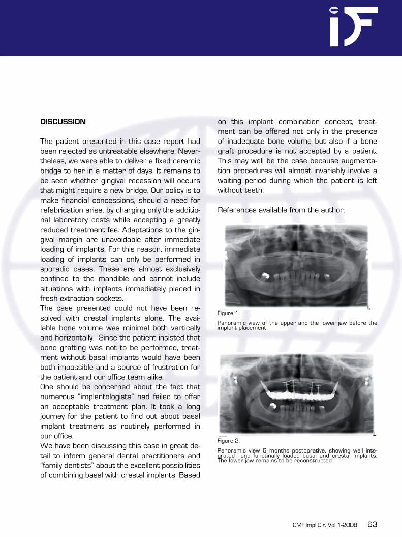
CMF.Impl.Dir. Vol 1-2008 63
DISCUSSION
The patient presented in this case report had been rejected as untreatable elsewhere. Never-theless, we were able to deliver a fixed ceramic bridge to her in a matter of days. It remains to be seen whether gingival recession will occurs that might require a new bridge. Our policy is to make financial concessions, should a need for refabrication arise, by charging only the additio-nal laboratory costs while accepting a greatly reduced treatment fee. Adaptations to the gin-gival margin are unavoidable after immediate loading of implants. For this reason, immediate loading of implants can only be performed in sporadic cases. These are almost exclusively confined to the mandible and cannot include situations with implants immediately placed in fresh extraction sockets. The case presented could not have been re-solved with crestal implants alone. The avai-lable bone volume was minimal both vertically and horizontally. Since the patient insisted that bone grafting was not to be performed, treat-ment without basal implants would have been both impossible and a source of frustration for the patient and our office team alike.One should be concerned about the fact that numerous “implantologists” had failed to offer an acceptable treatment plan. It took a long journey for the patient to find out about basal implant treatment as routinely performed in our office. We have been discussing this case in great de-tail to inform general dental practitioners and “family dentists” about the excellent possibilities of combining basal with crestal implants. Based
on this implant combination concept, treat-ment can be offered not only in the presence of inadequate bone volume but also if a bone graft procedure is not accepted by a patient. This may well be the case because augmenta-tion procedures will almost invariably involve a waiting period during which the patient is left without teeth.
References available from the author.
Figure 1.
Panoramic view of the upper and the lower jaw before the implant placement
Figure 2.
Panoramic view 6 months postoprative, showing well inte-grated and functinally loaded basal and crestal implants.The lower jaw remains to be reconstructed

64
Editor: Dr. Werner ManderManaging Editor: Dr. Sigmar Kopp
«CMF Implant Directions» is a valuable publication with a unique balance of scientific methodology and expert clinical ideas and opinion presented in a concise, easy to read manner.
Cranio-maxillofacial Implant Directions
Yes, I ́ d like to subscirbe the Journal: «CMF Implant Directions» I´d like to recieve only the next issue of «CMF Implant Directions»
30 Euro for a single issue 120 Euro for the annual subscription
Please send me an invoice to:
*Name:
*first Name:
*Address:
*City/State/Postal Code:
*Country:
*Phone:
E-mail:
Place, Date Signature
This order can be cancelled by giving written notice to the publisher within 30 days of subscribing. Cancellation must be mailed within this period (date of mailing verified by postal stamp).
The prices are for subscribers that are living in Germany or Switzerland! Please add a delivery charge of 16 Euro, when you live in foureign countrys. Thank you.
Please send this order form to: International Implant Foundation, Leopoldstr. 116, DE-80802 München
OR
DER
FO
RM
O
RD
ER F
OR
M
OR
DER
FO
RM
O
RD
ER F
OR
M
OR
DER
FO
RM
O
RD
ER F
OR
M
OR
DER
FO
RM
Cranio-maxillofacial
Implant Directions

CMF.Impl.Dir. Vol 1-2008 65
Research in Context – Part III
Random Assignment: Let chance be your friend.
Teaser
Not all study designs are created equal. Ran-domized controlled trials are the best study de-signs for minimizing bias. In this issue of Implant Directions, learn the characteristics (random al-location, concealment, and intent to treat) of a randomized controlled trial and why these are important in minimizing the potential bias that may occur in the other study designs described in the last edition of Implant Directions.
Text
The goal of a clinical trial assessing treatment is to obtain the most accurate and unbiased ef-fect of the treatment. One important way to help minimize bias is to select the best study design to accomplish your purpose. The randomized controlled trial, though often difficult and some-times unethical to perform, is the most effective way to minimize bias when comparing two treat-ment techniques.
Random allocation
Patients who are recruited to participate in studies should represent a relatively broad sample from the potential target population. It is important for the authors to describe the study population to ensure that conclu-
•
•
sions made are not only valid, but also apply to the patient population to which one is in-terested in making inference. Ideally, comparison groups are comprised of participants who are similar in all re-spects, with the exception of the particular intervention(s) that is being studied. The best method to achieve this similarity be-tween groups is that of random allocation or random assignment.The study design utilizing random allocation is the randomized controlled trial (RCT).
•
•
Ideally, comparison groups are comprised of participants who are similar in all re-spects, with the exception of the particu-lar intervention(s) that one is studying. The best method to achieve this similarity be-tween groups is that of random allocation.
Random allocation is a method of dividing subjects into groups in such a way that the characteristics of the subject do not affect the group to which they are allocated. To achieve this, we allow chance to decide to which group each subject is allocated. Thus, each subject is equally likely to be al-located to any of the available groups, and any differences between these groups hap-pen by chance.
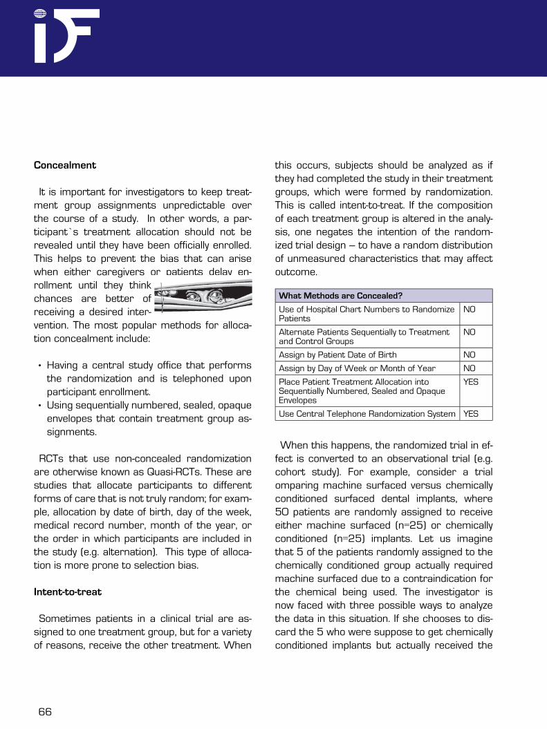
66
Concealment
It is important for investigators to keep treat-ment group assignments unpredictable over the course of a study. In other words, a par-ticipant’s treatment allocation should not be revealed until they have been officially enrolled. This helps to prevent the bias that can arise when either caregivers or patients delay en-rollment until they think chances are better of receiving a desired inter-vention. The most popular methods for alloca-tion concealment include:
Having a central study office that performs the randomization and is telephoned upon participant enrollment.Using sequentially numbered, sealed, opaque envelopes that contain treatment group as-signments.
RCTs that use non-concealed randomization are otherwise known as Quasi-RCTs. These are studies that allocate participants to different forms of care that is not truly random; for exam-ple, allocation by date of birth, day of the week, medical record number, month of the year, or the order in which participants are included in the study (e.g. alternation). This type of alloca-tion is more prone to selection bias.
Intent-to-treat
Sometimes patients in a clinical trial are as-signed to one treatment group, but for a variety of reasons, receive the other treatment. When
•
•
this occurs, subjects should be analyzed as if they had completed the study in their treatment groups, which were formed by randomization. This is called intent-to-treat. If the composition of each treatment group is altered in the analy-sis, one negates the intention of the random-ized trial design – to have a random distribution of unmeasured characteristics that may affect outcome.
When this happens, the randomized trial in ef-fect is converted to an observational trial (e.g. cohort study). For example, consider a trial omparing machine surfaced versus chemically conditioned surfaced dental implants, where 50 patients are randomly assigned to receive either machine surfaced (n=25) or chemically conditioned (n=25) implants. Let us imagine that 5 of the patients randomly assigned to the chemically conditioned group actually required machine surfaced due to a contraindication for the chemical being used. The investigator is now faced with three possible ways to analyze the data in this situation. If she chooses to dis-card the 5 who were suppose to get chemically conditioned implants but actually received the
What Methods are Concealed?
Use of Hospital Chart Numbers to Randomize Patients
NO
Alternate Patients Sequentially to Treatment and Control Groups
NO
Assign by Patient Date of Birth NO
Assign by Day of Week or Month of Year NO
Place Patient Treatment Allocation into Sequentially Numbered, Sealed and Opaque Envelopes
YES
Use Central Telephone Randomization System YES

CMF.Impl.Dir. Vol 1-2008 67
machine surfaced implants, or if she adds them to group randomly assigned to receive the ma-chine surfaced implants, the beneficial effects
Figure. Illustration of Intent-to-treat Analysis.
50 patients Keep togehter-for analysis
25
5
25
25
Machined
Chemical
Random Machined
Machined
Table 3. Evaluation of articles examining implant placement in patients with and without a histo-ry of periodontal disease
Type of Analysis Comparisons made
“Intent-to-treat”Correct analysis
Compare the 25 machined implant patients with the 25 randomized to the chemically conditioned implant group (20 chemical plus 5 machined)
“Per protocol”Biases results
Compare the 25 machined implant patients with 20 who received the chemically conditioned implants; discard the 5 machined implant patients who were randomized into the chemically conditioned implant group
“Treatment administered”Biased results
Compare 30 machined patients (5 machined implant patients who were randomized to the chemically conditioned implant group but who actually received the machined implants plus the 25 randomized to the machined implant group) with 20 chemically conditioned implant patients
of random assignment are diminished. The cor-rect analysis is the intent-to-treat analysis as demonstrated below.
Intent-to-treat analyses are the best way to as-sure bias due to an unequal distribution of cer-tain risk factors (i.e., confounding) will not play a role here. The price paid, however, is typically a reduction of any observed associations between treatment and outcome – any treatment effect found in an intent-to-treat analysis is likely to be
a conservative estimate of efficacy.
Next issue of Implant Directions….
Blinding…What does this mean and who should be blinded?

68
2
id iM
plA
nt
diR
Ecti
on
s E
du
cA
tio
nA
l v
idEo
sER
iEs
puBlishEd By if puBlishing, gERMAny oRdER no.: 6669
MAXILLARY IMPLANT PLACEMENT >>
REplAcing REplAcE®
Cranio-maxilofacial
Implant Directions®
Educational Video Series
EDUCATIONAL VIDEO SERIES
Maxillary Implant Placement
1 cREstAl & BAsAl iMplAnts Order Nr. 6667
2 And REplAcing REplAcE® Order Nr. 6669
Each DVD contains approx. 20 minutes of oral surgery.With explanations in english and ger-man language.
EuRo 35,00
Please send your order via e-mail to:[email protected]
or via regular postage mail to:International Implant Foundation Leopoldstr. 116, DE-80802 München
Guide for Authors
ID publishes articles, which contain information, that will impro-ve the quality of life, the treatment outcome, and the affordability of treatments.The following types of papers are published in the journal: Full length articles (maximum length abstract 250 words, to-tal 2000 words, references 25, no limit on tables and figures). Short communications including all case reports (maximum length abstract 150 words, total 600 words, references 10, fi-gures or tables 3) Technical notes (no abstract, no introduction or discussion, 500 words, references 5, figures or tables 3). Interesting cases/lessons learned (2 figures or tables, legend 100 words, maximum 2 references).
Literature Research and Review articles are usually commis-sioned.Critical appraisals on existing literature are welcome.
Direct submissions to:[email protected] text body (headline, abstract, keywords, article, conclusion), tables and figures should be submitted as separate documents. Each submission has to be accompanied by a cover letter. The cover letter must mention the names, addresses, e-mails of all authors and explain, why and how the content of the article will contribute to the improvement of the quality of life of patients.
id iM
plA
nt
diR
Ecti
on
s E
du
cA
tio
nA
l v
idEo
sER
iEs
id iM
plA
nt
diR
Ecti
on
s E
du
cA
tio
nA
l v
idEo
sER
iEs
id iM
plA
nt
diR
Ecti
on
s E
du
cA
tio
nA
l v
idEo
sER
iEs
id iM
plA
nt
diR
Ecti
on
s E
du
cA
tio
nA
l v
idEo
sER
iEs
puBlishEd By if puBlishing, gERMAny oRdER no.: 6667
MAXILLARY IMPLANT PLACEMENT >>
cREstAl & BAsAl iMplAnts
Cranio-maxilofacial
Implant Directions®
Educational Video Series
1


