Embed Size (px)
Citation preview
© 2014 Manaf Almatar and Zaidah Rahmat et al. This is an open access article distributed under the terms of the Creative Commons Attribution License -NonCommercial-ShareAlikeUnported License (http://creativecommons.org/licenses/by-nc-sa/3.0/).
Journal of Applied Pharmaceutical Science Vol. 4 (03), pp. 066-074, March, 2014 Available online at http://www.japsonline.com DOI: 10.7324/JAPS.2014.40314 ISSN 2231-3354
Identifying the developmental stages and optimizing the sample preparation for anatomical study of Orthosiphon stamineus Manaf Almatar* and Zaidah Rahmat
Faculty of Biosciences and Medical Engineering, Department of Biotechnology and Medical Engineering, Universiti Teknologi Malaysia (UTM),81310 Johor Bahru, Johor, Malaysia.
ARTICLE INFO
ABSTRACT
Article history: Received on: 30/11/2013 Revised on: 03/01/2014 Accepted on: 28/01/2014 Available online: 30/03/2014
This study focuses on the characterization of morphological and anatomical traits of Orthosiphon stamineus which belongs to the Lamiaceae family. Orthosiphon stamineus, better known as “Misai Kucing” or “cats whiskers” by the locals, contained active phenolics compounds such as flavanoids. Despite its wide usage as a medicinal plant, information regarding Orthosiphon stamineus specific developmental stages is relatively scarce. Furthermore, to date, no anatomical data of this plant is available. Therefore, this study aims to systematically identify the developmental stages and its anatomy which may provide more insight to its medical application. The result showed some distinct morphological and anatomical characteristics. In the morphological study, it was observed that Orthosiphon stamineus is a herbal shrub with well-developed creeping rootstock. The leaves are simple, green, and arranged in opposite pairs. The stem is approximately 54 cm in height at the oldest stage (62-64 days). In order to proceed with the anatomical study of Orthosiphon stamineus, the cross sections of different developmental stages of leaves and stems were examined. Several critical steps prior to viewing the prepared slides, which include dehydration, sectioning and staining, were optimized accordingly.
Key words: Orthosiphon stamineus, Morphology, Anatomy, Lamiaceae.
INTRODUCTION
Orthosiphon stamineus or locally named Misai Kuching belongs to Lamiaceae family and is widely used in herbal traditional medicine (Sim et al., 2003). Misai Kuching was used traditionally for centuries in southeast Asia by local people for the treatment of ailments of the bladder and kidney, diabetes mellitus and gout (Chin, 2007). Researchers gave much attention to this plant and many reports were published to describe the medical uses of this plant.Orthosiphon stamineus is also applied in traditional medicine to cure rheumatism, fever, hepatitis, gallstones, hypertension, diabetes, epilepsy and eruptive (Awale et al., 2003). The water extract of air-dried Orthosiphon stamineus leaves are used for renal diseases and urinary tract treatment in Myanmar (Awale et al., 2001).
This medical herb plant, Orthosiphon stamineus, contains several bioactive compounds such as ursolic acid, flavones, oleanolic acid, polyphenols, sterol, betulinic acid and rosmarinic acid (Hossain et al., 2008). Polyphenol, which is the most dominant compound in Orthosiphon stamineus leaves, prevents the formation of lipid peroxidation products in the .
.
* Corresponding Author E-mail: [email protected]
biological system, and has a considerable role in reducing oxidative stress (Hollman and Katan, 1999). Secoorthosi-phols A, B, and C, which are the three new highly oxygenated 2, 3-secoisopimarane-type diterpenes, have also been isolated from the aerial parts of this plant (Awale et al., 2002). Furthermore, high amount of flavonoids such as eupatorin (EUP), sinensetin (SEN), rosmarinic acid (RA) was also detected in different tissues of this plant (Akowuah et al., 2012). Morphologically, the plant is a perennial shrub tree with 4 angled stems which grows up to 1.5 meters. The leaves, which are used for medications, are arranged in opposite pairs with 0.3 cm in length with reddish purple petiole. The local name is derived from the flower morphology which looks like cat’s whiskers. The flowers are white to bluish with 16 cm length for verticils, and they have long far-exerted filaments (Fig 1) (Chin et al., 2009). A classification structure for Orthosiphon stamineus is represented in (Table 1). Despite its wide usage as a medicinal plant and its rich content of active phenolic compounds, information regarding Orthosiphon stamineus specific developmental stages is relatively scarce. Furthermore, to date, no systematic anatomical data of this plant is available in any online database. Therefore, this study aims to systematically identify the developmental stages and optimize the sample preparation for anatomical study of Orthosiphon stamineus as well.
Almatar and Rahmat / Journal of Applied Pharmaceutical Science 4 (03); 2014: 066-074 067
Fig. 1: General view of Orthosiphon stamineus.
Table. 1: A taxonomical classification for Orthosiphon stamineus.
Local Name Misai Kucing Kingdom Plantae Sub-kingdom
Tacheobionta (Vascular plants)
Supervision Spermatophyta (Seed plants) Division Magnoliophyta (Flowering plants) Class Magnoliopsida (Dicotyledons) Subclass Asteridae Order Lamiales Family Lamiaceae Genus Orthosiphon Species aristatus, labiatus, grandiflorum, spicatus, stamineus
MATERIALS AND METHODS
Plant Materials Orthosiphon stamineus plant was obtained from local
nursery, Pak Ali Nursery, located in Pulai, Johor and then was grown outdoor at the Faculty of Bioscience and Medical Engineering (FBME) under natural environment. Fresh samples from leaves and stems were collected to be used for anatomical study. Determination of morphological characterization for Orthosiphon stamineus
Morphological study of Orthosiphon stamineus was carried out at the local nursery (Pak Ali Nursery) in Pulai, Johor. Pictures were taken by using a DSLR camera (Nikon D3200) with white background, Meter scale and bar which was used to align the plants properly. Image lab Demo software was used for measuring the leaves area. The stem, leaf for plant (12-14), (32-34) and (62-64) days-old were examined. Histology technique
For anatomical studies of Orthosiphon stamineusin leaves and stems, plant tissues were subjected to several steps before analyzing the cross sections for histology characterization. For sample preparation, the samples must undergo fixation, dehydration, and embedding, prior to sectioning. Finally, the sectioned samples are stained before subjected to analysis. The details descriptions are as below; three methods were used for the
tissue slides preparation. Fresh specimens from leaves and stems were fixed with Formalin-Acetic-Alcohol fixing solution (50 mL absolute ethanol + 5 mL glacial acetic acid + 10 mL formaldehyde (37%-40%) + distilled water to a total volume of 100 mL) for 12 to 24 hours at room temperature. The dehydration was carried out by using a series of ethanol (30%, 50%, 70%, 80%, 90%, 95% and 100%).
In the first method, Formalin-Acetic-Alcohol and series of ethanol (30 to 95%) were used for fixation and dehydration.
For the second method, fixation and dehydration were also operated using Formalin-acetic-alcohol and series of ethanol (70 to 100%). In addition, the samples were immersed three times in excess xylene for 10 mins, one time in 50% xylene + 50% wax for 20 mins and 100% paraffin wax for 20 mins.
The processes in the third method were carried out as follows; Formalin-acetic-alcohol and series of ethanol (30 to 100%) were used for fixation and dehydration processes. The samples were soaked one time in xylene + ethanol at different ratio (3:1,1:3,1:1 followed by excess xylene for 30 mins), one time in 50% xylene + 50% wax for 20 mins and 100% paraffin wax for 30 mins for two times. The different compositions of chemicals used for the three dehydration methods are summarized in (Table 2).
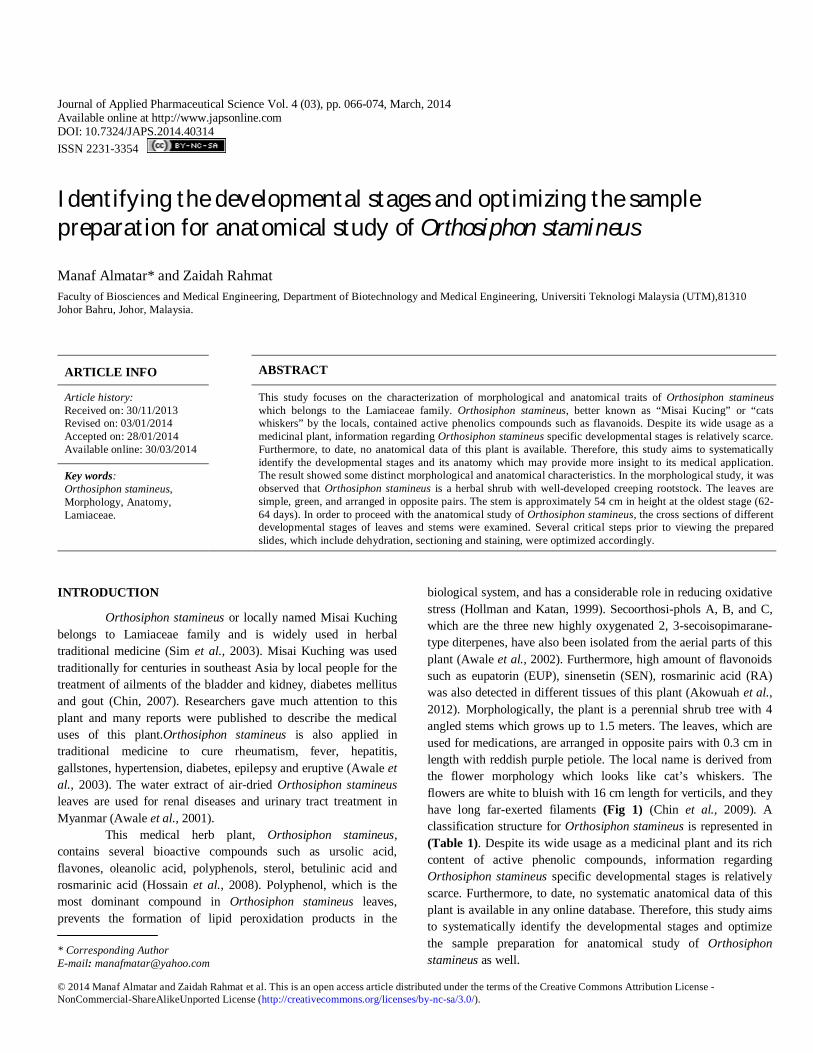
After the fixation and dehydration process, the samples were embedded using Tissue Embedding System 2900 (TEC) (Histo Line Laboratories Brand) (Fig 2A). The samples were embedded at the horizontally orientation using paraffin wax as embedding medium. Firstly, a mold was filled with melted paraffin wax and then tissues were oriented into the wax with warmed forceps, and the mold was chilled on a cold plate to speed up the embedding process. The embedded tissues were stored at 4ºC till sectioning. The samples could be stored at 4ºC for several months prior to sectioning.
The rotary microtome (Histo Line Laboratories Brand) (Fig 2B) was used for sectioning samples and should be set to cut between the range of 2-10 μM. The blade clearance angle should be at approximately ~5º (3-8º). After sectioning, the ribbons were carefully transferred into distilled water using forceps before transfer to the glass slides. The slides were dried at 40-45ºC overnight. Samples of 5.0 m – 7.0 m were stained by using hematoxylin and eosin staining.
Fig. 2: (A) Tissue Embedding Center (TEC 2900) (B) Rotary Microtome (Histo Line Laboratories Brand).
068 Almatar and Rahmat / Journal of Applied Pharmaceutical Science 4 (03); 2014: 066-074
Hematoxylin and Eosin staining To perform the staining step, the slides were first
deparaffinised using excess xylene three times for three mins each to remove the paraffin wax. Then the slides were exposed to gradual rehydration using a series of ethanol (80-95-100%) and deionized water as follows; (1) the slides were washed three times with 100% ethanol for three mins each, (2) the slides were rinsed with 95% ethanol for three mins, and then (3) they were immersed with 80% ethanol for three mins, and (4) sections were placed in deionized water for five mins.
Before staining the slides with hematoxylin, the excess water was removed by dabbing the slide holder. For hematoxylin, the staining time range was (1-3) mins (Table 3). Next, the slides were rinsed in deionized water followed by tap water for 5 mins in order to allow the stain to develop. Afterwards, they were destained using acid ethanol (99 mL 70% ethanol+ 1mL hydrochloric acid) by dipping them (8-12) times for (2-12 Sec each) (Table 3) and were rinsed two times with tap water for 1 min each. Then, the slides were immersed in deionized water for (3-5) mins (Table 3). Acid ethanol and deionized water were applied to remove the remaining stains.
After the hematoxylin-stained slides were rinsed, excess water was blotted to prepare slides for eosin staining. The slides were soaked in for (45 Sec-3 mins) (Table 3) and then were washed three times with 95 % ethanol for 5 mins each. They were also rinsed three times with 100 % ethanol for 5 mins each. Finally, excess ethanol was removed and the slides were immersed three times in 100 % xylene for 15 mins.The slides were covered with coverslip using permount mounting medium, where one drop was placed on the slide using glass rod, and gently covered with a coverslip. The permanent mounting medium was allowed to spread beneath the coverslip to cover all the tissues. Finally, the slides were allowed to dry overnight in the hood. The different parameters applied for the optimization the staining methods are summarized in (Table 3).
Microscopy
For stems, the slides were viewed under light microscopy (NIKON SMZ 800) and pictures were taken using attached camera (Nikon Smz800 TV Lens0.55.Ds Nikon) with 40X magnification. For leaves, the slides were viewed via light microscopy (LEICA Brand) and the pictures were taken using Samsung Galaxy camera S4 with 100X and 400X magnification. RESULTS AND DISCUSSION
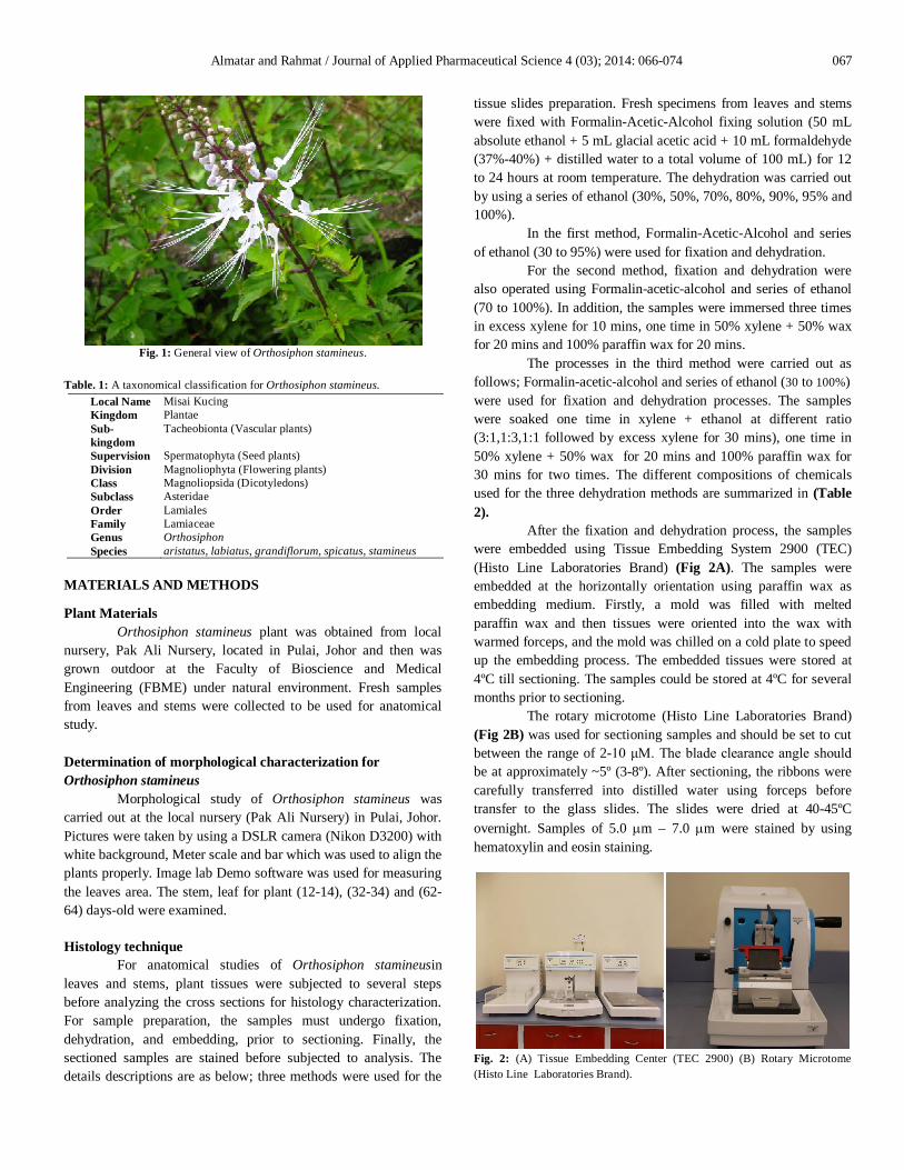
Vegetative growth stages of Orthosiphon stamineus This study was performed on three different stages of
Orthosiphon stamineus: stage A (12-14) days, stage B (32-34) days and stage C (62-64) days. Eighteen plants and twelve plants were studied for stage A (12-14) days age and stage B (32-34) days age, respectively. For stage C (62-64) days age, only two plants were used. The plants in stage C were the highest in height 54 cm while the stage A plants were the shortest at 30 cm and the stage B was in between 40 cm (Fig 3). Stage A (12-14) days Stage B (32-34) days Stage C (62-64) days
Fig. 3: Developmental stages for Orthosiphon stamineus. A (12-14)days, B (32-34) days and C (62-64) days.Bar=5cm for stage A, 7 cm for stage B, 10 cm for stage C.
The average height of those groups mentioned above
were 30.06, 35.67 and 50.50 cm for stage A, B and C, respectively
Table. 2: Dehydration parameters (concentration and immersion time of ethanol, xylene and paraffin) used in three histological methods.
Chemicals Method 1 Method 2 Method 3
Ethanol Samples were immersed in a series of ethanol (30%-50%-70%-80%-90%-95%) for 30 mins.
Samples were immersed in a series of ethanol (70%-80%-90%-95%-100%) for 5 mins followed by ethanol (100%) for 10 mins.
Samples were immersed in a series of ethanol (30%-50%-70%-80%-90%-95%-100%) for 30 mins.
Xylene / Xylene and Ethanol Not used Samples were immersed three times in excess xylene for 10 mins
Samples were immersed one time in xylene + ethanol at different ratio followed by excess xylene for 30 mins
50 % Xylene / 50 % ParraffinWax Not used Samples were immersed one time in 50% xylene
+ 50% wax for 20 mins. Samples were immersed one time with 50% xylene + 50% wax for 20 mins.
Paraffin Wax 100% Not used Samples were submerged one time in 100% paraffin wax for 20 mins
Samples were submerged tow times in 100% paraffin wax for 30 mins
Table. 3: Staining parameters (immersion time of hematoxylin,eosin, deionized water and acid ethanol) used in three histological method.
Parameter Method 1 Method 2 Method 3
Hematoxylin Slides were immersed in hematoxylin for 3 mins
Slides were immersed in hematoxylin for 1 min
Slides were immersed in hematoxylin for 2 mins
Eosin Slides were soaked in eosin for 45 Sec Slides were soaked in eosin for 2 mins Slides were soaked in eosin for 3 mins
Deionized water Slides were washed in deionized water for 3 mins
Slides were washed in deionized water for 5 mins
Slides were washed in deionized water for 5 mins
Acid ethanol Slides were rinsed 10 times in acid ethanol (2 Sec or each)
Slides were rinsed 12 times in acid ethanol (6 Sec or each)
Slides were rinsed 12 times in acid ethanol (12 Sec or each)
Almatar and Rahmat / Journal of Applied Pharmaceutical Science 4 (03); 2014: 066-074 069
(Appendix A, B, C and D). For leaves size, the different groups of studied plants mentioned above had three sizes of leaves ranging from small, medium and large but large leaves was observed on the stage C more than the other two stages (Fig 4). Leaves number was determined as follows; abundant numbers of leaves were available in the stage C plant more than the stage A and stage B plants. Regarding the branch color, stage C plant had more purple branches than the stages A and B plants (Fig 3). Comparison between morphological traits of three Orthosiphon stamineus stages were elucidated in (Table 4).
Fig. 4: Leaves size at different stages of Orthosiphon stamineus. StageA (12-14)days, B (32-34) days and C (62-64) days.
Table. 4: Stage A had the shortest height and a fewer number of leaves whereas the longest height occurred at stage C which had the highest number of leaves and stage B was in between. Regarding the leaves size, stage A had almost a smaller size comparing to the two other stages, B and C.
Plant age Height Number of leaves
Size of leaves (Large-Medium-Small)
Young stage A (12-14) days 30 cm 11 25 - 11 – 5 cm Medium stage B (32-34) days 40 cm 25 32 - 16 - 3 cm
Old stage C (62-64) days 54 cm 40 53 -18 – 10 cm
Optimization the sample preparation for anatomical study of Orthosiphon stamineus
In order to characterize the anatomical structures of Orthosiphon stamineus, cross sections of the stems and leaves of this plant were obtained via histological techniques. Three methods were attempted with several parameter manipulations for obtaining the best sections. The quality of the sections and identifications of specific structures of stems and leaves were observed under light microscope. Anatomical study of leaves and stems from Orthosiphon stamineus
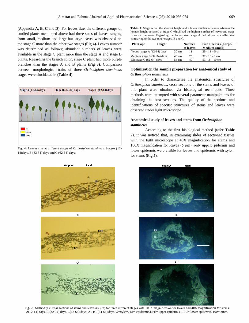
According to the first histological method (refer Table 2), it was noticed that, in examining slides of sectioned tissues with the light microscope at 40X magnification for stems and 100X magnification for leaves (5 μm), only uppere pidermis and lower epidermis were visible for leaves and epidermis with xylem for stems (Fig 5).
Fig. 5: Method (1) Cross sections of stems and leaves (5 μm) for three different stages with 100X magnification for leaves and 40X magnification for stems. A(12-14) days, B (32-34) days, C(62-64) days. A1-B1 (64-66) days. X=xylem, EP= epidermis,UPE= upper epidermis, LEU= lower epidermis, Bar= 2mm.
070 Almatar and Rahmat / Journal of Applied Pharmaceutical Science 4 (03); 2014: 066-074
The structures were only partially visible because of the tearing in the tissue sections and because of the fact that these sections were not clear enough.Two types of stain, hematoxylin and eosin, were used to stain tissues in blue and pink color, respectively. The eosin stain is not visible due to the prevailing effect of hematoxylin. These results are most probably because of the exclusion of the infiltration and clearing steps in the dehydration process in Method 1. These two steps were included in Method 2 (Table 2) as the first optimization step. Paraffined wax was used during the infiltration step which leads to the saturation of the tissue cavities and cells as well as preventing the tissues from tearing. Xylene is also used as a clearing agent to make tissues more translucent (Chong et al., 2012). Another problem observed in Method 1 is the dominant effect of hematoxylin that is caused by the long exposure (3 mins) which prevents the uptake of eosin entirely.
This is also due to the very short time allocated for destaining (Fig 5) (refer Table 3) (Gill, 2013).
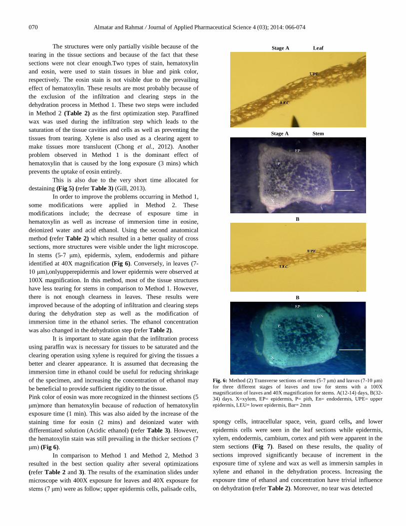
In order to improve the problems occurring in Method 1, some modifications were applied in Method 2. These modifications include; the decrease of exposure time in hematoxylin as well as increase of immersion time in eosine, deionized water and acid ethanol. Using the second anatomical method (refer Table 2) which resulted in a better quality of cross sections, more structures were visible under the light microscope. In stems (5-7 μm), epidermis, xylem, endodermis and pithare identified at 40X magnification (Fig 6). Conversely, in leaves (7-10 μm),onlyupperepidermis and lower epidermis were observed at 100X magnification. In this method, most of the tissue structures have less tearing for stems in comparison to Method 1. However, there is not enough clearness in leaves. These results were improved because of the adopting of infiltration and clearing steps during the dehydration step as well as the modification of immersion time in the ethanol series. The ethanol concentration was also changed in the dehydration step (refer Table 2).
It is important to state again that the infiltration process using paraffin wax is necessary for tissues to be saturated and the clearing operation using xylene is required for giving the tissues a better and clearer appearance. It is assumed that decreasing the immersion time in ethanol could be useful for reducing shrinkage of the specimen, and increasing the concentration of ethanol may be beneficial to provide sufficient rigidity to the tissue. Pink color of eosin was more recognized in the thinnest sections (5 μm)more than hematoxylin because of reduction of hematoxylin exposure time (1 min). This was also aided by the increase of the staining time for eosin (2 mins) and deionized water with differentiated solution (Acidic ethanol) (refer Table 3). However, the hematoxylin stain was still prevailing in the thicker sections (7 μm) (Fig 6).
In comparison to Method 1 and Method 2, Method 3 resulted in the best section quality after several optimizations (refer Table 2 and 3). The results of the examination slides under microscope with 400X exposure for leaves and 40X exposure for stems (7 μm) were as follow; upper epidermis cells, palisade cells,
Stage A Leaf
Stage A Stem
B
B
Fig. 6: Method (2) Transverse sections of stems (5-7 μm) and leaves (7-10 μm) for three different stages of leaves and tow for stems with a 100X magnification of leaves and 40X magnification for stems. A(12-14) days, B(32-34) days. X=xylem, EP= epidermis, P= pith, En= endodermis, UPE= upper epidermis, LEU= lower epidermis, Bar= 2mm
spongy cells, intracellular space, vein, guard cells, and lower epidermis cells were seen in the leaf sections while epidermis, xylem, endodermis, cambium, cortex and pith were apparent in the stem sections (Fig 7). Based on these results, the quality of sections improved significantly because of increment in the exposure time of xylene and wax as well as immersin samples in xylene and ethanol in the dehydration process. Increasing the exposure time of ethanol and concentration have trivial influence on dehydration (refer Table 2). Moreover, no tear was detected

Almatar and Rahmat / Journal of Applied Pharmaceutical Science 4 (03); 2014: 066-074 071
Stage B Leaf
Stage B Stem
C
C
Fig. 7: Method (3) Cross sections of stems (7μm) and leaves (7μm) for three different stages of leaves and tow for stems with a 400X magnification of leaves and 40X magnification for stems. B (32-34) days, C(62-64) days, UPE= upper epidermis, PP=palisade cells, SP=spongy cells,IS= intracellular space, GC=guard cells, LEC= lower epidermis, VS=vein, EP=epidermis, X= xylem, En endodermis, C=cambium, Co= cortex P=pith, Bar =2mm.
Table. 5: Summary of three histogical methods used in anatomical study of Orthosiphon stamineus stem. Method Stem obsevation Optimization Remark
Method (1)
Epidermis and xylem.
None Tear in all sections Hematoxylin was dominating with no trace of Eosin.
Method (2)
Epidermis, endodermis, pith and xylem.
Dehydration: inclusion of infiltration and clearing steps, timing and concentration of ethanol was modified. Staining: Hematoxylin, Eosin exposure time and deionized water with differentiated solution (Acidic ethanol).
A better observation of the structure occurred with little tear. Shown pink color of Eosin as a prevailing in the sections (5 μm).
Method (3)
Epidermis, endodermis, pith, cortex and xylem.
Dehydration: inclusion of infiltration, clearing, timing and concentration of ethanol. Staining: Hematoxylin, Eosin exposure time and deionized water with differentiated solution (Acidic ethanol).
No tear is detected in the tissues with more clearness for leaves sections, Eosin disappeared with evident domination of Hematoxylin.

072 Almatar and Rahmat / Journal of Applied Pharmaceutical Science 4 (03); 2014: 066-074
Table. 6: Summary of three histogical methods used in anatomical study of Orthosiphon stamineus leaves. Method Leaf observation Optimization Remark
Method (1)
Upper epidermis and lower epidermis.
None Tears in all sections. Hematoxylin was dominating with no trace of Eosin.
Method (2)
Upper epidermis and lower epidermis.
Dehydration: inclusion of infiltration and clearing steps, timing and concentration of ethanol was modified. Staining: Hematoxylin, Eosin exposure time and deionized water with differentiated solution (Acidic ethanol).
A better observation of the structure occurred with little tear. Shown pink color of Eosin as a prevailing in the sections (5 μm).
Method (3)
Upper epidermis cells, palisade cells, spongy cells, intracellular space, guard cells and lower epidermis cells.
Dehydration: inclusion of infiltration, clearing, timing and concentration of ethanol. Staining: Hematoxylin, Eosin exposure time and deionized water with differentiated solution (Acidic ethanol).
No tear is detected in the tissues with more clearness for leaves sections. Eosin disappeared with evident domination of Hematoxylin.
APPENDIX A
Stage (A) (12-14) days
Almatar and Rahmat / Journal of Applied Pharmaceutical Science 4 (03); 2014: 066-074 073
APPENDIX B
Stage (B) (32-34) days
APPENDIX C
Stage (C) (62-64) days
APPENDIX D The height of each plant in each group
Stage A (12-14) days [cm] Stage B (32-34) days [cm] Stage C (62-64) days [cm] 30 34 47 29 34 54 30 30 29 40 28 36 29 37 30 35 28 36 30 38 30 33 30 35 28 40 36 30 30 30 30 34
30.06 35.67 50.50
074 Almatar and Rahmat / Journal of Applied Pharmaceutical Science 4 (03); 2014: 066-074
and the tissues appeared to be quite clear. Nevertheless, the eosin stain fail to retain with evident domination of hematoxylin (Fig 7) in spite of the fact that the immersion time for eosin, deionized water and acidic ethanol has been increased (refer Table 3). These observations indicate; (1) the strong influence of hematoxylin, (2) staining time by hematoxylin was too long and (3) inadequate differentiation in HCl. In short, the various parameters optimized in (Table 2) (Dehydration parameters) and (Table 3) (Staining parameters) has highlighted the critical steps that improve the sections quality of Orthosiphon stamineus studied in this work, although further enhancement is needed. Briefly, dehydration parameters (concentration and immersion time in ethanol, xylene and paraffin) were optimized as shown in (Table 2). The results revealed that xylene and paraffin, which were used in Method 2 and 3, had important influence in improving the sections quality of Orthosiphon stamineus. On the other hand, Table 3 described the different parameters optimized for staining (immersion time in hematoxylin, eosin, deionized water and acid ethanol). The results indicated that decreasing the immersion time in hematoxylin led to the best appearance of eosin colour in the sections.The summary of the three histological methods applied in this study is displayed in Table5 and 6. CONCLUSIONS
The developmental stages and morphological characteristics of Orthosiphon stamineus for leaves, stems were determined in three different stages: stage A (12-14) days, stage B (32-34) days and stage C (62-64) days of this plant. For anatomical studies in stem and leaves, several critical steps, which include dehydration, sectioning and staining, were optimized. Three methods were performed for anatomical study. Method 3, which involves fixation, dehydration, embedding, sectioning and staining, was the best way of obtaining the high quality sections with no tear in tissue sections ranging from (5-7 μm). Thus, enabling the different structures including upper epidermis cells, palisade cells, spongy cells, intracellular space, guard cells and lower epidermis cells for leaves and epidermis, xylem, endodermis, cambium, cortex and pith for stems to be distinguished. Critical optimization steps during dehydration includes the infiltration, clearing step were carried out. For staining, the exposure time and destaining steps were modified accordingly to overcome the dominant stain by hematoxylin. However, the strong hematoxylin stain is still retained in the sections although after optimization.
ACKNOWLEDGEMENT
The authors wish to acknowledge the UTM for providing the financial assistance for the project.
REFERENCES
Akowuah GA, Ismail Z, Ahmad M. HPLC–TOF/MS profile and nitric oxide scavenging activity of Orthosiphon stamineus leaf extracts. Asia Pacific Journal of Tropical Biomedicine, 2012; 2:S1436-S1439.
Awale S, Tezuka Y, Banskota AH, Adnyana IK, Kadota S. Nitric oxide inhibitory isopimarane-type diterpenes from Orthosiphon stamineus of Indonesia. Journal of Natural Products, 2003; 66:255-258.
Awale S, Tezuka Y, Banskota AH, Kouda K, Kyaw MT, Kadota S. Four highly oxygenated isopimarane-type diterpenes of Orthosiphon stamineus. Planta Medica, 2002;68:286-288.
Awale S, Tezuka Y, Banskota AH, Kouda K, Tun KM, Kadota S. Five novel highly oxygenated diterpenes of Orthosiphon stamineus from Myanmar. Journal of Natural Products, 2001;64:592-596.
Chin JH. Effect of the Orthosiphon stamineus Benth on aminopyrine metabolism in rat hepatocytes.Malaysian Journal of Pharmaceutical Sciences, 2007;5:25-32.
Chin JH, Mahfoudh M, Abas HH. Interactions of Orthosiphon stamineus and Morinda citrifolia with hepatic aminopyrine metabolism by CYP3A in rats. Pharmacognosy Mag, 2009; 4:55-60.
Chong WC, Wu R, Tu AY. A Study on Tissue Processing. International Journal of Innovative Interdisciplinary Research, 2012;1:37-43.
Gill G. 2013. H&e stain. Cytopreparation. Springer New York. Hollman PCH, Katan MB. Dietary Flavonoids: Intake, Health
Effects and Bioavailability. Food and Chemical Toxicology, 1999; 37:937-942.
Hossain MA, Ismail Z, Rahman A, Kang SC. Chemical composition and anti-fungal properties of the essential oils and crude extracts of Orthosiphon stamineus Benth. Industrial Crops and Products, 2008; 27: 328-334.
Sim C, Ahmad M, Ismail Z, Othman A, Noor N, Zaihidee E. Chemometric Classification of Herb – Orthosiphon stamineus According to Its Geographical Origin Using Virtual Chemical Sensor Based Upon Fast GC. Sensors, 2003; 3: 458-471.
How to cite this article:
Manaf Almatar and Zaidah Rahmat. Identifying the developmental stages and optimizing the sample preparation for anatomical study of Orthosiphon stamineus. J App Pharm Sci, 2014; 4 (03): 066-074.