Embed Size (px)
Citation preview
Asken and Rabinovici acta neuropathol commun (2021) 9:96 https://doi.org/10.1186/s40478-021-01197-4
REVIEW
Identifying degenerative effects of repetitive head trauma with neuroimaging: a clinically-oriented reviewBreton M. Asken1* and Gil D. Rabinovici2
Abstract
Background and Scope of Review: Varying severities and frequencies of head trauma may result in dynamic acute and chronic pathophysiologic responses in the brain. Heightened attention to long-term effects of head trauma, par-ticularly repetitive head trauma, has sparked recent efforts to identify neuroimaging biomarkers of underlying disease processes. Imaging modalities like structural magnetic resonance imaging (MRI) and positron emission tomography (PET) are the most clinically applicable given their use in neurodegenerative disease diagnosis and differentiation. In recent years, researchers have targeted repetitive head trauma cohorts in hopes of identifying in vivo biomarkers for underlying biologic changes that might ultimately improve diagnosis of chronic traumatic encephalopathy (CTE) in living persons. These populations most often include collision sport athletes (e.g., American football, boxing) and military veterans with repetitive low-level blast exposure. We provide a clinically-oriented review of neuroimaging data from repetitive head trauma cohorts based on structural MRI, FDG-PET, Aβ-PET, and tau-PET. We supplement the review with two patient reports of neuropathology-confirmed, clinically impaired adults with prior repetitive head trauma who underwent structural MRI, FDG-PET, Aβ-PET, and tau-PET in addition to comprehensive clinical examina-tions before death.
Review Conclusions: Group-level comparisons to controls without known head trauma have revealed inconsistent regional volume differences, with possible propensity for medial temporal, limbic, and subcortical (thalamus, corpus callosum) structures. Greater frequency and severity (i.e., length) of cavum septum pellucidum (CSP) is observed in repetitive head trauma cohorts compared to unexposed controls. It remains unclear whether CSP predicts a particu-lar neurodegenerative process, but CSP presence should increase suspicion that clinical impairment is at least partly attributable to the individual’s head trauma exposure (regardless of underlying disease). PET imaging similarly has not revealed a prototypical metabolic or molecular pattern associated with repetitive head trauma or predictive of CTE based on the most widely studied radiotracers. Given the range of clinical syndromes and neurodegenerative pathologies observed in a subset of adults with prior repetitive head trauma, structural MRI and PET imaging may still be useful for differential diagnosis (e.g., assessing suspected Alzheimer’s disease).
Keywords: Chronic traumatic encephalopathy, Traumatic encephalopathy syndrome, Traumatic brain injury, Repetitive head trauma, Concussion, Neuroimaging, Positron emission tomography, Magnetic resonance imaging, Neurodegenerative disease
© The Author(s) 2021. Open Access This article is licensed under a Creative Commons Attribution 4.0 International License, which permits use, sharing, adaptation, distribution and reproduction in any medium or format, as long as you give appropriate credit to the original author(s) and the source, provide a link to the Creative Commons licence, and indicate if changes were made. The images or other third party material in this article are included in the article’s Creative Commons licence, unless indicated otherwise in a credit line to the material. If material is not included in the article’s Creative Commons licence and your intended use is not permitted by statutory regulation or exceeds the permitted use, you will need to obtain permission directly from the copyright holder. To view a copy of this licence, visit http:// creat iveco mmons. org/ licen ses/ by/4. 0/. The Creative Commons Public Domain Dedication waiver (http:// creat iveco mmons. org/ publi cdoma in/ zero/1. 0/) applies to the data made available in this article, unless otherwise stated in a credit line to the data.
IntroductionLifetime head trauma exposure is a risk factor for mul-tiple neurodegenerative diseases including Alzheimer’s disease (AD), Parkinson disease, amyotrophic lateral
Open Access
*Correspondence: [email protected] Department of Neurology, Memory and Aging Center, Weill Institute for Neurosciences, University of California, San Francisco, 675 Nelson Rising Lane, Suite 190, San Francisco, CA 94143, USAFull list of author information is available at the end of the article
Page 2 of 17Asken and Rabinovici acta neuropathol commun (2021) 9:96
sclerosis, and chronic traumatic encephalopathy (CTE) [1–8]. CTE is the only neurodegenerative disease occur-ring almost exclusively in individuals with prior repeti-tive head trauma exposure, which is often sustained in the context of collision sports and/or military service. Conversely, most individuals with non-CTE neurodegen-erative diseases have no documented head trauma expo-sure history, especially repetitive. Neuroimaging plays a key role in informing differential diagnoses of suspected neurodegenerative diseases, but researchers thus far have had little success identifying diagnostic imaging biomark-ers that are specific for CTE in living adults.
Several advanced neuroimaging techniques have helped rapidly advance our understanding of disease pathophysiology, though only a handful are utilized clini-cally. Structural magnetic resonance imaging (MRI) is routinely performed in clinical settings to rule out non-neurodegenerative causes of clinical impairment like mass lesions and stroke, but also provides topographic representation of atrophy patterns. In some neurode-generative conditions, atrophy patterns are sensitive and specific enough that they improve diagnostic certainty of syndromes like Alzheimer’s-type dementias [9–11], pri-mary progressive aphasia subtypes [12], and behavioral variant frontotemporal dementia [13]. However, frank volume loss observable on structural MRI is thought to represent the culmination of a complex process of pro-tein misfolding and deposition, neuronal and glial dys-function, and ultimately synaptic and neuronal loss. Structural MRI interpretation also can be complicated by image processing limitations. Brain structure visualiza-tion and volume quantification via MRI occurs indirectly through measurement of underlying physical–chemical properties of brain tissue. There is inherent risk of over-attributing group differences to true structural changes when other potential confounds like tissue perfusion or water content variability also significantly influence the measurement outcomes [14].
Other imaging techniques like positron emission tomography (PET) provide opportunities for detect-ing evidence of disease pathophysiology upstream of degeneration and potentially prior to symptom onset in individuals at risk for neurodegenerative disease. PET imaging is employed clinically to quantify meta-bolic brain changes or the burden and distribution of Aβ plaques and tau tangles using radiolabeled trac-ers. Uptake of fluorodeoxyglucose (FDG) is a marker of synaptic activity and brain metabolism. There is limited research applying FDG-PET in cases of repetitive head trauma or suspected CTE. Radiotracers have also been developed for binding Aβ plaques and tau tangles. Aβ tracers most often used in clinical practice include Flor-betapir, Florbetaben, and Flutemetamol. Regarding tau
PET, early small studies of repetitive head trauma cases used the FDDNP tracer, which has significant limitations, including nonselective binding to multiple insoluble protein aggregates (e.g., Aβ, tau, prion protein and oth-ers), low signal-to-noise ratio, and poor reproducibility across centers. Flortaucipir, the most studied tau PET tracer, was developed to bind paired helical filaments of tau that form neurofibrillary tangles (NFTs) in AD. AD research has rapidly facilitated development and clini-cal translation of Aβ and tau PET radiotracers. Together, these PET tracers can essentially confirm presence of AD plaque and tangle pathology in living patients. As we later discuss, initial excitement over the potential utility of tau PET tracers for identifying CTE tau pathology has significantly tempered in recent years. Other PET tracers detecting important disease processes like neuroinflam-mation (e.g., translocator protein, or TSPO-PET) [15] or synapse loss currently have relatively little data in repeti-tive head trauma populations.
The suspected higher prevalence of CTE in clinically impaired adults with repetitive head trauma exposure has motivated efforts targeting this population. Trau-matic encephalopathy syndrome (TES) refers to the clinical manifestations of cognitive and/or neurobehav-ioral changes in individuals with repetitive head trauma exposure [16, 17]. Research criteria for TES were revised in 2021 [17] and require “substantial” exposure to repeti-tive head impacts from collision sports, military service, or other causes to qualify for diagnosis. There must be a predominant cognitive (episodic memory and/or execu-tive functioning) and/or neurobehavioral syndrome (explosiveness, impulsivity, rage, etc.). Symptoms must be progressive and not fully accounted for by another neurologic, psychiatric, or medical condition, though suspicion of comorbid conditions (e.g., another neuro-degenerative disease) is not exclusionary. A provisional level of certainty is assigned based on degree of head impact exposure and specific symptom manifestations - “Suggestive of CTE,” “Possible CTE,” or “Probable CTE”. Fluid and neuroimaging biomarkers do not factor into these research diagnostic criteria for TES due to lack of available data demonstrating specific associations with CTE pathology. “TES with definite CTE” can only be diagnosed by autopsy.
TES criteria were designed initially to maximize sen-sitivity over specificity to underlying CTE pathology. Earlier criteria [16] included several core and support-ive symptoms also observed in relatively high frequency among adults without repetitive head trauma exposure or neurodegenerative disease (e.g., depression, anxiety, headaches) [18, 19]. Changes on structural neuroimag-ing (e.g., cavum septum pellucidum) or PET imaging of Aβ plaques and tau tangles previously were proposed to
Page 3 of 17Asken and Rabinovici acta neuropathol commun (2021) 9:96
inform the likelihood that CTE is the cause of TES. How-ever, neuroimaging correlates of repetitive head trauma, with or without presumed CTE, remain incompletely characterized. In particular, recent studies suggest that the proposed use of currently available tau PET tracers for increasing diagnostic certainty of CTE is premature.
The large number of review articles focused on neuro-imaging of repetitive head trauma reflects the scientific interest in establishing neuroimaging biomarkers of CTE or other neurodegenerative effects of repetitive head trauma [20–29]. There are two key concepts that must be emphasized when reviewing and interpreting this litera-ture: 1) clearly understanding the definition, frequency, severity, and timing of the head trauma exposure in any given study, and 2) resisting the temptation to assume that significant neuroimaging findings within repetitive head trauma cohorts reflect biomarkers specific to CTE. Despite CTE being highly associated with repetitive head trauma exposure [30, 31], CTE is only one possible neu-rodegenerative outcome of repetitive head trauma and often exists with other pathologies, such as Aβ plaques, alpha-synuclein, TDP-43 proteinopathies, and white matter rarefaction [5, 32]. In other words, the associa-tion of CTE with prior repetitive head trauma is much
stronger than the association of repetitive head trauma with underlying CTE.
Scope of the Review and Relevant TerminologyHere we provide an overview of structural MRI and PET neuroimaging data from repetitive head trauma popula-tions, with a focus on literature published within approxi-mately the past 5 years. We chose to highlight structural MRI and PET given their direct clinical applications. Data from advanced neuroimaging modalities like dif-fusion tensor imaging (DTI), functional MRI, cerebral perfusion, and other modalities advance our understand-ing of repetitive head trauma pathophysiology. However, these modalities currently have minimal clinical footprint and largely are not validated for informing differential diagnosis.
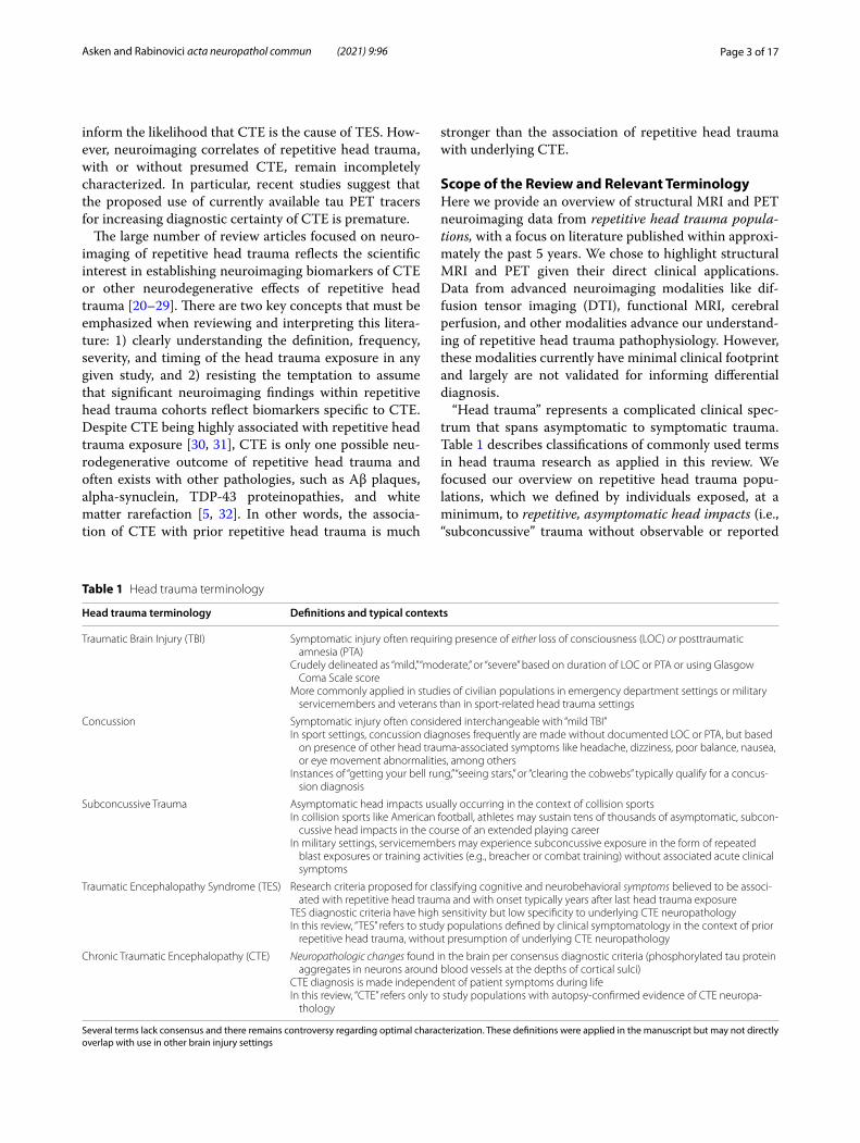
“Head trauma” represents a complicated clinical spec-trum that spans asymptomatic to symptomatic trauma. Table 1 describes classifications of commonly used terms in head trauma research as applied in this review. We focused our overview on repetitive head trauma popu-lations, which we defined by individuals exposed, at a minimum, to repetitive, asymptomatic head impacts (i.e., “subconcussive” trauma without observable or reported
Table 1 Head trauma terminology
Several terms lack consensus and there remains controversy regarding optimal characterization. These definitions were applied in the manuscript but may not directly overlap with use in other brain injury settings
Head trauma terminology Definitions and typical contexts
Traumatic Brain Injury (TBI) Symptomatic injury often requiring presence of either loss of consciousness (LOC) or posttraumatic amnesia (PTA)
Crudely delineated as “mild,” “moderate,” or “severe” based on duration of LOC or PTA or using Glasgow Coma Scale score
More commonly applied in studies of civilian populations in emergency department settings or military servicemembers and veterans than in sport-related head trauma settings
Concussion Symptomatic injury often considered interchangeable with “mild TBI”In sport settings, concussion diagnoses frequently are made without documented LOC or PTA, but based
on presence of other head trauma-associated symptoms like headache, dizziness, poor balance, nausea, or eye movement abnormalities, among others
Instances of “getting your bell rung,” “seeing stars,” or “clearing the cobwebs” typically qualify for a concus-sion diagnosis
Subconcussive Trauma Asymptomatic head impacts usually occurring in the context of collision sportsIn collision sports like American football, athletes may sustain tens of thousands of asymptomatic, subcon-
cussive head impacts in the course of an extended playing careerIn military settings, servicemembers may experience subconcussive exposure in the form of repeated
blast exposures or training activities (e.g., breacher or combat training) without associated acute clinical symptoms
Traumatic Encephalopathy Syndrome (TES) Research criteria proposed for classifying cognitive and neurobehavioral symptoms believed to be associ-ated with repetitive head trauma and with onset typically years after last head trauma exposure
TES diagnostic criteria have high sensitivity but low specificity to underlying CTE neuropathologyIn this review, “TES” refers to study populations defined by clinical symptomatology in the context of prior
repetitive head trauma, without presumption of underlying CTE neuropathology
Chronic Traumatic Encephalopathy (CTE) Neuropathologic changes found in the brain per consensus diagnostic criteria (phosphorylated tau protein aggregates in neurons around blood vessels at the depths of cortical sulci)
CTE diagnosis is made independent of patient symptoms during lifeIn this review, “CTE” refers only to study populations with autopsy-confirmed evidence of CTE neuropa-
thology
Page 4 of 17Asken and Rabinovici acta neuropathol commun (2021) 9:96
acute symptoms). Typically, these populations are current or former collision sport athletes exposed to up to tens of thousands of asymptomatic head blows throughout a playing career, or military servicemembers subjected to repeated blast exposures. These groups inherently are at high risk of sustaining multiple symptomatic events, often mild TBI or concussion, in addition to repetitive asymptomatic blows. Other research has focused on acute and chronic neuroimaging outcomes in groups defined by discrete, symptomatic TBI events. Occasion-ally, such studies incorporate “repeat TBI” groups defined by having more than one symptomatic TBI. Such popula-tions differ from those highlighted in our review based on the absence of repetitive, asymptomatic trauma, which currently is believed to be more strongly associated with TES and CTE.
In most cases, the repetitive head trauma studies refer-enced throughout include individuals who fulfill the min-imum exposure criterion proposed in the TES research diagnosis [17]. We reserve the use of “TES” for popula-tions defined by clinical symptomatology in the context of prior repetitive head trauma exposure, without pre-sumption of known underlying CTE pathology. “CTE” will be used when referring to neuropathologic changes found in the brain at autopsy per consensus diagnosis recommendations [33], without presumption of a specific clinical manifestation.
General Challenges and Limitations of Current ResearchValidating neuroimaging modalities as CTE biomarkers currently has significant challenges. Existing diagnostic criteria aimed at identifying living adults with underlying CTE are likely to capture many “false positive” patients given the criteria’s emphasis on sensitivity over specific-ity. Repetitive head trauma exposure places individuals at higher risk of CTE, but most will not develop CTE, so research cohorts defined by exposure alone may not have a high rate of CTE. Autopsy is currently the only gold standard for developing CTE biomarkers, and CTE cases with antemortem clinical and neuroimaging data are exceedingly rare [34–37]. There is also little research that has directly compared patients with and without prior head trauma with the cognitive and neurobehavio-ral features of the proposed TES criteria, which may help clarify syndrome profiles with greater specificity to head trauma-related neurodegenerative disease, like CTE.
Variability in acquisition of head trauma exposure data also complicates interpretation of current literature. Most neuroimaging studies of repetitive head trauma cohorts draw comparisons to either clinically normal or impaired controls considered free of lifetime head trauma exposure. Head trauma researchers frequently raise
concerns about inaccurate characterization of exposure, though discussions usually focus on improving accuracy of exposure estimates in the head trauma cohorts them-selves. Arguably, this issue is just as relevant for identify-ing appropriate control groups. Screening questionnaires inquiring about prior brain injury can be markedly insensitive and rarely query for lifetime participation in high-risk activities like collision sports [38]. Many questionnaires also require LOC or PTA for an event to qualify as a brain injury. There is therefore a high likeli-hood that many “control” groups used in these studies include some individuals with exposure to milder head trauma (e.g., concussion without LOC or PTA) or repeti-tive asymptomatic impacts, especially if drawn from existing study cohorts that were not recruited explicitly to serve as unexposed controls in comparison to a repeti-tive head trauma group. Inclusion of comparison groups with participants that have prior head trauma exposure may reduce the likelihood of identifying significant dif-ferences in various neuroimaging outcomes. However, matching controls on non-head trauma variables also presents challenges because high risk groups like elite athletes may disproportionately include individuals with sociodemographic, personality factors (e.g., risk-taking behaviors), and cognitive strengths (e.g., visuospatial or processing speed abilities) that are not representative of the general population.
Structural MRIBrain Volume Differences Associated with Repetitive Head TraumaHead trauma can result in diffuse axonal injury (DAI) resulting from the shear-strain forces imparted on white matter tracts [39]. Severe forms of TBI can result in DAI, focal contusions, or hemorrhages observable on conven-tional clinical MRI or CT. Prevailing theories suggest that repetitive asymptomatic head trauma, concussion, and mTBIs result in damage to cortical and subcortical microstructures despite observable findings on conven-tional MRI being rare [40]. Several studies of white mat-ter integrity using DTI support this assertion. Among long white matter tracts in the brain, the genu and body of the corpus callosum most consistently show evidence of microstructural changes associated with head trauma [26, 41]. Presumably, accumulated exposure to repetitive head trauma would therefore ultimately lead to brain tis-sue loss and measurable differences in brain volume com-pared to otherwise healthy individuals without repetitive head trauma. Multiple studies have compared groups across the adult lifespan with and without repetitive head trauma. Collision sport athletes are the most studied population.
Page 5 of 17Asken and Rabinovici acta neuropathol commun (2021) 9:96
Professional Collision Sport AthletesFormer professional American football players and box-ers represent the extreme of repetitive head trauma expo-sure and are a highly selected subgroup of collision sport athletes. As such, study samples are often small. Find-ings suggest that symptomatic (i.e., with cognitive and/or behavior and mood changes) former professional Ameri-can football athletes may have lower amygdala [42], hip-pocampus [42–44], cingulate gyrus [42], fronto-insular [43], and anterior temporal [43, 45] brain volumes than age-matched healthy controls without head trauma. A study of active and recently retired professional rugby players similarly found lower bilateral hippocampal and left amygdala volumes than controls; differences were attributed partially to alcohol use [46]. Hippocampal volume differences in particular may result from steeper age-related atrophy in those with repetitive head trauma [47]. Subcortically, lower thalamic volumes have been associated with earlier age of initiating American foot-ball participation among retired professionals [48]. The Professional Fighters Brain Health Study investigated 476 active and former professional fighters (boxers and mixed martial artists; 92% active fighters and otherwise healthy) compared to 63 unexposed controls and found lower thalamus and corpus callosum volumes among fighters [49].
Conversely, some studies of former professional Ameri-can football and hockey athletes without objective cog-nitive impairment showed no brain volume differences compared to controls [50]. Soccer participation has also raised concerns for brain health because of exposure to headers and high concussion risk [2], and one small study of former professional male soccer players noted areas of lower cortical thickness in inferior parietal, temporal, and occipital cortices [51]. Data from multi-site stud-ies targeting former professional collision sport athletes, like DETECT (Diagnosing and Evaluating Traumatic Encephalopathy using Clinical Tests) and DIAGNOSE CTE (Diagnostics, Imaging, and Genetics Network for the Objective Study and Evaluation of Chronic Traumatic Encephalopathy), are expected to advance development of clinically applicable neuroimaging biomarkers.
Non‑Professional Collision Sport Athletes (High School, Collegiate)Collegiate and high school collision sport athletes better represent general athlete population exposure levels to repetitive head trauma. Typically, these individuals have less overall lifetime head trauma exposure than profes-sionals given the earlier “retirement” from their sport. The Concussion Assessment, Research, and Education (CARE) Consortium is a national multi-site study of sport-related head trauma (concussion and repetitive
asymptomatic exposure) [52] that has produced several recent reports on structural brain changes in active col-legiate athletes [53–55]. Brett and colleagues found that active, healthy collision sport athletes showed an asso-ciation of more years of sport participation—a proxy for cumulative head trauma exposure—with lower thalamic volumes [56]. This effect was not observed in non-con-tact sport athletes. In a separate smaller study of active collegiate American football players, cortical thickness was lower than controls in several frontal lobe regions, but only in American football players who also had a history of symptomatic concussion [57]. This suggests a potential moderating or synergistic effect of symptomatic events with repetitive asymptomatic trauma on brain vol-ume development or tissue loss. A longitudinal investiga-tion of collegiate American football players found several regions of lower volume compared to non-contact ath-letes (volleyball) at baseline [58]. However, the American football athletes paradoxically exhibited less grey matter volume loss and cortical thinning over up to 4 years of follow-up than the non-contact athletes. This was inter-preted as a potential pathologic disruption to normal neurodevelopmental and myelination dynamics seen in adolescence and early adulthood [59].
Most American football participants do not play past high school. Data indicate that older adults with prior high school level exposure are indistinguishable on brain health metrics from older adults without prior head trauma exposure [60–62]. Former high school football players reporting multiple symptomatic concussions did not have significantly different brain volumes than for-mer high school players without prior concussion, but no pure control group without head trauma exposure was included [63]. While our focus in this review is on struc-tural MRI, multiple studies of active high school and col-legiate American football athletes have reported evidence for altered white matter microstructure and functional connectivity associated with repetitive head impacts even in the absence of symptomatic injuries [26, 41, 64–69]. The chronicity of these changes, relation to volume loss, and relevance for later-life brain health remain unclear.
Military Service and Repetitive Blast ExposureFew studies have directly evaluated brain volume changes associated with repetitive blast exposure in military ser-vicemembers, which contrasts the frequent study of acute and chronic outcomes of discrete TBI events [70–72]. Breachers are a unique subpopulation of military service-members (and of law enforcement) frequently exposed to repeated, low-intensity blasts during training and active duty. One small study of 20 breachers reporting between 100 and 35,000 estimated career blast exposures found greater cortical thickness in occipital lobe and default
Page 6 of 17Asken and Rabinovici acta neuropathol commun (2021) 9:96
mode network regions (medial frontal, medial temporal, inferior parietal, precuneus, posterior cingulate cortices) compared to unexposed controls [73]. Authors specu-lated that this finding may reflect alterations in cortical myelination, intracortical connections, or glial scarring at the gray-white matter junction that image process-ing pipelines miscalculate when distinguishing between tissue types [73]. Baseline (i.e., pre-exposure) group structural differences, as well as other factors that influ-ence volumetric measurements also cannot be ruled out. Another small study of 10 military veterans reporting frequent low-level blast exposures found no volumetric differences from unexposed controls, but noted areas of nonspecific white matter hyperintensity signal in 5 of the 10 blast-exposed veterans [74].
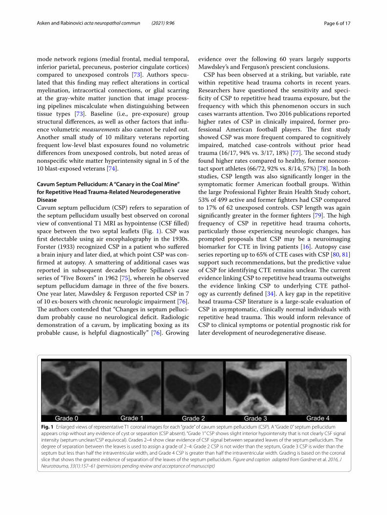
Cavum Septum Pellucidum: A “Canary in the Coal Mine” for Repetitive Head Trauma‑Related Neurodegenerative DiseaseCavum septum pellucidum (CSP) refers to separation of the septum pellucidum usually best observed on coronal view of conventional T1 MRI as hypointense (CSF filled) space between the two septal leaflets (Fig. 1). CSP was first detectable using air encephalography in the 1930s. Forster (1933) recognized CSP in a patient who suffered a brain injury and later died, at which point CSP was con-firmed at autopsy. A smattering of additional cases was reported in subsequent decades before Spillane’s case series of “Five Boxers” in 1962 [75], wherein he observed septum pellucidum damage in three of the five boxers. One year later, Mawdsley & Ferguson reported CSP in 7 of 10 ex-boxers with chronic neurologic impairment [76]. The authors contended that “Changes in septum pelluci-dum probably cause no neurological deficit. Radiologic demonstration of a cavum, by implicating boxing as its probable cause, is helpful diagnostically” [76]. Growing
evidence over the following 60 years largely supports Mawdsley’s and Ferguson’s prescient conclusions.
CSP has been observed at a striking, but variable, rate within repetitive head trauma cohorts in recent years. Researchers have questioned the sensitivity and speci-ficity of CSP to repetitive head trauma exposure, but the frequency with which this phenomenon occurs in such cases warrants attention. Two 2016 publications reported higher rates of CSP in clinically impaired, former pro-fessional American football players. The first study showed CSP was more frequent compared to cognitively impaired, matched case-controls without prior head trauma (16/17, 94% vs. 3/17, 18%) [77]. The second study found higher rates compared to healthy, former noncon-tact sport athletes (66/72, 92% vs. 8/14, 57%) [78]. In both studies, CSP length was also significantly longer in the symptomatic former American football groups. Within the large Professional Fighter Brain Health Study cohort, 53% of 499 active and former fighters had CSP compared to 17% of 62 unexposed controls. CSP length was again significantly greater in the former fighters [79]. The high frequency of CSP in repetitive head trauma cohorts, particularly those experiencing neurologic changes, has prompted proposals that CSP may be a neuroimaging biomarker for CTE in living patients [16]. Autopsy case series reporting up to 65% of CTE cases with CSP [80, 81] support such recommendations, but the predictive value of CSP for identifying CTE remains unclear. The current evidence linking CSP to repetitive head trauma outweighs the evidence linking CSP to underlying CTE pathol-ogy as currently defined [34]. A key gap in the repetitive head trauma-CSP literature is a large-scale evaluation of CSP in asymptomatic, clinically normal individuals with repetitive head trauma. This would inform relevance of CSP to clinical symptoms or potential prognostic risk for later development of neurodegenerative disease.
Fig. 1 Enlarged views of representative T1 coronal images for each “grade” of cavum septum pellucidum (CSP). A “Grade 0” septum pellucidum appears crisp without any evidence of cyst or separation (CSP absent). “Grade 1” CSP shows slight interior hypointensity that is not clearly CSF signal intensity (septum unclear/CSP equivocal). Grades 2–4 show clear evidence of CSF signal between separated leaves of the septum pellucidum. The degree of separation between the leaves is used to assign a grade of 2–4: Grade 2 CSP is not wider than the septum, Grade 3 CSP is wider than the septum but less than half the intraventricular width, and Grade 4 CSP is greater than half the intraventricular width. Grading is based on the coronal slice that shows the greatest evidence of separation of the leaves of the septum pellucidum. Figure and caption adapted from Gardner et al. 2016, J Neurotrauma, 33(1):157–61 (permissions pending review and acceptance of manuscript)
Page 7 of 17Asken and Rabinovici acta neuropathol commun (2021) 9:96
CSP has also been observed in other patient popu-lations and in variable rates among otherwise healthy individuals. A meta-analysis of CSP prevalence in psy-chiatric populations found 1.4 × greater likelihood of CSP compared to psychiatrically healthy controls and almost 2 × greater likelihood of a large CSP (≥ 6 mm length), though there was significant heterogeneity across included studies [82]. Schizophrenia, in particular, is classically linked to higher rates of CSP but data sug-gest that this association may be limited to risk for larger CSP than presence per se [83]. Similar associations with CSP enlargement have been noted in adolescent-onset opiate dependence [84] and obsessive–compulsive disor-der [85], though data are mixed [86]. It remains unclear whether CSP presence is congenital, develops over time, or is directly and meaningfully associated with clinical symptoms. Limited evidence from a small subset of seri-ally imaged boxers suggests CSP develops and increases in size in the course of repetitive trauma exposure [87, 88]. It is also essential to consider that other populations with high rates of CSP, like those with psychiatric illness, are at significantly higher risk of lifetime head trauma exposure [89–91]. As discussed previously, studies not focused on head trauma may not assess participants for possible exposure or may rely on insensitive methods to rule out exposure. Anecdotally, CSP with fenestrations or a “ratty” appearance may imply a traumatic etiology as some have speculated rapid acceleration-deceleration or fluid percussion force causes shearing of the two septal leaflets.
Structural White Matter AbnormalitiesWhite matter susceptibility to traumatic forces under-scores the potential for findings on clinically relevant structural MRI sequences. Active collegiate hockey play-ers were found to have a similar number of white mat-ter hyperintensity (WMH) counts to controls on T2/fluid attenuated inversion recovery (FLAIR) imaging [92]. However, WMH lesions in the athlete group were located more closely to the grey-white matter junction and to sul-cal depths, regions associated with early CTE pathology in affected brains. There was also a slight predominance for the frontal lobes (75% of all lesions) relative to con-trols (56%). Lesion counts did not change over the course of a single hockey season and did not increase acutely after concussion. In a small study of collegiate American football players, a subset of athletes exhibited general decreased susceptibility weighted imaging (SWI) signal after one season of participation, which was interpreted as potential evidence for asymptomatic, trauma-related microbleeds [64]. Alosco et al. reported higher frequency of white matter signal abnormalities on high-resolution T1 in former professional American football athletes
than controls, the number of which correlated with the estimated amount of head trauma exposure throughout their playing career [93]. To date, there is less research using clinically available MRI sequences to character-ize white matter abnormalities in repetitive head trauma cohorts than exists for acute brain injury patients.
It is unclear whether these structural white matter findings reflect neurodegenerative changes, especially in younger, asymptomatic participants. However, sev-eral studies document cerebrovascular pathophysiology associated with repetitive head trauma, including altered cerebral perfusion dynamics [64, 94–97] and blood–brain-barrier dysfunction [98, 99], which may produce white matter signal abnormalities on MRI. In CTE cases, more severe white matter rarefaction is associated with greater exposure to repetitive head trauma, severity of neurofibrillary tangle deposition, and likelihood of devel-oping dementia [32].
Take‑Home Points on Structural MRI in Repetitive Head TraumaNo characteristic atrophy pattern has emerged as specific to repetitive head trauma exposure. There are somewhat consistent findings of lower medial temporal and subcor-tical (thalamus, corpus callosum) volumes in repetitive head trauma cohorts compared to controls across the lifespan, though interpreting group level volume differ-ences in younger, active athletes is complicated by neu-rodevelopmental dynamics. We cannot readily attribute volumetric differences to a specific neurodegenerative pathology (e.g., CTE) but susceptibility of these regions to traumatic forces implicates repetitive head trauma regardless of the underlying pathophysiologic process. Collective evidence indicates that presence of CSP in clinically impaired adults with repetitive head trauma exposure should increase suspicion that the presence of any neurodegenerative disease, whether CTE or other-wise, is attributable at least in part to the patient’s repeti-tive head trauma. Emerging research suggests repetitive head trauma exposure may also lead to white matter alterations observable on clinically available structural MRI sequences (i.e., T2/FLAIR). White matter patholo-gies seen in CTE and their direct relevance for clinical impairment [80, 100–102] strongly implicate the impor-tance of further studying neuroimaging modalities that characterize white matter changes in repetitive head trauma cohorts.
Page 8 of 17Asken and Rabinovici acta neuropathol commun (2021) 9:96
Positron Emission Tomography (PET) Metabolic and Molecular NeuroimagingFDG‑PET Neuroimaging of Repetitive Head TraumaFDG-PET (2-deoxy-2-(18F)fluoro-deoxyglucose) pro-vides in vivo evidence of the severity and spatial distri-bution of changes in brain metabolism presumed to represent altered synaptic activity. Few studies have used FDG-PET to evaluate participants with a history of repetitive head trauma. Two small studies of active and former boxers found lower FDG uptake (i.e., hypome-tabolism) in multiple but inconsistent regions including posterior cingulate [103], bilateral frontal lobes [104], parieto-occipital cortex [103], and the cerebellum [103]. Former American football players had significantly lower frontotemporal metabolism than controls [43]. In one study of military veterans, higher number of prior blast exposures correlated with lower cerebellar metabolism [105]. An antemortem PET-to-autopsy case report (CTE stage IV with hippocampal sclerosis) showed mild FDG hypometabolism corresponding with medial temporal and frontal atrophy. Medial temporal structures con-tained multiple degenerative protein aggregates, while frontal lobe pathology was predominantly CTE (see [106] and description of Patient #1 below).
Aβ‑PET Neuroimaging of Repetitive Head TraumaIn 2012, Florbetapir became the first Aβ-PET tracer approved by the U.S. Food and Drug Administration (FDA) for detecting moderate to frequent neuritic Aβ plaques, a core neuropathological feature of AD. Two similar radiotracers received approval shortly thereafter (Flutemetamol, Florbetaben). Widespread clinical imple-mentation of Aβ-PET imaging remains limited in the U.S. and other countries due to lack of insurance reimburse-ment. However, emerging data from the Imaging Demen-tia – Evidence for Amyloid Scanning (IDEAS) study strongly support the relevance of Aβ-PET imaging in clinical management of cognitively impaired older adults [107]. Clinical feasibility of more routine Aβ-PET scans therefore may increase significantly, especially if tied to effective Aβ lowering therapeutic agents.
Most studies of repetitive head trauma do not spe-cifically analyze associations with cortical Aβ burden. Instead, Aβ-PET often is used to rule out or identify comorbid AD. A negative Aβ-PET scan previously was proposed as a “positive biomarker” supporting a “Prob-able CTE” diagnosis [16] because Aβ plaques are not a diagnostic feature of CTE and their absence strongly sug-gests AD is not driving symptoms [34]. Among 11 living patients with “Probable CTE,” 2 were Aβ-PET positive and also showed the most severe atrophy plus tau PET signal [43]. In brains with CTE at autopsy, Aβ deposition
is a common co-pathology (> 90% of former profes-sional American football players with advanced CTE) [5], occurs at an accelerated rate, and preferentially affects the depths of cortical sulci [108]. Aβ plaques in CTE usu-ally are diffuse rather than neuritic, which may explain lower affinity of Aβ-PET tracers.
Acute brain injury has been linked to upregulation of amyloid precursor protein, which is cleaved to form Aβ polypeptides [109–112]. Studies evaluating Aβ-PET acutely after TBI inconsistently note presence of corti-cal Aβ plaques [25]. One autoradiography study reported white matter accumulation of Aβ and amyloid precursor protein, but no binding of Aβ-radiotracer (Pittsburgh Compound B; PIB), which aligned with their finding of no differences in white matter PIB binding between TBI patients and controls [113]. Across two studies of moderate-severe TBI patients compared to controls, one showed greater cortical grey matter and striatum binding (< 1 yr post-TBI) [113] and one showed greater posterior cingulate and cerebellum binding (> 1 yr post-TBI) [114]. Conversely, a recent investigation of remote head trauma exposure (both mild TBI and a subset with repetitive asymptomatic exposure) found no association with later-life cortical Aβ burden using PET in clinically normal older adults [115]. Others similarly have reported a lack of association between remote, mild head trauma expo-sure and cortical Aβ burden [116–118].
Tau‑PET Neuroimaging of Repetitive Head TraumaFDDNPMost early tau-PET studies in repetitive head trauma patients used the FDDNP radiotracer. FDDNP binding properties severely limit its sensitivity and specificity to CTE pathology. FDDNP binds to different protein aggregates that form beta-pleated sheets (Aβ plaques, tau tangles, prion proteins, and others), has poor repro-ducibility, and has a low signal-to-noise ratio [119–122]. Regardless, several early studies found group-level differences between repetitive head trauma partici-pants and controls in the spatial pattern and degree of FDDNP tracer uptake [123, 124]. Data indicated that groups of blast-exposed veterans and former profes-sional American football players, albeit often with small numbers, showed FDDNP binding in white matter and subcortical structures [123] along with limbic and brain stem regions [123, 125]. Binding patterns seemingly dif-fered from AD cases as well. One case study reported a former American football player diagnosed with CTE at autopsy (also with frequent neuritic Aβ plaques) who underwent FDDNP-PET imaging about 4 years before death. The report showed that FDDNP binding levels correlated with the amount of tau deposition in
Page 9 of 17Asken and Rabinovici acta neuropathol commun (2021) 9:96
the brain at autopsy [126]. However, the FDDNP radi-otracer is not FDA approved and there is no support for clinical utility.
Flortaucipir (FTP) FTP was developed to detect paired helical filament tau in neurofibrillary tangles charac-teristic of AD (now FDA approved). Multiple investiga-tions consistently support FTP use for differentiation of AD from controls and non-AD tauopathies [127–129], but there is limited comparison to CTE. There were high hopes that the science of CTE and repetitive head trauma biomarker development could ride the wave of extremely promising research demonstrating strong affinity of the FTP tracer to AD tau [127]. Excitement over potential CTE diagnosis stemmed from known similarities in phos-phorylated tau isoforms between AD and CTE – mixed 3-repeat/4-repeat tau tangles with paired helical filament structures. However, an autoradiography study showed that FTP only weakly bound to brain tissue with dense CTE pathology compared to its strong binding to tissue with AD tau [130], again suggesting limited potential for sensitive or specific CTE detection. Newly identified dif-ferences in fibril folding microstructure between CTE and AD tau may explain differences in tau radiotracer binding affinity [131].
FTP exhibits “off-target” (i.e., non-tau related) binding to choroid plexus (often complicating medial temporal signal interpretation) [132, 133], caudate, putamen, pal-lidum, thalamus, and white matter [133–135], and cor-tically in some cases of tau-negative neurodegenerative disease [127]. An early case report speculating that FTP binding in the basal ganglia reflected a “novel variant” of CTE likely represented off-target tracer binding [133, 134, 136]. More recently, Stern et al. reported a group-level comparison of 26 predominantly Aβ-PET negative former professional American football athletes to 31 controls and found higher FTP binding in medial tem-poral, parietal, and superior frontal lobes [137]. Degree of FTP uptake correlated with number of years partici-pating in American football, but there was no associa-tion with cognitive outcomes [138]. Lesman-Segev et al. compared 11 clinically impaired TES patients to clinically impaired, biomarker-confirmed AD patients and unex-posed, Aβ-PET negative, clinically normal controls [43]. There was mildly elevated FTP binding in frontotempo-ral regions of TES patients relative to unexposed con-trols, and no regions with higher FTP signal than the AD group. Some patients exhibited FTP binding in a non-contiguous “dot-like” pattern, similar to data reported in a small group of veterans with history of multiple low-level blast exposures [74]. This pattern is also observed in some healthy controls [43] and may simply represent noise or imaging artifact [43, 127].
A recent case report compared antemortem FTP bind-ing in a former professional American football player to neuropathology observed at autopsy [106]. The patient had severe CTE (stage IV) and hippocampal sclerosis without comorbid AD. FTP uptake overlapped well with CTE tau pathology in the inferior temporal lobe and jux-tacortical frontal white matter, but there was weak FTP uptake on PET imaging in several areas of the brain with dense CTE tau deposition at autopsy and confirmed off-target binding subcortically.
The recent release of the FDA’s label for FTP provides essential context for application in clinical settings. FTP scans are indicated for estimating the density and distri-bution of aggregated tau neurofibrillary tangles in adults with cognitive impairment who are being evaluated for AD. “Positive” scans show visually apparent increased neocortical tracer uptake in the posterolateral temporal, occipital, or parietal/precuneus regions, with or without frontal uptake. FTP is not indicated for use in the evalua-tion of patients for CTE. This does not inherently rule out the potential clinical utility of FTP-PET scans for patients with a history of repetitive head trauma if their clinical profile raises suspicion for AD. In this scenario, a “posi-tive” FTP scan would implicate underlying Alzheimer’s disease as contributing to cognitive impairment (espe-cially if accompanied by elevated Aβ PET), but would not rule out comorbid CTE. A “negative” FTP scan might increase the likelihood that CTE is driving cognitive symptoms, contrary to prior research criteria proposing that positive tau PET findings fulfill the biomarker-based requirement for “Probable CTE” [16].
Other Tau PET Tracers and Considerations for Future Development Several additional tau PET tracers exist, and others are rapidly being developed [139], but most thus far have rarely been used in repetitive head trauma research. PBB3 is a family of tau PET compounds that appears to bind tau aggregates consisting of all isoforms [140, 141]. Takahata and colleagues found that patients with TES showed higher [11C]-PBB3 binding in white matter than individuals with single-event TBI [142]. Bind-ing to tau lesions at the depths of neocortical sulci (sug-gesting CTE pathology) was confirmed via in vitro assays [142]. The second-generation MK-6240 tracer is a highly selective paired helical filament tau tracer with less off-target binding in the brain, but with off-target meningeal binding [143, 144]. A recent case report of a former Aus-tralian rules football athlete described in vivo MK-6240 cortical uptake in a pattern resembling the spatial dis-tribution of CTE (bilateral superior frontal and medial temporal regions) and distinct from a typical AD pattern [145]. However, limited autoradiographic evidence sug-gests MK-6240, like FTP, may have high affinity for AD tau
Page 10 of 17Asken and Rabinovici acta neuropathol commun (2021) 9:96
tangles but not CTE tau [143]. Additional validation work is necessary. Other tau tracers include RO-948, PI-2620, and GTP-1. These tracers are derivatives of FTP and thus likely have similar binding characteristics, but this has not been tested empirically.
Temporal dynamics of the underlying disease process may also be particularly relevant for developing a diag-nostic PET biomarker for CTE. For example, FTP binds neurofibrillary tangles and autopsy studies suggest that a “positive” scan requires advanced AD tau pathology (Braak stage V-VI). Early-stage CTE involves sparse neu-rofibrillary tangle deposition often located at brain/CSF interfaces where PET signal can be washed out by partial volume effects. It is therefore likely that a CTE-specific PET tracer will be sensitive only to relatively advanced pathology (i.e., CTE Stage III-IV). Further complicating matters in CTE, which is a mixed 3R/4R tauopathy, the 4R tau isoform may be much more prevalent than the 3R isoform earlier in the disease process before shifting towards deposition of 3R tau and fully formed neurofi-brillary tangles [146]. Astrocytic tau inclusions are also a prominent feature of CTE despite being insufficient for formal diagnosis [34]. Recent work showed that the tau tangles within neurons are a mix of 3R and 4R isoforms while astrocytes predominantly contain 4R tau [146, 147].
Take‑Home Points on FDG‑PET, Aβ‑PET, Tau‑PET NeuroimagingFDG-PET study findings implicate inconsistent brain regions, which is not surprising given the heterogene-ous underlying diseases within clinically impaired repeti-tive head trauma cohorts. The main utility of Aβ-PET in repetitive head trauma research currently rests on rul-ing out concomitant AD pathology. Collective findings thus far unfortunately suggest limited utility of well-studied AD tau PET radiotracers for identifying CTE. A radiotracer sensitive and specific to CTE-tau must be developed and likely must account both for variations in relative presence of 3R versus 4R tau isoforms at different disease stages (e.g., mild or severe) and in different cell types (neurons versus astrocytes). Appreciation for off-target (i.e., non-tau) binding properties and nonspecific binding patterns (non-contiguous, “dot-like”) is critical to avoid potential false-positive diagnoses.
Clinico‑Pathologic Examples of Clinically Suspected CTE Patients With and Without CTE Pathology at AutopsyHere we present two research participants from the UCSF Memory and Aging Center’s Alzheimer’s Disease Research Center. Both patients were evaluated and dis-cussed by multidisciplinary consensus conference after comprehensive neurological and neuropsychological
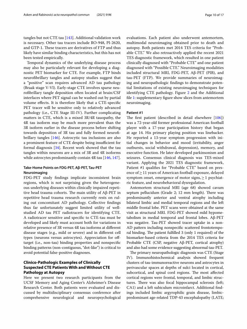
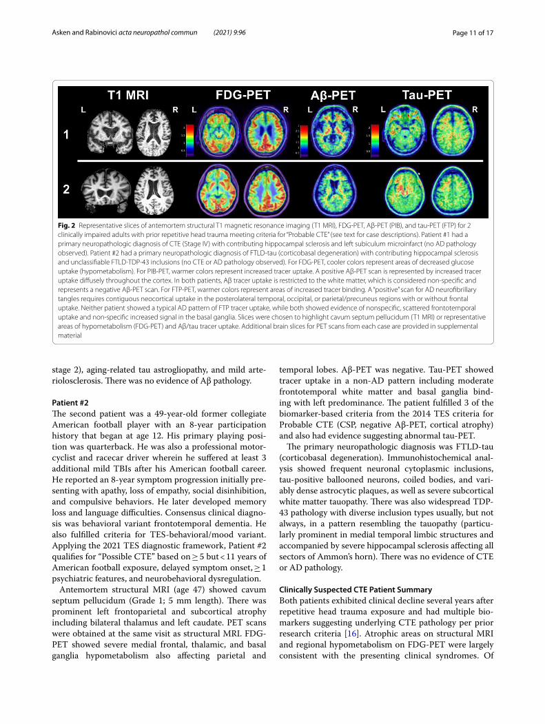
evaluations. Each patient also underwent antemortem, multimodal neuroimaging obtained prior to death and autopsy. Both patients met 2014 TES criteria for “Prob-able CTE.” We also retroactively applied the recent 2021 TES diagnostic framework, which resulted in one patient clinically diagnosed with “Probable CTE” and one patient diagnosed with “Possible CTE.” Neuroimaging modalities included structural MRI, FDG-PET, Aβ-PET (PIB), and tau-PET (FTP). We provide summaries of neuroimag-ing and neuropathologic findings to demonstrate poten-tial limitations of existing neuroimaging techniques for identifying CTE pathology. Figure 2 and the Additional file 1: supplementary figure show slices from antemortem neuroimaging.
Patient #1The first patient (described in detail elsewhere [106]) was a 72-year-old former professional American football player with a 17-year participation history that began at age 14. His primary playing position was linebacker. He reported a 13-year symptom progression with ini-tial changes in behavior and mood (irritability, anger outbursts, social withdrawal, depression), memory, and executive function. He later developed parkinsonism and seizures. Consensus clinical diagnosis was TES-mixed variant. Applying the 2021 TES diagnostic framework, Patient #1 qualifies for “Probable CTE” based on pres-ence of ≥ 11 years of American football exposure, delayed symptom onset, emergence of motor signs, ≥ 1 psychiat-ric feature, and neurobehavioral dysregulation.
Antemortem structural MRI (age 68) showed cavum septum pellucidum (Grade 2; 12 mm length). There was predominantly anterior and ventral atrophy including bilateral limbic and medial temporal regions and the left middle frontal lobe. PET scans were obtained at the same visit as structural MRI. FDG-PET showed mild hypome-tabolism in medial temporal and frontal lobes. Aβ-PET was negative. Tau-PET showed tracer uptake in a non-AD pattern including nonspecific scattered frontotempo-ral binding. The patient fulfilled 3 (only 1 required) of the biomarker-based criteria from the 2014 TES criteria for Probable CTE (CSP, negative Aβ-PET, cortical atrophy) and also had some evidence suggesting abnormal tau-PET.
The primary neuropathologic diagnosis was CTE (Stage IV). Immunohistochemical analysis showed frequent clusters of tau-immunoreactive neurons and astrocytes in perivascular spaces at depths of sulci located in cortical, subcortical, and spinal cord regions. The most affected cortical regions were frontal, temporal, and limbic struc-tures. There was also focal hippocampal sclerosis (left; CA1) and a left subiculum microinfarct. Additional find-ings included limbic argyrophilic grain disease, limbic-predominant age-related TDP-43 encephalopathy (LATE;
Page 11 of 17Asken and Rabinovici acta neuropathol commun (2021) 9:96
stage 2), aging-related tau astrogliopathy, and mild arte-riolosclerosis. There was no evidence of Aβ pathology.
Patient #2The second patient was a 49-year-old former collegiate American football player with an 8-year participation history that began at age 12. His primary playing posi-tion was quarterback. He was also a professional motor-cyclist and racecar driver wherein he suffered at least 3 additional mild TBIs after his American football career. He reported an 8-year symptom progression initially pre-senting with apathy, loss of empathy, social disinhibition, and compulsive behaviors. He later developed memory loss and language difficulties. Consensus clinical diagno-sis was behavioral variant frontotemporal dementia. He also fulfilled criteria for TES-behavioral/mood variant. Applying the 2021 TES diagnostic framework, Patient #2 qualifies for “Possible CTE” based on ≥ 5 but < 11 years of American football exposure, delayed symptom onset, ≥ 1 psychiatric features, and neurobehavioral dysregulation.
Antemortem structural MRI (age 47) showed cavum septum pellucidum (Grade 1; 5 mm length). There was prominent left frontoparietal and subcortical atrophy including bilateral thalamus and left caudate. PET scans were obtained at the same visit as structural MRI. FDG-PET showed severe medial frontal, thalamic, and basal ganglia hypometabolism also affecting parietal and
temporal lobes. Aβ-PET was negative. Tau-PET showed tracer uptake in a non-AD pattern including moderate frontotemporal white matter and basal ganglia bind-ing with left predominance. The patient fulfilled 3 of the biomarker-based criteria from the 2014 TES criteria for Probable CTE (CSP, negative Aβ-PET, cortical atrophy) and also had evidence suggesting abnormal tau-PET.
The primary neuropathologic diagnosis was FTLD-tau (corticobasal degeneration). Immunohistochemical anal-ysis showed frequent neuronal cytoplasmic inclusions, tau-positive ballooned neurons, coiled bodies, and vari-ably dense astrocytic plaques, as well as severe subcortical white matter tauopathy. There was also widespread TDP-43 pathology with diverse inclusion types usually, but not always, in a pattern resembling the tauopathy (particu-larly prominent in medial temporal limbic structures and accompanied by severe hippocampal sclerosis affecting all sectors of Ammon’s horn). There was no evidence of CTE or AD pathology.
Clinically Suspected CTE Patient SummaryBoth patients exhibited clinical decline several years after repetitive head trauma exposure and had multiple bio-markers suggesting underlying CTE pathology per prior research criteria [16]. Atrophic areas on structural MRI and regional hypometabolism on FDG-PET were largely consistent with the presenting clinical syndromes. Of
Fig. 2 Representative slices of antemortem structural T1 magnetic resonance imaging (T1 MRI), FDG-PET, Aβ-PET (PIB), and tau-PET (FTP) for 2 clinically impaired adults with prior repetitive head trauma meeting criteria for “Probable CTE” (see text for case descriptions). Patient #1 had a primary neuropathologic diagnosis of CTE (Stage IV) with contributing hippocampal sclerosis and left subiculum microinfarct (no AD pathology observed). Patient #2 had a primary neuropathologic diagnosis of FTLD-tau (corticobasal degeneration) with contributing hippocampal sclerosis and unclassifiable FTLD-TDP-43 inclusions (no CTE or AD pathology observed). For FDG-PET, cooler colors represent areas of decreased glucose uptake (hypometabolism). For PIB-PET, warmer colors represent increased tracer uptake. A positive Aβ-PET scan is represented by increased tracer uptake diffusely throughout the cortex. In both patients, Aβ tracer uptake is restricted to the white matter, which is considered non-specific and represents a negative Aβ-PET scan. For FTP-PET, warmer colors represent areas of increased tracer binding. A “positive” scan for AD neurofibrillary tangles requires contiguous neocortical uptake in the posterolateral temporal, occipital, or parietal/precuneus regions with or without frontal uptake. Neither patient showed a typical AD pattern of FTP tracer uptake, while both showed evidence of nonspecific, scattered frontotemporal uptake and non-specific increased signal in the basal ganglia. Slices were chosen to highlight cavum septum pellucidum (T1 MRI) or representative areas of hypometabolism (FDG-PET) and Aβ/tau tracer uptake. Additional brain slices for PET scans from each case are provided in supplemental material
Page 12 of 17Asken and Rabinovici acta neuropathol commun (2021) 9:96
note, tau PET in each participant showed abnormal, low-level tracer uptake in a clearly non-AD pattern. In the absence of tau PET, which is not widely available clini-cally, presence of CSP and negative Aβ-PET alone would likely have made both patients strong candidates for har-boring significant CTE pathology. It remains unknown if or how Patient #2’s head trauma exposure contributed to the onset, progression, or symptom manifestation of non-CTE neuropathologic processes. Beyond the limi-tations of available neuroimaging modalities for reliably identifying CTE, these cases highlight the diversity of prospectively documented symptom presentations and underlying neurodegenerative diseases among clinically impaired patients with repetitive head trauma exposure.
Future Considerations for Neuroimaging Research in Repetitive Head TraumaStructural NeuroimagingStructural MRI remains an important and relatively accessible component of clinical evaluations for patients with suspected neurodegenerative disease, but usually images are reviewed qualitatively. Incorporating quan-titative MRI methods and/or advanced sequences like DTI into clinical practice may improve sensitivity to head trauma-related brain changes. Systematically collecting different types of lifetime head trauma exposure, from repetitive asymptomatic impacts to severe TBI, will facil-itate our understanding of brain changes on structural MRI attributable to head trauma. While elite level colli-sion sport athletes represent an important study group, extending these efforts to the broader aging population will enhance generalizability and improve risk estimation along a wider spectrum of head trauma exposure.
Other PET RadiotracersWhile there is an understandable focus on in vivo CTE tau identification, measuring the degree and spatial dis-tribution of neuroinflammation [100, 148–150] and syn-aptic dysfunction are other potentially interesting PET applications for repetitive head trauma [151–153]. The translocator protein (TSPO) is a mitochondrial mem-brane protein that is upregulated in activated microglia, astroglia, and macrophages. Several TSPO-PET ligands have been developed as in vivo markers of neuroinflam-mation. One study of TSPO-PET using the [11C]DPA-713 tracer found higher signal in former American professional football players than controls in bilateral medial and superior temporal regions [15]. New molecu-lar targets for imaging activated microglia and astrocytes are currently under development [150]. To our knowl-edge, PET imaging of synaptic loss (e.g., synaptic vesicle glycoprotein 2A; SV2A-PET) [154, 155] has not been per-formed in repetitive head trauma cohorts. It is unclear if
these approaches will be clinically meaningful in isola-tion for specific disease identification (e.g., differentiating CTE from AD or other neurodegenerative diseases), but they may prove valuable for unlocking pathophysiologic mechanisms linking repetitive head trauma to increased neurodegenerative disease risk in general.
Sex‑Specific differences in Neuroimaging of Repetitive Head TraumaSex differences in neuroimaging findings among repeti-tive head trauma cohorts are unknown. Existing studies almost exclusively focus on male-predominant groups of collision sport athletes (e.g., American football) and mili-tary veterans. Reported sex differences in head impact bio-mechanics [156–158], brain injury outcomes [159–161], and risk for neurodegenerative disease [162, 163] under-score the critical importance of studying the role of sex in repetitive head trauma outcomes. Large-scale, longitudinal cohorts like the Professional Fighters Brain Health Study [164], CARE Consortium [52], Chronic Effects of Neu-rotrauma Consortium [165], and longitudinal follow-up of the Transforming Research and Clinical Knowledge in TBI cohort [166] (i.e., TRACK-TBI LONG) offer strong potential for studying sex-specific outcomes in the associa-tion of repetitive head trauma with brain volume changes or molecular/metabolic alterations on PET imaging. For-mer participants in presumably high-risk female collision sports like soccer/futbol, ice hockey, rugby, mixed mar-tial arts, etc. may be especially important study cohorts. Additionally, intimate partner violence survivors, who are usually female, are an often overlooked but critically important group to study [167]. Comprehensive evalua-tion of both repetitive, asymptomatic exposure and symp-tomatic brain injuries will be essential to these efforts.
Concluding RemarksRepetitive head trauma may increase risk for multiple neurodegenerative outcomes, with much recent focus on CTE. Structural MRI studies in repetitive head trauma cohorts do not clearly suggest a specific pattern of vol-ume loss, though subcortical structures like the thalamus and corpus callosum and medial temporal limbic region appear susceptible to repetitive traumatic forces. Cavum septum pellucidum is much more common in clinically impaired repetitive head trauma populations than both clinically normal and impaired cohorts without head trauma exposure. Presence of cavum septum in clini-cally impaired adults with repetitive head trauma should increase suspicion that head trauma exposure contrib-uted to the underlying disease. White matter abnor-malities occasionally are observable on conventional clinical MRI and may be spatially distinct in repetitive head trauma populations, but more work characterizing
Page 13 of 17Asken and Rabinovici acta neuropathol commun (2021) 9:96
these abnormalities is needed. FDG-PET studies have not identified a characteristic repetitive head trauma pattern, which likely reflects the diversity of underlying neuropathologies and associated clinical syndromes. Tau-PET remains a promising research avenue but will require development of CTE-tau specific radiotracers given the lack of support for current tracers with strong affinity for AD tau. PET imaging of Aβ plaques and AD tau tangles may still be clinically useful in ruling AD in or out. CTE is highly associated with prior repeti-tive head trauma. However, we caution against tenuous assumptions that CTE is present, or the sole or primary source of symptoms, in clinically impaired repetitive head trauma cohorts without other compelling clinical or biomarker data. Improving diagnostic precision for neurodegenerative disease within repetitive head trauma cohorts requires antemortem imaging-to-autopsy stud-ies and development of other in vivo biomarkers sensi-tive to the effects of repetitive trauma on brain health.
AbbreviationsAD: Alzheimer’s disease; Aβ: Beta-amyloid; CARE: Concussion Assessment, Research, and Education Consortium; CSF: Cerebrospinal fluid; CSP: Cavum septum pellucidum; CT: Computed tomography; CTE: Chronic traumatic encephalopathy; DAI: Diffuse axonal injury; DETECT: Diagnosing and Evaluat-ing Traumatic Encephalopathy using Clinical Tests; DIAGNOSE-CTE: Diagnos-tics, Imaging, and Genetics Network for the Objective Study and Evaluation of Chronic Traumatic Encephalopathy; DTI: Diffusion tensor imaging; FDA: Food & Drug Administration; FDDNP: 2-(1-{6-[(2-Fluoroethyl(methyl)amino]-2-naphthyl}ethylidene)malononitrile; FDG: Fluorodeoxyglucose; FLAIR: Fluid attenuated inversion recovery; FTP: Flortaucipir; IDEAS: Imaging Dementia – Evidence for Amyloid Scanning; LOC: Loss of consciousness; MRI: Magnetic resonance imaging; mTBI: Mild traumatic brain injury; NFT: Neurofibrillary tangle; PET: Positron emission tomography; PIB: Pittsburgh Compound B; PTA: Posttraumatic amnesia; SV2A: Synaptic vesicle glycoprotein 2A; SWI: Suscepti-bility weighted imaging; TBI: Traumatic brain injury; TES: Traumatic encepha-lopathy syndrome; TRACK-TBI: Transforming Research and Clinical Knowledge in TBI; TSPO: Translocator protein; WMH: White matter hyperintensity.
Supplementary InformationThe online version contains supplementary material available at https:// doi. org/ 10. 1186/ s40478- 021- 01197-4.
Additional file 1.
AcknowledgementsWe thank Corrina Fonseca, Renaud La Joie, Amelia Strom, and Nidhi Mundada for their assistance with preparation of structural MRI and PET images, with an additional thank you to Dr. La Joie for lending interpretations of the PET scans. We are especially thankful for the research participants who generously committed their time and effort to research projects at UCSF during life, and for their brain donation to the UCSF Neurodegenerative Disease Brain Bank. Neuropathologic findings for the two patient examples were derived from autopsy reports prepared by Drs. William Seeley or Salvatore Spina from the UCSF Memory and Aging Center Neurodegenerative Disease Brain Bank.
Authors’ contributionsBMA drafted the manuscript. GDR revised and was a major contributor in writ-ing the manuscript. Both authors read and approved the final manuscript.
FundingWe thank the UCSF Alzheimer’s Disease Research Center (NIA P50AG023501 and P30AG062422; PI: Bruce Miller) and Rainwater Charitable Foundation (PI: Gil Rabinovici) for their funding support. Funding agencies did not play a role in design or interpretation of data in this manuscript.
Availability of data and materialsData sharing is not applicable to this article as no datasets were generated or analysed during the current study.
Declarations
Ethics approval and consent to participateThe two patients described from the UCSF Alzheimer’s Disease Research Center consented to participate in all IRB-approved study protocols. Identifi-able information was redacted from reported data.
Competing interestsBMA and GDR declare that they have no competing interests relevant this review.
Consent for publicationIndividual consent was obtained from each participant at the time of study enrollment.
Author details1 Department of Neurology, Memory and Aging Center, Weill Institute for Neu-rosciences, University of California, San Francisco, 675 Nelson Rising Lane, Suite 190, San Francisco, CA 94143, USA. 2 Departments of Neurology, Radiology & Biomedical Imaging, Memory and Aging Center, Weill Institute for Neuro-sciences, University of California, San Francisco, 675 Nelson Rising Lane, Suite 190, San Francisco, CA 94143, USA.
Received: 20 January 2021 Accepted: 7 May 2021
References 1. Perry DC, Sturm VE, Peterson MJ, Pieper CF, Bullock T, Boeve BF et al
(2016) Association of traumatic brain injury with subsequent neurologi-cal and psychiatric disease: a meta-analysis. J Neurosurg 124(2):511–526
2. Mackay DF, Russell ER, Stewart K, MacLean JA, Pell JP, Stewart W (2019) Neurodegenerative disease mortality among former professional soc-cer players. N Engl J Med 381:1801–1808
3. Gardner RC, Yaffe K (2015) Epidemiology of mild traumatic brain injury and neurodegenerative disease. Mol Cell Neurosci 66:75–80
4. Nordström A, Nordström P (2018) Traumatic brain injury and the risk of dementia diagnosis: a nationwide cohort study. PLoS Med 15(1):e1002496
5. Mez J, Daneshvar DH, Kiernan PT, Abdolmohammadi B, Alvarez VE, Huber BR et al (2017) Clinicopathological evaluation of chronic traumatic encephalopathy in players of American football. JAMA 318(4):360–370
6. Gardner RC, Byers AL, Barnes DE, Li Y, Boscardin J, Yaffe K (2018) Mild TBI and risk of parkinson disease: a chronic effects of neurotrauma consor-tium study. Neurology 90(20):e1771–e1779
7. Lehman EJ, Hein MJ, Baron SL, Gersic CM (2012) Neurodegenerative causes of death among retired national football league players. Neurol-ogy 79(19):1970–1974
8. Crane PK, Gibbons LE, Dams-O’Connor K, Trittschuh E, Leverenz JB, Keene CD et al (2016) Association of traumatic brain injury with late-life neurodegenerative conditions and neuropathologic findings. JAMA Neurol 73(9):1062–1069
9. McKhann GM, Knopman DS, Chertkow H, Hyman BT, Jack CR Jr, Kawas CH et al (2011) The diagnosis of dementia due to Alzheimer’s disease: recommendations from the National Institute on Aging-Alzheimer’s Association workgroups on diagnostic guidelines for Alzheimer’s disease. Alzheimer’s Dementia 7(3):263–269
Page 14 of 17Asken and Rabinovici acta neuropathol commun (2021) 9:96
10. Crutch SJ, Schott JM, Rabinovici GD, Murray M, Snowden JS, van der Flier WM et al (2017) Consensus classification of posterior cortical atrophy. Alzheimer’s Dementia 13(8):870–884
11. Ossenkoppele R, Pijnenburg YA, Perry DC, Cohn-Sheehy BI, Scheltens NM, Vogel JW et al (2015) The behavioural/dysexecutive variant of Alzheimer’s disease: clinical, neuroimaging and pathological features. Brain 138(Pt 9):2732–2749
12. Gorno-Tempini ML, Hillis AE, Weintraub S, Kertesz A, Mendez M, Cappa SF et al (2011) Classification of primary progressive aphasia and its vari-ants. Neurology 76(11):1006–1014
13. Rascovsky K, Hodges JR, Knopman D, Mendez MF, Kramer JH, Neuhaus J et al (2011) Sensitivity of revised diagnostic criteria for the behavioural variant of frontotemporal dementia. Brain 134(Pt 9):2456–2477
14. Weinberger DR, Radulescu E (2021) Structural Magnetic Resonance Imaging All Over Again. JAMA Psychiat 78(1):11–12
15. Coughlin JM, Wang Y, Minn I, Bienko N, Ambinder EB, Xu X et al (2017) Imaging of glial cell activation and white matter integrity in brains of active and recently retired national football league players. JAMA Neurol 74(1):67–74
16. Montenigro PH, Baugh CM, Daneshvar DH, Mez J, Budson AE, Au R et al (2014) Clinical subtypes of chronic traumatic encephalopathy: literature review and proposed research diagnostic criteria for traumatic encephalopathy syndrome. Alzheimer’s Res Therapy 6(5):68
17. Katz DI, Bernick C, Dodick DW, Mez J, Mariani ML, Adler CH, et al (2021) National Institute of neurological disorders and stroke consensus diagnostic criteria for traumatic encephalopathy syndrome, Neurology (Epub Ahead of Print; PMID:33722990)
18. Iverson GL, Gardner AJ (2020) Risk for misdiagnosing chronic traumatic encephalopathy in men with anger control problems. Front Neurol 11:739
19. Iverson GL, Gardner AJ (2020) Risk of misdiagnosing chronic traumatic encephalopathy in men with depression. J Neuropsychiatry Clin Neuro-sci 32(2):139–146
20. Koerte IK, Lin AP, Willems A, Muehlmann M, Hufschmidt J, Coleman MJ et al (2015) A review of neuroimaging findings in repetitive brain trauma. Brain Pathol 25(3):318–349
21. Ng TS, Lin AP, Koerte IK, Pasternak O, Liao H, Merugumala S et al (2014) Neuroimaging in repetitive brain trauma. Alzheimers Res Ther 6(1):10
22. Sundman MH, Hall EE, Chen NK (2014) Examining the relationship between head trauma and neurodegenerative disease: a review of epidemiology, pathology and neuroimaging techniques. J Alzheimer’s Dis Parkin 4:137
23. Shetty T, Raince A, Manning E, Tsiouris AJ (2016) Imaging in chronic traumatic encephalopathy and traumatic brain injury. Sports Health 8(1):26–36
24. Lee BG, Leavitt MJ, Bernick CB, Leger GC, Rabinovici G, Banks SJ (2018) A Systematic review of positron emission tomography of tau, amyloid beta, and neuroinflammation in chronic traumatic encephalopathy: the evidence to date. J Neurotrauma 35(17):2015–2024
25. Ayubcha C, Revheim ME, Newberg A, Moghbel M, Rojulpote C, Werner TJ et al (2021) A critical review of radiotracers in the positron emission tomography imaging of traumatic brain injury: FDG, tau, and amyloid imaging in mild traumatic brain injury and chronic traumatic encepha-lopathy. Eur J Nucl Med Mol Imaging 48(2):623–641
26. Asken BM, DeKosky ST, Clugston JR, Jaffee MS, Bauer RM (2018) Diffu-sion tensor imaging (DTI) findings in adult civilian, military, and sport-related mild traumatic brain injury (mTBI): a systematic critical review. Brain Imaging Behav 12(2):585–612
27. Sparks P, Lawrence T, Hinze S (2020) Neuroimaging in the diagnosis of chronic traumatic encephalopathy: a systematic review. Clin J Sport Med 30(Suppl 1):S1-s10
28. Dallmeier JD, Meysami S, Merrill DA, Raji CA (2019) Emerging advances of in vivo detection of chronic traumatic encephalopathy and trau-matic brain injury. Br J Radiol 92(1101):20180925
29. Lin A, Charney M, Shenton ME, Koerte IK (2018) Chronic traumatic encephalopathy: neuroimaging biomarkers. Handb Clin Neurol 158:309–322
30. Asken BM, Sullan MJ, DeKosky ST, Jaffee MS, Bauer RM (2017) Research gaps and controversies in chronic traumatic encephalopathy: a review. JAMA Neurol 74(10):1255–1262
31. Bieniek KF, Ross OA, Cormier KA, Walton RL, Soto-Ortolaza A, Johnston AE et al (2015) Chronic traumatic encephalopathy pathology in a neu-rodegenerative disorders brain bank. Acta Neuropathol 130(6):877–889
32. Alosco ML, Stein TD, Tripodis Y, Chua AS, Kowall NW, Huber BR et al (2019) Association of white matter rarefaction, arteriolosclerosis, and tau with dementia in chronic traumatic encephalopathy. JAMA Neurol 76(11):1298–1308
33. Bieniek KF, Cairns NJ, Crary JF, Dickson DW, Folkerth RD, Keene CD et al (2021) The second NINDS/NIBIB consensus meeting to define neuro-pathological criteria for the diagnosis of chronic traumatic encepha-lopathy. J Neuropathol Exp Neurol 80(3):210–219
34. McKee AC, Cairns NJ, Dickson DW, Folkerth RD, Keene CD, Litvan I et al (2016) The first NINDS/NIBIB consensus meeting to define neuropatho-logical criteria for the diagnosis of chronic traumatic encephalopathy. Acta Neuropathol 131(1):75–86
35. Smith DH, Johnson VE, Trojanowski JQ, Stewart W (2019) Chronic traumatic encephalopathy—confusion and controversies. Nature Rev Neurol 15(3):179–183
36. Iverson GL, Gardner AJ, Shultz SR, Solomon GS, McCrory P, Zafonte R et al (2019) Chronic traumatic encephalopathy neuropathology might not be inexorably progressive or unique to repetitive neurotrauma. Brain 142(12):3672–3693
37. Iverson GL, Luoto TM, Karhunen PJ, Castellani RJ (2019) Mild chronic traumatic encephalopathy neuropathology in people with no known participation in contact sports or history of repetitive neurotrauma. J Neuropathol Exp Neurol 78(7):615–625
38. Gardner RC, Rivera E, O’Grady M, Doherty C, Yaffe K, Corrigan J et al (2020) Screening for lifetime history of traumatic brain injury among older American and Irish adults at risk for dementia: development and validation of a web-based survey. J Alzheimer’s Dis 74(2):699–711
39. Su E, Bell M (2016) Diffuse axonal injury. In: Laskowitz D, Grant G (eds) Translational research in traumatic brain injury. CRC Press/Taylor and Francis Group © (2016) by Taylor & Francis Group. LLC, Boca Raton, FL, pp 41–84
40. Klein AP, Tetzlaff JE, Bonis JM, Nelson LD, Mayer A, Huber DL et al (2019) Prevalence of potentially clinically significant MRI findings in athletes with and without sport-related concussion. J Neurotrauma 36(11):1776–1785
41. Champagne AA, Peponoulas E, Terem I, Ross A, Tayebi M, Chen Y et al (2019) Novel strain analysis informs about injury susceptibility of the corpus callosum to repeated impacts. Brain Commun 1(1):fcz021
42. Lepage C, Muehlmann M, Tripodis Y, Hufschmidt J, Stamm J, Green K et al (2019) Limbic system structure volumes and associated neu-rocognitive functioning in former NFL players. Brain Imaging Behav 13(3):725–734
43. Lesman-Segev OH, La Joie R, Stephens ML, Sonni I, Tsai R, Bourakova V et al (2019) Tau PET and multimodal brain imaging in patients at risk for chronic traumatic encephalopathy. NeuroImage: Clin 24:102025
44. Strain JF, Womack KB, Didehbani N, Spence JS, Conover H, Hart J et al (2015) Imaging correlates of memory and concussion history in retired National Football League athletes. JAMA Neurol 72(7):773–780
45. Goswami R, Dufort P, Tartaglia MC, Green RE, Crawley A, Tator CH et al (2016) Frontotemporal correlates of impulsivity and machine learning in retired professional athletes with a history of multiple concussions. Brain Struct Funct 221(4):1911–1925
46. Wojtowicz M, Gardner AJ, Stanwell P, Zafonte R, Dickerson BC, Iverson GL (2018) Cortical thickness and subcortical brain volumes in profes-sional rugby league players. NeuroImage Clin 18:377–381
47. Misquitta K, Dadar M, Tarazi A, Hussain MW, Alatwi MK, Ebraheem A et al (2018) The relationship between brain atrophy and cognitive-behavioural symptoms in retired Canadian football players with multiple concussions. NeuroImage Clin 19:551–558
48. Schultz V, Stern RA, Tripodis Y, Stamm J, Wrobel P, Lepage C et al (2018) Age at first exposure to repetitive head impacts is associated with smaller thalamic volumes in former professional american football play-ers. J Neurotrauma 35(2):278–285
49. Lee JK, Wu J, Bullen J, Banks S, Bernick C, Modic MT et al (2020) Associa-tion of cavum septum pellucidum and cavum vergae with cogni-tion mood, and brain volumes in professional fighters. JAMA Neurol 77(1):35–42
Page 15 of 17Asken and Rabinovici acta neuropathol commun (2021) 9:96
50. Zivadinov R, Polak P, Schweser F, Bergsland N, Hagemeier J, Dwyer MG et al (2018) Multimodal imaging of retired professional contact sport athletes does not provide evidence of structural and functional brain damage. J Head Trauma Rehabil 33(5):E24-e32
51. Koerte IK, Mayinger M, Muehlmann M, Kaufmann D, Lin AP, Steffinger D et al (2016) Cortical thinning in former professional soccer players. Brain Imaging Behav 10(3):792–798
52. Broglio SP, McCrea M, McAllister T, Harezlak J, Katz B, Hack D et al (2017) A national study on the effects of concussion in collegiate athletes and US military service academy members: the NCAA-DoD concussion assessment, research and education (CARE) consortium structure and methods. Sports Med 47(7):1437–1451
53. Wu YC, Harezlak J, Elsaid NMH, Lin Z, Wen Q, Mustafi SM et al (2020) Longitudinal white-matter abnormalities in sports-related concussion: A diffusion MRI study. Neurology 95(7):e781–e792
54. Mustafi SM, Harezlak J, Koch KM, Nencka AS, Meier TB, West JD et al (2018) Acute white-matter abnormalities in sports-related concussion: a diffusion tensor imaging study from the NCAA-DoD CARE consortium. J Neurotrauma 35(22):2653–2664
55. Klein AP, Tetzlaff JE, Bonis JM, Nelson LD, Mayer AR, Huber DL et al (2019) Prevalence of potentially clinically significant magnetic reso-nance imaging findings in athletes with and without sport-related concussion. J Neurotrauma 36(11):1776–1785
56. Brett BL, Bobholz SA, España LY, Huber DL, Mayer AR, Harezlak J et al (2020) Cumulative effects of prior concussion and primary sport partici-pation on brain morphometry in collegiate athletes: a study from the NCAA-DoD CARE Consortium. Front Neurol 11:673
57. Meier TB, Bellgowan PS, Bergamino M, Ling JM, Mayer AR (2016) Thinner cortex in collegiate football players with, but not without, a self-reported history of concussion. J Neurotrauma 33(4):330–338
58. Mills BD, Goubran M, Parivash SN, Dennis EL, Rezaii P, Akers C et al (2020) Longitudinal alteration of cortical thickness and volume in high-impact sports. Neuroimage 217:116864
59. Wierenga LM, Langen M, Oranje B, Durston S (2014) Unique develop-mental trajectories of cortical thickness and surface area. Neuroimage 87:120–126
60. Janssen PH, Mandrekar J, Mielke MM, Ahlskog JE, Boeve BF, Josephs K et al (2017) High school football and late-life risk of neurodegenerative syndromes, 1956–1970. Mayo Clin Proc 92(1):66–71
61. Savica R, Parisi JE, Wold LE, Josephs KA, Ahlskog JE (2012) High school football and risk of neurodegeneration: a community-based study. Mayo Clin Proc 87(4):335–340
62. Deshpande SK, Hasegawa RB, Rabinowitz AR, Whyte J, Roan CL, Taba-tabaei A et al (2017) Association of playing high school football with cognition and mental health later in life. JAMA Neurol 74(8):909–918
63. Terry DP, Miller LS (2018) Repeated mild traumatic brain injuries is not associated with volumetric differences in former high school football players. Brain Imaging Behav 12(3):631–639
64. Slobounov SM, Walter A, Breiter HC, Zhu DC, Bai X, Bream T et al (2017) The effect of repetitive subconcussive collisions on brain integrity in collegiate football players over a single football season: a multi-modal neuroimaging study. NeuroImage Clin 14:708–718
65. Mayinger MC, Merchant-Borna K, Hufschmidt J, Muehlmann M, Weir IR, Rauchmann BS et al (2018) White matter alterations in college football players: a longitudinal diffusion tensor imaging study. Brain Imaging Behav 12(1):44–53
66. Gong NJ, Kuzminski S, Clark M, Fraser M, Sundman M, Guskiewicz K et al (2018) Microstructural alterations of cortical and deep gray matter over a season of high school football revealed by diffusion kurtosis imaging. Neurobiol Dis 119:79–87
67. Murugesan G, Saghafi B, Davenport E, Wagner B, Urban J, Kelley M et al (2018) Single season changes in resting state network power and the connectivity between regions: distinguish head impact exposure level in high school and youth football players. Proc SPIE Int Soc Opt Eng 10575:105750F
68. Abbas K, Shenk TE, Poole VN, Breedlove EL, Leverenz LJ, Nauman EA et al (2015) Alteration of default mode network in high school football athletes due to repetitive subconcussive mild traumatic brain injury: a resting-state functional magnetic resonance imaging study. Brain Con-nect 5(2):91–101
69. Hirad AA, Bazarian JJ, Merchant-Borna K, Garcea FE, Heilbronner S, Paul D et al (2019) A common neural signature of brain injury in concussion and subconcussion. Sci Adv 5(8):eaau3460
70. Davenport ND, Gullickson JT, Grey SF, Hirsch S, Sponheim SR (2018) Longitudinal evaluation of ventricular volume changes associated with mild traumatic brain injury in military service members. Brain Inj 32(10):1245–1255
71. Bigler ED, Abildskov TJ, Eggleston B, Taylor BA, Tate DF, Petrie JA et al (2019) Structural neuroimaging in mild traumatic brain injury: a chronic effects of neurotrauma consortium study. Int J Methods Psychiat Res 28(3):1781
72. Martindale SL, Rostami R, Shura RD, Taber KH, Rowland JA (2020) Brain volume in veterans: relationship to posttraumatic stress disorder and mild traumatic brain injury. J Head Trauma Rehabil 35(4):E330-e341
73. Stone JR, Avants BB, Tustison NJ, Wassermann EM, Gill J, Polejaeva E et al (2020) Functional and structural neuroimaging correlates of repetitive low-level blast exposure in career breachers. J Neurotrauma 37(23):2468–2481
74. Dickstein DL, De Gasperi R, Gama Sosa MA, Perez-Garcia G, Short JA, Sosa H, et al (2020) Brain and blood biomarkers of tauopathy and neuronal injury in humans and rats with neurobehavioral syndromes following blast exposure. Mol Psychiatry (Online ahead of print; PMCID: PMC7484380)
75. Spillane JD (1962) Five boxers. BMJ 2(5314):1205–1210 76. Mawdsley C, Ferguson FR (1963) Neurological disease in boxers. Lancet
(London, England) 2(7312):795–801 77. Gardner RC, Hess CP, Brus-Ramer M, Possin KL, Cohn-Sheehy BI, Kramer
JH et al (2016) Cavum septum pellucidum in retired american pro-football players. J Neurotrauma 33(1):157–161
78. Koerte IK, Hufschmidt J, Muehlmann M, Tripodis Y, Stamm JM, Pasternak O et al (2016) Cavum septi pellucidi in symptomatic former professional football players. J Neurotrauma 33(4):346–353
79. Lee JK, Wu J, Banks S, Bernick C, Massand MG, Modic MT et al (2017) Prevalence of traumatic findings on routine MRI in a large cohort of professional fighters. AJNR Am J Neuroradiol 38(7):1303–1310
80. McKee AC, Stein TD, Nowinski CJ, Stern RA, Daneshvar DH, Alvarez VE et al (2013) The spectrum of disease in chronic traumatic encephalopa-thy. Brain 136(1):43–64
81. Smith DH, Johnson VE, Stewart W (2013) Chronic neuropathologies of single and repetitive TBI: substrates of dementia? Nat Rev Neurol 9(4):211–221
82. Wang LX, Li P, He H, Guo F, Tian P, Li C et al (2020) The prevalence of cavum septum pellucidum in mental disorders revealed by MRI: a meta-analysis. J Neuropsychiatry Clin Neurosci 32(2):175–184
83. Trzesniak C, Oliveira IR, Kempton MJ, Galvão-de Almeida A, Chagas MH, Ferrari MC et al (2011) Are cavum septum pellucidum abnormalities more common in schizophrenia spectrum disorders? A systematic review and meta-analysis. Schizophrenia Res 125(1):1–12
84. Hwang J, Kim JE, Kaufman MJ, Renshaw PF, Yoon S, Yurgelun-Todd DA, et al (2013) Enlarged cavum septum pellucidum as a neurodevelopmental marker in adolescent-onset opiate dependence. PLoS One 8(10):e78590.
85. Chon MW, Choi JS, Kang DH, Jung MH, Kwon JS (2010) MRI study of the cavum septum pellucidum in obsessive-compulsive disorder. Eur Arch Psychiatry Clin Neurosci 260(4):337–343
86. Kim KJ, Peterson BS (2003) Cavum septi pellucidi in Tourette syndrome. Biol Psychiat 54(1):76–85
87. Jordan BD, Jahre C, Hauser WA, Zimmerman RD, Zarrelli M, Lipsitz EC et al (1992) CT of 338 active professional boxers. Radiology 185(2):509–512
88. Zimmerman RD, Jordan BD (1992) Neuroradiology of boxing injuries. In: Jordan BD (ed) Medical aspects of boxing. CRC Press, Inc., Boca Raton, FL, pp 188–195
89. Greer N, Sayer NA, Spoont M, Taylor BC, Ackland PE, MacDonald R et al (2020) Prevalence and severity of psychiatric disorders and suicidal behavior in service members and veterans with and without traumatic brain injury: systematic review. J Head Trauma Rehabil 35(1):1–13
90. Phipps H, Mondello S, Wilson A, Dittmer T, Rohde NN, Schroeder PJ et al (2020) Characteristics and impact of U.S. military blast-related mild traumatic brain injury: a systematic review. Front Neurol 11:559318

Page 16 of 17Asken and Rabinovici acta neuropathol commun (2021) 9:96
91. Molloy C, Conroy RM, Cotter DR, Cannon M (2011) Is traumatic brain injury a risk factor for schizophrenia? A meta-analysis of case-controlled population-based studies. Schizophrenia Bull 37(6):1104–1110
92. Jarrett M, Tam R, Hernández-Torres E, Martin N, Perera W, Zhao Y et al (2016) A prospective pilot investigation of brain volume, white matter hyperintensities, and hemorrhagic lesions after mild traumatic brain injury. , Front Neurol 7:11
93. Alosco ML, Koerte IK, Tripodis Y, Mariani M, Chua AS, Jarnagin J et al (2018) White matter signal abnormalities in former National Football League players. Alzheimer’s Dementia 10:56–65
94. Amen DG, Willeumier K, Omalu B, Newberg A, Raghavendra C, Raji CA (2016) Perfusion neuroimaging abnormalities alone distinguish national football league players from a healthy population. J Alzheimer’s Diseas 53(1):237–241
95. Roby PR, Duquette P, Kerr ZY, Register-Mihalik J, Stoner L, Mihalik JP (2021) Repetitive head impact exposure and cerebrovascular function in adolescent athletes. J Neurotrauma 38(7):837–847
96. Hart J, Kraut MA, Womack KB, Strain J, Didehbani N, Bartz E et al (2013) Neuroimaging of cognitive dysfunction and depression in aging retired National Football League players: a cross-sectional study. JAMA Neurol 70(3):326–335
97. Bailey DM, Jones DW, Sinnott A, Brugniaux JV, New KJ, Hodson D et al (1979) (2013), Impaired cerebral haemodynamic function associated with chronic traumatic brain injury in professional boxers, Clinical sci-ence (London. England 124(3):177–189
98. Veksler R, Vazana U, Serlin Y, Prager O, Ofer J, Shemen N et al (2020) Slow blood-to-brain transport underlies enduring barrier dysfunction in American football players. Brain 143(6):1826–1842
99. Doherty CP, O’Keefe E, Wallace E, Loftus T, Keaney J, Kealy J et al (2016) Blood-brain barrier dysfunction as a hallmark pathology in chronic traumatic encephalopathy. J Neuropathol Exp Neurol 75(7):656–662
100. Cherry JD, Tripodis Y, Alvarez VE, Huber B, Kiernan PT, Daneshvar DH et al (2016) Microglial neuroinflammation contributes to tau accumula-tion in chronic traumatic encephalopathy. Acta Neuropathol Commun 4(1):112
101. Hsu ET, Gangolli M, Su S, Holleran L, Stein TD, Alvarez VE et al (2018) Astrocytic degeneration in chronic traumatic encephalopathy. Acta Neuropathol 136(6):955–972
102. Holleran L, Kim JH, Gangolli M, Stein T, Alvarez V, McKee A et al (2017) Axonal disruption in white matter underlying cortical sulcus tau pathology in chronic traumatic encephalopathy. Acta Neuropathol 133(3):367–380
103. Provenzano FA, Jordan B, Tikofsky RS, Saxena C, Van Heertum RL, Ichise M (2010) F-18 FDG PET imaging of chronic traumatic brain injury in boxers: a statistical parametric analysis. Nucl Med Commun 31(11):952–957
104. Bang SA, Song YS, Moon BS, Lee BC, Lee HY, Kim JM et al (2016) Neu-ropsychological, metabolic, and GABAA receptor studies in subjects with repetitive traumatic brain injury. J Neurotrauma 33(11):1005–1014
105. Meabon JS, Huber BR, Cross DJ, Richards TL, Minoshima S, Pagulayan KF et al (2016) Repetitive blast exposure in mice and combat veterans causes persistent cerebellar dysfunction. Sci Transl Med 8(321):321ra6
106. Mantyh WG, Spina S, Lee A, Iaccarino L, Soleimani-Meigooni D, Tsoy E et al (2020) Tau positron emission tomographic findings in a former US football player with pathologically confirmed chronic traumatic encephalopathy. JAMA Neurol 77(4):517–521
107. Rabinovici GD, Gatsonis C, Apgar C, Chaudhary K, Gareen I, Hanna L et al (2019) Association of amyloid positron emission tomography with subsequent change in clinical management among medicare beneficiaries with mild cognitive impairment or dementia. JAMA 321(13):1286–1294
108. Stein TD, Montenigro PH, Alvarez VE, Xia W, Crary JF, Tripodis Y et al (2015) Beta-amyloid deposition in chronic traumatic encephalopathy. Acta Neuropathol 130(1):21–34
109. Johnson VE, Stewart W, Smith DH (2012) Widespread tau and amyloid-beta pathology many years after a single traumatic brain injury in humans. Brain Pathol 22(2):142–149
110. Johnson VE, Stewart W, Smith DH (2010) Traumatic brain injury and amyloid-β pathology: a link to Alzheimer’s disease? Nat Rev Neurosci 11(5):361
111. Ikonomovic MD, Uryu K, Abrahamson EE, Ciallella JR, Trojanowski JQ, Lee VM et al (2004) Alzheimer’s pathology in human temporal cortex surgically excised after severe brain injury. Exp Neurol 190(1):192–203
112. Chen XH, Johnson VE, Uryu K, Trojanowski JQ, Smith DH (2009) A lack of amyloid beta plaques despite persistent accumulation of amyloid beta in axons of long-term survivors of traumatic brain injury. Brain Pathol 19(2):214–223
113. Hong YT, Veenith T, Dewar D, Outtrim JG, Mani V, Williams C et al (2014) Amyloid imaging with carbon 11–labeled Pittsburgh compound B for traumatic brain injury. JAMA Neurol 71(1):23–31
114. Scott G, Ramlackhansingh AF, Edison P, Hellyer P, Cole J, Veronese M et al (2016) Amyloid pathology and axonal injury after brain trauma. Neurology 86(9):821–828
115. Asken BM, Mantyh WG, La Joie R, Strom A, Casaletto KB, Staffaroni AM, et al (2021) Association of remote mild traumatic brain injury with cortical amyloid burden in clinically normal older adults. Brain Imaging Behav (Online ahead of print; PMID:33432536)
116. Weiner MW, Harvey D, Hayes J, Landau SM, Aisen PS, Petersen RC et al (2017) Effects of traumatic brain injury and posttraumatic stress disor-der on development of Alzheimer’s disease in Vietnam veterans using the Alzheimer’s Disease Neuroimaging Initiative: preliminary report. Alzheimer’s Dementia 3(2):177–188
117. Sugarman MA, McKee AC, Stein TD, Tripodis Y, Besser LM, Martin B et al (2019) Failure to detect an association between self-reported traumatic brain injury and Alzheimer’s disease neuropathology and dementia. Alzheimer’s Dementia 15(5):686–698
118. Wang ML, Wei XE, Yu MM, Li PY, Li WB (2017) Self-reported traumatic brain injury and in vivo measure of AD-vulnerable cortical thick-ness and AD-related biomarkers in the ADNI cohort. Neurosci Lett 655:115–120
119. Thompson PW, Ye L, Morgenstern JL, Sue L, Beach TG, Judd DJ et al (2009) Interaction of the amyloid imaging tracer FDDNP with hallmark Alzheimer’s disease pathologies. J Neurochem 109(2):623–630
120. Leung K (2004) 2-(4-(2-[(18)F]Fluoroethyl)piperidin-1-yl)benzo[4,5]imidazo[1,2-a]pyrimidine, Molecular Imaging and Contrast Agent Database (MICAD), National Center for Biotechnology Information (US), Bethesda (MD)
121. Agdeppa ED, Kepe V, Liu J, Flores-Torres S, Satyamurthy N, Petric A et al (2001) Binding characteristics of radiofluorinated 6-dialkylamino-2-naphthylethylidene derivatives as positron emission tomography imaging probes for beta-amyloid plaques in Alzheimer’s disease. J Neurosci 21(24):Rc189
122. Harada R, Okamura N, Furumoto S, Tago T, Yanai K, Arai H et al (2016) Characteristics of tau and its ligands in PET Imaging. Biomolecules 6(1):7
123. Barrio JR, Small GW, Wong KP, Huang SC, Liu J, Merrill DA et al (2015) In vivo characterization of chronic traumatic encephalopa-thy using [F-18]FDDNP PET brain imaging. Proc Natl Acad Sci USA 112(16):E2039–E2047
124. Omalu B, Small GW, Bailes J, Ercoli LM, Merrill DA, Wong KP et al (2018) Postmortem autopsy-confirmation of antemortem [F-18]FDDNP-PET scans in a football player with chronic traumatic encephalopathy. Neurosurgery 82(2):237–246
125. Chen ST, Siddarth P, Merrill DA, Martinez J, Emerson ND, Liu J et al (2018) FDDNP-PET tau brain protein binding patterns in military personnel with suspected chronic traumatic encephalopathy1. J Alzheimer’s Disease 65(1):79–88
126. Omalu B, Small G, Bailes J, Ercoli L, Merrill D, Wong K et al (2018) Post-mortem autopsy-confirmation of antemortem [F-18] FDDNP-PET scans in a football player with chronic traumatic encephalopathy. Neurosur-gery 82:237–246
127. Soleimani-Meigooni DN, Iaccarino L, La Joie R, Baker S, Bourakova V, Boxer AL et al (2020) 18F-flortaucipir PET to autopsy comparisons in Alzheimer’s disease and other neurodegenerative diseases. Brain 143(11):3477–3494
128. Ossenkoppele R, Rabinovici GD, Smith R, Cho H, Schöll M, Strandberg O et al (2018) Discriminative accuracy of [18F] flortaucipir positron emis-sion tomography for Alzheimer disease vs other neurodegenerative disorders. JAMA 320(11):1151–1162
Page 17 of 17Asken and Rabinovici acta neuropathol commun (2021) 9:96
129. Lowe VJ, Lundt ES, Albertson SM, Min HK, Fang P, Przybelski SA et al (2020) Tau-positron emission tomography correlates with neuropathol-ogy findings. Alzheimer’s Dementia 16(3):561–571
130. Marquié M, Agüero C, Amaral AC, Villarejo-Galende A, Ramanan P, Chong MST et al (2019) [18 F]-AV-1451 binding profile in chronic trau-matic encephalopathy: a postmortem case series. Acta Neuropathol Commun 7(1):164
131. Falcon B, Zivanov J, Zhang W, Murzin AG, Garringer HJ, Vidal R et al (2019) Novel tau filament fold in chronic traumatic encephalopathy encloses hydrophobic molecules. Nature 568(7752):420
132. Lee CM, Jacobs HIL, Marquié M, Becker JA, Andrea NV, Jin DS et al (2018) 18F-Flortaucipir binding in choroid plexus: related to race and hippocampus signal. J Alzheimer’s Dis 62(4):1691–1702
133. Baker SL, Harrison TM, Maass A, La Joie R, Jagust WJ (2019) Effect of off-target binding on (18)F-flortaucipir variability in healthy controls across the life span. J Nucl Med 60(10):1444–1451
134. Baker SL, Lockhart SN, Price JC, He M, Huesman RH, Schonhaut D et al (2017) Reference Tissue-Based Kinetic Evaluation of 18F-AV-1451 for Tau Imaging, Journal of nuclear medicine : official publication, Society of. Nuclear Medicine 58(2):332–338
135. Johnson KA, Schultz A, Betensky RA, Becker JA, Sepulcre J, Rentz D et al (2016) Tau positron emission tomographic imaging in aging and early Alzheimer disease. Ann Neurol 79(1):110–119
136. Mitsis EM, Riggio S, Kostakoglu L, Dickstein DL, Machac J, Delman B et al (2014) Tauopathy PET and amyloid PET in the diagnosis of chronic traumatic encephalopathies: studies of a retired NFL player and of a man with FTD and a severe head injury. Transl Psychiatry 4(9):e441
137. Stern RA, Adler CH, Chen K, Navitsky M, Luo J, Dodick DW et al (2019) Tau positron-emission tomography in former national football league players. N Engl J Med 380(18):1716–1725
138. Tanner JA, Rabinovici GD (2021) Relationship between tau and cogni-tion in the evolution of Alzheimer’s disease: new insights from tau PET. J Nucl Med 62(5):612–613
139. Wang YT, Edison P (2019) Tau imaging in neurodegenerative diseases using positron emission tomography. Curr Neurol Neurosci Rep 19(7):45
140. Endo H, Shimada H, Sahara N, Ono M, Koga S, Kitamura S et al (2019) In vivo binding of a tau imaging probe, [(11) C]PBB3, in patients with progressive supranuclear palsy. Mov Disord 34(5):744–754
141. Tagai K, Ono M, Kubota M, Kitamura S, Takahata K, Seki C et al (2021) High-contrast in vivo imaging of tau pathologies in Alzheimer’s and non-Alzheimer’s disease tauopathies. Neuron 109(1):42–58
142. Takahata K, Kimura Y, Sahara N, Koga S, Shimada H, Ichise M et al (2019) PET-detectable tau pathology correlates with long-term neu-ropsychiatric outcomes in patients with traumatic brain injury. Brain 142(10):3265–3279
143. Aguero C, Dhaynaut M, Normandin MD, Amaral AC, Guehl NJ, Neelamegam R et al (2019) Autoradiography validation of novel tau PET tracer [F-18]-MK-6240 on human postmortem brain tissue. Acta Neuropathol Commun 7(1):37
144. Betthauser TJ, Cody KA, Zammit MD, Murali D, Converse AK, Barnhart TE et al (2019) In vivo characterization and quantification of neurofibrillary tau PET radioligand 18F-MK-6240 in humans from Alzheimer disease dementia to young controls. J Nucl Med 60(1):93–99
145. Krishnadas N, Doré V, Lamb F, Groot C, McCrory P, Guzman R et al (2020) Case Report: (18)F-MK6240 tau positron emission tomography pattern resembling chronic traumatic encephalopathy in a retired australian rules football player. Front Neurol 11:
146. Cherry JD, Kim SH, Stein TD, Pothast MJ, Nicks R, Meng G et al (2020) Evolution of neuronal and glial tau isoforms in chronic traumatic encephalopathy. Brain pathology (Zurich, Switzerland) 30(5):913–925
147. Arena JD, Smith DH, Lee EB, Gibbons GS, Irwin DJ, Robinson JL et al (2020) Tau immunophenotypes in chronic traumatic encepha-lopathy recapitulate those of ageing and Alzheimer’s disease. Brain 143(5):1572–1587
148. Palleis C, Sauerbeck J, Beyer L, Harris S, Schmitt J, Morenas-Rodriguez E et al (2020) In vivo assessment of neuroinflammation in 4-repeat tauopathies. Mov Disord 36(4):883–894
149. Needham EJ, Helmy A, Zanier ER, Jones JL, Coles AJ, Menon DK (2019) The immunological response to traumatic brain injury. J Neuroimmu-nol 332:112–125
150. Kreisl WC, Kim MJ, Coughlin JM, Henter ID, Owen DR, Innis RB (2020) PET imaging of neuroinflammation in neurological disorders. The Lancet Neurology 19(11):940–950
151. Wilson H, Politis M, Rabiner EA, Middleton LT (2020) Novel PET biomark-ers to disentangle molecular pathways across age-related neurodegen-erative diseases. Cells 9(12):2581
152. Holland N, Jones PS, Savulich G, Wiggins JK, Hong YT, Fryer TD et al (2020) Synaptic loss in primary tauopathies revealed by [(11) C]UCB-j positron emission tomography. Mov Disord 35(10):1834–1842
153. Mansur A, Rabiner EA, Comley RA, Lewis Y, Middleton LT, Huiban M et al (2020) Characterization of 3 PET tracers for quantification of mitochon-drial and synaptic function in healthy human brain: (18)F-BCPP-EF, (11)C-SA-4503, and (11)C-UCB-J. J Nucl Med 61(1):96–103
154. Heurling K, Ashton NJ, Leuzy A, Zimmer ER, Blennow K, Zetterberg H et al (2019) Synaptic vesicle protein 2A as a potential biomarker in synaptopathies. Mol Cell Neurosci 97:34–42
155. Mecca AP, Chen MK, O’Dell RS, Naganawa M, Toyonaga T, Godek TA et al (2020) In vivo measurement of widespread synaptic loss in Alzheimer’s disease with SV2A PET. Alzheimer’s Dementia 16(7):974–982
156. Mihalik JP, Amalfe SA, Roby PR, Ford CB, Lynall RC, Riegler KE et al (2020) Sex and sport differences in college lacrosse and soccer head impact biomechanics. Med Sci Sports Exerc 52(11):2349–2356
157. Reyes J, Mitra B, McIntosh A, Clifton P, Makdissi M, Nguyen JVK et al (2020) An investigation of factors associated with head impact expo-sure in professional male and female australian football players. Am J Sports Med 48(6):1485–1495
158. Alsalaheen B, Johns K, Bean R, Almeida A, Eckner J, Lorincz M (2019) Women and men use different strategies to stabilize the head in response to impulsive loads: implications for concussion injury risk. J Orthop Sports Phys Ther 49(11):779–786
159. Gallagher V, Kramer N, Abbott K, Alexander J, Breiter H, Herrold A et al (2018) The effects of sex differences and hormonal contraception on outcomes after collegiate sports-related concussion. J Neurotrauma 35(11):1242–1247
160. Dougan BK, Horswill MS, Geffen GM (2014) Athletes’ age, sex, and years of education moderate the acute neuropsychological impact of sports-related concussion: a meta-analysis. J Int Neuropsychol Soc 20(1):64–80
161. Iverson GL, Gardner AJ, Terry DP, Ponsford JL, Sills AK, Broshek DK et al (2017) Predictors of clinical recovery from concussion: a systematic review. Br J Sports Med 51(12):941–948
162. Dubal DB (2020) Sex difference in Alzheimer’s disease: An updated, balanced and emerging perspective on differing vulnerabilities. Handb Clin Neurol 175:261–273
163. Ullah MF, Ahmad A, Bhat SH, Abu-Duhier FM, Barreto GE, Ashraf GM (2019) Impact of sex differences and gender specificity on behavioral characteristics and pathophysiology of neurodegenerative disorders. Neurosci Biobehav Rev 102:95–105
164. Bernick C, Banks S, Phillips M, Lowe M, Shin W, Obuchowski N et al (2013) Professional fighters brain health study: rationale and methods. Am J Epidemiol 178(2):280–286
165. Walker WC, Carne W, Franke LM, Nolen T, Dikmen SD, Cifu DX et al (2016) The chronic effects of neurotrauma consortium (CENC) multi-centre observational study: description of study and characteristics of early participants. Brain Inj 30(12):1469–1480
166. Yue JK, Vassar MJ, Lingsma HF, Cooper SR, Okonkwo DO, Valadka AB et al (2013) Transforming research and clinical knowledge in traumatic brain injury pilot: multicenter implementation of the common data elements for traumatic brain injury. J Neurotrauma 30(22):1831–1844
167. Valera EM, Cao A, Pasternak O, Shenton ME, Kubicki M, Makris N et al (2019) White matter correlates of mild traumatic brain injuries in women subjected to intimate-partner violence: a preliminary study. J Neurotrauma 36(5):661–668
Publisher’s NoteSpringer Nature remains neutral with regard to jurisdictional claims in pub-lished maps and institutional affiliations.