Embed Size (px)
Citation preview
Identifying Constraints that Govern CellBehavior: A Key to Converting Conceptualto Computational Models in Biology?
Markus W. Covert, Iman Famili, Bernhard O. Palsson
Department of Bioengineering, University of California, San Diego, 9500Gilman Drive, La Jolla, California 92093; telephone: 858-534-5668;fax: 858-882-3120; e-mail: [email protected]
Pubilshed online 24 November 2003 in Wiley InterScience (www.interscience.wiley.com). DOI: 10.1002/bit.10849
Abstract: Cells must abide by a number of constraints. Theenvironmental constraints of cellular behavior and phys-icochemical limitations affect cellular processes. To regu-late and adapt their functions, cells impose constraintson themselves. Enumerating, understanding, and applyingthese constraints leads to a constraint-based modelingformalism that has been helpful in converting conceptualmodels to computational models in biology. The continuedsuccess of the constraint-based approach depends uponidentification and incorporation of new constraints tomore accurately define cellular capabilities. This reviewconsiders constraints in terms of environmental, physico-chemical, and self-imposed regulatory and evolutionaryconstraints with the purpose of refining current constraint-basedmodels of cell phenotype. B 2003Wiley Periodicals, Inc.
Keywords: systems biology; flux balance analysis (FBA);extreme pathway analysis; constraints; computationalmodeling
INTRODUCTION
The complex composition of a biological system requires the
use of computational tools to describe its integrated func-
tion. As a result, more biologists are turning to engineers,
physicists, and mathematicians, who in turn are scrambling
to learn biological fundamentals. Such cross-disciplinary
fertilization has led to important studies on regulation of
galactose utilization in yeast (Ideker et al., 2001), control of
the InB–NF–nB signaling module (Hoffmann et al., 2002),
and mammalian cell cycle regulation (Qu et al., 2003),
among others. Taken together, these developments signal an
important shift in biology from conceptual modeling
to computational modeling. Conceptual models describe a
system in qualitative terms, whereas computational models
can quantitatively simulate systemic properties to ana-
lyze, interpret, and predict cell behavior. Thanks to the high-
throughput generation of ‘‘omics’’ data (Blaine-Metting
and Romine, 1997), biologists find themselves well posi-
tioned to reconstruct fairly complicated conceptual models
of metabolic, regulatory, and signaling networks (in order of
difficulty) (Davidson et al., 2002a, 2002b; Karp et al., 2002;
Salgado et al., 2001), culminating in the development of
databases such as KEGG and MetaCyc (Kanehisa and Goto,
2000; Karp et al., 2000).
One current challenge in systems biology is to translate
these conceptual models into genome-scale computational
models. It is clear that the complexity of biological systems,
the difficulty of obtaining kinetic parameters, and the enor-
mous generation of data will require the development of new
analytical methods in in silico biology (Bailey, 2001; Kitano,
2002) (Fig. 1). The constraint-based approach (Palsson,
2000) to the deduction of phenotype from the genotype and
environment directly addresses these challenges. In the
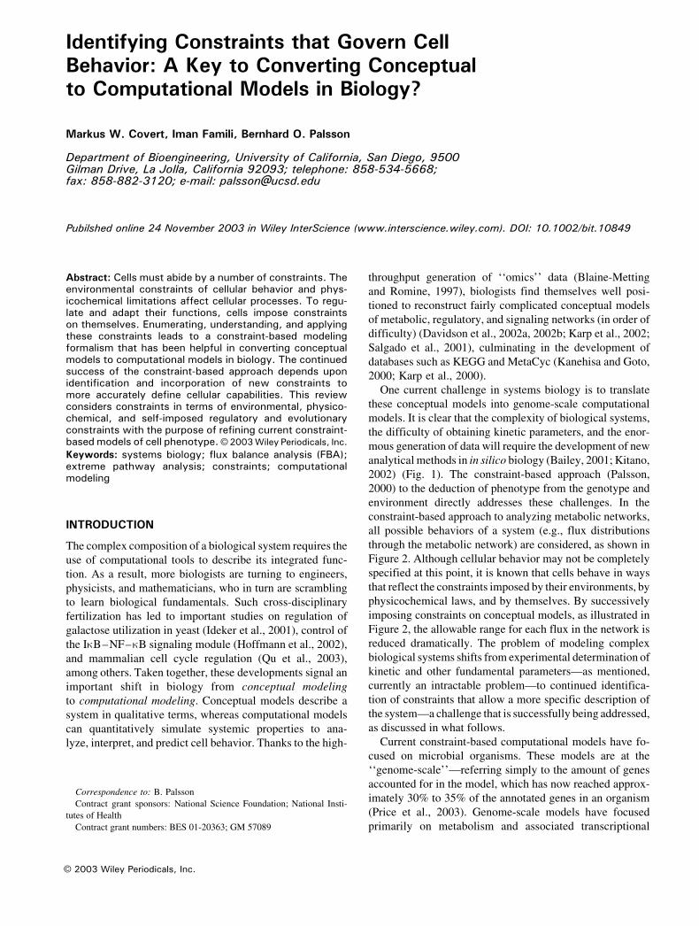
constraint-based approach to analyzing metabolic networks,
all possible behaviors of a system (e.g., flux distributions
through the metabolic network) are considered, as shown in
Figure 2. Although cellular behavior may not be completely
specified at this point, it is known that cells behave in ways
that reflect the constraints imposed by their environments, by
physicochemical laws, and by themselves. By successively
imposing constraints on conceptual models, as illustrated in
Figure 2, the allowable range for each flux in the network is
reduced dramatically. The problem of modeling complex
biological systems shifts from experimental determination of
kinetic and other fundamental parameters—as mentioned,
currently an intractable problem—to continued identifica-
tion of constraints that allow a more specific description of
the system—a challenge that is successfully being addressed,
as discussed in what follows.
Current constraint-based computational models have fo-
cused on microbial organisms. These models are at the
‘‘genome-scale’’—referring simply to the amount of genes
accounted for in the model, which has now reached approx-
imately 30% to 35% of the annotated genes in an organism
(Price et al., 2003). Genome-scale models have focused
primarily on metabolism and associated transcriptional
B 2003 Wiley Periodicals, Inc.
Correspondence to: B. Palsson
Contract grant sponsors: National Science Foundation; National Insti-
tutes of Health
Contract grant numbers: BES 01-20363; GM 57089

regulation, but are aimed at a complete representation of an
organism and have already been used to simulate cell
behavior under a variety of conditions (Reed and Palsson,
2003). In addition, because these models attempt to capture
systems-level behaviors as completely as possible, they are
instrumental in identifying and characterizing emergent
properties of biological networks, where an observed
behavior has been difficult or even impossible to interpret
from the cellular ‘‘parts list’’ alone (Papin et al., 2003; Price
et al., 2003).
The success of the constraint-based approach depends
upon identification and incorporation of new constraints to
define the cellular capabilities more accurately. This review
considers constraints in terms of environmental, physico-
chemical, and self-imposed regulatory and evolutionary
constraints with the purpose of refining current constraint-
based models of cell phenotype. Some of the constraints may
seem intuitive or basic; however, we aim to illustrate how
their consideration leads to nonintuitive modeling conse-
quences (Fig. 3, Table I).
ENVIRONMENTAL CONSTRAINTS
The constraints imposed on cells by their environments—
both external and internal—have a major influence on cell
behavior. External environments impose constraints on cells
in terms of nutrients, physical factors, and neighboring influ-
ences. Whether a cell can grow in a given environment
depends in part on its ability to obtain or synthesize all nec-
essary biomass components (Neidhardt et al., 1990). The
presence or absence of necessary compounds thus represents
an environmental constraint on the cell. As an example,
development of a ‘‘minimal gene set’’ to sustain life must be
kept in the context of environmental constraints, as the
minimal gene set for life in a complex medium would differ
significantly from that required for life in glucose minimal
Figure 1. The shift from studying components to studying systems requires the use of computational tools to integrate conceptual data and simulate
systemic behavior. The importance of computation has become apparent in the biological sciences, wherein generation of experimental data far outpaces
efforts to reconcile these data in terms of a comprehensive model. New mathematical approaches will be required to describe such systems. Such a
challenge is not new; the development of statistical mechanics originated as an attempt to integrate the known chemical properties of molecules to simulate
the properties of a bulk fluid. Escherichia coli image courtesy of www.denniskunkel.com.
764 BIOTECHNOLOGY AND BIOENGINEERING, VOL. 84, NO. 7, DECEMBER 30, 2003
medium (Burgard et al., 2001; Koonin, 2000). The impor-
tance of the nutrient constraints imposed by the external
environment also underscores the importance of defined
media in mathematical simulation of cell behavior. Without
adequate knowledge of the nutritional content of the exter-
nal environment, significant constraints must be ignored or
grossly approximated, resulting in incorrect or misleading
predictions of cell behavior. The development of high-
throughput phenotyping technologies has addressed the
inadequacies of studies on undefined media, enabling char-
acterization of the environmental effects on organism growth
under thousands of well-defined conditions (Bochner et al.,
2001). Physical characteristics of the external environment,
such as temperature, pressure, pH, and exposure to light or
water, can also limit possible cell behavior and survival.
Physical environmental constraints have been used to inves-
tigate the possibility of life on Mars (Cockell et al., 2000).
The environmental conditions experienced by a cell gen-
erally change over time. They may change by the presence
of new harmful products (such as the cell’s own waste),
depletion of nutrients (by the cell itself or by competitors), or
other dynamic forces. A tightly packed cellular community
necessitates competition for, or exchange of, nutrients and
adhesion sites, evolving mechanisms to survive toxin expo-
sure or to move toward scarce nutrients, but also raises the
possibility of obtaining new cellular capabilities via gene
transfer, or of cooperation and specialization, such as in a
tissue, where signaling molecules allow cells to communi-
cate and cooperate (DeLisa et al., 2001). To account for such
interactions in a model, the cellular community must there-
fore be accurately represented (Tsuchiya et al., 1966). Forces
such as fluid flow (wind on a plant leaf, juices through the
intestines) necessitate that cells adhere to their chosen envi-
ronments or develop systems that are resilient to changing
environments (sporulation, broad substrate utilization). The
cell is therefore constrained to the development of biological
functions that allow it to thrive in a dynamic environment.
The intracellular environment of a cell also imposes con-
straints on cellular behavior, notably in terms of its internal
components and the physical properties of its interior. Cells
are obviously limited by the biochemical components of
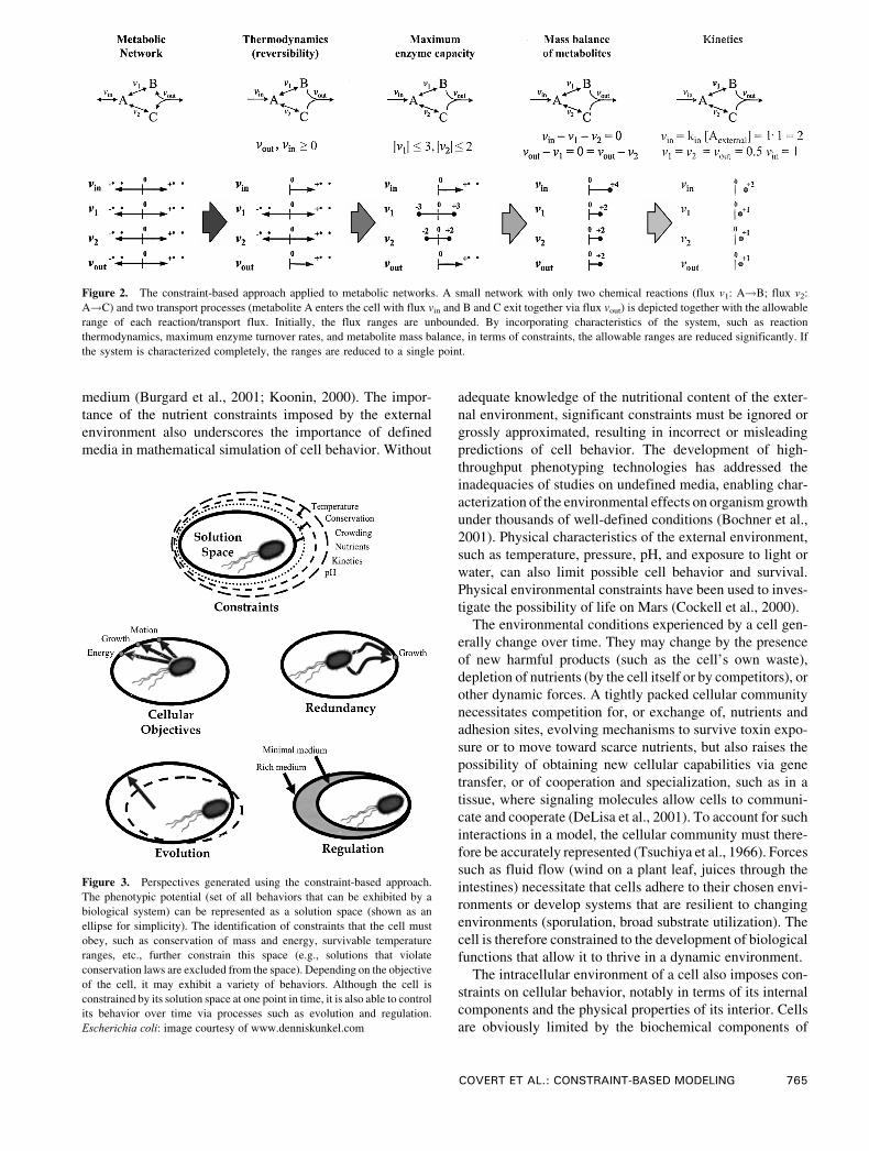
Figure 3. Perspectives generated using the constraint-based approach.
The phenotypic potential (set of all behaviors that can be exhibited by a
biological system) can be represented as a solution space (shown as an
ellipse for simplicity). The identification of constraints that the cell must
obey, such as conservation of mass and energy, survivable temperature
ranges, etc., further constrain this space (e.g., solutions that violate
conservation laws are excluded from the space). Depending on the objective
of the cell, it may exhibit a variety of behaviors. Although the cell is
constrained by its solution space at one point in time, it is also able to control
its behavior over time via processes such as evolution and regulation.
Escherichia coli: image courtesy of www.denniskunkel.com
Figure 2. The constraint-based approach applied to metabolic networks. A small network with only two chemical reactions (flux v1: A!B; flux v2:
A!C) and two transport processes (metabolite A enters the cell with flux vin and B and C exit together via flux vout) is depicted together with the allowable
range of each reaction/transport flux. Initially, the flux ranges are unbounded. By incorporating characteristics of the system, such as reaction
thermodynamics, maximum enzyme turnover rates, and metabolite mass balance, in terms of constraints, the allowable ranges are reduced significantly. If
the system is characterized completely, the ranges are reduced to a single point.
COVERT ET AL.: CONSTRAINT-BASED MODELING 765

which they are comprised; the glycolytic genes must not
only be located in the genome but also must be expressed as
functional proteins for glycolysis to occur in an organism.
The components may be thought of as a ‘‘toolbox’’ that the
cell requires to use the resources found in the environment
to perform necessary functions (growth, signaling, chemo-
taxis, etc.). This toolbox constantly changes over time,
either enabling or limiting the function of the cell. For
example, the maximum transport rate of a particular cell
moiety will be determined in part by the number of transport
proteins specific to that moiety located in the cell
membrane. The total number of components that can be
contained in the cell are limited by a generalized bounding
constraint, cell volume (i.e., the toolbox is also quite small).
The last several decades of biological research has focused
on identifying cellular components, culminating in the dev-
elopment of high-throughput methods to study the genome
(Gaasterland and Oprea, 2001), transcriptome (Devaux
et al., 2001), proteome (Naaby-Hansen et al., 2001), and
metabolome (Raamsdonk et al., 2001) of organisms under
various conditions. Because cell function depends on the
action and interaction of various components, the ‘‘omics’’
data are of fundamental importance in the effort to model
cell behavior.
Physical factors of the cellular interior also impose
constraints on the cell. Pictorial models of the interior of
E. coli depict a crowded, tightly packed, nonhomogeneous
cytoplasm (Ellis, 2001; Goodsell, 1993). Such a dense
environment has a constraining effect on solute and macro-
molecular diffusion. One way in which cells may overcome
diffusion-related constraints in a crowded environment is
via compartmentalization of major cell processes and
metabolic channeling (Verkman, 2002). Furthermore, the
crowded internal environment of the cell creates an osmotic
pressure in relation to the often aqueous external environ-
ment that must be balanced. Cells achieve this balance by
exchanging molecules with the external environment. The
balance of osmotic pressure must be achieved while
maintaining an electroneutral environment on both sides
of the membrane. Osmotic and electroneutrality constraints
can affect the total volume of the cell, and the need to meet
these constraints imposes significant energy demands on
the cell.
PHYSICOCHEMICAL CONSTRAINTS
Physicochemical laws place demanding constraints on cel-
lular behavior (see examples in Appendix). Cells balance
Table I. Some biological constraints with important modeling consequences. The constraints listed at the left may seem intuitive, but the consideration of
such constraints in modeling biological systems can be vital in terms of correct prediction and simulation.
766 BIOTECHNOLOGY AND BIOENGINEERING, VOL. 84, NO. 7, DECEMBER 30, 2003
mass and energy, conform to the laws of thermodynamics
and kinetics, and operate under limited enzyme turnover
rates and activity of gene products. Physicochemical con-
straints are generally considered to be ‘‘hard’’ constraints
and are thought to remain unchanged.
Mass and energy are never created or destroyed in the
cell. Elements entering the cell are either incorporated into
biosynthetic material for cell growth and replication,
utilized to generate energy required for cellular functions,
or secreted into the extracellular environment. Excess
biochemical byproducts that remain internal to the cell
may accumulate over time and result in cellular toxicity and
death. Complex systems have evolved to sense and respond
to imbalances of mass within the cell. Energy imbalance
also has detrimental consequences. Eukaryotic loss of
mitochondrial function to generate energy prohibits the cell
from driving cellular functions and causes death (Scheffler,
1999). The balance of mass and energy thus poses critical
constraints on how the cell must allocate its resources. Mass
balance of reactions also imposes stoichiometric constraints
on the network. The stoichiometric coefficients of any
biochemical reaction are such that the number of elements
and charge is conserved in a conversion. Stoichiometric
constraints impose restrictions on the network that, apart
from the nature of kinetics, define what combinations of
species must be present or absent in a steady state (Feinberg,
1987). For example, for a set of compounds with zero
concentrations, it is possible to determine which reactions
have zero rates (i.e., ‘‘switched off’’) and which have
positive rates (i.e., ‘‘switched on’’) (Feinberg, 1987). The
requirement of mass balance exerts such a strong constraint
on metabolic network function that flux balance analysis
requires virtually only these constraints, with only a handful
of strain-specific parameters, for detailed qualitative sim-
ulations (Edwards et al., 2002).
The thermodynamics of internal reactions can signifi-
cantly affect the overall capability and phenotypic properties
of the cell. The direction in which reactions proceed is a
function of energetic properties of the biochemical conver-
sions and may determine the ability of the cell to reach
diverse metabolic states in a variety of internal and external
conditions. Furthermore, chemical turnover of uncatalyzed
reactions is often very slow. In the presence of enzymes,
however, substrates are quickly converted into products.
Enzymes also provide means for fast-responding control
mechanisms. Feedback and feedforward control mecha-
nisms, including inhibition and activation, and effect of pH
and temperature on enzyme activity can influence the rate of
biochemical reactions, either linearly or nonlinearly (Bailey,
1998; Desai et al., 1999; Lee et al., 1999; Savageau, 1998).
Kinetic constraints are especially important in cells with
little or no other means of regulation. Upon maturation, the
red blood cell loses its DNA and, consequently, lacks any
means of transcriptional regulation. The sole form of regu-
lation in the red cell is thus kinetic regulation, which makes
the red cell a suitable model for studying kinetics (Jamshidi
et al., 2001).
The maximum throughput or enzyme capacity of bio-
chemical reactions can also force the cell to exhibit more
limited behaviors than otherwise. The enzyme turnover rate
of reactions in biochemical pathways can impose ‘‘bottle-
neck’’ constraints on the maximum allowable flow achieved
in a pathway (Bailey, 2001). Such bottlenecks have been
identified and analyzed using metabolic flux analysis and
metabolic control analysis (Stephanopoulos et al., 1998).
Metabolic engineering of microbial organisms has centered
around the premise of removing such bottleneck constraints
and achieving a higher production rate of desirable com-
pounds. Such efforts have been successful in a number of
cases (Stephanopoulos et al., 1998).
The balance of osmotic pressure and maintenance of
electroneutrality also impose constraints on cells. For exam-
ple, the constraints of osmotic pressure and charged mole-
cule requirements (e.g., nutrients) were found to be driving
forces in evolution of cell walls in bacteria (Koch, 2000).
Although cell volume regulation is relatively well charac-
terized (Hallows and Knauf, 1994), it is routinely ignored by
most models. The consequences of such constraints have
been studied in detail in the human red blood cell, because
such cells are relatively simple, lacking chromosomes, and
are therefore not capable of replication (Joshi and Palsson,
1989; Werner and Heinrich, 1985). Initial assessment of
the importance of these constraints using metabolic control
analysis shows that they represent dominant regulatory ef-
fects (Lee and Palsson, 1991). Clearly, much work is re-
quired to ascertain the importance of these constraints and
implement them in mathematical models.
SELF-IMPOSED CONSTRAINTS
We have previously discussed the environmental and phys-
icochemical constraints imposed on cells, which are beyond
the cells’ direct control. To respond to these constraints and
still carry out their desired functions (e.g., growth, nitrogen/
carbon dioxide fixation, development), cells must impose
constraints upon themselves to direct their behavior, select-
ing the ‘‘best’’ or most suitable option from a range of al-
lowable alternatives. Self-imposed constraints are different
from other constraints because they respond to—and often
change—internal or external environments. Unlike physico-
chemical constraints, they are time-dependent. Such adap-
tive constraints may entail regulation in the short term and
evolution over longer time scales.
Evolutionary Constraints
Although cells are constrained by the contents of their ge-
nome, they are able to change their genome sequence via
evolution. The evolutionary process is associated with cer-
tain constraints. For example, E. coli’s overall error rate for
DNA replication is between 1 in 1010 and 1 in 1011 basepairs
(Neidhardt et al., 1990); this error rate may change some-
COVERT ET AL.: CONSTRAINT-BASED MODELING 767

what in stressful conditions to increase the rate of beneficial
mutation and adaptation. Evolution is also constrained by the
number of possible mutation sites, as can be illustrated by
rolling a six-sided die. With one roll, the probability of
rolling a six is only one in six, or 16.7%. However, if the die
is rolled 36 times, the probability of rolling at least one six
becomes 99.9%. Similarly, genome size and growth rate in-
fluence the probability of a favorable mutation (see Appen-
dix, section iii). Although most mutations are selected
against, the number of mutations that occur in each gen-
eration can drive the occurrence of an evolved mutant to
virtual inevitability over time (de Duve, 1996). Thus, after a
period of only decades, bacteria, malarial parasites, insects,
and weeds have been found to resist man-made antibiotics
and toxins (de Duve, 1996). More quantitatively, Esche-
richia coli has been shown to overcome thymineless death by
experimental evolution over 10,000 generations (de Crecy-
Lagard et al., 2001), and also to exhibit parallel changes in
expression of 59 genes after 20,000 generations of evolution
on a glucose-limiting medium (Cooper et al., 2003). Con-
sideration of evolutionary constraints also led to the predic-
tion that E. coli would optimize its metabolic network for
growth on a glycerol minimal medium, which was then
demonstrated experimentally over 700 generations (Ibarra
et al., 2002).
Regulatory Constraints
Beyond reconfiguring their genomes via mutation and
adaptive evolution, organisms are able to control, to a cer-
tain extent, which genes are expressed, which proteins are
present, and even the activity of proteins in cells. The in-
corporation of regulatory constraints in considerations of
biological systems is vital (Liao andOh, 1999). For example,
recent studies of cellular metabolic networks (Jeong et al.,
2000; Wagner and Fell, 2001) have characterized such
networks as scale-free or ‘‘small world.’’ These studies were
based on genomic constraints only (i.e., any gene located in
the genome was considered a part of the network). However,
the entire metabolic network of a cell is never completely
expressed at a given time. For this reason, transcriptomic
and proteomic studies are necessary to further constrain and
thereby more accurately characterize these networks.
Furthermore, many molecules, notably those involved in
the control of regulatory networks, are present and act at
nanomolar concentrations in the cell (McAdams and Arkin,
1999). As a result of these low concentrations, there is a
substantial amount of noise in gene expression, as has been
recently demonstrated (Elowitz et al., 2002). Stochasticmod-
eling approaches have been used to incorporate the con-
straints imposed by gene noise in models of the lambda
phage (McAdams and Arkin, 1999), and experiments have
demonstrated how such noisy systems may be controlled by
negative feedback (Gardner and Collins, 2000). Recent work
has quantified the efficacy of regulatory constraints in re-
ducing metabolic solution spaces using extreme pathway
analysis to illustrate the potential dominant effect of regu-
lation on cell phenotype (Covert and Palsson, 2003).
APPLICATION OF CONSTRAINTS TOGENOME-SCALE MODELS
As mentioned earlier, constraint-based approaches have
enabled the development of genome-scale models of micro-
organisms. Thus far, the constraints that have been incorpo-
rated into genome-scale simulative models of metabolism,
such as those that exist for E. coli, H. influenzae, H. pylori,
and S. cerevisiae, have been stoichiometric, thermodynamic,
enzyme capacity, and energy balance constraints (Price et al.,
2003a). Transcriptional regulatory constraints have also
recently been added to enable combined simulation of
regulatory and metabolic networks (Covert et al., 2001b;
Covert and Palsson, 2002, 2003).
The constraint-based approach to modeling metabolic
systems has been described by Covert et al. (2001b) and
Palsson (2000). A metabolic network is reconstructed for a
microorganism, yielding a set of all metabolic reactions
available to the cell (Covert et al., 2001a). Once defined,
this set of reactions represents the stoichiometric constraints
on a cell. The reaction list also contains certain thermo-
dynamic constraints, in the effective irreversibility of some
reactions. A useful mathematical representation of all pos-
sible cell behaviors is one established geometrically as a
solution space, which is effectively capped and reduced as
constraints are incorporated (Fig. 3). We are then left with a
smaller solution space having general properties that can be
studied, or in which certain points (i.e., cellular behaviors)
may be examined in more detail.
Pathway analysis, a method of relating the structure of a
metabolic network in terms of an organism’s overall meta-
bolic capabilities, is based solely on identification of the
aforementioned stoichiometric and thermodynamic con-
straints and requires no other parameters (Papin et al., in
press). Using pathway analysis it is possible to determine,
for example, possible network ‘‘dead-ends’’ (Schilling and
Palsson, 2000; Schilling et al., 2002), key regulatory con-
trol points (Price et al., 2003b, and the level of redundancy
in a metabolic network (Papin et al., 2002a, 2002b; Price
et al., 2002).
Incorporation of enzyme capacity constraints enables flux
balance analysis (FBA), which may be used to study specific
flux distributions in more detail. FBA is associated with
linear programming (Chvatal, 1983), wherein the maximum
value of an objective function (e.g., synthesis of biomass
precursors) is located in the solution space. Some notable
recent work in FBA focused on identification and characteri-
zation of objective functions (Burgard and Maranas, 2003;
Lee et al., 2000; Mahadevan et al., 2002; Segre et al., 2002).
Metabolic flux analysis (MFA), closely related to FBA,
attempts to constrain the space more completely by deter-
mining certain internal fluxes experimentally (Bonarius
et al., 1998; Wiechert and de Graaf, 1996); however, it must
be noted that such measurements do not represent ‘‘hard’’
768 BIOTECHNOLOGY AND BIOENGINEERING, VOL. 84, NO. 7, DECEMBER 30, 2003

constraints. It has been shown both computationally using
extreme pathway analysis (Papin et al., 2002a; Price et al.,
2002) and experimentally (Flores et al., 2002) that a high
degree of redundancy exists in metabolic networks; orga-
nisms may use any of a number of routes to generate pre-
cursors or byproducts (see Fig. 3). The uses of MFA and
FBA have been reviewed in detail elsewhere (Edwards et al.,
2002; Stephanopoulos et al., 1998).
The elimination of systemically (i.e., not reaction by
reaction) thermodynamically infeasible solutions yields an
additional set of useful physicochemical constraints to deter-
mine allowable behaviors of biochemical reaction networks.
Incorporation of the systemic thermodynamic constraints
leads to energy balance analysis (EBA) (Beard and Liang,
2002). The importance of adding these constraints into
genome-scale models needs to be evaluated. EBA, as
presented by Beard et al. (2002), provides a foundation for
constraints-based analysis of reaction free energies in large-
scale biochemical systems and thus expands the scope of
information available from constraints-based modeling of
biochemical networks.
Thus far, the constraints noted in connectionwith genome-
scale models have focused primarily onmetabolic processes.
Such models fail to accurately predict cell behavior when
transcriptional regulatory processes have a dominant effect
on phenotype (Edwards and Palsson, 2000; Varma and
Palsson, 1994). To expand the scope and predictive
capability of these models, a formalism for incorporation
of regulatory constraints was developed (Covert et al.,
2001b). Boolean rules to describe regulation and a time delay
for protein synthesis/degradation are specified for each gene
in the model. The resulting regulatory network determines
the constraint imposed on every reaction in the metabolic
network over time. Because the time constants that describe
regulation and cell growth are significantly slower than those
for metabolism (milliseconds to tens of seconds vs. tens of
minutes to hours or days), metabolism may be considered to
be at quasi-steady state for appropriately short time periods
(Varma and Palsson, 1994). Simulation of cellular metab-
olism leads to calculation of flux changes and external
metabolite/biomass concentrations; these outputs lead to a
reinterpretation of the Boolean rules in the regulatory
network and, possibly, to a change in the expressed
metabolic network (Covert and Palsson, 2002). Integration
of a metabolism simulation module (i.e., FBA) and a
transcriptional regulation simulationmodule (i.e., evaluation
of Boolean logic) is greatly facilitated by the recognition that
regulatory events simply impose constraints.
This framework, which has been called regulatory flux
balance analysis (rFBA), has been applied to a central meta-
bolic/regulatory model of E. coli, accounting for 149 genes,
including 16 regulatory proteins and 73 enzymes (Covert and
Palsson, 2002). Comparisonwith experimentally determined
mutant phenotypes and gene expression data has shown that
the application of regulatory constraints to regulatorymodels
led not only to more accurate predictive capabilities, but also
to a broader predictive scope. For example,mutant regulatory
gene phenotypesmay nowbe predicted, as well as qualitative
gene expression (Covert and Palsson, 2002). The metabolic/
regulatory model ofE. coli has recently been expanded to the
genome scale and now accounts for over 1000 genes in this
organism (Reed et al., 2003; Covert et al., submitted).
CONSTRAINTS-BASED MODELS IN THE FUTURE
Several constraints relevant to cells, especially microor-
ganisms, have been enumerated. A framework has been
presented whereby impossible behaviors are eliminated and
the resulting solution space is used to analyze, interpret, and
predict cell phenotype in the context of genotype and exter-
nal environment. We have attempted to show how the recent
incorporation of new constraints, such as energy balance and
transcriptional regulation, results in both a broader scope of
simulation and more accurate predictive capability.
This leads us to the following question: Which constraints
can we incorporate in the future to add more functionality to
our models? The primary challenges to integrating new
constraints into cell models include obtaining the necessary
data to define a constraint mathematically and developing
the framework to incorporate the constraint in an existing
model (for an insightful discussion and review of such
approaches, see Bailey [1998]). For example, most of the
constraints discussed herein have involved fluxes through
biochemical reactions. One can anticipate that similar
analyses will be developed for defining limits on allowable
ranges of intracellular concentration as well as bracketing
numerical values of kinetic parameters (Lee et al., 1999;
Ronen et al., 2002). Importantly, not all constraints are of
equal value with respect to modeling, and determination of
the factors that are most constraining of cell function under
various conditions will result in more accurate and mean-
ingful simulations.
These challenges will likely be answered in a dialogue
between experimental biologists, who are generally more
aware of biological constraints, and computational biolo-
gists, who are equipped to describe constraints mathe-
matically. As such dialogues multiply and grow, we believe
that genome-scale constraints-based models will develop
to include all major cellular processes and enable predic-
tion and interpretation of large-scale data sets. Such models
will be of fundamental importance in understanding bio-
logical systems.
APPENDIX 1
Some representative calculations illustrating certain physic-
ochemical constraints in biology are given in what follows.
These examples are computationally simple, but have much
biological importance.
(i) Growth rate constraints and chromosome replication.
The growth rate of a cell is limited by the time required to
synthesize all necessary cellular components, such as cell
COVERT ET AL.: CONSTRAINT-BASED MODELING 769
membrane, proteins, and chromosome. Accordingly, the
synthesis time of each set of components can be calculated
and interpreted as a constraint on growth rate. Chromosome
replication in E. coli K-12 is considered here. Replication
rate, rrep, can be calculated as:
rrep ¼f � rflg
� �
where rf is the rate of replication for one replication fork, f is
the number of replication forks, and lg is the genome length.
Given a genome size of roughly 4.3 � 106 bp, two replicationforks, and a fork replication rate of approximately 750 bp per
second (Marians, 1996), the average time required for
genome replication is approximately 45 minutes. E. coli is
also capable of increasing its growth rate by multiplexing
DNA replication, resulting in an average of 4.2 replication
forks per cell and an estimated doubling time of about
23 minutes (Neidhardt et al., 1990). As another example, the
S, G2, and M phases of the cell cycle in mammalian systems
last about 8, 3, and 1 hours, respectively, resulting in a
doubling time of approximately 12 hours (Alberts et al.,
2002). The outcome of this computation leads to the fol-
lowing question: Which constraint dominates when E. coli
grows at maximum or near-maximum growth rates?
(ii) Maximum enzyme capacity. Physicochemical con-
straints impose an upper limit on the maximum enzyme
turnover rate. The enzyme association rate:
Sþ E!k1 SE
where S and E are the substrate and enzyme, respectively, is
generally the limiting step in the enzymatic conversions of
metabolites. The maximum turnover rate of this step, Vmax =
k1[S][E], is determined by the collision rate constant, k1, as
well as metabolite and enzyme concentrations in the cell.
The substrate concentration may be approximated to be
about 100 AM, assuming that the average compound
concentration for 1000 different metabolites is about
100 g/mol molecular weight, and that 1% of the dry weight
is composed of metabolic intermediates and that the cell
density is about 1 g/cm3 (Ingraham et al., 1983). The
average enzyme concentration can be estimated similarly,
assuming 1000 different enzymes in the cell with an average
molecular weight of 40,000 g/mol, composing 15% of the
dry weight (Ingraham et al., 1983). The enzyme concen-
tration is thereby estimated to be about 1 AM. Using a
typical collision rate constant of k1 = 108 M�1 s�1 and the
average enzyme and metabolite concentrations calculated
earlier, a maximum enzymatic rate limit of 106 molecules
Am�3 s�1 is computed. This upper limit implies that the
enzymatic reactions may never exceed rates faster than
106 molecules Am�3 s�1. Measured experimental fluxes
appear to coincide with this calculation; for example, in E.
coli, a glucose uptake rate of 15 mmol g�1 h�1 corresponds
to about 0.7 � 106 molecules Am�3 s�1, which falls within
the estimated maximum rate.
(iii) Evolution and chance. As explained in the text, the
probability of rolling a particular number may be low for one
roll, but is driven to virtual inevitability as the number of
rolls increases (de Duve, 1996). Similarly, we can ask the
question: To what extent does ‘‘chance’’ govern the evo-
lutionary process in a cell? A rough estimate may be
obtained using simple statistics. The probability of obtaining
a mutation in a particular base, Pm, after n replications, may
be calculated using the replication error rate, Perr, by:
Pm ¼ 1� ð1� PerrÞn
In other words, the chance of not getting the mutation of
interest after n generations is subtracted from one. Given that
the replication error rate of E. coli is generally about one
error in 1010 bases (but can be increased under stress)
(Neidhardt et al., 1990), the number of replications required
for the mutation to occur with 99.9% probability is 7 � 1010,corresponding to only 36 generations. Therefore, the
appearance of any particular mutation in a population is
extremely likely after a relatively short time (e.g., hours
to days with E. coli), indicating that the environment may
be a more constraining factor than the probability of a de-
sired mutation.
The authors thank Markus Herrgard and Jason Papin for helpful
discussions and reviews.
References
Alberts B, Johnson A, et al. 2002. Molecular biology of the cell. New
York: Garland. 1463 p.
Bailey JE. 1998. Mathematical modeling and analysis in biochemical
engineering: Past accomplishments and future opportunities. Biotech-
nol Progr 14:8–20.
Bailey JE. 2001. Complex biology with no parameters. Nat Biotechnol
19:503–504.
Beard DA, Liang SD, et al. 2002. Energy balance for analysis of complex
metabolic networks. Biophys J 83:79–86.
Blaine-Metting F, Romine MF. 1997. Microbial genomics: The floodgates
open. Trends Microbiol 5:91–92.
Bochner BR, Gadzinski P, et al. 2001. Phenotype microarrays for high-
throughput phenotypic testing and assay of gene function. Genome
Res 11:1246–1255.
Bonarius HPJ, Timmerarends B, et al. 1998. Metabolite-balancing
techniques vs. 13C tracer experiments to determine metabolite fluxes
in hybridoma cells. Biotechnol Bioeng 58:258–262.
Burgard AP, Maranas CD. 2003. Optimization-based framework for in-
ferring and testing hypothesized metabolic objective functions. Bio-
technol Bioeng 82:670–677.
Burgard AP, Vaidyaraman S, et al. 2001. Minimal reaction sets for Esch-
erichia coli metabolism under different growth requirements and up-
take environments. Biotechnol Progr 17:791–797.
Chvatal V. 1983. Linear programming. New York: W.H. Freeman and Co.
Cockell CS, Catling DC, et al. 2000. The ultraviolet environment of
Mars: Biological implications past, present, and future. Icarus 146:
343–359.
Cooper TF, Rozen DE, et al. 2003. Parallel changes in gene expression
after 20,000 generations of evolution in Escherichia coli. Proc Natl
Acad Sci USA 100:1072–1077.
770 BIOTECHNOLOGY AND BIOENGINEERING, VOL. 84, NO. 7, DECEMBER 30, 2003

Covert M, Palsson BO. 2003. Constraints-based models: Regulation of
gene expression reduces the steady-state solution space. J Theor Biol
221:309–325.
Covert MW, Palsson BO. 2002. Transcriptional regulation in constraints-
based metabolic models of Escherichia coli. J Biol Chem 277:
28058–28064.
Covert MW, Reed JL, Knight EM, Herrgard MJ, Palsson BO. Com-
bining high-throughput data and network-based computational mod-
els leads to testable hypothesis about transcriptional regulation in
E. coli. (submitted).
Covert MW, Schilling CH, et al. 2001a. Metabolic modeling of
microbial strains in silico. Trends Biochem Sci 26:179–186.
Covert MW, Schilling CH, et al. 2001b. Regulation of gene expression in
flux balance models of metabolism. J Theor Biol 213:73–88.
Davidson EH, Rast JP, et al. 2002a. A genomic regulatory network for
development. Science 295:1669–1678.
Davidson EH, Rast JP, et al. 2002b. A provisional regulatory gene network
for specification of endomesoderm in the sea urchin embryo. Dev Biol
246:162–190.
de Crecy-Lagard VA, Bellalou J, et al. 2001. Long term adaptation of a
microbial population to a permanent metabolic constraint: Overcoming
thymineless death by experimental evolution of Escherichia coli. BMC
Biotechnol 1:10.
de Duve C. 1996. The constraints of chance. Sci Am 274:112.
DeLisa MP, Valdes JJ, et al. 2001. Quorum signaling via AI-2
communicates the ‘‘metabolic burden’’ associated with heterolo-
gous protein production in Escherichia coli. Biotechnol Bioeng 75:
439–450.
Desai RP, Nielsen LK, et al. 1999. Stoichiometric modeling of Clostridium
acetobutylicum fermentations with non-linear constraints. J Biotechnol
71:191–205.
Devaux F, Marc P, et al. 2001. Transcriptomes, transcription activators and
microarrays. FEBS Lett 498:140–144.
Edwards JS, Covert M, et al. 2002. Metabolic modelling of microbes: The
flux-balance approach. Environ Microbiol 4:133–140.
Edwards JS, Palsson BO. 2000. The Escherichia coli MG1655 in silico
metabolic genotype: Its definition, characteristics, and capabilities.
Proc Natl Acad Sci USA 97:5528–5533.
Ellis RJ. 2001. Macromolecular crowding: Obvious but underappreciated.
Trends Biochem Sci 26:597–604.
Elowitz MB, Levine AJ, et al. 2002. Stochastic gene expression in a single
cell. Science 297:1183–1186.
Feinberg M. 1987. Chemical reaction network structure and the stability of
complex isothermal reactors—I. The deficiency zero and deficiency
one theorems. Chem Eng Sci 42:2229–2268.
Flores S, Gosset G, et al. 2002. Analysis of carbon metabolism in
Escherichia coli strains with an inactive phosphotransferase
system by (13)C labeling and NMR spectroscopy. Metab Eng
4:124–137.
Gaasterland T, Oprea M. 2001. Whole-genome analysis: Annotations and
updates. Curr Opin Struct Biol 11:377–381.
Gardner TS, Collins JJ. 2000. Neutralizing noise in gene networks. Nature
405:520–521.
Goodsell DS. 1993. The machinery of life. New York: Springer.
Hallows K, Knauf P. 1994. Principles of cell volume regulation. In:
Strange K, editor. Cellular and molecular physiology of cell volume
regulation. Boca Raton, FL: CRC Press. p 3–29.
Hoffmann A, Levchenko A, et al. 2002. The IkappaB-NF-kappaB signaling
module: Temporal control and selective gene activation. Science
298:1241–1245.
Ibarra RU, Edwards JS, et al. 2002. Escherichia coli K-12 undergoes
adaptive evolution to achieve in silico predicted optimal growth. Nature
420:186–189.
Ideker T, Thorsson V, et al. 2001. Integrated genomic and proteomic
analyses of a systematically perturbed metabolic network. Science 292:
929–934.
Ingraham JL, Maaloe O, et al. 1983. Growth of the bacterial cell.
Sunderland, MA: Sinauer Associates.
Jamshidi N, Edwards JS, et al. 2001. Dynamic simulation of the human red
blood cell metabolic network. Bioinformatics 17:286–287.
Jeong H, Tombor B, et al. 2000. The large-scale organization of metabolic
networks. Nature 407:651–654.
Joshi A, Palsson BO. 1989. Metabolic dynamics in the human red cell.
Part II—Interactions with the environment. J Theor Biol 141:
529–545.
Kanehisa M, Goto S. 2000. KEGG: Kyoto encyclopedia of genes and
genomes. Nucl Acids Res 28:27–30.
Karp PD, Riley M, et al. 2002. The EcoCyc database. Nucl Acids Res
30:56–58.
Karp PD, Riley M, et al. 2000. The EcoCyc and MetaCyc databases. Nucl
Acids Res 28:56–59.
Kitano H. 2002. Systems biology: A brief overview. Science 295:
1662–1664.
Koch AL. 2000. The bacterium’s way for safe enlargement and division.
Appl Environ Microbiol 66:3657–3663.
Koonin EV. 2000. How many genes can make a cell: The minimal-gene-
set concept. Annu Rev Genom Hum Genet 1:99–116.
Lee B, Yen J, et al. 1999. Incorporating qualitative knowledge in enzyme
kinetic models using fuzzy logic. Biotechnol Bioeng 62:722–729.
Lee I-D, Palsson BO. 1991. A comprehensive model of human erythrocyte
metabolism: Extensions to include pH effects. Biomed Biochim Acta
49:771–789.
Lee S, Phalakornkule C, et al. 2000. Recursive MILP model for finding all
the alternate optima in LP models for metabolic networks. Compr
Chem Eng 24:711–716.
Liao JC, Oh MK. 1999. Toward predicting metabolic fluxes in met-
abolically engineered strains. Metab Eng 1:214–223.
Mahadevan R, Edwards JS, et al. 2002. Dynamic flux balance analysis of
diauxic growth in Escherichia coli. Biophys J 83:1331–1340.
Marians KJ. 1996. Replication fork propagation. In: Neidhardt FC, editor.
Escherichia coli and Salmonella: Cellular and molecular biology.
Washington, DC: ASM Press. p 749–763.
McAdams HH, Arkin A. 1999. It’s a noisy business! Genetic regulation at
the nanomolar scale. Trends Genet 15:65–69.
Naaby-Hansen S, Waterfield MD, et al. 2001. Proteomics—post-genomic
cartography to understand gene function. Trends Pharmacol Sci
22:376–384.
Neidhardt FC, Ingraham JL, et al. 1990. Physiology of the bacterial cell.
Sunderland, MA: Sinauer Associates.
Palsson BO. 2000. The challenges of in silico biology. Nat Biotechnol
18:1147–1150.
Papin JA, Price ND, et al. 2002a. The genome-scale metabolic extreme
pathway structure inHaemophilus influenzae shows significant network
redundancy. J Theor Biol 215:67–82.
Papin JA, Price ND, et al. 2002b. Extreme pathway lengths and reaction
participation in genome-scale metabolic networks. Genome Res 12:
1889–1900.
Papin JA, Price ND, et al. 2003. Metabolic pathways in the post-genome
era. Trends Biochem Sci 28:250–258.
Price ND, Papin JA, et al. 2002. Determination of redundancy and systems
properties of Helicobacter pylori’s metabolic network using genome-
scale extreme pathway analysis. Genome Res 12:760–769.
Price ND, Papin JA, et al. 2003. Genome-scale microbial in silico models:
The constraints-based approach. Trends Biotechnol 21:162–169.
Price ND, Reed JL, et al. 2003. Analysis of metabolic capabilities using
singular value decomposition of extreme pathway matrices. Biophys J
84:794–804.
Qu Z, Weiss JN, et al. 2003. Regulation of the mammalian cell cycle:
A model of the G1-to-S transition. Am J Physiol Cell Physiol 284:
C349–C364.
Raamsdonk LM, Teusink B, et al. 2001. A functional genomics strategy
that uses metabolome data to reveal the phenotype of silent mutations.
Nat Biotechnol 19:45–50.
Reed JL, Palsson BO. 2003. Thirteen years of building constraint-based in
silico models of Escherichia coli. J Bacteriol 185:2692–2699.
Reed JL, Vo TD, Schilling CH, Palsson BO. 2003. An expanded genome-
COVERT ET AL.: CONSTRAINT-BASED MODELING 771
scale model of Escherichia coli K-12 (iJR904 GSM/GPR). Genome
Biol. 4:R54. Epub 2003 Aug 28.
Ronen M, Rosenberg R, et al. 2002. Assigning numbers to the arrows:
Parameterizing a gene regulation network by using accurate expression
kinetics. Proc Natl Acad Sci USA 99:10555–10560.
Salgado H, Santos-Zavaleta A, et al. 2001. RegulonDB (version 3.2):
transcriptional regulation and operon organization in Escherichia coli
K-12. Nucl Acids Res 29:72–74.
Savageau MA. 1998. Development of fractal kinetic theory for enzyme-
catalysed reactions and implications for the design of biochemical
pathways. Biosystems 47:9–36.
Scheffler IE. 1999. Mitochondria. New York: Wiley-Liss. 367 p.
Schilling CH, Covert MW, et al. 2002. Genome-scale metabolic model of
Helicobacter pylori 26695. J Bacteriol 184:4582–4593.
SchillingCH, PalssonBO. 2000. Assessment of themetabolic capabilities of
Haemophilus influenzae Rd through a genome-scale pathway analysis.
J Theor Biol 203:249–283.
Segre D, Vitkup D, et al. 2002. Analysis of optimality in natural and
perturbed metabolic networks. Proc Natl Acad Sci USA 99:
15112–15117.
Stephanopoulos G, Nielsen J, et al. 1998.Metabolic engineering. SanDiego,
CA: Academic Press.
Tsuchiya HM, Fredrickson AG, et al. 1966. Dynamics of microbial cell
populations. Adv Chem Eng 6:125–206.
Varma A, Palsson BO. 1994. Stoichiometric flux balance models
quantitatively predict growth and metabolic by-product secretion in
wild-type Escherichia coli W3110. Appl Environ Microbiol 60:
3724–3731.
Verkman AS. 2002. Solute and macromolecule diffusion in cellular aque-
ous compartments. Trends Biochem Sci 27:27–33.
Wagner A, Fell DA. 2001. The small world inside large metabolic net-
works. Proc R Soc Lond B Biol Sci 268:1803–1810.
Werner A, Heinrich R. 1985. A kinetic model for the interaction of
energy metabolism and osmotic states of human erythrocytes. Anal-
ysis of the stationary ‘‘in vivo’’ state and of time dependent varia-
tions under blood preservation conditions. Biomed Biochim Acta 44:
185–212.
Wiechert W, de Graaf AA. 1996. In vivo stationary flux analysis
by 13C labeling experiments. Adv Biochem Eng/Biotechnol 54:
109–54.
772 BIOTECHNOLOGY AND BIOENGINEERING, VOL. 84, NO. 7, DECEMBER 30, 2003