Embed Size (px)
Citation preview
1
Identification of two binding regions for the Suppressor of Hairless protein
within the intracellular domain of Drosophila Notch
Maude Le Gall and Edward Giniger*
Fred Hutchinson Cancer Research Center, 1100 Fairview Avenue North,
Seattle, Washington 98109-1024, USA
* to whom correspondence should be addressed
Phone : 1 206 667 6842
Fax : 1 206 667 3308
e-mail: [email protected]
Running Title
Two Su(H)-binding regions in the Notch intracellular domain
JBC Papers in Press. Published on April 29, 2004 as Manuscript M404589200
Copyright 2004 by The American Society for Biochemistry and Molecular Biology, Inc.
by guest on July 10, 2018http://w
ww
.jbc.org/D
ownloaded from

2
Summary
Notch is a phylogenetically conserved transmembrane receptor that is required for many aspects
of animal development. Upon ligand stimulation, a fragment of Notch is released proteolytically,
and enters the nucleus to form a complex with the DNA-binding protein CSL and activate
transcription of Notch/CSL target genes. The physical structure of the Notch/CSL complex
remains unclear, however, clouding the interpretation of previous efforts to correlate Notch
structure and function. We have therefore characterized the binding of Drosophila CSL (called
Suppressor of Hairless, or Su(H)) to the intracellular domain of Drosophila Notch, both in vitro
and in vivo. We report identification of two Su(H) binding regions in Notch. The first is in the
juxtamembrane region (the “RAM” domain). The second is just C-terminal to the Notch ankyrin
repeats, overlapping or identical to two previously proposed nuclear localization sequences
(NLS). The ankyrin repeats themselves do not bind to Su(H), however, they substantially
enhance binding of Su(H) to the more C-terminal region. Consistent with this picture, removal of
either the Ram or PPD binding sites, separately, modestly reduces Notch activity in vivo, while
removal of both renders Notch severely defective. These results clarify the relationship between
Notch and CSL, help to explain the importance of the ankyrin repeats in Notch signaling and
reconcile many apparently contradictory results from previous Notch structure/function studies.
Moreover, they suggest a second function for the Notch “NLS” elements.
by guest on July 10, 2018http://w
ww
.jbc.org/D
ownloaded from
3
Introduction
The Notch pathway is a phylogenetically conserved signaling mechanism that mediates cell-cell
communication events in the development of nearly all metazoan organisms that have been
examined. Homologues of Notch have been found in echinoderms, ascidians, nematodes, insects
and vertebrates. In all those organisms, the Notch pathway is essential for a variety of
developmental processes involving intercellular communication, including asymmetric cell-fate
decisions, formation of boundaries in developmental fields, cell proliferation, and apoptosis [1].It
is thought that the Notch pathway exerts these pleiotropic effects through a common signaling
pathway involving the interaction of a few core elements: a signal, DSL1 (Delta, Serrate, LAG-
2), a receptor, Notch (sometimes called LNG for LIN-12, Notch, GLP-1), and a DNA binding
protein, CSL (CBF-1, Suppressor of Hairless (Su(H)), LAG-1) with its co-activator, Mam
(Mastermind, MAML). Interaction of Notch with its ligand DSL initiates a series of proteolytic
events at the membrane, which ultimately result in the release of the intracellular domain of
Notch (Nicd) (for review see [2]). The free Notch intracellular domain then translocates into the
nucleus where it forms a transcription control complex in association with CSL and Mam,
binding to target genes that bear CSL binding sites and activating their transcription.
The intracellular domain of Drosophila Notch is large, 937 amino acids, and contains a variety
of signaling domains and binding sites for regulators and effectors. (for a schematic
representation see Figure 1A). It starts with the juxtamembrane RAM domain, which contains a
conserved nuclear localization signal (NLS) sequence. RAM also contains a high affinity binding
site for the PTB domain of Numb [3], involved in the trafficking of the receptor, particularly for
endocytosis [4] and degradation [5]. C-terminal to RAM, seven ankyrin repeats [6-9] constitute
a domain (ANK) that is essential to all Notch functions described so far, and which is highly
conserved throughout the entire metazoan Notch family [1]. The molecular function of ANK
remains elusive, but it includes a binding site for Deltex , a protein that is required for full Notch
activity in vivo [10-12]. A domain that we have called in this study PPD for Potential
Phosphorylated Domain follows ANK. It contains a large number of serines and threonines in
consensus phosphorylation motifs for various kinases [13, 14], two stretches of basic residues
with NLS homologies, and a transcriptional activation capability [15, 16]. The more C-terminal
by guest on July 10, 2018http://w
ww
.jbc.org/D
ownloaded from
4
part of the Notch intracellular domain contains a Glutamine rich region (OPA) and a PEST
region involved in regulating the half-life of the protein [17]. In mouse and nematode the PEST
region is phosphorylated in vivo and becomes a binding site for the E3 ubiquitin ligase Sel-10
(for review [18]).
The association of Notch with CSL is thought to be the key to Notch signaling, since it is CSL
that determines what genes are bound and activated by the Notch/Mam/CSL complex [1], but the
physical basis for this association remains unclear. The RAM domain was first identified as the
primary binding domain for CSL in mammalian Notch [19]. Mutation of a span of conserved
residues in this region prevents in vitro binding of CBF-1 with mNotch [19]. However, in
mammalian cultured cells, RAM-deleted Notch derivatives retain Notch activity, suggesting
either the presence of other CBF-1 sites or some CBF-1-independent activity [15, 20, 21]. More
definitively, in Drosophila, RAM-deleted Notch derivatives still provide significant Su(H)-
dependent activity [22, 23]. It has been suggested that Su(H) may interact in vitro with the
ankyrin repeats of Drosophila Notch [24], and indeed, in contrast to the RAM domain, the ANK
repeats are essential for all CSL-dependent Notch functions in all studied organisms. However,
the binding of CSL proteins to the ANK region seems to be very much weaker than the binding
to the RAM domain [25], and has not even been detectable in all studies. Moreover, no CSL
binding site has been specifically mapped to this part of the protein. Very recently, it has been
shown in mammalian cells that the presence of the protein Mastermind (Mam) interacting with
the ANK domain can reinforce the interaction between CBF-1 and RAM-deleted Notch
derivatives [26].
To clarify the structure of the Notch/Su(H) signaling complex and its relationship to the
sequence of the receptor, we have mapped Su(H) binding sites on Drosophila Notch in vitro, in
Drosophila cells and in extract of Drosophila embryos. We identified two Su(H) binding regions
within the intracellular domain of Drosophila Notch. One is in the RAM domain, and confirms
the conservation of a strong CSL binding site in the juxtamembrane region of the intracellular
domain. A second Su(H) binding region is more complex and was found just C-terminal to the
ankyrin repeats, within the domain we called PPD. The ankyrin repeats themselves do not bind
directly to Su(H) but they substantially enhance binding of Su(H) to the PPD region, and may be
by guest on July 10, 2018http://w
ww
.jbc.org/D
ownloaded from
5
linked to Su(H)indirectly in vivo, via a bridging adaptor protein [27, 28]. The Ram and PPD
sites each contribute to Notch function in vivo, and deletion of both regions severely reduces
Notch activity. Together, these data allow us to reconcile a great deal of seemingly conflicting
data on the function of Notch domains and the effects of Notch mutations.
by guest on July 10, 2018http://w
ww
.jbc.org/D
ownloaded from
6
Experimental ProceduresPlasmid constructionsBriefly, constructs were obtained as followed:
GST-Su(H): To create a baculovirus construct for the expression of GST fusion proteins, the
GST coding sequence, polylinker region, and stop codons of pGEX-2T (Pharmacia Biotech Inc.)
were amplified by PCR and subcloned into the baculovirus transfer vector pVL1393 transfer
vector (pharmingen) to create the vector pVL-GST. pGEX-KG-Su(H) (provided by Posakony,
[29]) was cut EcoRI and HindIII (blunt) and subcloned into pVL-GST cut EcoRI and XbaI
(blunt).
Notch fragments: His tagged Notch fragments RAM and OPA were PCR amplified and
subcloned between the NheI and BglII sites of pRSET A and have been previously described
[30] . The others similar constructs of this study: RAMDA, RAMDB, RAMDC, RAMDD, ANK,
ANK-PPD, the different PPD-OPA, N1766-2262 and N1779-2262 were made the same way. In
each case, a stop codon was introduced after the final amino acid and the amplified fragment was
fully sequenced.
pT7/C1 EGFP and pT7/C1 EGFP-PPD: pT7/C1 EGFP was derived from pEGFP-C1 (Clontech)
with addition of a T7 promoter. pT7/C1 EGFP-PPD was obtained by subcloning pRSET-ANK-
PPD cut BamHI and PstI into pT7/C1 EGFP cut BglII and PstI.
pUAS Flag-N1766 and pUAS Flag-N1779: The constructs were obtained after amplification by
PCR of a fragment from pUAS N1-2262 (provided by C. S. Wesley [31] ) with addition in 5’ of
a BglII site, a Drosophila initiation of translation sequence and 3 Flag tags in frame with amino
acid 1766 or 1779 and the sequence ending in 1962 at the XhoI site. The amplified fragments
were sequenced and then subcloned into pUAS N1790 (provided by S. Kidd [16]) cut by BglII
and XhoI.
pUAS NFL WT : To get the pUAS NFL WT we cleaned the stop mutation of pUAS N1-2262
[31] by replacing its XhoI/XbaI fragment by the one from pUAS N1790 [16].
pUAS Su(H)-HA: The triple HA tag was derived from IRES hrGFP2a (Stratagene), cut XhoI and
BstEII (blunt) and subcloned into pBS-SK cut XhoI and KpnI (blunt). Su(H) was PCR amplified
from pBS-KS-Su(H) (provided by F. Schweisguth, [32]) with a KpnI site in front of the ATG
and a XhoI site in place of the stop codon putting it in frame with the three HA tags. The KpnI-
XbaI insert was fully sequenced before being subcloned into pUAS.
by guest on July 10, 2018http://w
ww
.jbc.org/D
ownloaded from
7
Mutagenesis
All the deletions or mutations in the Notch derivatives were done by directed mutagenesis
according to the protocol of the Quick Change Directed mutagenesis kit (Stratagene).
Briefly, for all the mutations in the RAM domain, the NheI-NcoI fragment of pUAS N1-2262
was subcloned into pRSET and then mutagenised. For all the mutations in the PPD domain, the
Xho-NarI fragment of pUAS Nintra1790 was subcloned into BS-SK and then mutagenised. All
the mutagenised fragments were fully sequenced before being re-subcloned in their appropriate
backbones (pUAS NFL WT, pUAS N1766 or N1779, pRSET N1766 or N1779, etc.).
Recombinant protein
Baculovirus: Baculovirus stocks were produced by cotransfecting into SF9-insect cells pVL-GST
or pVL-GST-Su(H) plasmids along with a mixture of AcNPV DNA according to the
manufacturer procedures (BaculoGoldTM Pharmingen).
Purification: GST and GST-Su(H) fusion protein expressed in SF9 were purified on glutathione-
sepharose beads (Pharmacia). Briefly, 36 hours post-infection with baculovirus expressing pVL-
GST or pVL-GST-Su(H), SF9 cells were harvested, washed in phosphate-buffered saline and
lysed in 25mM Hepes (pH 7.5), 300mM NaCl, 0.5% NP40, 15mM b-mercaptoethanol, 5mM
NaF, 10mMNa pyrophosphate and 25mM b-glycerophosphate. After 30min of gentle agitation at
4oC and 30min of centrifugation at 14,000rpm, the supernatant was diluted with lysis buffer
lacking NaCl to reduce NaCl to 125mM. Preblocked glutathione-sepharose beads were added to
the lysate and incubate for 1hour with gentle rocking. Beads were washed 4 times in lysis buffer
50mM NaCl and stored in presence of azide 2mM. GST and GST-PTB purified from bacteria
have been previously described [30].
In vitro binding assay
Notch fusion proteins were in vitro transcribed and translated (IVT) in the presence of 35S
methionine using rabbit reticulocyte lysates (TNT, Promega) and following the manufacturer
protocol. As indicated, some of the constructs (ANK-PPD derivatives) were digested and
purified before being IVT. Binding of in vitro translated polypeptides or fly extracts to GST
fusion proteins was performed as previously described [30].
Cell culture and cell extracts
by guest on July 10, 2018http://w
ww
.jbc.org/D
ownloaded from
8
Drosophila Schneider 2 (S2) cells were obtained from Invitrogen and maintained in Schneider’s
Drosophila Medium supplemented in 10% heat-inactivated Fetal Calf Serum, 50 units/ml
penicillin and 50mg/ml streptomycin according to the manufacturer’s instructions.
The cells were transfected with calcium phosphate as recommended by the manufacturer.
Briefly, 3 106 S2 cells were plated in a 35mm plate in 3ml, grow for 20 hours before being
tranfected with 1mg of each plasmid, as indicated, and 1mg of pRK241 (Actin-Gal4, from R.
Kostriken and P. O’Farrell) to allow the expression of the UAS constructs. Twenty four hours
after transfection, the cells were harvested, washed with phosphate-buffered saline and
suspended in 300ml of ice-cold 25mM Hepes (pH 7.5), 100mM NaCl, 0.5%NP40 (v:v), 10%
glycerol (v:v), 15mM b-mercaptoethanol and 1mM Phenylmethylsulfonyl fluoride. After 30min
of gentle agitation at 4°C and 30min of centrifugation at 14000rpm, the supernatants were
collected as whole cell extracts.
Transgenic fly strains and fly extracts
Transgenic strain: Germ line transformation was performed using standard procedures. w1118
flies were injected with UAS constructs at 1mg/ml and Delta2-3 helper plasmid at 0.5mg/ml.
Other fly strains: The strong mutant allele Notch[55e11] (from E.H. Grell and Y.N. Jan) was
used as the background for transgene rescue experiment. GAL4 drivers for various experiments
were 69B (A. Brand): expression throughout embryonic ectoderm; scabrous-GAL4 (C.S.
Goodman): expression in neuroectoderm and its derivatives; hairy-GAL4 (S. Parkhurst):
expression in every other segment of the early embryo.
Whole embryo lysates : UAS expressing flies were crossed to 69B to obtain expression in
embryos. F1 embryos were collected on grape juice plates O/N (~18 hours at 25oC), harvested
with 0.7% NaCl, 0.3% Triton (NaCl/Tx), dechorionated with 50% bleach, washed with NaCl/Tx
and transferred to an ice-cold Dounce homogenizer. Embryos were then washed twice with cold
H2O and once with cold lysis buffer. Embryos were suspended with 3 volumes of 25mM Hepes
(pH 7.5), 100mM NaCl, 0.5mM DTT, 0.5% NP-40 (v:v), 10% glycerol (v:v), 1mM
Phenylmethylsulfonyl fluoride and lysed with 25 strokes of an ice-cold A pestle, followed by 25
strokes with a cold B pestle. Embryo lysates were transferred to microfuge tubes and centrifuged
at 14,000rpm for 10min at 4oC. The supernatants were collected as whole embryo extracts.
Immunoprecipitation and western blot
by guest on July 10, 2018http://w
ww
.jbc.org/D
ownloaded from
9
Antibodies: antibodies, their sources, and the concentrations used for western blotting (WB) and
immunoprecipitation (IP) were as follows: anti-Notch (C17.9C6, Developmental Studies
Hybridoma Bank (Iowa)), 1:50 (WB), 30ml/IP, Anti-HA.11 (16B12, Covance) 1/2,000(WB)
0.5ml /IP, anti-Flag (M2, Sigma) 1/1,000 (WB), 0.5ml /IP. Peroxidase-conjugated anti-mouse
secondary (Jackson) was used at 1:10,000 (WB), Rb anti-mouse IgG (Jackson) was used at
1mg/5ml packed Protein A sepharose beads.
Immunoprecipitation: Protein A sepharose beads (Amersham) were blocked with lysis buffer
containing 0.5% BSA, prebound with Rb anti-mouse IgG and washed 2x. Extract was thawed on
ice (if previously frozen), and 5ml of protein A/Rb anti-mouse beads was added per IP sample
(typically 200 ml of extract, diluted to 350 ml final volume) for pre-clearing. Sample was rocked
at 4oC for 1hr, then cleared by centrifugation at 14,000rpm 10min. Supernatant was removed,
added to appropriate primary antibody and rocked at 4oC for 1h, 5ml of protein A/Rb anti-mouse
beads were added per IP sample and rocked for 2 hours. Beads were spun down for 5sec in a
microfuge, unbound material was removed, and the beads were washed 3x with lysis buffer.
Beads were then boiled in 20ml Laemmli buffer.
Western blot: Proteins separated by SDS-PAGE (6.5% 29:1 acrylamide:bis) were
electrophoretically transferred to nitrocellulose membranes. Membranes were blocked for 90min
in PBST (PBS with 0.05% tween 20) containing 3% non fat milk and were incubated with
primary antibodies diluted in blocking solution for 3 hours. After washing with PBST,
secondaries diluted in PBST were applied for 30min. The membranes were finally washed with
PBST, developed with Supersignal West Pico chemoluminescent substrate (Pierce) and exposed
to Kodak Biomax MR Films.
For densitometry quantification, non-saturated chemoluminescence exposed film were scanned
and densitometry analysis was performed with NIH Image Software.
Immunohistochemistry and immunofluorescence
Embryos were collected at 25o, fixed with 4% formaldehyde and stained with antibodies by
standard methods. Whole mount embryos visualized by HRP histochemistry were examined
with a Nikon Optiphot microscope using Nomarski optics and photographed with a digital
camera (Coolscan). Fluorescently-labelled embryos were examined by confocal microscopy
(Leica TCS). Antibodies used were: anti-Notch C17.9C6 (1:50), rat anti-Elav (1: 20) and anti-
Sex Lethal (1:50): Developmental Studies Hybridoma Bank; anti-FLAG M2 (Kodak; 1:300);
by guest on July 10, 2018http://w
ww
.jbc.org/D
ownloaded from
10
anti-b-galactosidase (Cappel; 1:10,000). For in situ detection of nuclear Notch protein, we used
TSA amplification (PerkinElmer) of the Notch signal, detected with DTAF-streptavidin.
by guest on July 10, 2018http://w
ww
.jbc.org/D
ownloaded from
11
ResultsSu(H) interacts with two separate regions of Nicd in vitro
To map the Su(H) interaction region(s) within the Drosophila Notch intracellular domain (Nicd)
in vitro, we purified GST-Su(H) fusion protein produced in SF9 cells infected by recombinant
baculovirus and we in vitro translated and 35S-labeled different functional regions of the Notch
intracellular domain (Figure 1A). We tested those various regions for their ability to bind to
GST-Su(H) on glutathione beads (Figure 1B). The RAM domain is sufficient to bind efficiently
to GST-Su(H) (Figure 1B, lane 1) whereas it does not bind to GST alone (data not shown). This
indicates that the RAM domain of Drosophila Notch, just like the RAM domain of vertebrate
Notch [19], contains an interaction surface for CSL. We also found that a fragment containing
both ANK and PPD bound to Su(H) (Figure 1B, lane 3) with similar efficiency as RAM.
Furthermore, a fragment containing both PPD and OPA bound to GST-Su(H), though very
weakly (Figure 1B, lane 4). Since neither the ANK domain alone (containing the full seven
ankyrin repeats [8, 9]) nor the OPA domain alone bound to Su(H) (lane 2 and 5), we suspected
that the PPD domain was the region of interaction and tested that hypothesis by creating a fusion
protein between PPD and EGFP (Figure 1C). Whereas EGFP did not bind at all to GST-Su(H),
EGFP-PPD bound GST-Su(H) about as well as did PPD-OPA (Figure 1C, compare lanes 1 and
2). These data suggest that the PPD domain can interact with Su(H) but somehow this interaction
is strongly improved by the presence of the ANK domain. Together, these data suggest the
existence of two separate Su(H) binding sites within Drosophila Nicd : one in the PPD domain
that is sensitive to the presence of the ankyrin repeats, and one in the RAM domain.
Molecular characterization of the Su(H) binding site within the RAM domain of NotchTo narrow down the location of the Su(H) binding site within the RAM domain of Notch, we
divided that domain into four subregions (A, B, C , and D, see Figure 2A) and tested the ability
of deleted forms of RAM to bind to GST-Su(H) (Figure 2B). All subdomains except A were
dispensable for Su(H) binding (Figure 2B). The A subdomain is highly conserved between
species and includes a strong match to the previously described CBF-1 binding region in the
RAM domain of mammalian Notch [19]. Therefore, we introduced a triple point mutant WFP to
LLA at position 1776-1778 in the Drosophila RAM domain. As in mammals, this mutation in
the RAM domain of Drosophila Notch prevented binding to Su(H) (Figure 2B, lane 8, middle
by guest on July 10, 2018http://w
ww
.jbc.org/D
ownloaded from
12
panel). In contrast, it did not prevent the binding of a different Notch-interacting protein, a fusion
protein between GST and the PTB domain of Disabled (Dab) that we have shown previously to
interact with RAM [30] and that also binds to the RAM A subregion (Figure 2B, lanes 2 and 7
and 8, lower panel).
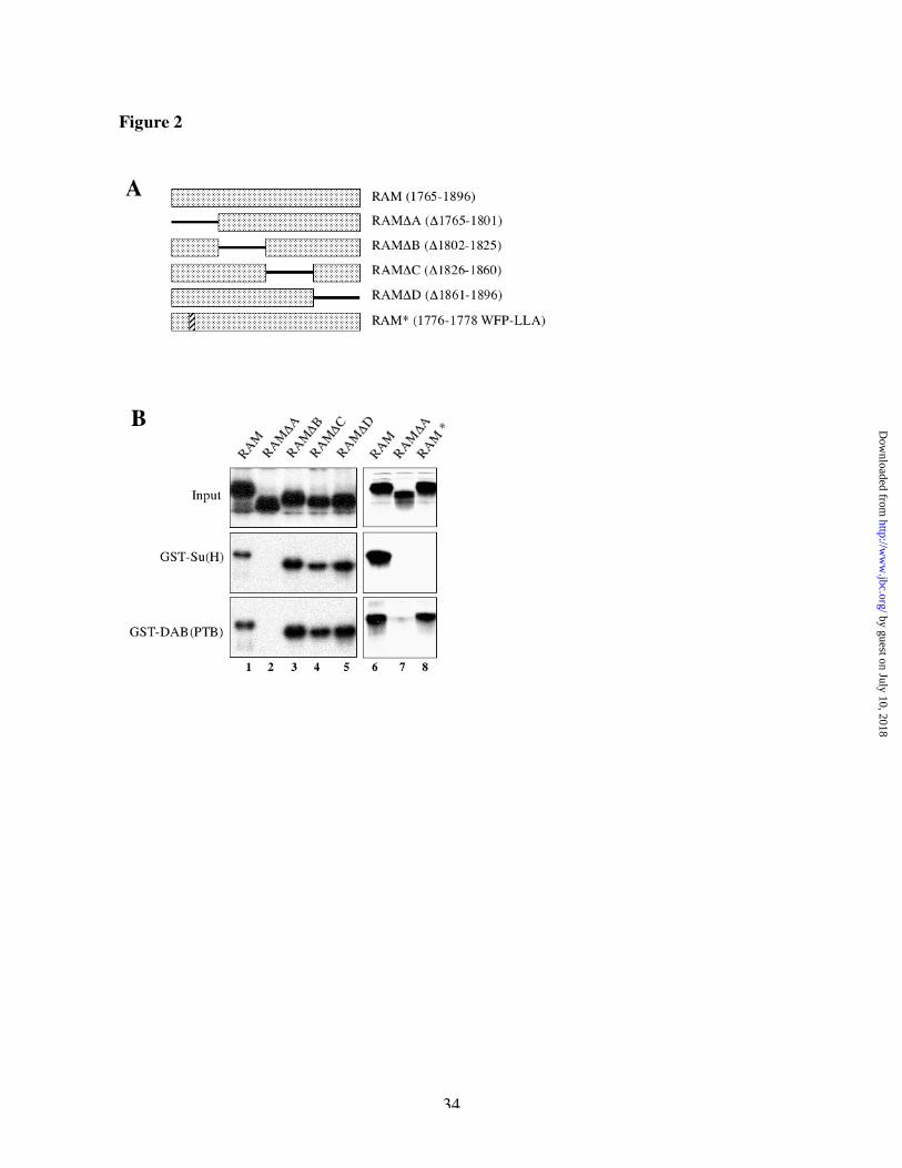
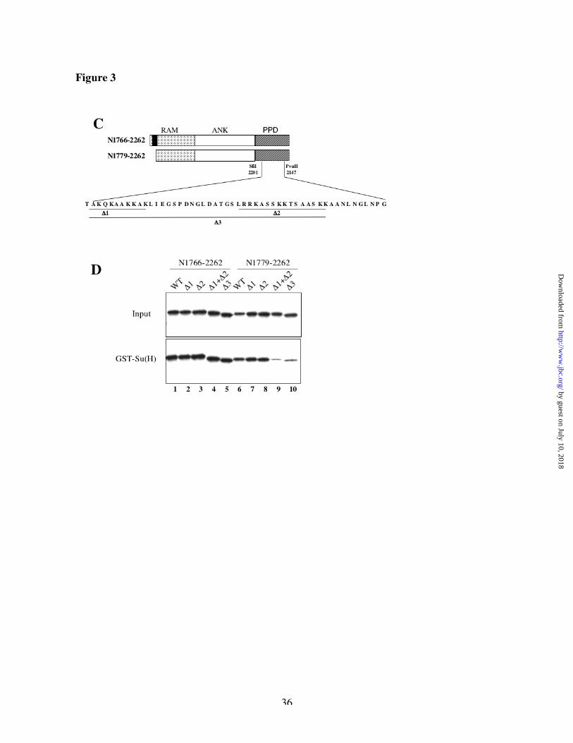
Molecular characterization of the Su(H) binding site within the PPD domain of NotchWe set out to characterize binding of Su(H) to the PPD domain, using the same strategy that we
had used for RAM. We started with the ANK-PPD fragment, digested its coding DNA with 5
restriction enzymes deleting progressively the C-terminus part of that encoded peptide and
produced the encoded protein by in vitro transcription and translation (Figure 3A). Whereas
Su(H) can bind to the peptide encoded by the constructs cut at the EcoRI or PvuII site (Figure
3B, lanes 1-2), it cannot bind to any shorter peptide (Figure 3B, lanes 3-5). Conversely, we
generated different versions of the PPD-OPA fragment starting at those different restriction sites
(Figure 3A) and showed that whereas a construct starting at the SfiI site bound as well as full
length PPD-OPA (Figure 3B, lanes 8-9), another construct starting at the PvuII site interacted
little, if at all, with Su(H) (Figure 3B, lane 7). Those results suggest that amino acids 2201-2247,
coded by the DNA sequence between the SfiI and PvuII sites, are essential for the interaction of
Su(H) with the PPD domain of Notch.
We next mutated all the codons between SfiI and PvuII in groups of four adjacent codons and to
our surprise, none of these mutations prevented binding to Su(H) (data not shown). This
suggested that the binding region might be complex or redundant. Looking at the primary amino
acid sequence of the PPD region, we noticed two stretches of basic residues (lysines and
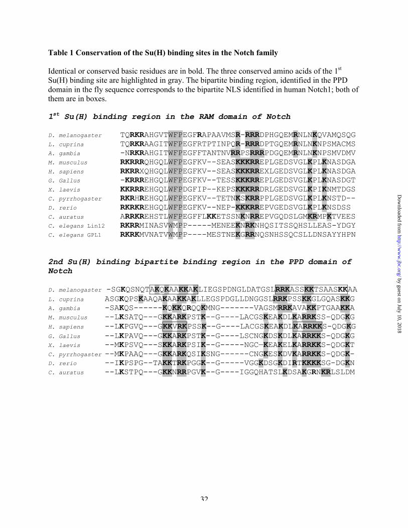
arginines) that are strongly conserved within the Notch family (Figure 3C and Table 1). These
peptides were previously suggested in human Notch to be Nuclear Localization Signals (NLS)
[20]. The Su(H) interaction site in the RAM domain is also in a region with a block of positively
charged residues [19]. We therefore wondered whether those blocks of basic residues might be
important for the binding of Su(H) to the PPD region. We made deletions in the PPD region of
Drosophila Notch that removed each of those stretches of charged residues separately (D1 or D
2) or both together (D1+D2 and D3) (Figure 3C) and we tested their effect on the binding to
GST-Su(H). We made those deletions in a construct starting at codon 1779 (N1779-2262)
by guest on July 10, 2018http://w
ww
.jbc.org/D
ownloaded from
13
lacking the Ram site, as well as in a construct starting at codon 1766 (N1766-2262) that retains
the Ram site and acts as a positive control for the activity of the construct. As shown in Figure
3D, deletion of one (D1, lane 7) or the other (D2, lane 8) block of basic residues does not affect
the binding to GST-Su(H). However, deletion of both blocks (D1+D2 or D3, lanes 9-10) strongly
reduces the interaction of Su(H) with N1779-2262. All proteins bearing the RAM site (N1766-
2262 derivatives) still bound Su(H) (lanes 1-5), suggesting that the deletions did not grossly
inactivate the proteins. These results suggest that the PPD region contains two small peptides
either of which is sufficient to allow binding of Su(H) to N1779-2262. The deletion D3 was used
in the experiments below as a mutant form of the PPD region that prevents binding to GST-
Su(H).
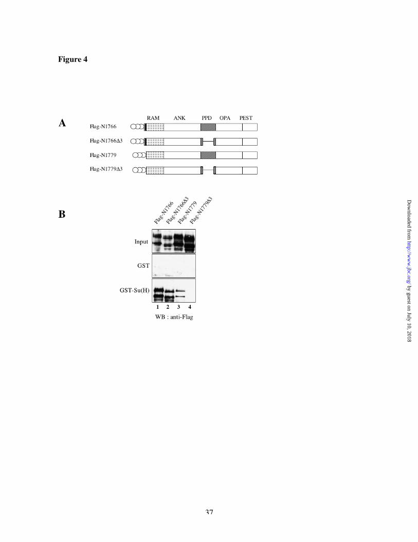
Binding of Su(H) to Nicd in fly extracts
We further tested the significance of the two apparent Su(H) binding regions by performing pull
downs of Notch derivatives from Drosophila embryo extract. We used the GAL4/UAS system to
express in fly embryos [33, 34] different intracellular domains of Notch containing deletions or
mutations in the two binding regions described above. Those constructs were tagged at the N-
terminus with three FLAG epitopes to distinguish them from cleavage products of endogenous
Notch (Figure 4A). Flag-N1766 has the entire intracellular portion of Notch, starting at the
amino acid 1766 up to the last amino acid of Notch (2703) and contains the two Su(H) binding
regions described above, whereas Flag-N1779 is missing the Su(H) binding site within the RAM
domain. Incorporating the D3 deletion in those two constructs allows us to eliminate the second
Su(H) binding region within the PPD domain. We tested the ability of these Nicd derivatives to
interact with GST or GST-Su(H) in vitro. As shown on Figure 4B middle panel, none of the
constructs bound to GST alone. Deletion of either the RAM site or the PPD region reduced the
binding to GST-Su(H) (lanes 2-3). Deletion of both sites prevented binding completely (lane 4).
We always noticed a strong difference in expression levels of the constructs having or lacking
the RAM Su(H) binding site (compare Figure 4B lanes 1-2 with 3-4, upper panel): proteins
bearing the Ram site were present at much lower levels in vivo. This difference was reproducible
with independent insertion lines and with expression through different GAL4 drivers (data not
by guest on July 10, 2018http://w
ww
.jbc.org/D
ownloaded from
14
shown) . This observation may suggest a link between interaction of Su(H) at the RAM domain
of Notch and degradation and/or stability of the Notch protein.
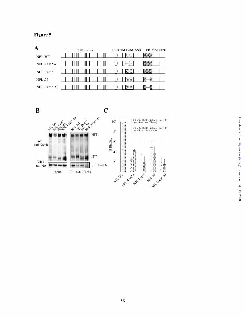
Su(H) interacts with two separate domains of Nicd in vivo
To test the binding of Notch to Su(H) in vivo, we co-transfected Drosophila S2 cells with C-
terminus HA-tagged Su(H) and non-tagged full-length derivatives of Notch bearing or lacking
the various Su(H) binding regions (Figure 5A). Drosophila S2 cells do not express endogenous
Notch. Upon immunoprecipitation of Notch derivatives with an anti-Notch antibody, we detected
both full-length as well as cleaved products of Notch (Figure 5B, top panels), and we tested
whether we could detect co-immunoprecipitated Su(H)-HA (Figure 5B, bottom panels). Because
of the different levels of expression of Notch derivatives (see above), the level of co-
immunoprecipitation of Su(H) was normalized to the levels of full-length Notch and/or cleaved
Notch in each experiment (Figure 5C). We found that removing either the Su(H) binding site in
the RAM domain (with the triple point mutations or the DA deletion, respectively) or the Su(H)
binding region in the PPD (with the D3 deletion) domain reduces the binding to Su(H),
suggesting that both binding regions contribute to the association of these proteins in vivo. We
noted that some residual binding of Su(H) to Notch was detected even in the double mutant.
Based just on the biochemical data, we cannot distinguish unambiguously whether this reflects
non-specific binding or authentic association of Su(H) with Notch in some other way (see
below).
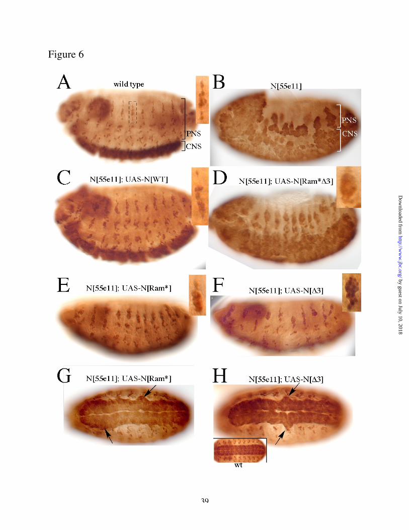
Both Su(H) binding regions contribute to Notch activity in vivo
We employed a functional assay of Notch activity in vivo to assess the physiological significance
of the Su(H) binding sites identified biochemically, above. Notch mutant embryos display
severe hyperplasia of the central nervous system (CNS) and peripheral nervous system (PNS)
(Fig 6 A,B), and this phenotype can be rescued almost completely by expression of wild type
Notch throughout the neuroectoderm under control of an appropriate GAL4 driver (scabrous-
GAL4; Fig 6 C). We therefore used this driver to express Notch derivatives lacking the Ram
binding site for Su(H), the PPD sites, or both (Fig 6 D-H). Embryos that expressed Notch
by guest on July 10, 2018http://w
ww
.jbc.org/D
ownloaded from
15
proteins lacking either Su(H) binding region alone showed residual neural hyperplasia in the
rescued embryos (Fig 6 E-H). Since the mutant proteins are expressed at levels equivalent to or
greater than that of wild type Notch (Fig 5 and data not shown), this demonstrates that the
modified proteins had reduced activity relative to wild type Notch . A Notch derivative lacking
both Su(H) binding regions provided only very weak rescue of the mutant phenotype (Fig 6D),
demonstrating that it is severely reduced in Notch activity. The phenotype of embryos rescued
with the double mutant was clearly less severe than that of the original Notch allele, however,
indicating that the multiply mutant gene still retains some activity. These phenotypic data
therefore are consistent with the co-IP experiment of Fig 5, in which we observed residual
association of Su(H) with the multiply mutant Notch protein.
While the data above show that the basic motifs in the PPD region contribute to Notch activity in
vivo, they do not distinguish whether these sequences are required for Su(H) binding or for
nuclear targeting. We therefore prepared embryos expressing mutated Notch derivatives in
alternate segments (driven by hairy-GAL4) and used immunofluorescence to investigate the
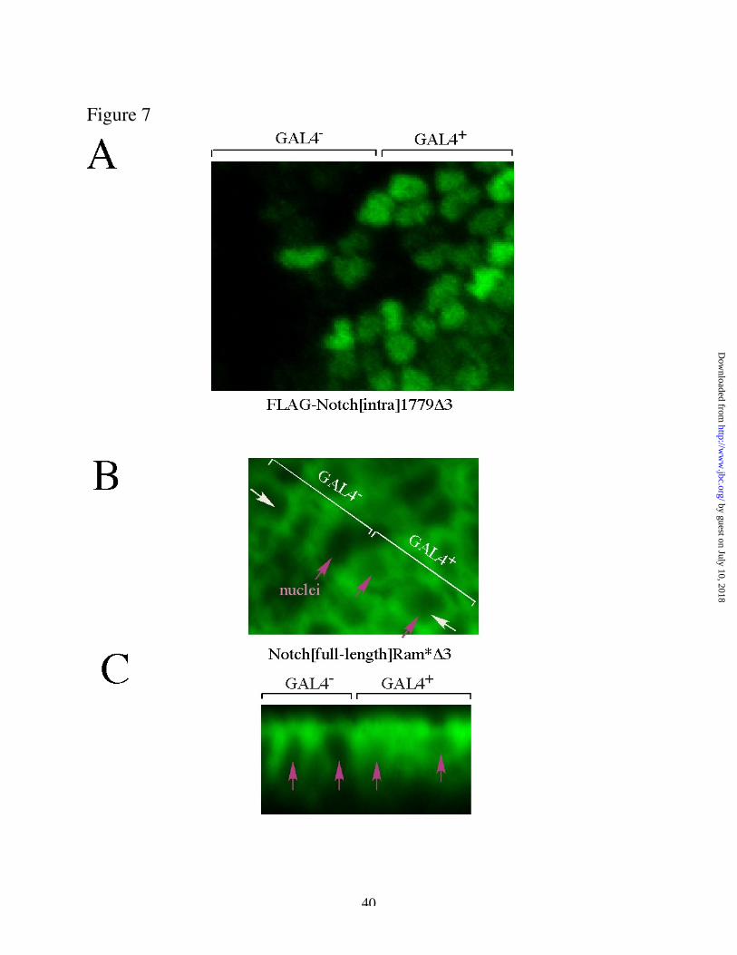
subcellular localization of Notch. Embryos expressing a FLAG-tagged form of the Notch
intracellular domain lacking the PPD Su(H) sites (N[intra1779 D3]) showed clear nuclear
concentration of FLAG immunofluorescence (Fig 7A). We futher verified this result by analysis
of untagged, full-length Notch bearing the D3 deletion (Notch[full-length, Ram*D3]; Fig 7 B, C).
In cells lacking expression of the transgene, endogenous Notch can be clearly detected around
the cell periphery, but consistent with previous studies, the level of protein in the nucleus is
below the limit of detection by immunofluorescence. In cells in which the transgene is
expressed, in contrast, a modest increase is evident in labeling of the cell periphery, and labeling
by guest on July 10, 2018http://w
ww
.jbc.org/D
ownloaded from
16
is now also detectable in the center of the cell, in the position of the nucleus (Fig 7B).
Examination of a cross section of this sample (Fig 7C) further supports the interpretation that this
represents staining of the cell nucleus. In some experiments, this was verified by double-
labelling with an authentic nucelar marker (anti-Lola; data not shown). We infer, therefore, that
the expressed Notch protein lacking the two PPD basic motifs is present in the nucleus in these
embryos.
by guest on July 10, 2018http://w
ww
.jbc.org/D
ownloaded from
17
Discussion
We have shown in this study that Su(H) binds to Drosophila Nicd through two separate binding
regions. The first is in the RAM domain and was previously suspected by analogy with
mammalian Notch. The second is bipartite, located downstream of the ankyrin repeats and
overlapping if not identical to the second Notch “nuclear localization signal”. The efficiency of
Su(H) binding to this second region is strongly enhanced by the presence of the ankyrin repeats.
The two binding regions were identified in vitro, suggesting a direct interaction between Su(H)
and Notch, and were confirmed in vivo both biochemically, by co-immunoprecipitation from
Drosophila cell extracts, and functionally, by demonstrating that each contributes to Notch
activity in vivo.
Fortini and Artavanis-Tsakonas were the first to report a direct physical interaction between
Su(H) and Drosophila Notch [24]. Using the yeast 2-hybrid system, they detected an interaction
between Su(H) and a portion of Notch centered on the ANK repeats; the deletion of the ANK
repeats strongly reduced that interaction. Co-localization of transfected Notch and Su(H) in the
nucleus of S2 cells was also dependent on the integrity of the ANK repeats. Surprisingly,
however, Tamura et al. reported compelling evidence based on yeast 2-hybrid experiments and
GST pull-downs showing that CSL bound strongly within the RAM domain of Notch [19], but
only weakly, if at all, with an extended region including the ANK repeats [25]. These data of
Tamura et al. were also difficult to reconcile with earlier genetic data in flies showing a strong
gain of function phenotype of Drosophila Notch intracellular domains missing the RAM Su(H)
binding domain [22, 23]. Subsequent studies in nematodes and vertebrates recapitulate the same
contradictions, indicating a strong binding site within the Ram domain that could not always be
linked with functional activity and variable and inconsistent binding to more C-terminal
sequences, which failed to correlate simply with the activity of truncated derivatives and were
not precisely localized. [15, 21, 27, 35, 36].
by guest on July 10, 2018http://w
ww
.jbc.org/D
ownloaded from
18
Our data now help to reconcile these earlier studies. We confirm in Drosophila the interaction
domain identified by Tamura et al. within the RAM region, both in vitro and in vivo in fly cells.
Moreover, we describe the functional conservation of the three amino acids shown to be essential
to the interaction of mNotch with CBF-1[19]. We also identify a second binding region in the
PPD domain immediately downstream of the 7th ANK repeat. The PPD binding site is bipartite,
with its two elements being redundant in most experimental contexts, explaining why it has
previously been difficult to identify by mutation or deletion. The previously described
requirement for the ANK repeats in CSL-dependent Notch signaling [15, 25, 36] is consistent
with our data showing that the presence of the ANK repeats greatly increases the effectiveness of
Su(H) binding to PPD. Perhaps ANK holds PPD in a conformation that is favorable for Su(H)
binding. We also find that the expression levels of Drosophila Notch derivatives in vivo are
systematically dependent on the ability of those derivatives to interact with Su(H), complicating
the functional mapping of Notch domains unless such experiments are normalized for Notch
expression levels.
Previous studies offer independent support for the interpretation we propose. Notch derivatives
lacking the RAM binding site that were shown to bind CSL in vitro and in vivo and to activate
the CSL pathway retained one or both of the PPD sites (for example [37]; summarized in [9]).
Moreover, Jeffries and Capobianco used both a neoplastic transformation assay and a luciferase
reporter assay to show for human Notch1 the importance of what we are calling the PPD domain:
the minimal transformation domain (TFD) they identified in human Notch1 corresponds to the
ANK repeats together with the PPD domain we define in Drosophila Notch, and a deletion that
removed only a portion of the PPD domain gave an intermediate level of activity in their
transformation assay [38]. Finally, Oswald, et al [39] characterized transcriptional activation by
a series of mouse Notch1 intracellular domain derivatives with C-terminal truncations that ended
in and around the PPD domain. In that study, the shortest derivative that provided full Notch
activity deleted one of the basic peptides in the PPD, but retained the other. A derivative that
was just 22 codons shorter and lacked both of the basic peptides was reduced in activity by
~50% compared with wild type Notch, even though both expression level and nuclear
localization of the derivatives tested appeared to be unimpaired in that assay system.
by guest on July 10, 2018http://w
ww
.jbc.org/D
ownloaded from
19
It is striking that the Su(H) binding region that we have identified within the PPD domain is the
previously described “double NLS region” of Notch [20, 38]. This region contains two small
basic peptides that resemble nuclear localization signals and are believed to contribute to nuclear
accumulation of Nicd. We find that these same peptides are essential for in vitro binding of
Su(H). While our data do not exclude the possibility that those residues may also provide a
classical NLS signal, we find that deletion of these sequences does not prevent nuclear entry of
Notch, in agreement with previous data from mouse Notch [39, 40]. We note that it has not been
demonstrated whether these basic peptides promote nuclear localization by direct interaction
with the nuclear targeting machinery or by some other mechanism. For example, they could in
principle promote nuclear retention by allowing interaction with a nuclear protein like Su(H).
Our data, however, provide a plausible rationale for the strong conservation of the “double NLS”
motif even though it is not essential for nuclear targetting of Nicd.
We note that we still observed residual co-immunoprecipitation of Su(H) with Notch derivatives
deleted for both the Ram and PPD binding sites (Fig 5 B,C), even though those mutations
abolished binding in vitro (Fig 4 B). While we cannot fully exclude the possibility that the
residual interaction reflects nonspecific background in the assay, an alternative possibility
consistent with our functional data is that Su(H) is capable of associating with Notch indirectly,
for example, through a “bridging” protein that itself provides the direct contact to Notch. Such a
model has been proposed previously for Notch orthologs in nematodes and mammals. Roehl et
al. provided compelling data that just the ankyrin repeats of GLP-1 by themselves have
substantial Notch pathway activity [27, 35]. Since they observed colocalisation of GLP-1 and the
CSL homologue Lag-1 in vivo but did not observe direct binding in vitro, they postulated that
some other protein(s) might provide a link between them, perhaps the Mastermind (Mam)
homologue Lag-3 [41]. Similarly, in vertebrates, it has been proposed that the presence of Mam
may stabilize or induce interaction of Notch with CBF-1 independently of the RAM domain
[42], mediated perhaps by interaction of Mam with both CBF-1 and the Notch ankyrin repeats
[26, 28]. Our evidence that the presence of the ANK repeats enhances binding of Notch to the
PPD site in vitro suggests that ANK may also play a more direct role promoting Notch(PPD) and
Su(H) association, while the co-immunoprecipitation data supports the idea that the importance
by guest on July 10, 2018http://w
ww
.jbc.org/D
ownloaded from
20
of the ANK repeats in signaling is reinforced by their role in also mediating an indirect
association through Mam, and perhaps other proteins.
In summary, our biochemical characterization of the binding of Su(H) to two regions within the
Notch intracellular domain clarify the molecular role of the Notch ANK repeats, suggest an
additional function for the two conserved nuclear localization sequences just downstream of
those repeats and help to explain the phenotypes of a number of Notch mutations and Notch
derivatives from a wide variety of experimental systems.
AcknowledgementsWe are grateful to D. Crowner and L. Luke for technical assistance, and to L. Arnaud and all the
members of our laboratory for useful discussions. We thank D. Barrick, S. Carlson, W. Carter, T.Reh and V. Vasioukhin for critically reading the manuscript. We also thank C.S. Wesley, F.
Schweisguth, J. Posakony and R. Kostriken for providing DNA constructs. MLG was supported,
in part, by a fellowship from the Association pour la Recherche Contre le Cancer. Theseexperiments were supported by NIH grant GM 57830.
by guest on July 10, 2018http://w
ww
.jbc.org/D
ownloaded from
21
References
1. Artavanis-Tsakonas, S., M.D. Rand, and R.J. Lake, Notch signaling: cell fate control and
signal integration in development. Science, 1999. 284(5415): p. 770-6.
2. Lai, E.C., Notch cleavage: Nicastrin helps Presenilin make the final cut. Curr Biol, 2002.
12(6): p. R200-2.
3. Guo, M., L.Y. Jan, and Y.N. Jan, Control of daughter cell fates during asymmetric
division: interaction of Numb and Notch. Neuron, 1996. 17(1): p. 27-41.
4. Berdnik, D., T. Torok, M. Gonzalez-Gaitan, and J.A. Knoblich, The endocytic protein
alpha-Adaptin is required for numb-mediated asymmetric cell division in Drosophila.
Dev Cell, 2002. 3(2): p. 221-31.
5. McGill, M.A. and C.J. McGlade, Mammalian numb proteins promote Notch1 receptor
ubiquitination and degradation of the Notch1 intracellular domain. J Biol Chem, 2003.
278(25): p. 23196-203.
6. Zweifel, M.E. and D. Barrick, Studies of the ankyrin repeats of the Drosophila
melanogaster Notch receptor. 1. Solution conformational and hydrodynamic properties.
Biochemistry, 2001. 40(48): p. 14344-56.
7. Zweifel, M.E. and D. Barrick, Studies of the ankyrin repeats of the Drosophila
melanogaster Notch receptor. 2. Solution stability and cooperativity of unfolding.
Biochemistry, 2001. 40(48): p. 14357-67.
8. Zweifel, M.E., D.J. Leahy, F.M. Hughson, and D. Barrick, Structure and stability of the
ankyrin domain of the Drosophila Notch receptor. Protein Sci, 2003. 12(11): p. 2622-32.
by guest on July 10, 2018http://w
ww
.jbc.org/D
ownloaded from
22
9. Lubman, O.Y., S.V. Korolev, and R. Kopan, Anchoring notch genetics and biochemistry;
structural analysis of the ankyrin domain sheds light on existing data. Mol Cell, 2004.
13(5): p. 619-26.
10. Matsuno, K., M. Ito, K. Hori, F. Miyashita, S. Suzuki, N. Kishi, S. Artavanis-Tsakonas,
and H. Okano, Involvement of a proline-rich motif and RING-H2 finger of Deltex in the
regulation of Notch signaling. Development, 2002. 129(4): p. 1049-59.
11. Matsuno, K., M.J. Go, X. Sun, D.S. Eastman, and S. Artavanis-Tsakonas, Suppressor of
Hairless-independent events in Notch signaling imply novel pathway elements.
Development, 1997. 124(21): p. 4265-73.
12. Matsuno, K., R.J. Diederich, M.J. Go, C.M. Blaumueller, and S. Artavanis-Tsakonas,
Deltex acts as a positive regulator of Notch signaling through interactions with the Notch
ankyrin repeats. Development, 1995. 121(8): p. 2633-44.
13. Foltz, D.R., M.C. Santiago, B.E. Berechid, and J.S. Nye, Glycogen synthase kinase-3beta
modulates notch signaling and stability. Curr Biol, 2002. 12(12): p. 1006-11.
14. Foltz, D.R. and J.S. Nye, Hyperphosphorylation and association with RBP of the
intracellular domain of Notch1. Biochem Biophys Res Commun, 2001. 286(3): p. 484-
92.
15. Kurooka, H., K. Kuroda, and T. Honjo, Roles of the ankyrin repeats and C-terminal
region of the mouse notch1 intracellular region. Nucleic Acids Res, 1998. 26(23): p.
5448-55.
16. Kidd, S., T. Lieber, and M.W. Young, Ligand-induced cleavage and regulation of
nuclear entry of Notch in Drosophila melanogaster embryos. Genes Dev, 1998. 12(23):
p. 3728-40.
by guest on July 10, 2018http://w
ww
.jbc.org/D
ownloaded from
23
17. Rechsteiner, M., Regulation of enzyme levels by proteolysis: the role of pest regions. Adv
Enzyme Regul, 1988. 27: p. 135-51.
18. Lai, E.C., Protein degradation: four E3s for the notch pathway. Curr Biol, 2002. 12(2): p.
R74-8.
19. Tamura, K., Y. Taniguchi, S. Minoguchi, T. Sakai, T. Tun, T. Furukawa, and T. Honjo,
Physical interaction between a novel domain of the receptor Notch and the transcription
factor RBP-J kappa/Su(H). Curr Biol, 1995. 5(12): p. 1416-23.
20. Aster, J.C., E.S. Robertson, R.P. Hasserjian, J.R. Turner, E. Kieff, and J. Sklar,
Oncogenic forms of NOTCH1 lacking either the primary binding site for RBP-Jkappa or
nuclear localization sequences retain the ability to associate with RBP-Jkappa and
activate transcription. J Biol Chem, 1997. 272(17): p. 11336-43.
21. Nofziger, D., A. Miyamoto, K.M. Lyons, and G. Weinmaster, Notch signaling imposes
two distinct blocks in the differentiation of C2C12 myoblasts. Development, 1999.
126(8): p. 1689-702.
22. Fortini, M.E., I. Rebay, L.A. Caron, and S. Artavanis-Tsakonas, An activated Notch
receptor blocks cell-fate commitment in the developing Drosophila eye. Nature, 1993.
365(6446): p. 555-7.
23. Lieber, T., S. Kidd, E. Alcamo, V. Corbin, and M.W. Young, Antineurogenic phenotypes
induced by truncated Notch proteins indicate a role in signal transduction and may point
to a novel function for Notch in nuclei. Genes Dev, 1993. 7(10): p. 1949-65.
24. Fortini, M.E. and S. Artavanis-Tsakonas, The suppressor of hairless protein participates
in notch receptor signaling. Cell, 1994. 79(2): p. 273-82.
by guest on July 10, 2018http://w
ww
.jbc.org/D
ownloaded from
24
25. Tani, S., H. Kurooka, T. Aoki, N. Hashimoto, and T. Honjo, The N- and C-terminal
regions of RBP-J interact with the ankyrin repeats of Notch1 RAMIC to activate
transcription. Nucleic Acids Res, 2001. 29(6): p. 1373-80.
26. Nam, Y., A.P. Weng, J.C. Aster, and S.C. Blacklow, Structural requirements for
assembly of the CSL.intracellular Notch1.Mastermind-like 1 transcriptional activation
complex. J Biol Chem, 2003. 278(23): p. 21232-9.
27. Roehl, H., M. Bosenberg, R. Blelloch, and J. Kimble, Roles of the RAM and ANK
domains in signaling by the C. elegans GLP-1 receptor. Embo J, 1996. 15(24): p. 7002-
12.
28. Wu, L., J.C. Aster, S.C. Blacklow, R. Lake, S. Artavanis-Tsakonas, and J.D. Griffin,
MAML1, a human homologue of Drosophila mastermind, is a transcriptional co-
activator for NOTCH receptors. Nat Genet, 2000. 26(4): p. 484-9.
29. Bailey, A.M. and J.W. Posakony, Suppressor of hairless directly activates transcription
of enhancer of split complex genes in response to Notch receptor activity. Genes Dev,
1995. 9(21): p. 2609-22.
30. Giniger, E., A role for Abl in Notch signaling. Neuron, 1998. 20(4): p. 667-81.
31. Wesley, C.S. and L. Saez, Analysis of notch lacking the carboxyl terminus identified in
Drosophila embryos. J Cell Biol, 2000. 149(3): p. 683-96.
32. Schweisguth, F. and J.W. Posakony, Suppressor of Hairless, the Drosophila homolog of
the mouse recombination signal-binding protein gene, controls sensory organ cell fates.
Cell, 1992. 69(7): p. 1199-212.
33. Fischer, J.A., E. Giniger, T. Maniatis, and M. Ptashne, GAL4 activates transcription in
Drosophila. Nature, 1988. 332: p. 853-856.
by guest on July 10, 2018http://w
ww
.jbc.org/D
ownloaded from
25
34. Brand, A.H. and N. Perrimon, Targeted gene expression as a means of altering cell fates
and generating dominant phenotypes. Development, 1993. 118: p. 401-415.
35. Roehl, H. and J. Kimble, Control of cell fate in C. elegans by a GLP-1 peptide consisting
primarily of ankyrin repeats. Nature, 1993. 364(6438): p. 632-5.
36. Aster, J.C., L. Xu, F.G. Karnell, V. Patriub, J.C. Pui, and W.S. Pear, Essential roles for
ankyrin repeat and transactivation domains in induction of T-cell leukemia by notch1.
Mol Cell Biol, 2000. 20(20): p. 7505-15.
37. Kato, H., Y. Taniguchi, H. Kurooka, S. Minoguchi, T. Sakai, S. Nomura-Okazaki, K.
Tamura, and T. Honjo, Involvement of RBP-J in biological functions of mouse Notch1
and its derivatives. Development, 1997. 124(20): p. 4133-41.
38. Jeffries, S. and A.J. Capobianco, Neoplastic transformation by Notch requires nuclear
localization. Mol Cell Biol, 2000. 20(11): p. 3928-41.
39. Oswald, F., B. Tauber, T. Dobner, S. Bourteele, U. Kostezka, G. Adler, S. Liptay, and
R.M. Schmid, p300 acts as a transcriptional coactivator for mammalian Notch-1. Mol
Cell Biol, 2001. 21(22): p. 7761-74.
40. Kopan, R., J.S. Nye, and H. Weintraub, The intracellular domain of mouse Notch: a
constitutively activated repressor of myogenesis directed at the basic helix-loop-helix
region of MyoD. Development, 1994. 120(9): p. 2385-96.
41. Petcherski, A.G. and J. Kimble, LAG-3 is a putative transcriptional activator in the C.
elegans Notch pathway. Nature, 2000. 405(6784): p. 364-8.
42. Kitagawa, M., T. Oyama, T. Kawashima, B. Yedvobnick, A. Kumar, K. Matsuno, and K.
Harigaya, A human protein with sequence similarity to Drosophila mastermind
by guest on July 10, 2018http://w
ww
.jbc.org/D
ownloaded from
26
coordinates the nuclear form of notch and a CSL protein to build a transcriptional
activator complex on target promoters. Mol Cell Biol, 2001. 21(13): p. 4337-46.
by guest on July 10, 2018http://w
ww
.jbc.org/D
ownloaded from
27
Footnotes
1: Abbreviations are as follows: CSL: CBF1/Suppressor of Hairless/Lag1; Su(H): Suppressor of
Hairless protein. Nicd: intracellular domain of Notch; NLS: nuclear localisation signal, PTB:
phosphorylation binding domain EGF repeats: epidermal growth factor-like repeats; LNG: LIN-
12, Notch, GLP-1 motif; TM: transmembrane domain; ANK: ankyrin repeats; PPD: domain
bearing conserved potential phosphorylation motifs; OPA: poly-glutamine repeat-containing
region: PEST: domain rich in proline, aspartate, serine and threonine residues; EGFP: enhanced
green fluorescent protein.
by guest on July 10, 2018http://w
ww
.jbc.org/D
ownloaded from
28
Figure legends
Fig 1: Su(H) interacts with two separate regions of Nicd in vitro.
(A) schematic representation of the different regions of the Notch intracellular domain (with
their corresponding amino acid numbers) in vitro translated in the following binding assays. (B
and C) comparison of the binding of various regions of Notch to GST-Su(H). In each binding
assay, 35S-labeled in vitro translated Notch proteins were mixed with GST-Su(H) glutathione
sepharose beads, bound proteins were eluted in Laemmli buffer and analyzed by SDS-PAGE
followed by fluorography. The input lanes correspond to 10% of the total proteins.
Fig 2: Molecular characterization of the Su(H) binding site in the RAM domain of Notch
(A) schematic representation of the different constructs tested in B. The RAM domain was
subdivided in 4 regions and derivatives deleted for those regions (“D” with corresponding amino
acid numbers) were tested for their binding to GST-Su(H) and to a fusion of GST to the PTB
domain of the Drosophila Disabled protein (GST-DAB(PTB)). Binding was performed as
described for the experiments of Fig 1. The amino acid sequence of the triple point mutation
RAM* is also shown, and its position indicated by a vertical, striped bar. (B) comparison of the
binding of various deletions and mutations in the RAM domain of Notch to GST-Su(H) or GST-
DAB(PTB).
Fig 3: Molecular characterization of the Su(H) binding region within the PPD domain of
Notch
(A) schematic representation of the different PPD derivatives tested in B. All the ANK-PPD
derivatives were obtained by digestion of the coding DNA of ANK-PPD with the indicated
by guest on July 10, 2018http://w
ww
.jbc.org/D
ownloaded from
29
restriction enzymes before 35S-labeled in vitro translation, whereas all the PPD-OPA derivatives
are independent constructs. B, comparison of the binding of the various deletions in ANK-PPD
or PPD-OPA regions to GST-Su(H). Binding was assayed as for the experiments of Fig 1. The
region between amino acid 2201 and 2247 is necessary and sufficient to allow binding of GST-
Su(H) with ANK-PPD or PPD-OPA. Lanes 6-9 are reproduced from an experiment with a rather
longer fluorographic exposure than lanes 1-5, due to the less efficient binding of PPD-OPA as
compared with ANK-PPD. C, schematic representation of the two sets of constructs tested in D.
N1779-2262 is deleted for the Su(H) binding site in the RAM domain (black rectangle), as
compared with N1766-2262. The amino acid sequence encoded by the SfiI–PvuII region is also
indicated, together with the deletions used in D. Note the presence of two blocks of basic
residues in the Su(H)-binding interval. D, comparison of the binding of GST-Su(H) with Notch
derivatives deleted for the putative Su(H) binding sites. Deletion of the Su(H) binding site in the
RAM domain in combination with deletion of both stretches of basics residues strongly reduces
the binding to Su(H).
Fig 4: Su(H) interacts with two separate domains of Nicd in flies
(A) schematic representation of the different constructs used in (B). The complete Notch
intracellular domain and derivatives of it were labeled with three FLAG tags (indicated by open
circles) and expressed in the ectoderm of Drosophila embryos, using the GAL4 driver 69B.
Vertical black bar represents the position of the N-terminal Su(H) binding site; thin, horizontal
lines represent the location of deleted sequences. (B) Binding of Notch derivatives, made in
Drosophila embryos, to GST or GST-Su(H). Bound proteins were eluted in Laemmli buffer and
by guest on July 10, 2018http://w
ww
.jbc.org/D
ownloaded from
30
analyzed by SDS-PAGE followed by Western Blot analysis (WB) with anti-FLAG antibody.
Input lanes represent 5% of total proteins.
Fig 5: Su(H) interacts with two separate domains of Nicd in vivo
(A) Schematic representation of different Notch full-length derivatives expressed in Drosophila
S2 cells. Vertical, striped bar represents the position of the N-terminal Su(H) binding site; thin
horizontal lines depict the locations of sequences that are deleted in particular derivatives. (B)
Co-immunoprecipitation of Su(H)-HA with Notch full-length derivatives in S2 cells. S2 cells,
co-transfected with Su(H)-HA and Notch Full-length derivatives, were lysed and incubated with
anti-Notch antibody (IP), then with rabbit anti-mouse-bound Protein A sepharose.
Immunoprecipitated proteins were eluted in Laemmli Buffer and subjected to SDS-PAGE and
Western Blot (WB) analysis with anti-Notch (top) or anti-HA (bottom). Input lanes represent 1%
of total proteins. (C) Quantification of binding data. Su(H)-HA signal was quantified by
densitometry and normalized for the total amount of immunoprecipitated Notch (light speckles)
or for the amount of Nicd (dark speckles). Data shown are the average of 4 independent
experiments. DA is a deletion of Notch amino acids 1765-1801; Ram* bears a triple point
mutation of the Ram binding site for Su(H); D3 is a deletion of both basic peptides of the PPD;
Ram*D3 bears both the Ram site point mutation and the D3 deletion.
Fig. 6: Su(H) binding sites are required for Notch function in vivo
Embryos of the indicated genotypes were allowed to develop to embryonic stage 16/17, fixed,
stained with anti-Elav antibodies to label all neuronal nuclei and visualized by peroxidase
histochemistry. (A) wild type embryo, (B) Notch[55e11]. Central nervous system (CNS) and
by guest on July 10, 2018http://w
ww
.jbc.org/D
ownloaded from
31
peripheral nervous system (PNS) are indicated. Inset in (A) shows a higher magnification view
of the dorsal sensory neuron cluster of segment A3 (indicated by a dashed box).
(C-H) N[55e11] embryos in which a Notch transgene has been expressed throughout the
neuroectoderm under the control of scabrous-GAL4, as follows:
(C) UAS-wild type Notch transgene; (D) UAS-Notch[Ram*, D3]; (E) UAS-Notch[Ram*] (PNS
view only); (F) UAS-Notch[D3] (PNS only); (G) UAS-Notch[Ram*] (CNS only); (H) UAS-Notch
[D3] (CNS only). (A-F) show lateral views of embryos (anterior to the left; dorsal up). Insets in
(C-F) show the segment A3 dorsal sensory cluster from those embryos. (E, F) are focused on
dorsal and lateral sensory clusters; v’ and ventral clusters are largely out of view. The severity of
the PNS phenotype is not fully evident in (B, D-F) since labelled cells are piled-up in multiple
focal planes. (G, H) ventral views of embryos. Inset in (H) shows the CNS of a wild type embryo
for comparison. Arrows in (G, H) point to bulges in the CNS indicative of neuronal hyperplasia.
Fig. 7: PPD “ NLS” motifs are not essential for nuclear entry in vivo.
Embryos were prepared that express the specified transgenes in alternate segments, under control
of hairy-GAL4. Stage 9-11 embryos were fixed and stained with the indicated antibodies, and
visualized by confocal microscopy. (A) Embryo expressing FLAG-Notch[intra1779D3],
visualized with anti-FLAG. (B) Embryo expressing Notch[full length, RAM* D3], visualized
with anti-Notch. Endogenous Notch does not accumulate in the nucleus to detectable levels, but
Notch immunoreactivity is clearly observed in the position of the nucleus in cells expressing the
transgene. Nuclear positions are indicated with magenta arrows. (C) Cross section of a z-series
of the same embryo in (B), taken at the position of the white arrows.
by guest on July 10, 2018http://w
ww
.jbc.org/D
ownloaded from
32
Table 1 Conservation of the Su(H) binding sites in the Notch family
Identical or conserved basic residues are in bold. The three conserved amino acids of the 1st
Su(H) binding site are highlighted in gray. The bipartite binding region, identified in the PPDdomain in the fly sequence corresponds to the bipartite NLS identified in human Notch1; both ofthem are in boxes.
1st Su(H) binding region in the RAM domain of Notch
D. melanogaster TQRKRAHGVTWFPEGFRAPAAVMSR-RRRDPHGQEMRNLNKQVAMQSQGL. cuprina TQRKRAAGITWFPEGFRTPTINPQR-RRRDPTGQEMRNLNKNPSMACMSA. gambia -NRKRAHGITWFPEGFFTANTNVRRPSRRRPDGQEMRNLNKNPSMVDMVM. musculus RKRRRQHGQLWFPEGFKV--SEASKKKRREPLGEDSVGLKPLKNASDGAH. sapiens RKRRXQHGQLWFPEGFKV--SEASKKKRREXLGEDSVGLKPLKNASDGAG. Gallus -KRRREHGQLWFPEGFKV--TESSKKKRREPLGEDSVGLKPLKNASDGTX. laevis KKRRREHGQLWFPDGFIP--KEPSKKKRRDRLGEDSVGLKPIKNMTDGSC. pyrrhogaster RKRHREHGQLWFPEGFKV--TETNKSKRRPPLGEDSVGLKPLKNSTD--D. rerio RKRKREHGQLWFPEGFKV--NEP-KKKRREPVGEDSVGLKPLKNSDSSC. auratus ARRKREHSTLWFPEGFFLKKETSSNKNRREPVGQDSLGMKRMPKTVEESC. elegans Lin12 RKRRMINASVWMPP-----MENEEKNRKNHQSITSSQHSLLEAS-YDGYC. elegans GPL1 RKRKMVNATVWMPP----MESTNEKGRRNQSNHSSQCSLLDNSAYYHPN
2nd Su(H) binding bipartite binding region in the PPD domain ofNotch
D. melanogaster -SGKQSNQTAKQKAAKKAKLIEGSPDNGLDATGSLRRKASSKKTSAASKKAAL. cuprina ASGKQPSKAAQAKAAKKAKLLEGSPDGLLDNGGSLRRKPSSKKGLGQASKKGA. gambia -SAKQS------KQKKQRQQKMNG-------VAGSMRRKAVAKKPTGAAKKAM. musculus --LKSATQ---GKKARKPSTK--G----LACGSKEAKDLKARRKSS-QDGKGH. sapiens --LKPGVQ---GKKVRKPSSK--G----LACGSKEAKDLKARRKKS-QDGKGG. Gallus --LKPAVQ---GKKARKPSTK--G----LSCNGKDSKDLKARRKKS-QDGKGX. laevis --MKPSVQ---SKKARKPSIK--G-----NGC—KEAKELKARRKKS-QDGKTC. pyrrhogaster --MKPAAQ---GKKARKQSIKSNG------CNGKESKDVKARRKKS-QDGK-D. rerio --IKPSPG--TAKKTRKPGGK--G-----VGGKDSGKDIRTKKKKSG-DGKNC. auratus --LKSTPQ---GKKNRRPGVK--G----IGGQHATSLKDSAKGRNKRLSLDM
by guest on July 10, 2018http://w
ww
.jbc.org/D
ownloaded from
Maude Le Gall and Edward Ginigerintracellular domain of drosophila notch
Identification of two binding regions for the suppressor of hairless protein within the
published online April 29, 2004J. Biol. Chem.
10.1074/jbc.M404589200Access the most updated version of this article at doi:
Alerts:
When a correction for this article is posted•
When this article is cited•
to choose from all of JBC's e-mail alertsClick here
by guest on July 10, 2018http://w
ww
.jbc.org/D
ownloaded from