Embed Size (px)
Citation preview

Life Science Journal 2012;9(4) http://www.lifesciencesite.com
4472
Identification of three fish species of genus Plectorhynchus from the Red Sea by their scale characteristics
Ahmed S. A. Harabawy1, 3; Imam A. A. Mekkawy2, 3 and Ali Alkaladi1
1Biology Department, Faculty of Science, North Jeddah, King Abdulaziz University, Jeddah, Saudi Arabia. 2Biology Department, Faculty of Science, Taif University, Taif, Saudi Arabia.
3Zoology Department, Faculty of Science, Assiut University, Assiut, Egypt. [email protected]
Abstract: Scale characteristics and its detailed structure from different body regions were studied in three Plectorhynchus species in terms of morphometry and Scanning Electron Microscopic techniques. The present work has been suggested to the rarity of taxonomic and biological information about the three Plectorhynchus species, P. gaterinus, P. pictus and P. schotaf from the Red Sea. A wide spectrum of size-free intraspecific and interspecific variations was recorded and documented concerning the overall form of the scales and their morphometrics, scale surface morphology, the primary, secondary and tertiary radii counts, shape of interradial tongues and the first circuli, the interradial circuli, most outer lateral circuli, inner lateral circuli, denticles on circuli, interradial and intercircular grooves, segmentation and granulation pattern of the caudal field and the shape of ctenii. The quantitative scale characters of Plectorhynchus species are subjected to cluster analysis that revealed two main subclusters, the first includes P. gaterinus and P. schotaf, and the second subcluster includes P. pictus. In addition to the intraspecific variations, the highly significant interspecific variations in the morphometric indices also were evident from one body region to another (P<0.01). The pattern of variations reflected by CVA referred to the partial discrimination between the three Plectorhynchus species considered. Among the scale characteristics used to differentiate between Plectorhynchus species are the overall shape of the lateral line canal, the relative position of its anterior and posterior openings and the cantilevered anterior extension of the canal. [Ahmed S. A. Harabawy, Imam A. A. Mekkawy and Ali Alkaladi. Identification of three fish species of genus Plectorhynchus from the Red Sea by their scale characteristics. Life Sci J 2012;9(4):4472-4485]. (ISSN: 1097-8135). http://www.lifesciencesite.com. 673 Keywords: Plectorhynchus, scale morphology, morphometrics, scanning electron microscopy. 1. Introduction
The Red Sea has a unique environment as it is exposed to extreme natural influences, viz. intensive sun radiation, constant hot winds and subsequent high evaporation rate with negligible inflow of rivers. Like other tropical and subtropical seas, the Red Sea is characterized by a rich and highly diversified fauna and flora (Shiekh-Eldin, 1988; Ghisotti, 1995).
Coral reefs provide a home to thousands of species of different flora and fauna and found to be one of the most diverse and productive natural ecosystems. In spite of presence of a large variety of bony fishes living in and around the coral reefs in the Red Sea, so far, little is known about these fishes.
Lepidology, the use of scale morphology and squamation in fish classification, can be dating back to the mid-nineteenth century where the first use of fish scales in taxonomy; in this time the fishes were divided into four groups according to the structure of their scales: Placoidei, Ganoidei, Ctenoidei, and Cycloidei (Jawad, 2005).
The importance of scale structure as a research material in fish taxonomy and fisheries increased with great developments in the field of microscopy. Application of scanning electron
microscopy (SEM) to reveal morphology, ultrastructure and surface ornamentation of fish scales had facilitated its utility to distinguish the taxonomic groups over a continuum ranging from higher taxa to species.
Detailed structure of the fish scale can be helpful in describing the species, nomenclature, phylogeny, sexual dimorphism, age determination, growth studies, population dynamics, stock assessment, past environment experienced by the fish, migration, taxonomy, discriminating between hatchery-reared and wild populations, in understanding the pathology of a fish scale due to pollution of a water body, in addition to use the scales as a biosorbent to remove heavy metals from contaminated waste streams (Mekkawy et al., 2007; Dapar et al., 2012; Ganzon et al., 2012; Prabu et al., 2012).
Many authors have strongly emphasized on the validity of scale morphology and ultrastructural characteristics for fish taxonomy and phylogeny (Lippitsch, 1990, 1993; Mekkawy et al., 1999, 2003, 2006, 2011; Mahmoud et al., 2005; Harabawy et al., 2007; Reza et al., 2009; Esmaeili and Gholami, 2011; Dapar et al., 2012 and Ganzon et al., 2012). In addition to the systematic status, the functional

Life Science Journal 2012;9(4) http://www.lifesciencesite.com
4473
approach of the ultrastructure and superficial ornamentation of teleost scales also attracted the attention of the aforementioned recent authors.
Studies that use the morphometric characteristics of scales from different body regions in fish taxonomy were rare (e.g. Mekkawy et al., 1999, 2003, 2006, 2011; Mahmoud et al., 2005 and Harabawy et al., 2007). Most of these studies aimed to establish a wide range of valuable scale characters that can reflect a well-defined taxonomic status and a well-founded phylogenic tree of fish taxa.
The family Haemulidae (order Perciformes) represents one of the valuable and economical fish families in the Red Sea. Its members (Haemulids or grunts) are bottom-feeding, carnivorous, nocturnal fishes and use the reefs for shelter (Randall, 1992). Taxonomically, Haemulids appear as one of the problematic groups of marine fish families due to the fact that the traditional characters used in identification of fishes are relatively constant among certain species of haemulids. From a taxonomic point of view, the Red Sea region seems to be one of the least studied areas in concern with haemulids.
The present work aimed to screening and documenting the diversity of scale characteristics of three Plectorhynchus species: P. gaterinus, P. pictus and P. schotaf from the Red Sea at Jeddah and to answer the questions that were previously mentioned by Mekkawy et al. (2011) concerning the most useful scales characters for systematic purposes, the validity of the use of scale morphometrics in fish taxonomy, possibility to give an interpretations for the surface scale ornamentation in terms of systematic and functional approaches. In addition, what is the difference in scale characteristics of marine and freshwater fishes? The answers of such questions should present the bases for further work on scale characteristics and their exploitation for phylogenetic investigations for Haemulidae. Among the interesting scale characteristics considered in the present work were the overall form of the scales and their morphometrics, counts of radii, shape of interradial tongues and the first circuli, the interradial circuli, most outer lateral circuli, inner lateral circuli, denticles and grooves among circuli , granulation pattern of caudal field and form of ctenii. Do such characters reflect functional morphological aspects? This is also a question to be answered. 2. Materials and Methods
In the present work, 1160 scales from 27 specimens of three Plectorhynchus species, namely: P. gaterinus (Forsskål, 1775) (150 – 267 mm standard length (SL)), P. pictus (Tortonese, 1936) (200 – 280 mm SL) and P. schotaf (Forsskål, 1775) (188 – 245 mm SL) were examined to elucidate their scale characteristics. These specimens were collected
from the Red Sea at Jeddah, Saudi Arabia through the period from March to April 2012.
The scales were gently removed with fine forceps from the left side of the body from the following positions on the body: 1)- Region A, directly below the anterior part of the dorsal fin (BDFS). 2)- Region B, postoberculum (POS). 3)- Region C, below the lateral line, between the pectoral and pelvic fins (BLLS). 4)- Region D, caudal peduncle directly above the lateral line (CPS). In addition, anterior lateral line scales (ALLS), middle lateral line scales (MLLS) and posterior lateral line scales (PLLS) from caudal peduncle region. Morphometric measurements and radii counts were taken only from the scales of the first four regions (regions A, B, C and D). While scales forming the lateral line were examined to show the lateral line pattern, shape and characters of the lateral line canal.
Scales examined were cleaned by physical careful removing of the adhering tissues debris without damage in the scale surface. Then they were immersed in a solution of 10% ammonia for 24-36 hr to soften adhering tissues and to clean them. Cleaned scales were dried on a filter paper and kept between two glass slides.
Fig. 1 shows the structure of a sectioned scale, types of radii and the morphometric measurements considered. The primary, secondary and tertiary radii were counted to reveal intraspecific and interspecific variations. The morphometric measurements were treated in terms of indices (L1/L, L2/L, L1/L2 and W/L) whereas, L: scale length; L1: rostral field length; L2: caudal field length; W: scale width.
To clarify intraspecific and interspecific variations of three Plectorhynchus species, ANOVA (Design:Species+Region+Species*Region, (R2=0.97-0.99)), canonical variate analysis and clustering analysis were applied on the morphometric indices of scales using SPSS package, release 9.0.0 (1998) with the assumption of homogeneity of variance.
Scanning Electron Microscopy (SEM) was used to study the morphology and microstructures of the scales in the rostral, lateral, and caudal regions. The cleaned and dried scales that are used for Scanning Electron Microscope (SEM) examination were mounted and fixed by sticker tape on a specimen holder and coated with a 30-nm layer of gold. The electron micrographs were produced on GAOL, GSM5400LV, SEM in back scattering mode and on a Stereo Scan Cambridge Mark 2A (15 KV ) in Assiut University Electron Microscope Center, Assiut, Egypt.
Also the shape of overall granulation area in the caudal field was studied according to the shape of rostro-caudal separation line, the posterior rim and the
Life Science Journal 2012;9(4) http://www.lifesciencesite.com
4474
shape of granulation area in the caudal field by using light microscope. 3. Results Scale surface morphology and morphometrics:
All scales of Plectorhynchus species are mainly of the ctenoid and sectioned type (i. e. with well-developed radii) on all parts of the body of the three species investigated. No simple scales (i. e. without or with only weakly developed radii) were recorded. The surface of scales is divided into three distinct fields, anterior or rostral field from the focus to anterior margin, posterior or caudal field from the focus to the posterior margin and lateral fields (Fig. 1). The scales of Plectorhynchus species studied show a characteristic surface ornamentation which in its simplest case consists of ridges (circuli) and grooves, forming nearly circular rings around a center called focus except in the caudal region that have no circuli and alternatively, contains ctenii and granulation segments. The variable scale shape has its effect on the arrangement of the circuli. The regenerated scales (without any ornamentation at least at the central part of the scale) were recorded. The largest scales are recorded in post operculum region (POS) below the lateral line, while the smallest ones are found in caudal peduncle region and on the belly.
In the rostral part of the scale, the circuli are partitioned by deep and narrow grooves that run radialy (radii) between the focus and anterior rim. The radii on the scales of Plectorhynchus can be categorized into three types depending on their origin and end on the scale including: primary, secondary and tertiary (Fig. 1). In comparison to the tertiary radii, the relative number of primary and secondary radii is more in number. Morphometrics and counts of radii:
Table 1 shows the basic statistics of the scale morphometric indices (relative to scale length, L) from four different body regions. This table reveals the interspecific variations among the three Plectorhynchus species considered. Based on the size-free morphometric indices clustering of these species (Fig. 2) refers to their division into two main subclusters, the first includes P. gaterinus and P. schotaf, and the second subcluster includes P. pictus. Highly significant interspecific variations in these indices were evident in diffirent body regions scales (P<0.01) (Table 1).
In each species, Tables 2-4 reveal intraspicific variations in these scale morphometric characteristics of the four body regions considered. From Table 2, one can note that, in P. gaterinus, the indices L1/L and L1/L2 show significant difference between scales of the four body regions (P�0.05). But W/L index revealed highly significant variations (P<0.01). While L2/L index reflected insignificant
variation (P>0.05). Tables 3 and 4 showed that in both P. pictus and P. schotaf respectively, only one index (W/L) revealed highly significant variations (P<0.01) while the rest of indices show insignificant differences (P>0.05) between scales of the four body regions of both.
The pattern of variations reflected by CVA referred to the partial discrimination between the three Plectorhynchus species on CVI only (Fig. 3) due to the discriminating power of L1/L index that has the higher loading value on CVI (Table 5).
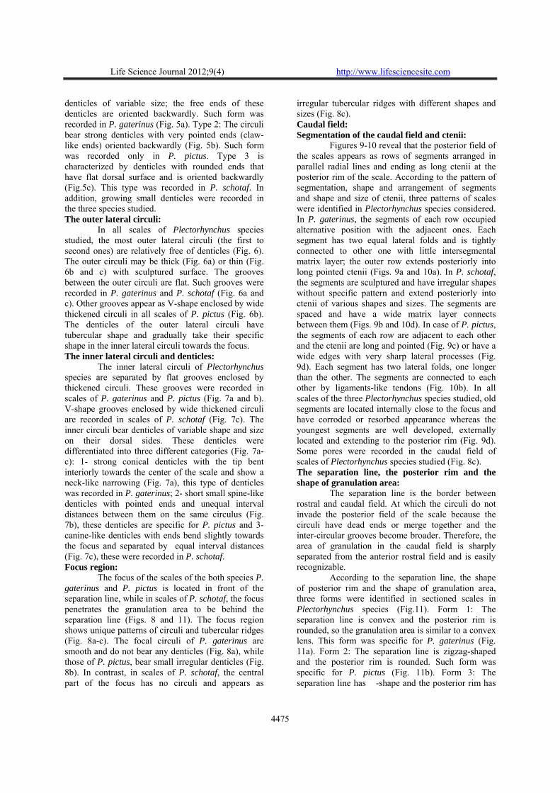
The percentages of occurrence and basic statistics of the primary, secondary and tertiary radii counts are given in Tables 6, 7 and 8. Table 6 shows variations in the primary radii counts among the three Plectorhynchus species studied. These counts are ranged between 2–7, 1-9 and 4-9 in P. gaterinus, P. pictus and P. schotaf respectively. The secondary radii counts varied between 0-7, 0-7 and 0-3 in P. gaterinus, P. pictus and P. schotaf respectively. The tertiary radii ranged between 0-2, 0-5 and 0-2 in P. gaterinus, P. pictus and P. schotaf respectively. Such counts were size free in species considered (P>0.05). The patterns of distribution of such size-free counts revealed significant scale interspecific variations of Plectorhynchus species using G – statistics test (P<0.01). Scanning electron microscope studies: Rostral field:
At the rostral rim of all scales of Plectorhynchus species studied, tongue-like projections are found in the inter-radial space and are free of circuli near the rim (Fig. 4a). On the same scale, such tongues may be convex or straight or both are found; the 1st inter-radial circulus is rounded or relatively straight (Fig. 4a).
The radial grooves in the rostral field of the scales of Plectorhynchus species have two different forms. In the first form, the groove appeared as a deep irregular groove, some strong ridges on each side of the groove are touched and some others are short. This form was recorded in scales of P. gaterinus and P. pictus (Fig. 4b). In the second form, the groove is relatively shallow with narrow split; this form was recorded in P. schotaf (Fig. 4c). Inter-radial circuli, grooves and denticles: The spaces between the circuli are called intercircular spaces (grooves). Such grooves were flat and wide relative to the circulus thickness in most of scales of Plectorhynchus species studied (Fig. 5). The circuli found between radii (in the interradial space) bear small denticles or tooth-like structures that can be seen only under high magnification and are called lepidonts. Three different characteristic types of denticles or lepidonts were identified on the inter-radial circuli (Fig. 5a-c). Type 1: The circuli bear
Life Science Journal 2012;9(4) http://www.lifesciencesite.com
4475
denticles of variable size; the free ends of these denticles are oriented backwardly. Such form was recorded in P. gaterinus (Fig. 5a). Type 2: The circuli bear strong denticles with very pointed ends (claw-like ends) oriented backwardly (Fig. 5b). Such form was recorded only in P. pictus. Type 3 is characterized by denticles with rounded ends that have flat dorsal surface and is oriented backwardly (Fig.5c). This type was recorded in P. schotaf. In addition, growing small denticles were recorded in the three species studied. The outer lateral circuli: In all scales of Plectorhynchus species studied, the most outer lateral circuli (the first to second ones) are relatively free of denticles (Fig. 6). The outer circuli may be thick (Fig. 6a) or thin (Fig. 6b and c) with sculptured surface. The grooves between the outer circuli are flat. Such grooves were recorded in P. gaterinus and P. schotaf (Fig. 6a and c). Other grooves appear as V-shape enclosed by wide thickened circuli in all scales of P. pictus (Fig. 6b). The denticles of the outer lateral circuli have tubercular shape and gradually take their specific shape in the inner lateral circuli towards the focus. The inner lateral circuli and denticles: The inner lateral circuli of Plectorhynchus species are separated by flat grooves enclosed by thickened circuli. These grooves were recorded in scales of P. gaterinus and P. pictus (Fig. 7a and b). V-shape grooves enclosed by wide thickened circuli are recorded in scales of P. schotaf (Fig. 7c). The inner circuli bear denticles of variable shape and size on their dorsal sides. These denticles were differentiated into three different categories (Fig. 7a-c): 1- strong conical denticles with the tip bent interiorly towards the center of the scale and show a neck-like narrowing (Fig. 7a), this type of denticles was recorded in P. gaterinus; 2- short small spine-like denticles with pointed ends and unequal interval distances between them on the same circulus (Fig. 7b), these denticles are specific for P. pictus and 3- canine-like denticles with ends bend slightly towards the focus and separated by equal interval distances (Fig. 7c), these were recorded in P. schotaf. Focus region:
The focus of the scales of the both species P. gaterinus and P. pictus is located in front of the separation line, while in scales of P. schotaf, the focus penetrates the granulation area to be behind the separation line (Figs. 8 and 11). The focus region shows unique patterns of circuli and tubercular ridges (Fig. 8a-c). The focal circuli of P. gaterinus are smooth and do not bear any denticles (Fig. 8a), while those of P. pictus, bear small irregular denticles (Fig. 8b). In contrast, in scales of P. schotaf, the central part of the focus has no circuli and appears as
irregular tubercular ridges with different shapes and sizes (Fig. 8c). Caudal field: Segmentation of the caudal field and ctenii:
Figures 9-10 reveal that the posterior field of the scales appears as rows of segments arranged in parallel radial lines and ending as long ctenii at the posterior rim of the scale. According to the pattern of segmentation, shape and arrangement of segments and shape and size of ctenii, three patterns of scales were identified in Plectorhynchus species considered. In P. gaterinus, the segments of each row occupied alternative position with the adjacent ones. Each segment has two equal lateral folds and is tightly connected to other one with little intersegmental matrix layer; the outer row extends posteriorly into long pointed ctenii (Figs. 9a and 10a). In P. schotaf, the segments are sculptured and have irregular shapes without specific pattern and extend posteriorly into ctenii of various shapes and sizes. The segments are spaced and have a wide matrix layer connects between them (Figs. 9b and 10d). In case of P. pictus, the segments of each row are adjacent to each other and the ctenii are long and pointed (Fig. 9c) or have a wide edges with very sharp lateral processes (Fig. 9d). Each segment has two lateral folds, one longer than the other. The segments are connected to each other by ligaments-like tendons (Fig. 10b). In all scales of the three Plectorhynchus species studied, old segments are located internally close to the focus and have corroded or resorbed appearance whereas the youngest segments are well developed, externally located and extending to the posterior rim (Fig. 9d). Some pores were recorded in the caudal field of scales of Plectorhynchus species studied (Fig. 8c). The separation line, the posterior rim and the shape of granulation area:
The separation line is the border between rostral and caudal field. At which the circuli do not invade the posterior field of the scale because the circuli have dead ends or merge together and the inter-circular grooves become broader. Therefore, the area of granulation in the caudal field is sharply separated from the anterior rostral field and is easily recognizable.
According to the separation line, the shape of posterior rim and the shape of granulation area, three forms were identified in sectioned scales in Plectorhynchus species (Fig.11). Form 1: The separation line is convex and the posterior rim is rounded, so the granulation area is similar to a convex lens. This form was specific for P. gaterinus (Fig. 11a). Form 2: The separation line is zigzag-shaped and the posterior rim is rounded. Such form was specific for P. pictus (Fig. 11b). Form 3: The separation line has �-shape and the posterior rim has

Life Science Journal 2012;9(4) http://www.lifesciencesite.com
4476
V-shape, therefore the granulation area is homboidal in shape, this form was specific for P.
schotaf (Fig. 11c). Lateral line canal:
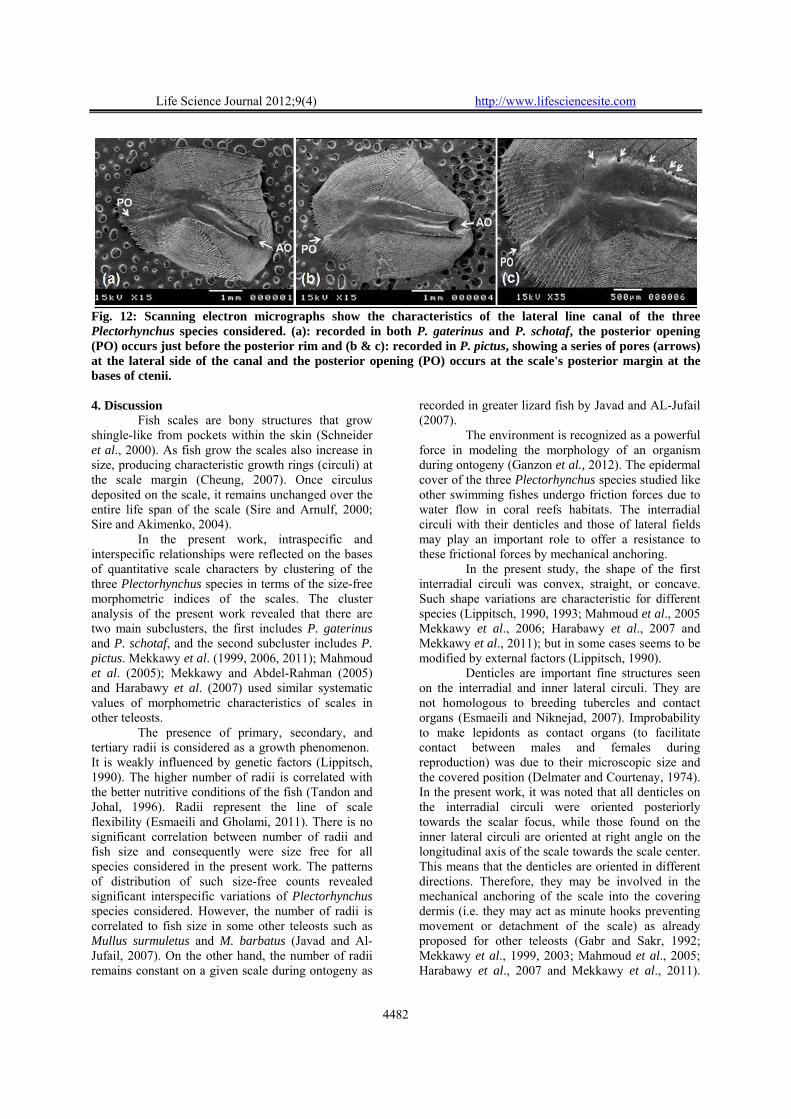
Figure 12 shows the characteristics of the lateral line canal of Plectorhynchus species studied. The lateral line canal is a simple tube embodied within the matrix of the scale. It is divided into a rostral wide tube extend anteriorly and a caudal narrow tube that extend posteriorly to end just before the posterior margin (P. gaterinus and P. schotaf) (Fig. 12a) or reach the posterior margin of the scale (P. pictus) (Fig. 12b and c). The rostral wide tube slightly deviates anteriorly from the anteroposterior axis of the scale (oblique). The anterior opening is
hidden by a cap-like projection cantilevered over it. The anteriorly projecting cap-like extensions are adorned by two forward-projecting spines from the antero-dorsal and antero-ventral corners of the cover. The posterior opening of caudal narrow tube occurs on the inner surface of the caudal region. In P. pictus, a series of pores are found at the lateral side of the wide rostral canal and the posterior opening occurs at the scale’s posterior margin at the bases of ctenii (Fig. 12c), but in P. gaterinus and P. schotaf, no pores are found and the posterior opening occurs just before the posterior margin (Fig. 12a). The length of the lateral-line canals vary along the lateral line and gradually decreases to be just a simple short straight tube in caudal peduncle lateral line scales.
Table 1: Basic statistics, mean ± SD (Range) of the morphometric indices of scales from four body regions of the three Plectorhynchus species from the Red Sea, Jeddah, Saudi Arabia. The postopercular scales (POS), below dorsal-fin scales (BDFS), below lateral line scales (BLLS) and caudal peduncle scales (CPS). Region Index Mean±SD (Range) Mean±SD (Range) Mean±SD (Range)
P. gaterinus (N=40) P. pictus (N=50) P. schotaf (N=45)
Region A (BDFS)
L1/L** 66.7±5.28(57.03–74.07) a 72.03±3.46(62.44–78.98) b 68.52±2.29(63.73–72.43) c L2/L** 33.31±5.3(25.93–42.97) a 27.98±3.47(21.02–37.56) b 31.48±2.27(27.37–36.08) c L1/L2** 205.9±52.74(60.61–285.61) a 262.91±46.22(166.22–375.81) b 219.25±22.89(176.63–264.66) a W/L** 84.92±4.73(77.62–100) a 94.51±7.42(78.68–107.33) b 80.08±7.17(67.02–93.7) c
P. gaterinus (N=40) P. pictus (N=50) P. schotaf (N=45)
Region B
(POS)
L1/L** 66.36±3.1(61.57–76.9) a 71.42±3.68(61.87–79.05) b 69.66±3.07(64.97–77.08) c L2/L** 33.64±3.09(23.1–38.43) a 28.57±3.67(21.17–38.13) b 30.35±3.06(22.92–35.03) c L1/L2** 200.08±31.92(160.2–332.87) a 256.13±49.56(162.28–373.47) b 233.05±36.23(185.5–336.26) c W/L** 98.64±5.02(89.45–108.99) a 109.69±8.18(92.43–131.23) b 93.42±4.6(81.72–104.13) c
P. gaterinus (N=40) P. pictus (N=50) P. schotaf (N=45)
Region C (BLLS)
L1/L** 64.89±4(60.03–72.36) a 71.6±2.6(66.67–77.88) b 69.02±4.24(63.28–81.82) c L2/L** 35.13±3.94(27.92–39.97) a 28.38±2.59(22.14–33.41) b 30.97±4.25(18.18–36.93) c L1/L2** 188.46±34.46(150.21–259.18) a 255.34±34.66(200–350.53) b 229.85±53.54(171.35–450) c W/L** 86.25±6.58(66.61–98.42) a 102.54±8.5(88.84–122.57) b 87.05±4.77(76.86–100) a
P. gaterinus (N=40) P. pictus (N=50) P. schotaf (N=45)
Region D
(CPS)
L1/L** 63.9±4.97(51.74–71.46) a 72.34±2.33(67.42–75.83) b 68.25±3.3(61.97–75.61) c L2/L** 36.13±4.97(28.54–48.26) a 27.71±2.29(24.17–32.58) b 31.77±3.3(24.39–38.03) c L1/L2** 181.91±38.15(107.2–250.41) a 263.48±29.76(206.9–313.7) b 218.35±35.11(162.98–310.08) c W/L** 78.42±6.62(66.67–95.66) a 87.72±5.45(75.13–100) b 75.4±4.02(65.82–82.99) c
**= The differences are highly significant at 0.01 level. Table 2: Basic statistics, mean ± SD (Range) of the morphometric indices of scales from four body regions of P. gaterinus from the Red Sea, Jeddah, Saudi Arabia. The postopercular scales (POS), below dorsal-fin scales (BDFS), below lateral line scales (BLLS) and caudal peduncle scales (CPS). Index Region A (BDFS) Region B (POS) Region C (BLLS) Region D (CPS)
Mean±SD (Range) (N=40) Mean±SD (Range) (N=40) Mean±SD (Range) (N=40) Mean±SD (Range) (N=40)
L1/L* 66.7±5.28 (57.03–74.07) a
66.36±3.1 (61.57–76.9) a
64.89±4 (60.03–72.36) ab
63.9±4.97 (51.74–71.46) b
L2/Ln 33.31±5.30 (25.93–42.97) a
33.64±3.09 (23.1–38.43) a
35.13±3.94 (27.92–39.97) a
36.13±4.97 (28.54–48.26) a
L1/L2* 205.9±52.74 (60.61–285.61) a
200.08±31.92 (160.2–332.87) ab
188.46±34.46 (150.21–259.18) ab
181.91±38.15 (107.2–250.41) b
W/L** 84.92±4.73 (77.62–100) a
98.64±5.02 (89.45–108.99) b
86.25±6.58 (66.61–98.42) a
78.42±6.62 (66.67–95.66) c
n= Insignificant difference at 0.05 level. *= The differences are significant at 0.05 level. **= The differences are highly significant at 0.01 level.

Life Science Journal 2012;9(4) http://www.lifesciencesite.com
4477
Table 3: Basic statistics, mean ± SD (Range) of the morphometric indices of scales from four body regions of P. pictus from the Red Sea, Jeddah, Saudi Arabia. The postopercular scales (POS), below dorsal-fin scales (BDFS), below lateral line scales (BLLS) and caudal peduncle scales (CPS).
Index
Region A (BDFS) Region B (POS) Region C (BLLS) Region D (CPS) Mean±SD (Range) (N=50) Mean±SD (Range) (N=50) Mean±SD (Range) (N=50) Mean±SD (Range) (N=50)
L1/Ln 72.03±3.46 (62.44–78.98) a
71.42±3.68 (61.87–79.05) a
71.6±2.60 (66.67–77.88) a
72.34±2.33 (67.42–75.83) a
L2/Ln 27.98±3.47 (21.02–37.56) a
28.57±3.67 (21.17–38.13) a
28.38±2.59 (22.14–33.41) a
27.71±2.29 (24.17–32.58) a
L1/L2n 262.91±46.22 (166.22–375.81) a
256.13±49.56 (162.28–373.47) a
255.34±34.66 (200–350.53) a
263.48±29.76 (206.9–313.7) a
W/L** 94.51±7.42 (78.68–107.33) a
109.69±8.18 (92.43–131.23) b
102.54±8.50 (88.84–122.57) c
87.72±5.45 (75.13–100) d
n= Insignificant difference at 0.05 level. **= The differences are highly significant at 0.01 level. Table 4: Basic statistics, mean ± SD (Range) of the morphometric indices of scales from four body regions of P. schotaf from the Red Sea, Jeddah, Saudi Arabia. The postopercular scales (POS), below dorsal-fin scales (BDFS), below lateral line scales (BLLS) and caudal peduncle scales (CPS).
Index
Region A (BDFS) Region B (POS) Region C (BLLS) Region D (CPS) Mean±SD (Range) (N=45) Mean±SD (Range) (N=45) Mean±SD (Range) (N=45) Mean±SD (Range) (N=45)
L1/Ln 68.52±2.29 (63.73–72.43) a
69.66±3.07 (64.97–77.08) a
69.02±4.24 (63.28–81.82) a
68.25±3.3 (61.97–75.61) a
L2/Ln 31.48±2.27 (27.37–36.08) a
30.35±3.06 (22.92–35.03) a
30.97±4.25 (18.18–36.93) a
31.77±3.30 (24.39–38.03) a
L1/L2n 219.25±22.89 (176.63–264.66) a
233.05±36.23 (185.5–336.26) a
229.85±53.54 (171.35–450) a
218.35±35.11 (162.98–310.08) a
W/L** 80.08±7.17 (67.02–93.7) a
93.42±4.60 (81.72–104.13) b
87.05±4.77 (76.86–100) c
75.4±4.02 (65.82–82.99) d
n= Insignificant difference at 0.05 level. **= The differences are highly significant at 0.01 level. Table 5: Standardized canonical variates derived from CVA of certain morphometric characters of the scales of the three Plectorhynchus species from the Red Sea, Jeddah, Saudi Arabia.
characters CVI CVII L1/L 0.737 -2.535 L2/L 0.057 0.368 L1/L2 0.036 2.344 W/L 0.642 0.686
Eigenvalue 0.772 0.193 % of Variance 80% 20%
Table 6: Percentages of occurrence and basic statistics of the primary radii counts of the postopercular scales (POS), below dorsal-fin scales (BDFS), below lateral line scales (BLLS) and caudal peduncle scales (CPS) of the three Plectorhynchus species from the Red Sea at Jeddah, Saudi Arabia.
Primary radii counts Species Scales N 1 2 3 4 5 6 7 8 9 Mean±SD
P. gaterinus
BDFS 82 0 0 6.1 20.73 59.76 12.2 1.22 0 0 4.82±0.77 POS 69 0 4.35 17.39 31.88 40.58 4.35 1.45 0 0 4.28±1
BLLS 68 0 1.47 7.35 39.71 44.12 7.35 0 0 0 4.49±0.8 CPS 55 0 0 1.82 16.36 50.91 29.09 1.82 0 0 5.13±0.77
P. pictus
BDFS 90 0 0 1.11 8.89 35.56 38.89 13.33 2.22 0 5.61±0.94 POS 99 2.02 3.03 12.12 27.27 26.26 24.24 5.05 0 0 4.66±1.3
BLLS 96 0 0 5.21 11.46 25 40.63 12.5 3.13 2.08 5.61±1.22 CPS 87 0 0 0 2.3 14.94 41.38 32.18 6.9 2.3 6.33±0.97
P. schotaf
BDFS 79 0 0 0 2.53 39.24 45.57 12.66 0 0 5.68±0.73 POS 69 0 0 0 2.9 24.64 50.72 21.74 0 0 5.91±0.76
BLLS 70 0 0 0 0 27.14 52.86 17.14 1.43 1.43 5.97±0.8 CPS 53 0 0 0 0 26.42 49.06 24.53 0 0 5.98±0.72
Life Science Journal 2012;9(4) http://www.lifesciencesite.com
4478
Table 7: Percentages of occurrence and basic statistics of the secondary radii counts of the postopercular scales (POS), below dorsal-fin scales (BDFS), below lateral line scales (BLLS) and caudal peduncle scales (CPS) of the three Plectorhynchus species from the Red Sea at Jeddah, Saudi Arabia.
Secondary radii counts Species Scales N 0 1 2 3 4 5 6 7 Mean±SD
P. gaterinus
BDFS 81 4.94 18.52 33.33 32.1 11.11 0 0 0 2.26±1.05 POS 69 0 0 4.35 27.54 40.58 17.39 8.7 1.45 4.03±1.06
BLLS 68 0 10.29 20.59 47.06 17.65 4.41 0 0 2.85±0.98 CPS 55 3.64 10.91 45.45 34.55 5.45 0 0 0 2.27±0.87
P. pictus
BDFS 90 77.78 17.78 3.33 1.11 0 0 0 0 0.28±0.58 POS 99 5.05 20.2 22.22 24.24 14.14 10.1 3.03 1.01 2.7±1.55
BLLS 96 22.92 36.46 23.96 7.29 9.38 0 0 0 1.44±1.19 CPS 87 78.16 17.24 4.6 0 0 0 0 0 0.26±0.54
P. schotaf
BDFS 79 81.01 17.72 1.27 0 0 0 0 0 0.2±0.43 POS 69 59.42 28.99 10.14 1.45 0 0 0 0 0.54±0.74
BLLS 70 85.71 12.86 1.43 0 0 0 0 0 0.16±0.4 CPS 53 83.02 13.21 3.77 0 0 0 0 0 0.21±0.49
Table 8: Percentages of occurrence and basic statistics of the tertiary radii counts of the postopercular scales (POS), below dorsal-fin scales (BDFS), below lateral line scales (BLLS) and caudal peduncle scales (CPS) of the three Plectorhynchus species from the Red Sea at Jeddah, Saudi Arabia.
Tertiary radii counts Species Scales N 0 1 2 3 4 5 Mean±SD
P. gaterinus
BDFS 81 70.37 27.16 2.47 0 0 0 0.32±0.52 POS 69 78.26 21.74 0 0 0 0 0.22±0.42
BLLS 68 97.06 2.94 0 0 0 0 0.03±0.17 CPS 55 100 0 0 0 0 0 0±0
P. pictus
BDFS 90 34.44 35.56 21.11 4.44 1.11 3.33 1.12±1.17 POS 99 77.78 17.17 5.05 0 0 0 0.27±0.55
BLLS 96 68.75 19.79 11.46 0 0 0 0.43±0.69 CPS 87 54.02 37.93 5.75 2.3 0 0 0.56±0.71
P. schotaf
BDFS 79 92.41 7.59 0 0 0 0 0.08±0.27 POS 69 81.16 14.49 4.35 0 0 0 0.23±0.52
BLLS 70 94.29 4.29 1.43 0 0 0 0.07±0.31 CPS 53 88.68 7.55 3.77 0 0 0 0.15±0.46
Fig. 1: A diagrammatic structure of Plectorhynchus scale showing the different regions, terms and morphometric measurements used in the present work; L: scale length; L1: rostral field length; L2: caudal field length; W: scale width.

Life Science Journal 2012;9(4) http://www.lifesciencesite.com
4479
Fig. 2 : Clustering (Single Linkage, squared Mahalanobis distance) of three Plectorhynchus species from the Red Sea, Jeddah, Saudi Arabia based on scale morphometric indices. P. gaterinus (PG), P. pictus (PP) and P. schotaf (PS).
Fig. 3: Plots of scores of CVI and CVII derived from CVA carried out on indices of certain morphometric characters of the scales of three Plectorhynchus species from the Red Sea, Jeddah, Saudi Arabia. P. gaterinus (PG), P. pictus (PP) and P. schotaf (PS).
Fig. 4: Scanning electron micrographs show (a): the inter-radial tongues (IRT), 1st inter-radial circulus (1st IRC), inter-radial circuli (IRC) and radii (R) in the rostral field of the scales recorded in the three Plectorhynchus species considered; (b): deep radial grooves (DRG) with irregular split recorded in P. gaterinus and P. pictus and (c): shallow radial grooves (SRG) with very narrow split recorded in P. schotaf only.

Life Science Journal 2012;9(4) http://www.lifesciencesite.com
4480
Fig. 5: Scanning electron micrographs show the inter-radial circuli (C), lepidonts or denticles (D), growing denticles (GD) and grooves (G) of the three Plectorhynchus species considered (a): recorded in P. gaterinus, (b): in P. pictus and (c): in P. schotaf.
Fig. 6: Scanning electron micrographs show the most outer lateral circuli (OLC) and grooves (G) of the three Plectorhynchus species considered. (a): recorded in P. gaterinus, (b): in P. pictus and (c): in P. schotaf.
Fig. 7: Scanning electron micrographs show the inner lateral circuli (C), denticles (D) and grooves (G) of the three Plectorhynchus species considered. (a): recorded in P. gaterinus, (b): in P. pictus and (c): in P. schotaf.
Fig. 8: Scanning electron micrographs show the focus region (F) in the scales of the three Plectorhynchus species considered. (a): the focus with arranged smooth circuli recorded in P. gaterinus, (b): the arranged focal circuli bear small irregular denticles recorded in P. pictus and (c): the focus has no circuli and appears as tubercular ridges with different shapes and sizes in P. schotaf, note the presence of pores (P).

Life Science Journal 2012;9(4) http://www.lifesciencesite.com
4481
Fig. 9: Scanning electron micrographs (a-c) and photomicrograph (d) show the different forms of ctenii (Ct) and granulation segments (Sg) identified in the caudal field of the scales of the three Plectorhynchus species; (a): ctenii recorded in P. gaterinus; (b): in P. schotaf and (c&d): in P. pictus, old segments (OSg) are located internally and have a corroded appearance, the youngest segments (YSg) are well developed and located externally.
Fig. 10: Scanning electron micrographs show the different forms of segments (Sg) identified in the caudal field of the scales of the three Plectorhynchus species; (a): recorded in P. gaterinus, the segments occupied alternative position with the adjacent ones, each segment has two equal lateral folds (LF) and tightly connected to each other , (b): in P. pictus, the segments are adjacent to each other, has one long lateral fold and connected by ligaments-like tendons (LT) and (c): in P. schotaf, irregular sculptured segments are spaced and connected by a wide matrix layer (stars).
Fig. 11: Forms of scales identified in three Plectorhynchus species from the Red Sea, Jeddah, Saudi Arabia; according to the rostro-caudal separation line (SL), the posterior rim (PR) and the shape of granulation area (GA) in the caudal field. (A): specific for P. gaterinus; (B): specific for P. pictus and (C): specific for P. schotaf, note the location of the focus (F).
Life Science Journal 2012;9(4) http://www.lifesciencesite.com
4482
Fig. 12: Scanning electron micrographs show the characteristics of the lateral line canal of the three Plectorhynchus species considered. (a): recorded in both P. gaterinus and P. schotaf, the posterior opening (PO) occurs just before the posterior rim and (b & c): recorded in P. pictus, showing a series of pores (arrows) at the lateral side of the canal and the posterior opening (PO) occurs at the scale's posterior margin at the bases of ctenii. 4. Discussion
Fish scales are bony structures that grow shingle-like from pockets within the skin (Schneider et al., 2000). As fish grow the scales also increase in size, producing characteristic growth rings (circuli) at the scale margin (Cheung, 2007). Once circulus deposited on the scale, it remains unchanged over the entire life span of the scale (Sire and Arnulf, 2000; Sire and Akimenko, 2004).
In the present work, intraspecific and interspecific relationships were reflected on the bases of quantitative scale characters by clustering of the three Plectorhynchus species in terms of the size-free morphometric indices of the scales. The cluster analysis of the present work revealed that there are two main subclusters, the first includes P. gaterinus and P. schotaf, and the second subcluster includes P. pictus. Mekkawy et al. (1999, 2006, 2011); Mahmoud et al. (2005); Mekkawy and Abdel-Rahman (2005) and Harabawy et al. (2007) used similar systematic values of morphometric characteristics of scales in other teleosts.
The presence of primary, secondary, and tertiary radii is considered as a growth phenomenon. It is weakly influenced by genetic factors (Lippitsch, 1990). The higher number of radii is correlated with the better nutritive conditions of the fish (Tandon and Johal, 1996). Radii represent the line of scale flexibility (Esmaeili and Gholami, 2011). There is no significant correlation between number of radii and fish size and consequently were size free for all species considered in the present work. The patterns of distribution of such size-free counts revealed significant interspecific variations of Plectorhynchus species considered. However, the number of radii is correlated to fish size in some other teleosts such as Mullus surmuletus and M. barbatus (Javad and Al-Jufail, 2007). On the other hand, the number of radii remains constant on a given scale during ontogeny as
recorded in greater lizard fish by Javad and AL-Jufail (2007).
The environment is recognized as a powerful force in modeling the morphology of an organism during ontogeny (Ganzon et al., 2012). The epidermal cover of the three Plectorhynchus species studied like other swimming fishes undergo friction forces due to water flow in coral reefs habitats. The interradial circuli with their denticles and those of lateral fields may play an important role to offer a resistance to these frictional forces by mechanical anchoring.
In the present study, the shape of the first interradial circuli was convex, straight, or concave. Such shape variations are characteristic for different species (Lippitsch, 1990, 1993; Mahmoud et al., 2005 Mekkawy et al., 2006; Harabawy et al., 2007 and Mekkawy et al., 2011); but in some cases seems to be modified by external factors (Lippitsch, 1990).
Denticles are important fine structures seen on the interradial and inner lateral circuli. They are not homologous to breeding tubercles and contact organs (Esmaeili and Niknejad, 2007). Improbability to make lepidonts as contact organs (to facilitate contact between males and females during reproduction) was due to their microscopic size and the covered position (Delmater and Courtenay, 1974). In the present work, it was noted that all denticles on the interradial circuli were oriented posteriorly towards the scalar focus, while those found on the inner lateral circuli are oriented at right angle on the longitudinal axis of the scale towards the scale center. This means that the denticles are oriented in different directions. Therefore, they may be involved in the mechanical anchoring of the scale into the covering dermis (i.e. they may act as minute hooks preventing movement or detachment of the scale) as already proposed for other teleosts (Gabr and Sakr, 1992; Mekkawy et al., 1999, 2003; Mahmoud et al., 2005; Harabawy et al., 2007 and Mekkawy et al., 2011).

Life Science Journal 2012;9(4) http://www.lifesciencesite.com
4483
This explains why isolated scales always have a well defined strip of skin adhering to it after removal (Lanzing and Bower, 1973). Jawad (2005) stated that the small-sized processes located on the circuli cannot anchor the scale in the dermis as securely as can the well-developed denticles. On the other hand, Zylberberg and Meunier (1981) proposed that the scale is anchored in the surrounding tissue by the bundles of collagen fibers connecting the upper part of the scale to the overlying dermis.
Concentration of the denticles on the interradial and inner lateral circuli of scales of Plectorhynchus species considered in addition to the absence or rarity of such denticles in the most outer lateral circuli may suggest that the anchoring is more important in the radial part of the rostral field and near the focus and is less important in the outer part of the outer lateral field (the newly formed circuli) as proposed by Mekkawy et al. (2011). Also, as all of the denticles are not oriented to a specific direction, they constitute multidirectional anchoring. These findings agree with those reported by Mekkawy et al. (1999, 2003, and 2011) and Harabawy et al. (2007).
The surface structures of the overlapped region of the scales are stable after their formation (Esmaeili and Gholami, 2011). This stability may suggest that the shape, size and spacing of the circuli and denticles and other surface characteristics may provide useful criteria for systematic purposes. But, this hypothesis requires test and refinement by comparisons of such characteristics in different related species. Concerning this point, a wide spectrum of lepidonts (denticles) of different size and shape in many fish genera and species were recorded by many authors (e.g. Lippitsch, 1990, 1992, 1993; Mekkawy et al., 1999, 2003, 2006; Mahmoud et al., 2005; Harabawy et al., 2007 and Mekkawy et al., 2011). These authors suggested that such denticles might characterize genera and may even distinguish some taxa at the specific level reaching to species level. Inspite of being carnivores inhabiting the bottoms and use the reefs for shelter, Plectorhynchus species are subjected to various environmental factors. Their size-free scale characters and ornamentation also were stable. So, the impacts of such environmental factors were omitted and the size-free fixed scale characters are expressions of their divergent evolution and were not controlled by the environmental factors. Accordingly, the intraspecific and interspecific variations in Plectorhynchus species could be genetically controlled.
The position of the focus on the scale remains the same throughout the life of the individual species (Ganzon et al., 2012). The focus area sculpture was among the fine features of scales studied by SEM. This area may be smooth or have a
granulate sculpture that varies in shape and size between teleosts (Lippitsch, 1992). In the present work the focus of scales has different locations and different shapes among Plectorhynchus species studied. The focus shows unique patterns of circuli and tubercular ridges in P. gaterinus and P. pictus or has no circuli and appears as irregular tubercular ridges with different shapes and sizes in P. schotaf. Lippitsch (1992) believed such focus area to be formed by disintegration of circuli.
Granulation patterns, shape and size of segments and ctenii and overall caudal field of the scales of Plectorhynchus species studied were constant with fish size and exhibited interspecific variations. The large degree of morphological variations exhibited between species and genera of the same family and a complete change in morphology for several species outside the family were evident (Mekkawy et al., 2006; Mahmoud et al., 2005; Harabawy et al., 2007 and Mekkawy et al., 2011). These findings suggest and emphasized on the importance of the caudal field of scales as a taxonomic character not only at the level of species or genera but also at families level (Delamater and Courtenay, 1974; Mekkawy et al., 1999, 2003, 2006; Mahmoud et al., 2005; Harabawy et al., 2007 and Mekkawy et al., 2011). In contrast, Esmaeili and Gholami (2011) stated that the tubercles and ridges of the free caudal region change in shape and increase in surface and thickness during ontogeny. Consequently, the ornamentation patterns are specific to different ontogenetic stages and this should be taken into account in systematic studies.
In the present work, SEM observations revealed that the reduction in length or resorption process of the ctenial spines (ctenii) is not severe and almost stops after the tip of the spine length has been resorbed as recorded by Jawad (2005). Therefore, such resorption process may lead to segmentation of the caudal field of the scales of Plectorhynchus species considered. Hughes (1981) and Roberts (1993) interpreted the mechanism of spine loss as a progressive resorption rather than a sudden amputation. A severe resorption leading to complete disappearance of the spine was recorded in some other teleost fishes (Roberts, 1993). Jawad (2005) reported that the resorption of ctenial spines, as a character, seems to be a characteristic of broader taxonomic groups such as genera and families rather than isolated species within these groups.
The caudal field of the scales of the three Plectorhynchus species was found perforated by a number of pores. A similar finding was recorded by Harabawy (2002), Mahmoud et al. (2005), Mekkawy and Abdel-Rahman (2005) and Mekkawy et al., 2006 and 2011). Harabawy (2002) postulated that such

Life Science Journal 2012;9(4) http://www.lifesciencesite.com
4484
pores may act as minute canaliculi through which the mucus can pass to cover the fish body and other substances can be released into the water when the skin is damaged. The mucus is significantly decreasing the friction between the fish and water and it promotes the healing of wounds (Holčík, 1989). Mahmoud et al. (2005) added that these pores may represent a part of the lateral line system.
The anterior opening of the lateral line canal of Plectorhynchus species studied is hidden by an evelike extension cantilevered over it. Anterior opening is wider than the posterior one. Delamater and Courtenay (1973) reported as a possible working hypothesis that the cantilevered anterior extension of the canal could help in detection of water motion speed and direction. A wide range of structural variation of lateral line canal was recorded in different teleost species by many authors (e.g. Mekkawy, 1980; Khalil et al., 1982; Mekkawy et al., 1999, 2003, 2006; Mahmoud et al., 2005; Harabawy et al., 2007 and Mekkawy et al., 2011). It ranged from a simple direct or slightly oblique perforation to an extended canal with or without simple to highly complex cantilevered extensions acting as covers anterior opening. Also, it may be a complex branched canal (Harabawy et al., 2007). In contrast, no definite structural lateral line canal was recorded in Epinephelus species studied by Mekkawy et al. (1999) or in Cephalopholis species and variola louti studied by Mekkawy et al. (2006). Such canals formed by some type of body scale arrangement to appear as a false lateral line canal.
It is apparent that both gross differences and details of such variation may be important in identification of groups (orders, families, genera and species), especially when combined with other equally impressive characters of scale structure (Delamater and Courtenay, (1972). Delamater and Courtenay (1974), Mekkawy (1980), Khalil et al. (1982) and Mahmoud, et al. (2005) also used the variable position of the anterior and posterior openings of the lateral line canal to differentiate between some fish species belonging to the same genus or different genera. Khalil et al. (1982) and Mahmoud, et al. (2005) recorded some pattern of displacement of the lateral line canal, and in turn their openings on the scales directed toward the caudal end. The latter authors found such pattern of displacement to be valuable in species identification of seven Lethrinus species but not in an obvious way.
In the present work it was noted that the qualitative and quantitative (morphometrics and radii counts) characters of the scales exhibit a lot of species-specific characters for each species of the three Plectorhynchus species studied. This may indicate that such qualitative and quantitative scale
characters are genetically fixed and more stable. In contrast, Mahmoud, et al. (2005) and Harabawy et al. (2007) recorded that such qualitative characters exhibit species-specific characters for each species studied by them than that of morphometric ones. These authors mentioned that this situation may indicate that the qualitative scale characters are genetically fixed and more stable than the quantitative ones.
References 1. Cheung C.H.Y.; Chaillé P.M.; Randall D.J.; Gray
J.S.; Au D.W.T. (2007): The use of scale increment as a means of indicating fish growth and growth impairment. Aquaculture, 266: 102–111.
2. Dapar M.L.G.; Torres M.A.J.; Fabricante P.K. and Demayo C.G. (2012): Scale morphology of the indian goatfish, Parupeneus indicus (Shaw, 1803) (Perciformes: Mullidae). Adv. Environ. Biol., 6(4): 1426-1432.
3. Delamater, E.D. and Courtenay, W.R. (1972): Fish scales as seen by scanning electron microscopy. Florida Sci., 37(3): 141-149.
4. Delamater, E.D. and Courtenay, W.R. (1973): Variations in structure of the lateral line canal on scales of teleostean fishes. Zeifur Morphologie, der Tiere, 75: 259-266.
5. Delamater, E.D. and Courtenay, W.R. (1974): Fish scales as seen by Scanning Electron Microscopy. Florida Sci., 37: 141-149.
6. Esmaeili, H.R. and Gholami, Z. (2011): Scanning Electron Microscopy of the scale morphology in Cyprinid fish, Rutilus frisii kutum Kamenskii, 1901 (Actinopterygii: Cyprinidae). Iranian J. Fisher. Sci., 10(1): 155-166.
7. Esmaeili, H.R. and Niknejad, V. (2007): Scale morphology of 4 loaches from Fars Province. J. Sci., 19 (2): 1-10.
8. Gabr, S.A and Sakr, S.A. (1992): A Scanning Electron Microscopic Study on the Scales of Tilapia nilotica. J. Egypt. Ger. Soc. Zool., 7(c): 41-48.
9. Ganzon M.A.M.; Torres M.A.J.; Gorospe J.J. and Demayo C.G. (2012): Variations in scale morphology between sexes of the spotted barb, Puntius binotatus (Valenciennes, 1842) (Actinopterygii: Cyprinidae). 2nd International Conference on Environment and BioScience, IPCBEE vol.44 (2012) © (2012) IACSIT Press, Singapore: 80-84.
10. Ghisotti, A. (1995): The Red Sea. Casa Editrice Bonechi-Florence-Haly, 128pp.
11. Harabawy, A.S.A. (2002): Biological and taxonomic studies on some fish species of Genus Lethrinus (Family Lethrinidae) from the Red Sea, Egypt and genus Abramis (Family Syprinidae) from the Baltic Drainage. Ph. D. Thesis, Fac. Sci., Assiut Univ., Egypt.
12. Harabawy, A.S.A.; Mekkawy, I.A.A.; Mahmoud, U.M. (2007): A comparative study of scale characteristics and their functional morphology of four goatfishes (Family Mullidae) from the Red Sea. J. Egypt. Soc. Biotech. Environ. Sci., 9(A): 123-163.

Life Science Journal 2012;9(4) http://www.lifesciencesite.com
4485
13. Holčík, J. (Ed.). (1989): The Freshwater Fishes of Europe. Volume 1, Part II. General Introduction to Fishes. Acipenseriformes. AULA-Verlag, Wiesbaden, 469 pp.
14. Hughes, D.R. (1981): Development and organization of the posterior field of ctenoid scales in the Platycephalidae. Copeia, 1981(3): 596-606.
15. Javad, L.A. and AL-Jufaili, S.M. (2007): Scale morphology of grater lizardfish Saurida tumbil (Bloch, 1795) (Pisces: Synodontidae). J. Fish Biol., 70: 1185-1212.
16. Jawad L.A. (2005): Comparative morphology of scales of four teleost fishes from Sudan and Yemen. J. Natur. Hist., 39(28): 2643–2660.
17. Khalil, A.; Yoakim, E.G. and Mekkawy, I.A.A. (1982): Identification of two Nile fish species of the genus Alestes by scale characteristics. Bull. Fac. Sci. Assiut Univ., 11(1-C): 185-207.
18. Lanzing, W.J.R. and Bower, C.C. (1973): Development of colour patterns in relation to behavior in Tilapia mossambica (Peter). J. Fish. Biol., 6: 29-41.
19. Lippitsch, E. (1990): Scale morphology and squamation patterns in Cichlids (Teleostei, Perciformes): A comparative study. J. Fish Biol., 37: 265-291.
20. Lippitsch, E. (1992): Squamation and scale character stability in Cichlids, examined in Sarotherodon galilaeus (Linnaeus, 1758) (Perciformes, Cichlidae). J. Fish Biol., 41: 355-362.
21. Lippitsch, E. (1993): A phyletic study on lacustrine haplochromine fishes (Perciformes, Cichlidae) of East Africa, based on scale and squamation characters. J. Fish Biol., 42: 903-946.
22. Mahmoud, U.M.; Mekkawy, I.A.A. and Harabawy, A.S.A. (2005): Scale characteristics of seven species of genus Lethrinus (Family Lethrinidae) from the Red Sea, Egypt. J. Zool., 44: 545-580.
23. Mekkawy, I.A.A. (1980): Taxonomic studies on the Nile fishes, family Characidae. Unpublished M. Sc. Thesis, Assiut Univ. Egypt.
24. Mekkawy, I.A.A. and Abdel-Rahman, G.H. (2005): Comparative study of scales of five species of parrotfishes (Family Scaridae) from the Red Sea, Hurghada, Egypt with emphasis on their functional morphology. Egypt. J. Zool., 44: 421-443.
25. Mekkawy, I.A.A.; Harabawy, A.S.A.; Adam, E.A. and Ibrahim, M.A. (2007): Biology and population dynamics of Lates niloticus (Linnaeus 1758) (Family Centropomidae) from Lake Nasser, Egypt. J. Egypt. Soc. Biotech. Environ. Sci., 10(A): 191-234.
26. Mekkawy, I.A.A; Mahmoud, U.M. and Harabawy, A.S.A. (2003): Identification of four Labeo fish species from the Nile, Egypt by their scale characteristics and scanning electron microscopy. J. Union Arab Biol. Cairo. 19(A): 81-104.
27. Mekkawy, I.A.A; Mahmoud, U.M.; Khidr, B.M. and Mohammad, A.S. (2006): Scale characteristics of three species of grouper fishes (family: Serranidae, subfamily: Epinephelinae) from the Red Sea, Egypt. J. Union Arab Biol. Cairo, 26(A): 65-100.
28. Mekkawy, I.A.A; Shehata, S.M.A; Saber, S.A. and Osman, A.G.M. (1999): Scale characteristics of five species of genus Epinephelus (Family: Serranidae) from the Red Sea, Egypt. J. Egypt. Ger. Soc. Zool., 30(B): 71-102.
29. Mekkawy I.A.A.; Wassif E.T. and Basmidi A.A.M., (2011): Scale characteristics of three Lutjanus species (Family: Lutjanidae) from the Red Sea. Egypt. J. Fisher. Aqu. Sci., 6: 506-522.
30. Prabu, K.; Shankarlal, S. and Natarajan, E. (2012): A Biosorption of Heavy Metal Ions from Aqueous Solutions Using Fish Scale (Catla catla). World Journal of Fish and Marine Sciences, 4 (1): 73-77.
31. Randall, J.E. (1992): Red Sea Reef Fishes. IMMEL Publishing, London. 192 pp.
32. Reza, E.H.; Somayeh B.; Halimeh, Z. and Fatemeh, S. (2009): Scale morphology of tank goby Glossogobius giuris (Hamilton-Buchanan, 1822) (Perciformes: Goblidae) using scanning electron microscope. J. Biol. Sciences, 9: 899-903.
33. Roberts, C.D. (1993): Comparative morphology of spined scales and their phylogenetic significance in the Teleostei. Bull. Mar. Sci., 52: 60-113.
34. Schneider, J.C.; Laarman, P.W. and Gowing, H. (2000): Age and growth methods and state averages. In: Schneider, J.C. (Ed.), Manual of Fisheries Survey Methods II: With Periodic Updates. Fisheries Special Report, vol. 25. Michigan Department of Natural Resources, Ann Arbor.
35. Shiekh-Eldin, M.Y. (1988): Biological studies on certain marine teleosts. M.Sc. Thesis, Faculty of Sci, Ain Shams Univ.
36. Sire, J.Y. and Akimenko, M. A. (2004): Scale development in fish: a review, with description of sonic hedgehog (shh) expression in the zebrafish (Danio rerio). Int. J. Dev. Biol., 48: 233–247.
37. Sire, J.Y.; Arnulf, I. (2000):. Structure and development of the ctenial spines on the scales of a teleost fish, the cichlid Cichlasoma nigrofasciatum. Acta Zool., 81 (2): 139–158.
38. SPSS, RELEASE 9.0.0, INC. (1998): Statistica for windows, standard version.
39. Tandon, K.K. and Johal, M.S. (1996): Age and Growth in Indian Freshwater Fishes. New Delhi, Narendra Publishing House. 232p.
40. Zylberberg, L. and Meunier, F.J. (1981): Evidence of denticles and attachment fibres in the superficial layer of scale in two fishes: Carassius auratus and Cyprinus carpio (Cyprinidae, Teleostei). J. Zool. Lond., 195: 459-471.
12/1/2012


![Phylogeny [phylo = tribe, family; gen = creation, birth] taxonomy: classifying and naming organisms taxon, taxa (pl.): classification unit (e.g. genus,](https://img.pdfslide.us/doc/110x75/56649d0b5503460f949defc7/phylogeny-phylo-tribe-family-gen-creation-birth-taxonomy-classifying.jpg)