Embed Size (px)
Citation preview

VTT CREATES BUSINESS FROM TECHNOLOGY�Technology�and�market�foresight�•�Strategic�research�•�Product�and�service�development�•�IPR�and�licensing�•�Assessments,�testing,�inspection,�certification�•�Technology�and�innovation�management�•�Technology�partnership
Satu Hilditch
Identification of the fungal catabolic D-galacturonate pathway
Many microorganisms that live on decaying plant material can use D-galacturonate for growth. Eukaryotic catabolic D-galacturonate pathway was not known before. In this thesis work the pathway for D-galacturonate catabolism was identified in the filamentous fungus Trichoderma reesei. The pathway consisted of four enzymes: NADPH- dependent D-galacturonate reductase (GAR1), L-galactonate dehydratase (LGD1), L-threo-3-deoxy-hexulosonate aldolase (LGA1) and NADPH-dependent glycer-aldehyde reductase (GLD1). In this pathway D-galacturonate was converted to pyruvate and glycerol via L-galactonate, L-threo-3-deoxy-hexulosonate and L- glyceraldehyde. The enzyme activities of GAR1, LGD1 and LGA1 were present in crude mycelial extract only when T. reesei was grown on D-galacturonate. The corresponding genes were identified and cloned. They were functionally expressed in Saccharomyces cerevisiae, and the enzymes were characterised. GAR1 and LGA1 catalysed reversible reactions, whereas only the forward reactions were observed for LGD1 and GLD1. When gar1, lgd1 or lga1 was deleted in T. reesei the deletion strain was unable to grow with D-galacturonate as the only carbon source, demonstrating that all the corresponding enzymes were essential for D-galacturonate catabolism and that no alternative D-galacturonate pathway exists in T. reesei.
Dissertation VTT PUBLICATIONS 739
•�•�•��VTT�PUB
LICA
TION
S�739��IDEN
TIFICA
TION
�OF�TH
E�FUN
GA
L�CA
TAB
OLIC
�D-G
ALA
CTU
RO
NA
TE�PATH
WAY�
ISBN 978-951-38-7398-1 (soft back ed.) ISBN 978-951-38-7399-8 (URL: http://www.vtt.fi/publications/index.jsp)ISSN 1235-0621 (soft back ed.) ISSN 1455-0849 (URL: http://www.vtt.fi/publications/index.jsp)


VTT PUBLICATIONS 739
Identification of the fungal catabolic D-galacturonate pathway
Satu Hilditch
Faculty of Biosciences Department of Biological and Environmental Sciences
Division of Biochemistry University of Helsinki
Helsinki, Finland
To be presented with the permission of the Faculty of Biosciences of the University of Helsinki, for public criticism in the Auditorium 2041 at the
Department of Biosciences, Viikki Biocenter, Viikinkaari 5, Helsinki, on June 11th, 2010 at 12 o’clock noon.
2
ISBN 978-951-38-7398-1 (soft back ed.) ISSN 1235-0621 (soft back ed.)
ISBN 978-951-38-7399-8 (URL: http://www.vtt.fi/publications/index.jsp) ISSN 1455-0849 (URL: http://www.vtt.fi/publications/index.jsp)
Copyright © VTT 2010
JULKAISIJA – UTGIVARE – PUBLISHER
VTT, Vuorimiehentie 5, PL 1000, 02044 VTT puh. vaihde 020 722 111, faksi 020 722 4374
VTT, Bergsmansvägen 5, PB 1000, 02044 VTT tel. växel 020 722 111, fax 020 722 4374
VTT Technical Research Centre of Finland, Vuorimiehentie 5, P.O. Box 1000, FI-02044 VTT, Finland phone internat. +358 20 722 111, fax + 358 20 722 4374
Technical editing Leena Ukskoski Text preparing Raija Sahlstedt Edita Prima Oy, Helsinki 2010
3
Satu Hilditch. Identification of the fungal catabolic D-galacturonate pathway. Espoo 2010. VTT Publications 739. 74 p. + app. 38 p.
Keywords Trichoderma reesei, filamentous fungus, D-galacturonate, catabolic pathway, D-galacturonate reductase, L-galactonate dehydratase, L-threo-3-deoxy-hexulosonate aldolase, glyceraldehyde reductase, enzyme activity
Abstract
Pectin is a natural polymer consisting mainly of D-galacturonic acid monomers. Microorganisms living on decaying plant material can use D-galacturonic acid for growth. Although bacterial pathways for D-galacturonate catabolism had been described previously, no eukaryotic pathway for D-galacturonate catabolism was known at the beginning of this work. The aim of this work was to identify such a pathway.
In this thesis the pathway for D-galacturonate catabolism was identified in the filamentous fungus Trichoderma reesei. The pathway consisted of four enzymes: NADPH-dependent D-galacturonate reductase (GAR1), L-galactonate dehydratase (LGD1), L-threo-3-deoxy-hexulosonate aldolase (LGA1) and NADPH-dependent glyceraldehyde reductase (GLD1). In this pathway D-galacturonate was converted to pyruvate and glycerol via L-galactonate, L-threo-3-deoxy-hexulosonate and L-glyceraldehyde.
The enzyme activities of GAR1, LGD1 and LGA1 were present in crude mycelial extract only when T. reesei was grown on D-galacturonate. The activity of GLD1 was equally present on all the tested carbon sources. The corresponding genes were identified either by purifying and sequencing the enzyme or by expressing genes with homology to other similar enzymes in a heterologous host and testing the activities. The new genes that were identified were expressed in Saccharomyces cerevisiae and resulted in active enzymes. The GAR1, LGA1 and GLD1 were also produced in S. cerevisiae as active enzymes with a polyhistidine-tag, and purified and characterised. GAR1 and LGA1 catalysed reversible reactions, whereas only the forward reactions were observed for LGD1 and GLD1. When gar1, lgd1 or lga1 was deleted in T. reesei the deletion strain was unable to grow with D-galacturonate as the only carbon

4
source, demonstrating that all the corresponding enzymes were essential for D-galacturonate catabolism and that no alternative D-galacturonate pathway exists in T. reesei.
A challenge for biotechnology is to convert cheap raw materials to useful and more valuable products. Filamentous fungi are especially useful for the conversion of pectin, since they are efficient producers of pectinases. Identification of the fungal D-galacturonate pathway is of fundamental importance for the utilisation of pectin and its conversion to useful products.
5
Preface
This study was carried out at VTT Biotechnology in the Metabolic Engineering team. The financial support from Maj and Tor Nessling Foundation, Academy of Finland and the University of Helsinki is greatly appreciated.
I am indebted to former Vice President Prof. Juha Ahvenainen, Vice President Prof. Anu Kaukovirta-Norja, Research Professor Hans Söderlund and Technology Manager Dr. Richard Fagerström for providing excellent working facilities. I am grateful to Technology Manager Dr. Tiina Nakari-Setälä, Team Leader Dr. Laura Ruohonen and Research Professor Merja Penttilä for their supportive and encouraging attitude towards this thesis work.
My warmest thanks are due to my supervisor Doc. Peter Richard for being the inexhaustible source of ideas and the driving force of this work. I highly admire his enthusiastic and devoted attitude towards science. His constant support and encouragement in all situations have been invaluable over the years.
I wish to thank my co-authors Janis Liepins, Suvi Berghäll, Dr. Paula Jouhten, Dos. Peter Richard, Dos. Hannu Maaheimo, Dos. John Londesborough, Prof. Merja Penttilä and Dos. Nisse Kalkkinen for their contributions to the research work and writing of the manuscripts. Additional thanks are addressed to John and Peter for their constructive criticism on the thesis manuscript.
Emeritus Prof. Pekka Mäntsälä and Doc. Taina Lundell are warmly thanked for the careful pre-examination of the thesis and for their valuable comments. Michael Bailey is acknowledged for revising the English language.
I sincerely thank everyone working in the yeast/mould lab for the friendly and supportive working atmosphere and for all the help they have readily provided on various matters. It has been a privilege to work with such skilled colleagues. Being part of this awesome group of people has meant a lot to me. From the other labs of VTT Biotechnology I especially want to thank Birgit Hillebrandt-Chellaoui for technical assistance with the protein purification and Eila Leino who often helped me with the enzyme activity measurements. I heartily thank

6
Anne, Anu, Eija, Jari, Laura, Mari, Mervi, Mikko, Outi, Ritva and Virve for the companionship and refreshing discussions over the lunch table.
I wish to thank all my friends and relatives for encouragement along the way. My special loving thanks are due to my parents, Keijo and Riitta, for their wonderful support which I will cherish always. Above all, I want to thank my dearest ones, Kai and Leo, for their understanding and love and for being the most important source of joy and strength to me. Espoo, May 2010 Satu Hilditch
7
Contents
Abstract ................................................................................................................. 3
Preface.................................................................................................................. 5
List of publications................................................................................................. 9
List of symbols .................................................................................................... 10
1. Introduction ................................................................................................... 13 1.1 Pectin and D-galacturonate .............................................................................................. 14 1.2 Pectin degradation and D-galacturonate utilisation .......................................................... 18
1.2.1 Bacterial D-galacturonate pathways................................................................. 19 1.2.2 Metabolism of D-galacturonate in plants and animals ..................................... 23 1.2.3 Earlier observations concerning D-galacturonate catabolism in mould fungi... 25
1.3 Examples of non-phosphorylated catabolic pathways...................................................... 28 1.4 Hypotheses and objectives of the work ............................................................................ 30
2. Materials and methods.................................................................................. 31
3. Results .......................................................................................................... 33 3.1 D-Galacturonate reductase (I) .......................................................................................... 33
3.1.1 Cloning the D-galacturonate reductase............................................................ 33 3.1.2 Characterisation of the D-galacturonate reductase.......................................... 35
3.2 L-Galactonate dehydratase (II) ......................................................................................... 36 3.2.1 Cloning the L-galactonate dehydratase............................................................ 36 3.2.2 Characterisation of the L-galactonate dehydratase ......................................... 37
3.3 L-Threo-3-deoxy-hexulosonate aldolase (IV) ................................................................... 38 3.3.1 Cloning the L-threo-3-deoxy-hexulosonate aldolase........................................ 39 3.3.2 Characterisation of the L-threo-3-deoxy-hexulosonate aldolase...................... 39
3.4 Glyceraldehyde reductase and DHA reductase (III) ......................................................... 40 3.4.1 Cloning the glyceraldehyde reductase and DHA reductase............................. 41 3.4.2 Characterisation of the glyceraldehyde reductase and DHA reductase........... 41
4. Discussion..................................................................................................... 42 4.1 The fungal D-galacturonate pathway................................................................................ 42 4.2 The D-galacturonate reductase ........................................................................................ 46 4.3 The L-galactonate dehydratase ........................................................................................ 47 4.4 The L-threo-3-deoxy-hexulosonate aldolase .................................................................... 54 4.5 The glyceraldehyde reductase and DHA reductase ......................................................... 57 4.6 Biotechnological applications of the D-galacturonate pathway......................................... 62

8
5. Conclusions and future prospects ................................................................ 64
References.......................................................................................................... 66
Appendices
Publications I–IV
Publication I is not included in the PDF version. Please order the printed version to get the complete publication (http://www.vtt.fi/publications/index.jsp).
9
List of publications I Kuorelahti, S., Kalkkinen, N., Penttilä, M., Londesborough, J. and
Richard, P. (2005). Identification in the mold Hypocrea jecorina of the first fungal D-galacturonic acid reductase. Biochemistry 44, 11234–11240.
II Kuorelahti, S., Jouhten, P., Maaheimo, H., Penttilä, M. and Richard, P. (2006). L-Galactonate dehydratase is part of the fungal path for D-galacturonic acid catabolism. Mol. Microbiol. 61, 1060–1068.
III Liepins, J., Kuorelahti, S., Penttilä, M. and Richard, P. (2006). Enzymes for the NADPH-dependent reduction of dihydroxyacetone and D-glyceraldehyde and L-glyceraldehyde in the mould Hypocrea jecorina. FEBS J. 273, 4229–4235.
IV Hilditch, S., Berghäll, S., Kalkkinen, N., Penttilä, M. and Richard, P. (2007). The missing link in the fungal D-galacturonate pathway: identification of the L-threo-3-deoxy-hexulosonate aldolase. J. Biol. Chem. 282, 26195–26201.

1. Introduction
10
List of symbols
DHA dihydroxyacetone
DHAP dihydroxyacetone phosphate
DHDPS dihydrodipicolinate synthase
DNS 3,5-dinitrosalicylate
EC Enzyme Commission
ESI-MS electrospray ionisation mass spectrometry
FucD L-fuconate dehydratase of Xanthomonas campestris
gaaA D-galacturonate reductase-encoding gene of Aspergillus niger
GAAA D-galacturonate reductase of A. niger or Aspergillus nidulans
GAAB L-galactonate dehydratase of A. niger
gaaC 2-keto-3-deoxy-L-galactonate aldolase-encoding gene of A. niger
GAAC 2-keto-3-deoxy-L-galactonate aldolase of A. niger
gaaD glyceraldehyde reductase-encoding gene of A. niger
GAAD glyceraldehyde reductase of A. niger
gar1 D-galacturonate reductase-encoding gene of Trichoderma reesei
GAR1 D-galacturonate reductase of T. reesei
gar2 a putative D-galacturonate reductase-encoding gene of T. reesei
GAR2 a putative D-galacturonate reductase of T. reesei
Gcy1p glyceraldehyde reductase of Saccharomyces cerevisiae
gld1 glyceraldehyde reductase-encoding gene of T. reesei
11
GLD1 glyceraldehyde reductase of T. reesei
Gld1 DHA reductase of Trichoderma atroviride
gld2 DHA reductase-encoding gene of T. reesei
GLD2 DHA reductase of T. reesei
GldB DHA reductase of A. nidulans
HPLC high performance liquid chromatography
KER alkyl 4-halo-3-oxobutyrate reductase of Penicillium citrinum
lga1 L-threo-3-deoxy-hexulosonate aldolase-encoding gene of T. reesei
LGA1 L-threo-3-deoxy-hexulosonate aldolase of T. reesei
lgd1 L-galactonate dehydratase-encoding gene of T. reesei
LGD1 L-galactonate dehydratase of T. reesei
Mbp mega base pairs
MR mandelate racemase of Pseudomonas putida
MS-MS tandem mass spectrometry
NCBI National Center for Biotechnology Information
NMR nuclear magnetic resonance
TBA thiobarbituric acid
TIM triose phosphate isomerase
Ypr1p glyceraldehyde reductase of S. cerevisiae

1. Introduction
12
1. Introduction
13
1. Introduction Mould fungi, yeast fungi and mushrooms are the most commonly known members of the fungal kingdom. Characteristic to fungi is their ability efficiently to degrade organic materials. They secrete a wide range of extracellular enzymes to break down complex substrates into simple components such as sugars and amino acids that can then be taken up by the fungus and further metabolised by numerous specialised pathways for growth and energy. This osmotrophic growth habit has enabled fungi to compete successfully with other organisms and to colonise diverse habitats. For example, they are important in degrading plant debris and in that way recycle carbon and other elements in the environment. On the other hand they also contaminate crops and cause many diseases in both plants and animals.
The filamentous fungus Trichoderma reesei belongs to the subphylum Pezizomycotina of the phylum Ascomycota (NCBI Taxonomy Browser: http://www.ncbi.nlm.nih.gov/Taxonomy/Browser/wwwtax.cgi). It is non-pathogenic and has a long history of being safe to use for enzyme production in industrial scale (Nevalainen et al., 1994). It has an exceptionally efficient protein secretion system and it is one of the most important industrial producers of cellulases and hemicellulases that are used for hydrolysis of plant cell wall polysaccharides. It was one of the first fungi for which the entire genome was sequenced. The seven chromosomes of T. reesei comprise a genome of 34 Mbp. Although T. reesei has the lowest number of cellulase and hemicellulase genes of all sequenced plant cell wall-degrading fungi, it breaks down cellulose and hemicellulose efficiently. It has also very few pectinase genes in its genome and therefore it has been suggested that for pectin degradation T. reesei may largely depend on other fungi and bacteria in the soil (Martinez et al., 2008). T. reesei was originally discovered in the Solomon Islands during World War II, where it was found to be responsible for the rapid decomposition of US Army military
1. Introduction
14
uniforms and cotton canvas tents (Reese, 1976). All T. reesei strains used nowadays in biotechnology and basic research have been derived from that single isolate QM6a (Reese et al., 1950). T. reesei was long believed to propagate only asexually and was shown to be an anamorph derived from the sexual (teleomorphic) ancestor Hypocrea jecorina (Kuhls et al., 1996). Recently, T. reesei QM6a was found to carry only the mating type MAT1-2, whereas some other H. jecorina isolates from different geographical locations contained only the opposite mating type MAT1-1. Heterothallic fungi require both types to be present for sexual reproduction. In mating experiments between strains QM6a and H. jecorina MAT1-1, sexual reproduction could be induced (Seidl et al., 2009).
Pectin is a common biopolymer in nature. Its backbone is a linear chain of D-galacturonate molecules. Many bacterial species are able to utilise D-galacturonate, and some bacterial catabolic pathways are known. Several fungal species, such as T. reesei, are able to grow using D-galacturonate as the only carbon and energy source. It was well established that fungi do not have the same set of enzymes to break down D-galacturonate as bacteria. However, it was not known how eukaryotic species catabolise D-galacturonate. In this thesis the first eukaryotic pathway for D-galacturonate catabolism was identified using T. reesei as the model organism.
1.1 Pectin and D-galacturonate
Pectin is a common component of the primary cell wall and middle lamella of all higher plants. The primary cell wall may contain approximately 35% pectin, 30% cellulose, 25% hemicellulose and 10% structural protein, but the proportions vary greatly depending on the species and cell type (Brownleader et al., 1999). The amounts of pectin in some fruit and vegetable tissues are listed in Table 1.

1. Introduction
15
Table 1. Pectin contents of some fruit and vegetable tissues. The amount of pectin is approximately tenfold higher in the dry matter compared to the fresh substance. Adapted from Jayani et al., 2005; Prasanna et al., 2007.
Fruit/vegetable Tissue Substance Pectin content (%) African Mango Pulp Fresh 0.72 Apple Pulp Fresh 0.5–1.6 Apple Pomace Fresh 1.5–2.5 Avocado Pulp Fresh 0.73 Banana Pulp Fresh 0.7–1.2 Cashew Pomace Fresh 1.28 Cherry Pulp Fresh 0.24–0.54 Guava Pulp Fresh 0.26–1.2 Lemon Pulp Fresh 2.5–4.0 Lemon Peel Fresh 5.0 Litchi Pulp Fresh 0.42 Mango Pulp Fresh 0.66–1.5 Orange Pulp Fresh 1.35 Orange Peel Fresh 3.5–5.5 Papaya Pericarp Fresh 0.66–1.0 Passion fruit Pulp Fresh 0.5 Passion fruit Rind Fresh 2.1–3.0 Pea Pulp Fresh 0.9–1.4 Peach Pulp Fresh 0.1–0.9 Pineapple Pulp Fresh 0.04–0.13 Strawberry Pulp Fresh 0.14–0.7 Tomato Pulp Fresh 0.2–0.6 Carrot Pulp Dry matter 6.9–18.6 Orange Pulp Dry matter 12.4–28.0 Potato Pulp Dry matter 1.8–3.3 Tomato Pulp Dry matter 2.4–4.6 Sugar beet Pulp Dry matter 10.0–30.0
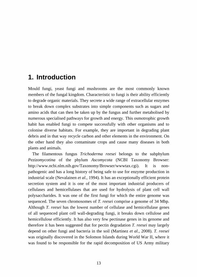
Pectin is a general name for a family of complex, highly heterogeneous polysaccharides with molecular masses ranging from 25 to 360 kDa (Jayani et al., 2005). Homogalacturonan, rhamnogalacturonan I, rhamnogalacturonan II and xylogalacturonan are some of the structural domains that form pectic

1. Introduction
16
polysaccharides and may be covalently joined to each other (Perez et al., 2003). Their structures are presented in Figure 1. Approximately 70% of pectin is composed of D-galacturonic acid residues which are mainly found in the backbone of pectin (Mohnen, 2008).
= 3-deoxy-D-lyxo-heptulosaric acid
= D-Galacturonic acid
= L-Rhamnose
= D-Xylose
= D-Galactose
= L-Galactose
= L-Arabinose
= L-Aceric acid
= D-Apiose
= D-Glucuronic acid
= L-Fucose
= 3-deoxy-D-manno-octulosonic acid
= Methyl
= Acetyl
Homogalacturonan
Rhamnogalacturonan I
Rhamnogalacturonan II
Xylogalacturonan
Arabinogalactanside chain
Arabinanside chain
Figure 1. The structures of homogalacturonan, rhamnogalacturonan I, rhamnogalacturonan II and xylogalacturonan.
In the classical pectin structure long “smooth” regions may occasionally be interrupted by “hairy” regions. The “smooth” regions are composed of homogalacturonan, in which about 100 α-(1->4)-linked D-galacturonic acid residues form linear chains. In some plants the hydroxyl groups on C2 and C3 of the D-galacturonic acid residues may carry acetyl groups (Renard et al., 1995). The carboxyl groups at C6 of D-galacturonic acids can be methyl-esterified, which removes the negative charge and makes the homogalacturonan more hydrophobic. In native pectins the homogalacturonan is highly methyl-esterified,
1. Introduction
17
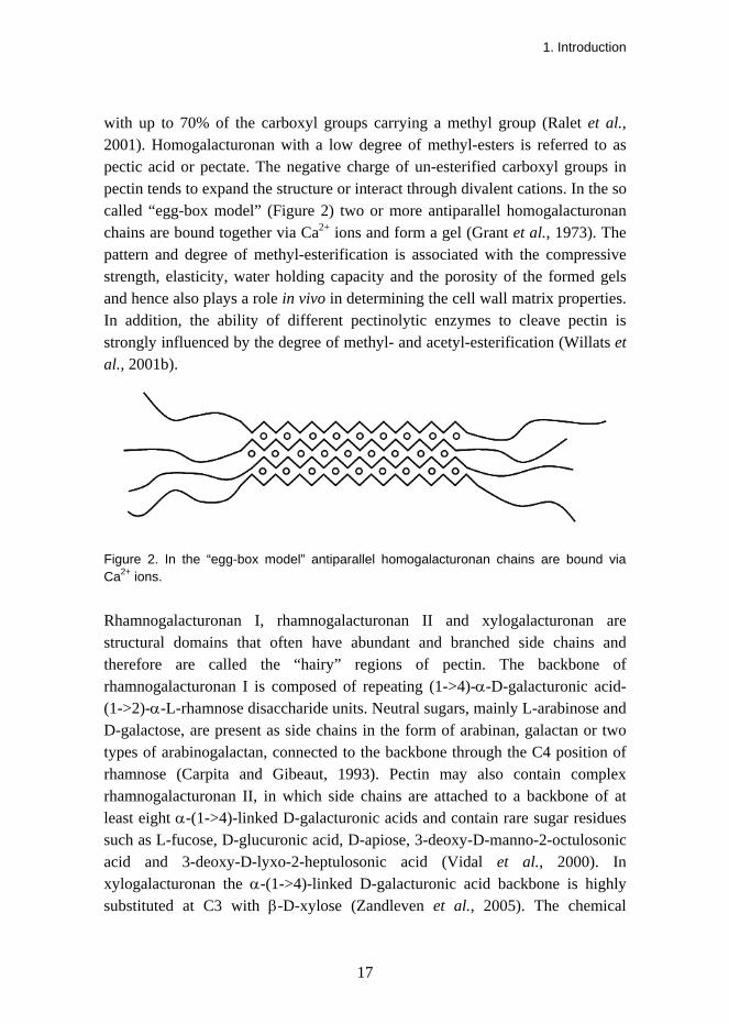
with up to 70% of the carboxyl groups carrying a methyl group (Ralet et al., 2001). Homogalacturonan with a low degree of methyl-esters is referred to as pectic acid or pectate. The negative charge of un-esterified carboxyl groups in pectin tends to expand the structure or interact through divalent cations. In the so called “egg-box model” (Figure 2) two or more antiparallel homogalacturonan chains are bound together via Ca2+ ions and form a gel (Grant et al., 1973). The pattern and degree of methyl-esterification is associated with the compressive strength, elasticity, water holding capacity and the porosity of the formed gels and hence also plays a role in vivo in determining the cell wall matrix properties. In addition, the ability of different pectinolytic enzymes to cleave pectin is strongly influenced by the degree of methyl- and acetyl-esterification (Willats et al., 2001b).
Figure 2. In the “egg-box model” antiparallel homogalacturonan chains are bound via Ca2+ ions.
Rhamnogalacturonan I, rhamnogalacturonan II and xylogalacturonan are structural domains that often have abundant and branched side chains and therefore are called the “hairy” regions of pectin. The backbone of rhamnogalacturonan I is composed of repeating (1->4)-α-D-galacturonic acid-(1->2)-α-L-rhamnose disaccharide units. Neutral sugars, mainly L-arabinose and D-galactose, are present as side chains in the form of arabinan, galactan or two types of arabinogalactan, connected to the backbone through the C4 position of rhamnose (Carpita and Gibeaut, 1993). Pectin may also contain complex rhamnogalacturonan II, in which side chains are attached to a backbone of at least eight α-(1->4)-linked D-galacturonic acids and contain rare sugar residues such as L-fucose, D-glucuronic acid, D-apiose, 3-deoxy-D-manno-2-octulosonic acid and 3-deoxy-D-lyxo-2-heptulosonic acid (Vidal et al., 2000). In xylogalacturonan the α-(1->4)-linked D-galacturonic acid backbone is highly substituted at C3 with β-D-xylose (Zandleven et al., 2005). The chemical
1. Introduction
18
composition, structure and amount of pectin vary depending on the plant material. Pectin has various functions in plants, for example in physiology, growth, development and defence mechanisms. Pectin is by no means an inert substance but is dynamically synthesised, modified and degraded during different development stages of the plant. Pectin is the major adhesive material between the plant cells and is for example involved in the control of cell wall porosity. When plant pathogens degrade pectin the released oligosaccharides may function as signalling molecules for the plant’s defence mechanisms. The rare sugars in the pectin structure may also have a defensive role against pathogens because they might make pectin more difficult to degrade (Willats et al., 2001a).
1.2 Pectin degradation and D-galacturonate utilisation
In order to gain access to the significant carbon and energy supply stored in pectin, organisms must first break it down to its components. A variety of pectinolytic enzymes such as pectin methyl-esterases, polygalacturonases, endo-pectate lyases, endo-pectin lyases, rhamnogalacturonases, arabinases and galactanases are required to biodegrade completely the complex and heterogeneous structure of pectin. Pectinolytic activity is mainly found in mould fungi and bacteria. Aspergillus niger is one of the best producers of pectinases and almost all the commercially available pectin-degrading enzymes are produced by this mould fungus (Zandleven et al., 2005). Among the other extensively studied pectin-degrading mould fungi are the saprotrophs Aspergillus nidulans and T. reesei and the plant pathogens Botrytis cinerea, Fusarium oxysporum and Sclerotium sclerotiorum (Gamauf et al., 2007). Some Erwinia and Bacillus species are examples of bacteria with high pectinolytic activity (Ried and Collmer, 1986; Soares et al., 1999). Some yeast fungi, for example Cryptococcus albidus, Kluyveromyces marxianus and some strains of Saccharomyces cerevisiae also produce exocellular polygalacturonases and are therefore capable of degrading pectin (Blanco et al., 1999). In addition to bacteria and fungi, pectinolytic enzymes are also found in plants. For example the degradation of pectin is responsible for tissue softening of many fruits during the ripening process. For a review of pectinolytic enzymes see Jayani et al. (2005).
D-Galacturonate is the main component of pectin and is therefore an important carbon source for many pectin-degrading microorganisms. However, the ability to
1. Introduction
19
degrade pectin does not always mean that the organism is able to catabolise D-galacturonate. Depolymerising pectin might aim only at exposing other cell wall polymers to degradation and utilisation, or at using some monosaccharides of pectin other than D-galacturonate as the carbon source. On the other hand, the coexistence of different species on pectic substances may explain why some species do not have strong pectinolytic activity but can catabolise D-galacturonate.
Many bacterial and fungal species are able to grow using D-galacturonate as the only carbon and energy source. Different species have different kinds of catabolic D-galacturonate pathways. Furthermore, some plant and animal cells are able to metabolise D-galacturonate but their pathways are not catabolic but lead to production of L-ascorbate (vitamin C). These pathways are discussed below.
1.2.1 Bacterial D-galacturonate pathways
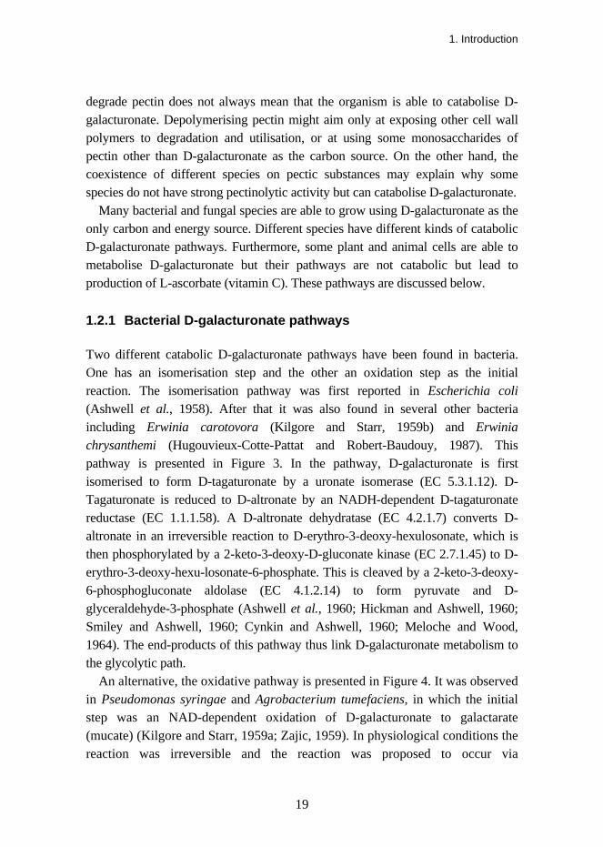
Two different catabolic D-galacturonate pathways have been found in bacteria. One has an isomerisation step and the other an oxidation step as the initial reaction. The isomerisation pathway was first reported in Escherichia coli (Ashwell et al., 1958). After that it was also found in several other bacteria including Erwinia carotovora (Kilgore and Starr, 1959b) and Erwinia chrysanthemi (Hugouvieux-Cotte-Pattat and Robert-Baudouy, 1987). This pathway is presented in Figure 3. In the pathway, D-galacturonate is first isomerised to form D-tagaturonate by a uronate isomerase (EC 5.3.1.12). D-Tagaturonate is reduced to D-altronate by an NADH-dependent D-tagaturonate reductase (EC 1.1.1.58). A D-altronate dehydratase (EC 4.2.1.7) converts D-altronate in an irreversible reaction to D-erythro-3-deoxy-hexulosonate, which is then phosphorylated by a 2-keto-3-deoxy-D-gluconate kinase (EC 2.7.1.45) to D-erythro-3-deoxy-hexu-losonate-6-phosphate. This is cleaved by a 2-keto-3-deoxy-6-phosphogluconate aldolase (EC 4.1.2.14) to form pyruvate and D-glyceraldehyde-3-phosphate (Ashwell et al., 1960; Hickman and Ashwell, 1960; Smiley and Ashwell, 1960; Cynkin and Ashwell, 1960; Meloche and Wood, 1964). The end-products of this pathway thus link D-galacturonate metabolism to the glycolytic path.
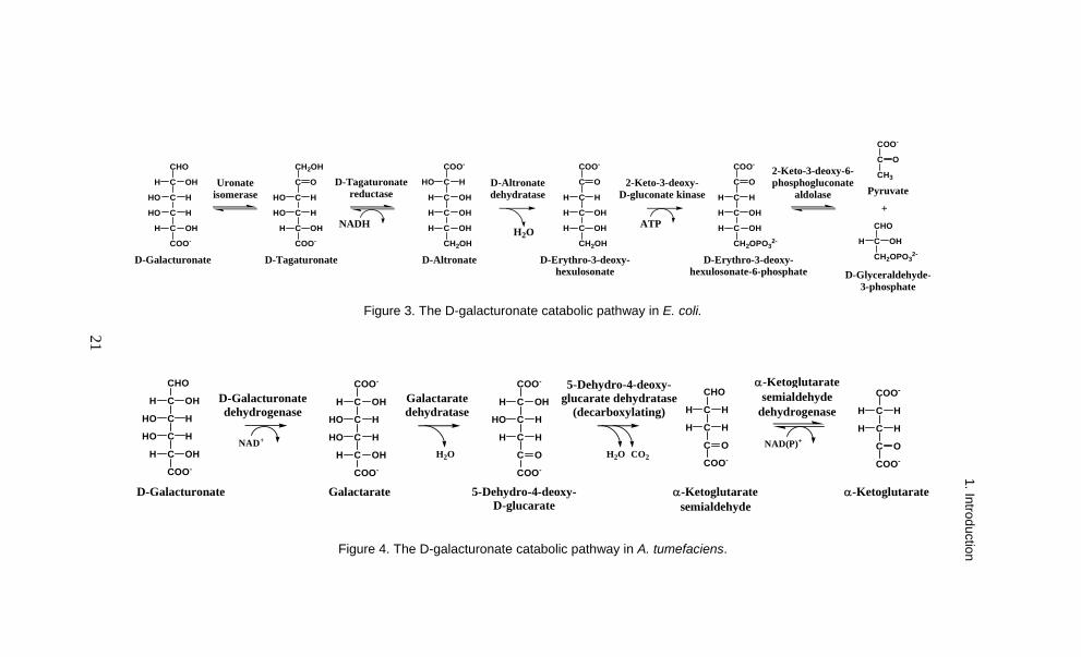
An alternative, the oxidative pathway is presented in Figure 4. It was observed in Pseudomonas syringae and Agrobacterium tumefaciens, in which the initial step was an NAD-dependent oxidation of D-galacturonate to galactarate (mucate) (Kilgore and Starr, 1959a; Zajic, 1959). In physiological conditions the reaction was irreversible and the reaction was proposed to occur via

1. Introduction
20
galactarolactone that is converted to galactarate by a spontaneous hydration step (Chang and Feingold, 1969; Wagner and Hollmann, 1976). Galactarate is more stable than its lactone form in neutral pH and the reverse reaction of the D-galacturonate dehydrogenase (EC 1.1.1.203) was only observed at acidic pH, when some of the galactarate was in the lactone form (Wagner and Hollmann, 1976). The D-galacturonate dehydrogenase was induced when cells were grown on D-galacturonate but not when grown on D-glucose (Chang and Feingold, 1969; Bateman et al., 1970). In the following steps a dehydratase (EC 4.2.1.42) converted galactarate to 5-dehydro-4-deoxy-D-glucarate, then via the action of dehydratase-decarboxylase (EC 4.2.1.41) to α-ketoglutarate semialdehyde, possibly via a 2,5-diketoadipic acid intermediate and finally an NAD(P)-dependent dehydrogenase (EC 1.2.1.26) completed the conversion to α-ketoglutarate (Dagley and Trudgill, 1965; Chang and Feingold, 1969; Chang and Feingold, 1970). α-Ketoglutarate is an intermediate of the citric acid cycle. Azospirillum brasilense also has another NAD-preferring α-ketoglutarate semialdehyde dehydrogenase, the expression of which was induced during growth on galactarate (Watanabe et al., 2006b).
1. Introduction
21
CHO
C
C
C
C
COO-
OH
HO
HHO
H
H
OHH
D-Galacturonate
CH2OH
C
C
C
C
COO-
HO
HHO
H
OHH
O
D-TagaturonateCH2OH
C
C
C
C
COO-
OH
H OH
H
HO H
OHH
D-AltronateCH2OH
C
C
C
C
COO-
OH
H H
H
OHH
O
D-Erythro-3-deoxy-hexulosonate
CH2OPO32-
C
C
C
C
COO-
OH
H H
H
OHH
O
D-Erythro-3-deoxy-hexulosonate-6-phosphate
CHO
C
CH2OPO32-
OHH
D-Glyceraldehyde-3-phosphate
COO-
C
CH3
O
Pyruvate
NADHH2O
ATP
Uronate isomerase
D-Tagaturonate reductase
D-Altronate dehydratase
2-Keto-3-deoxy-D-gluconate kinase
2-Keto-3-deoxy-6-phosphogluconate
aldolase+
Figure 3. The D-galacturonate catabolic pathway in E. coli.
CHO
C
C
C
C
COO-
OH
HO
HHO
H
H
OHH
D-Galacturonate
COO-
C
C
C
C
COO-
OH
HO
HHO
H
H
OHH
Galactarate
COO-
C
C
C
C
COO-
OH
HO
HH
H
H
O
5-Dehydro-4-deoxy-D-glucarate
CHO
C
C
C
COO-
H
HH
H
O
α-Ketoglutaratesemialdehyde
COO-
C
C
C
COO-
H
HH
H
O
α-Ketoglutarate
H2O H2O CO2
NAD(P)+NAD+
D-Galacturonate dehydrogenase
Galactarate dehydratase
5-Dehydro-4-deoxy-glucarate dehydratase
(decarboxylating)
α-Ketoglutaratesemialdehyde
dehydrogenase
Figure 4. The D-galacturonate catabolic pathway in A. tumefaciens.
21
1. Introduction
1. Introduction
22
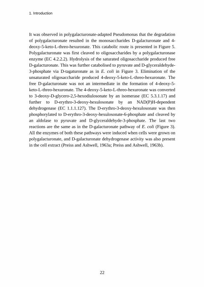
It was observed in polygalacturonate-adapted Pseudomonas that the degradation of polygalacturonate resulted in the monosaccharides D-galacturonate and 4-deoxy-5-keto-L-threo-hexuronate. This catabolic route is presented in Figure 5. Polygalacturonate was first cleaved to oligosaccharides by a polygalacturonase enzyme (EC 4.2.2.2). Hydrolysis of the saturated oligosaccharide produced free D-galacturonate. This was further catabolised to pyruvate and D-glyceraldehyde-3-phosphate via D-tagaturonate as in E. coli in Figure 3. Elimination of the unsaturated oligosaccharide produced 4-deoxy-5-keto-L-threo-hexuronate. The free D-galacturonate was not an intermediate in the formation of 4-deoxy-5-keto-L-threo-hexuronate. The 4-deoxy-5-keto-L-threo-hexuronate was converted to 3-deoxy-D-glycero-2,5-hexodiulosonate by an isomerase (EC 5.3.1.17) and further to D-erythro-3-deoxy-hexulosonate by an NAD(P)H-dependent dehydrogenase (EC 1.1.1.127). The D-erythro-3-deoxy-hexulosonate was then phosphorylated to D-erythro-3-deoxy-hexulosonate-6-phosphate and cleaved by an aldolase to pyruvate and D-glyceraldehyde-3-phosphate. The last two reactions are the same as in the D-galacturonate pathway of E. coli (Figure 3). All the enzymes of both these pathways were induced when cells were grown on polygalacturonate, and D-galacturonate dehydrogenase activity was also present in the cell extract (Preiss and Ashwell, 1963a; Preiss and Ashwell, 1963b).
1. Introduction
23
O
OH
OH
COO-
O
OH
OH
COO-
O O
OH
OH
COO-
O
OH
OH
COO-
OO O R2OR1
O
OH
OH
COO-
O
OH
OH
COO-
O O
OH
OH
COO-
O
OH
OH
COO-
O O R2OR1 +
POLYGALACTURONASE
O
OH
OH
COO-
OR1 +O
OH
OH
COO-
O R2+
HCHC OH
CHHOCHHO
HCCOO-
O
OHHCHC OH
CHHOCHCCOO-
O
OH
CHOHC OH
CHHOCH2CCOO-
HYDROLYSIS
CHOHC OH
CHHOCHHO
HC OHCOO-
D-Galacturonate 4-Deoxy-5-keto-L-threo-hexuronate
ELIMINATION
COO-
C OCH2
HCCCH2OH
OHO
COO-
C OCH2
HCHC
CH2OH
OHOH
COO-
C OCH2
HCHC
CH2OPO32-
OHOH CHO
HCCH2OPO3
2-OH
COO-
C OCH3
3-Deoxy-D-glycero-2,5-hexodiulosonate
D-Erythro-3-deoxy-hexulosonate
D-Erythro-3-deoxy-hexulosonate-6-phosphate
D-Glyceraldehyde-3-phosphate
Pyruvate+
O
1 2 3 4
Figure 5. Polygalacturonate degradation in polygalacturonate-adapted Pseudomonas. The residues of the saturated oligosaccharide do not have double bonds, whereas the non-reducing terminal residue of the unsaturated oligosaccharide has a double bond between C4 and C5. 1 4-Deoxy-5-keto-uronate isomerase; 2 2-dehydro-3-deoxy-D-gluconate 5-dehydrogenase; 3 2-keto-3-deoxy-D-gluconate kinase; 4 2-keto-3-deoxy-6-phosphogluconate aldolase. Revised from the figure of Preiss and Ashwell 1963a.
1.2.2 Metabolism of D-galacturonate in plants and animals
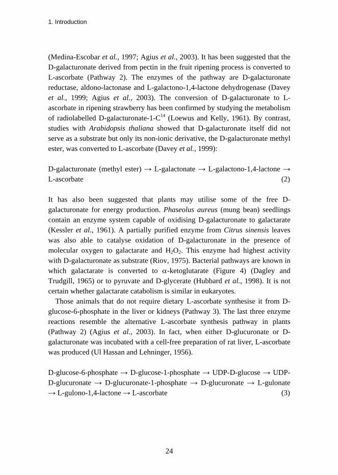
Higher plants produce L-ascorbate mainly from D-glucose-6-phosphate via a pathway that has GDP-D-mannose and GDP-L-galactose as two of the intermediates (Pathway 1) (Wheeler et al., 1998):
D-glucose-6-phosphate → D-fructose-6-phosphate → D-mannose-6-phosphate → D-mannose-1-phosphate → GDP-D-mannose → GDP-L-galactose → L-galactose-1-phosphate → L-galactose → L-galactono-1,4-lactone → L-ascorbate (1) However, there is evidence for alternative biosynthetic L-ascorbate pathways, one of which has D-galacturonate as an intermediate. A correlation between fruit softening and ripening, disassembling of pectin in the cell walls and elevation of the L-ascorbate content has been shown for example in strawberry fruit
1. Introduction
24
(Medina-Escobar et al., 1997; Agius et al., 2003). It has been suggested that the D-galacturonate derived from pectin in the fruit ripening process is converted to L-ascorbate (Pathway 2). The enzymes of the pathway are D-galacturonate reductase, aldono-lactonase and L-galactono-1,4-lactone dehydrogenase (Davey et al., 1999; Agius et al., 2003). The conversion of D-galacturonate to L-ascorbate in ripening strawberry has been confirmed by studying the metabolism of radiolabelled D-galacturonate-1-C14 (Loewus and Kelly, 1961). By contrast, studies with Arabidopsis thaliana showed that D-galacturonate itself did not serve as a substrate but only its non-ionic derivative, the D-galacturonate methyl ester, was converted to L-ascorbate (Davey et al., 1999): D-galacturonate (methyl ester) → L-galactonate → L-galactono-1,4-lactone → L-ascorbate (2) It has also been suggested that plants may utilise some of the free D-galacturonate for energy production. Phaseolus aureus (mung bean) seedlings contain an enzyme system capable of oxidising D-galacturonate to galactarate (Kessler et al., 1961). A partially purified enzyme from Citrus sinensis leaves was also able to catalyse oxidation of D-galacturonate in the presence of molecular oxygen to galactarate and H2O2. This enzyme had highest activity with D-galacturonate as substrate (Riov, 1975). Bacterial pathways are known in which galactarate is converted to α-ketoglutarate (Figure 4) (Dagley and Trudgill, 1965) or to pyruvate and D-glycerate (Hubbard et al., 1998). It is not certain whether galactarate catabolism is similar in eukaryotes.
Those animals that do not require dietary L-ascorbate synthesise it from D-glucose-6-phosphate in the liver or kidneys (Pathway 3). The last three enzyme reactions resemble the alternative L-ascorbate synthesis pathway in plants (Pathway 2) (Agius et al., 2003). In fact, when either D-glucuronate or D-galacturonate was incubated with a cell-free preparation of rat liver, L-ascorbate was produced (Ul Hassan and Lehninger, 1956).
D-glucose-6-phosphate → D-glucose-1-phosphate → UDP-D-glucose → UDP-D-glucuronate → D-glucuronate-1-phosphate → D-glucuronate → L-gulonate → L-gulono-1,4-lactone → L-ascorbate (3)
1. Introduction
25
1.2.3 Earlier observations concerning D-galacturonate catabolism in mould fungi
D-Galacturonate catabolism in the filamentous fungus A. nidulans was concluded to be different from that in E. coli because the enzyme activities of the bacterial pathway were not found in this mould fungus after growing on D-galacturonate. Furthermore, A. nidulans does not catabolise D-galacturonate through one of the main carbohydrate degradation pathways, which was shown by growing several D-galacturonate non-utilising mutant strains on different carbon sources. For example on D-glucose and D-xylose these mutants grew normally (Uitzetter et al., 1986). The pentose phosphate pathway appeared to have some role in D-galacturonate catabolism because an A. nidulans mutant with an inoperative transaldolase showed almost no growth on D-galacturonate. Mutants of A. nidulans with inoperative pyruvate carboxylase or pyruvate dehydrogenase had poor or no growth on D-galacturonate. Therefore pyruvate was predicted to be one of the degradation products of D-galacturonate. A mutant lacking pyruvate kinase activity grew on D-galacturonate, which indicated that pyruvate was not produced from D-galacturonate via phosphoenol pyruvate (Uitzetter et al., 1982). The relationship between these enzyme reactions is presented in Figure 6.
In addition, the A. nidulans mutants that were unable to utilise glycerol were also unable to grow on D-galacturonate (Uitzetter et al., 1982). Furthermore, when a wild-type strain of A. nidulans was transferred from D-fructose to glycerol or D-galacturonate medium the glycerol kinase and FAD-dependent mitochondrial glycerol-3-phosphate dehydrogenase activities were induced. Induction of NADH-dependent D-glyceraldehyde reductase and alcohol dehydrogenase activities was also observed. However, it was suggested that the alcohol dehydrogenase is responsible for both these activities. After transfer to D-galacturonate, NAD-dependent cytosolic glycerol-3-phosphate dehydrogenase and weak dihydroxyacetone (DHA) kinase and D-glyceraldehyde kinase activities were also induced (Hondmann et al., 1991). These results indicated that the glycerol and D-galacturonate pathways are linked in some way. It was suggested that D-glyceraldehyde is the common intermediate. It was also predicted that in A. nidulans D-galacturonate is metabolised through a non-phosphorylating pathway, because glyceraldehyde-3-phosphate did not appear to be one of the end products (Uitzetter et al., 1986). Studies with A. niger, a related filamentous fungus species, supported the suggested connection between

1. Introduction
26
the glycerol and D-galacturonate pathways. A glycerol kinase mutant strain of A. niger grew poorly on glycerol and on D-galacturonate. When the mutant strain was transferred from D-glucose to D-galacturonate it accumulated glycerol in the culture. Therefore, it was concluded that in this filamentous fungus D-galacturonate is converted to glycerol, which cannot be further metabolised by the glycerol kinase mutant strain.
It was proposed that the D-galacturonate catabolic pathway in A. nidulans contains at least two enzymes. This was because two separate gene sites with point mutations produced strains that were unable to grow specifically on D-galacturonate (Uitzetter et al., 1986). It was not until 22 years later that these genes were reported to code for D-galacturonate reductase and L-galactonate dehydratase (Martens-Uzunova and Schaap, 2008). A microarray analysis some years earlier had shown that when A. niger was grown on D-galacturonate the genes possibly coding for an aldo-keto reductase, a racemase and an aldolase were co-expressed (Martens-Uzunova et al., 2005). The degradation of D-galacturonate was proposed also to involve an NADP+-dependent glycerol dehydrogenase-catalysed reaction because this activity was induced on D-galacturonate. The enzyme was proposed to reduce D-glyceraldehyde in the reverse reaction but not to use DHA as a substrate (Witteveen et al., 1990). An NADP+-dependent glycerol dehydrogenase activity was also specifically induced in A. nidulans during growth on D-galacturonate (Sealy-Lewis and Fairhurst, 1992)

1. Introduction
27
Glucose
Glucose-6-phosphate
Fructose-6-phosphate
Fructose-1,6-bisphosphate
Glyceraldehyde-3-phosphate
Dihydroxyacetone phosphate
1,3-Bisphosphoglycerate
3-Phosphoglycerate
2-Phosphoglycerate
Phosphoenolpyruvate
Pyruvate
Glycerol Dihydroxyacetone
Glycerol-3-phosphate
Glyceraldehyde
12
3
4
5
6
7 8
Acetyl-CoAOxaloacetate9 10
11
Figure 6. Relationship between different enzyme reactions that may be involved in D-galacturonate catabolism in mould fungi (adapted from Tom et al., 1978). 1 Glycerol kinase; 2 NAD+- or NADP+-dependent glycerol dehydrogenase; 3 alcohol dehydrogenase/NADH-dependent D-glyceraldehyde reductase or NADP+-dependent glycerol dehydrogenase; 4 NAD+- or FAD-dependent glycerol-3-phosphate dehydrogenase; 5 DHA kinase; 6 D-glyceraldehyde kinase; 7 triose phosphate isomerase; 8 aldolase; 9 pyruvate carboxylase; 10 pyruvate dehydrogenase; 11 pyruvate kinase.

1. Introduction
28
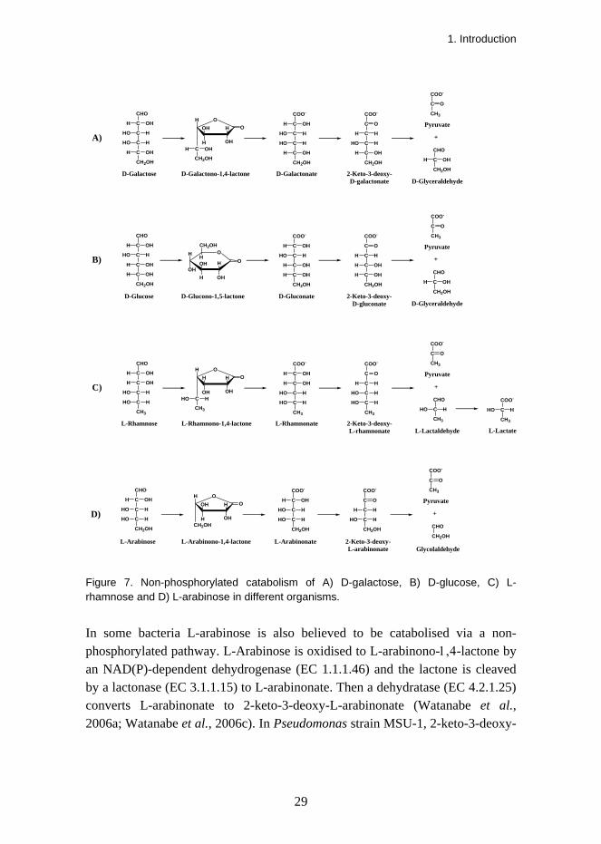
1.3 Examples of non-phosphorylated catabolic pathways
Some known catabolic pathways in which no phosphorylation step is involved and in which a 2-keto-3-deoxy sugar acid is one of the intermediates are presented in Figure 7. A. niger converts D-galactose to pyruvate and D-glyceraldehyde via a non-phosphorylated route. In this pathway D-galactose is first oxidised to D-galactono-1,4-lactone in the presence of NAD+ by a D-galactose dehydrogenase (EC 1.1.1.48). The D-galactono-1,4-lactone is then linearised by a lactonase (EC 3.1.1.25) to D-galactonate. An inducible D-galactonate dehydratase (EC 4.2.1.6) was proposed to convert the D-galactonate to 2-keto-3-deoxy-D-galactonate, after which a constitutive 2-keto-3-deoxy-D-galactonate aldolase converts this metabolite further to pyruvate and D-glyceraldehyde (Elshafei and Abdel-Fatah, 2001). The last two reactions had also been detected in Aspergillus terreus (Elshafei and Abdel-Fatah, 1991).
A. niger is also able to catabolise D-glucose to D-glyceraldehyde and pyruvate through a similar pathway. A. niger oxidises D-glucose to D-glucono-1,5-lactone by a glucose oxidase (EC 1.1.3.4) in the presence of molecular oxygen. This filamentous fungus also has a gluconolactonase enzyme (EC 3.1.1.17) that converts D-glucono-1,5-lactone to D-gluconate (Ogawa et al., 2002). D-Gluconate is dehydrated to 2-keto-3-deoxy-D-gluconate by a D-gluconate dehydratase (EC 4.2.1.39). This is further cleaved to D-glyceraldehyde and pyruvate by a 2-keto-3-deoxy-D-gluconate aldolase (EC 4.1.2.20) (Elzainy et al., 1973). A similar pathway, the non-phosphorylated Entner-Doudoroff pathway, also operates in thermophilic archaea (Buchanan et al., 1999; Siebers and Schönheit, 2005). Here the only difference is in the first reaction from D-glucose to D-glucono-1,5-lactone, which is catalysed by a glucose dehydrogenase (EC 1.1.1.47) instead of a glucose oxidase.
L-Rhamnose catabolism in the yeast fungus Pichia stipitis and the bacterium Azotobacter vinelandii also involves non-phosphorylated intermediates. The enzymes of the pathway are NAD(P)+-dependent L-rhamnose dehydrogenase (EC 1.1.1.173), L-rhamnono-1,4-lactonase (EC 3.1.1.65), L-rhamnonate dehydratase (EC 4.2.1.90), 2-keto-3-deoxy-L-rhamnonate aldolase and NAD(P)+-dependent L-lactaldehyde dehydrogenase (EC 1.2.1.22). These enzymes convert L-rhamnose to pyruvate and L-lactate via the intermediates L-rhamnono-1,4-lactone, L-rhamnonate, 2-keto-3-deoxy-L-rhamnonate and L-lactaldehyde (Twerdochlib et al., 1994; Watanabe et al., 2008a, Watanabe et al., 2008b).
1. Introduction
29
D-Galactonate 2-Keto-3-deoxy-D-galactonate
CHO
C
CH2OH
OHH
D-Glyceraldehyde
COO-
C
CH3
O
Pyruvate
+
CH2OH
C
C
C
C
COO-
H
HO H
HO
H OH
OHH
CH2OH
C
C
C
C
COO-
H
H H
HO
OHH
O
CHO
C
C
C
C
CH2OH
OH
HO
HHO
H
H
OHH
D-Galactose D-Galactono-1,4-lactone
A)
D-Gluconate 2-Keto-3-deoxy-D-gluconate
CHO
C
CH2OH
OHH
D-Glyceraldehyde
COO-
C
CH3
O
Pyruvate
+
CH2OH
C
C
C
C
COO-
OH
HO H
H
H OH
OHH
CH2OH
C
C
C
C
COO-
OH
H H
H
OHH
O
CHO
C
C
C
C
CH2OH
OH
HO
OHH
H
H
OHH
D-Glucose D-Glucono-1,5-lactone
B)OH
OHH
OHH
OH
CH2OH
HO
C
H
H OH
OH HO
O
OHH
CH2OH
L-Rhamnonate 2-Keto-3-deoxy-L-rhamnonate
CHO
C
CH3
HHO
L-Lactaldehyde
COO-
C
CH3
O
Pyruvate
+
CH3
C
C
C
C
COO-
H
H OH
HO
H OH
HHO
CH3
C
C
C
C
COO-
H
H H
HO
HHO
O
CHO
C
C
C
C
CH3
OH
H
HHO
OH
H
HHO
L-Rhamnose L-Rhamnono-1,4-lactone
C)C
H
OH OH
H HO
O
HHO
CH3
L-Lactate
COO-
C
CH3
HHO
L-Arabinonate 2-Keto-3-deoxy-L-arabinonate
CHO
CH2OH
Glycolaldehyde
COO-
C
CH3
O
Pyruvate
+
CH2OH
C
C
C
COO-
H
HO H
HO
H OH
CH2OH
C
C
C
COO-
H
H H
HO
O
CHO
C
C
C
CH2OH
OH
HO
HHO
H
H
L-Arabinose L-Arabinono-1,4-lactone
D)CH2OH
H
H OH
OH HO
O
Figure 7. Non-phosphorylated catabolism of A) D-galactose, B) D-glucose, C) L-rhamnose and D) L-arabinose in different organisms.
In some bacteria L-arabinose is also believed to be catabolised via a non-phosphorylated pathway. L-Arabinose is oxidised to L-arabinono-1,4-lactone by an NAD(P)-dependent dehydrogenase (EC 1.1.1.46) and the lactone is cleaved by a lactonase (EC 3.1.1.15) to L-arabinonate. Then a dehydratase (EC 4.2.1.25) converts L-arabinonate to 2-keto-3-deoxy-L-arabinonate (Watanabe et al., 2006a; Watanabe et al., 2006c). In Pseudomonas strain MSU-1, 2-keto-3-deoxy-

1. Introduction
30
L-arabinonate is cleaved to pyruvate and glycolaldehyde by an aldolase (EC 4.1.2.18) (Dahms and Anderson, 1969).
1.4 Hypotheses and objectives of the work
D-Galacturonate is the major component of pectin and a potential carbon source for microorganisms living on decaying plant material. Bacterial catabolic D-galacturonate pathways are known but similar pathways have not been reported in eukaryotes. However, many species of yeast and mould fungi are able to utilise and grow on D-galacturonate. This indicates that in eukaryotic microorganisms a catabolic D-galacturonate pathway exists which is different from the bacterial pathways. The aim of this work was to determine how eukaryotes catabolise D-galacturonate and what enzymes and intermediate metabolites form the pathway. The filamentous fungus T. reesei was chosen as the model organism.
Some plant materials such as the peel of citrus fruits and sugar beet pulp are especially rich in pectin. Large amounts of these pectin-rich residues are produced as side products of the food industry. They are mainly used as animal feed after drying and pelletising. However, these residues have relatively low feed value and drying consumes energy. Therefore it would be desirable to convert this cheap and renewable raw material to products of higher value.

2. Materials and methods
31
2. Materials and methods The filamentous fungus Trichoderma reesei was able to grow on D-galacturonate. Its genome sequence was known and was publicly available at the Joint Genome Institute website (http://genome.jgi-psf.org/Trire2/Trire 2.home.html). Furthermore, T. reesei has been extensively studied in our laboratory and many genetic engineering techniques have been developed for working with this mould fungus. For example, a good cDNA library of the strain Rut C-30 had already been made, which facilitated cloning of the genes. T. reesei was therefore chosen as the model organism in this study. The strains that were used to study the different enzymes of the D-galacturonate pathway are listed in Table 2.
Table 2. The T. reesei strains used in this work.
Enzyme Enzyme activity observed in strain
Gene cloned from strain
Gene deleted from strain
GAR1 LGD1 LGA1 GLD1 GLD2
Rut C-30 Rut C-30 VTT-D-80133 Rut C-30 and QM6a Rut C-30 and QM6a
Rut C-30 Rut C-30 Rut C-30 Rut C-30 QM6a
QM6a (Mojzita et al., 2010) Rut C-30 QM6a
T. reesei was grown in liquid culture that contained 20 g/l of the main carbon source, 2 g/l (in GAR1 and GLD1 experiments) or 0.5 g/l (in LGD1 and LGA1 experiments) of proteose peptone and also 15 g/l KH2PO4, 5 g/l (NH4)2SO4, 0.6 g/l MgSO4*7H2O, 0.6 g/l CaCl2*2H2O, and trace elements (Mandels and Weber, 1969) at 28ºC. Before harvesting T. reesei was typically grown on D-galacturonate for five days. Growth on this substrate was slower than on all the

2. Materials and methods
32
other used carbon sources. Difference in the growth rate on D-galacturonate was not observed between the used T. reesei host strains.
The work proceeded stepwise along the pathway starting by finding the pathway’s first enzyme exhibiting activity with D-galacturonate. When the reaction product of the previous enzyme was identified it was then used as the reaction substrate for the next enzyme. Some of the intermediate substrates had to be prepared chemically or enzymatically, since they were not commercially available. The enzyme activities were measured using crude mycelial extracts of T. reesei grown aerobically on D-galacturonate, and for comparison on other carbon sources. Enzyme activities were measured as U/mg, i.e. micromoles of substrate per minute per milligram of protein. The gene coding for the enzyme was identified either by purifying and sequencing the enzyme or by directly cloning a gene from the T. reesei cDNA library based on sequence homology to other similar enzymes. The cloned genes were expressed in S. cerevisiae, the enzyme activities were verified and the enzymes were further characterised. Some of the pathway’s genes were deleted from T. reesei by replacing them with a hygromycin B resistance gene by homologous recombination. The effect of the gene deletion was then studied by growing the deletion strains on D-galacturonate. The materials and methods are described in more detail in the original articles. The used methods are listed in Table 3.
Table 3. Methods used in the original articles (I–IV).
Method Used in Chemical or enzymatic preparation of reaction substrates III, IV Cloning a fungal gene based on homology II, III Constructing a fungal deletion strain II, IV Dry mass measurements II, IV Enzyme activity measurements I–IV Expressing fungal genes in S. cerevisiae I–IV Metabolite analysis, HPLC or NMR II, IV Protein purification and partial sequencing I, IV RNA extraction and Northern hybridisation I, II

3. Results
33
3. Results A novel catabolic D-galacturonate pathway was identified in the filamentous fungus Trichoderma reesei. It consisted of four enzymes which were described in four separate publications. The enzymes were NADPH-dependent D-galacturonate reductase (GAR1), L-galactonate dehydratase (LGD1), L-threo-3-deoxy-hexulosonate aldolase (LGA1) and NADPH-dependent glyceraldehyde reductase (GLD1). This fungal pathway converted D-galacturonate to pyruvate and glycerol as presented in Figure 8.
3.1 D-Galacturonate reductase (I)
T. reesei grown on D-galacturonate had an NADPH-dependent D-galacturonate reductase activity in the crude mycelial extract. When the mould fungus was grown on D-glucose, D-fructose, D-galactose, D-xylose, or glycerol this activity was absent.
3.1.1 Cloning the D-galacturonate reductase
In order to identify the gene sequence of the T. reesei D-galacturonate reductase, the enzyme was purified from mycelial extract after growth on D-galacturonate. The purified enzyme with an estimated molecular mass of 40 kDa was digested with trypsin and the amino acid sequences of two fragments were obtained. These peptide sequences corresponded to a T. reesei cDNA sequence in public databases where only the 3’ end of the open reading frame was reported. The genomic sequence of T. reesei upstream from this partial open reading frame was compared to previously reported aldo-keto reductase sequences and six possible start codons were detected. T. reesei cDNA library and six different sense primers were used with an antisense primer to identify the correct start codon. In this way the open reading frame was identified and cloned. It coded

3. Results
34
CHO
C
C
C
C
COO-
OH
HO
HHO
H
H
OHH
D-Galacturonate L-Galactonate L-Threo-3-deoxy-hexulosonate
CHO
C
CH2OH
HHO
L-Glyceraldehyde
COO-
C
CH3
O
Pyruvate
NADPHH2O
D-Galacturonate reductase(GAR1)
L-Galactonate dehydratase
(LGD1)
L-Threo-3-deoxy-hexulosonate aldolase
(LGA1)+
CH2OH
C
C
C
C
COO-
OH
H OH
H
HO H
HHO
CH2OH
C
C
C
C
COO-
OH
H H
H
HHO
O
CH2OH
C
CH2OH
OHH
Glycerol
Glyceraldehyde reductase(GLD1)
NADPH
Figure 8. The D-galacturonate catabolic pathway in T. reesei.
34
3. Results

3. Results
35
for a 309 amino acid protein with a molecular mass of 33 940 Da. The corresponding genomic DNA sequence had five introns. The open reading frame was expressed in S. cerevisiae in a multicopy plasmid under a constitutive promoter which resulted in the production of an active enzyme in this heterologous host. The gene was called gar1 and the sequence was deposited in NCBI GenBank with accession number AY862503. In the T. reesei genome database v2.0 it has a protein ID 22004.
3.1.2 Characterisation of the D-galacturonate reductase
The partially purified T. reesei D-galacturonate reductase used NADPH as a cofactor. The reverse reaction was observed with L-galactonate and NADP+. No activity was observed with L-galactono-1,4-lactone as a substrate.
To facilitate purification the D-galacturonate reductase was produced in S. cerevisiae with a C-terminal polyhistidine tag. The tagged and the nontagged protein had the same activity in the crude yeast extract and the purified tagged protein was used for kinetic analysis. The D-galacturonate reductase exhibited activity in the forward direction with D-galacturonate, D-glucuronate and DL-glyceraldehyde. The Michaelis-Menten constants were Km = 6 mM and Vmax = 40 U/mg, Km = 11 mM and Vmax = 25 U/mg, and Km = 6 mM and Vmax = 7 U/mg, respectively. The cofactor was always NADPH, for which the Michaelis-Menten constants were Km = 30 µM and Vmax = 40 U/mg when D-galacturonate was used as a substrate. No activity was observed when D-glucose, D-fructose, D-xylose, D-galactose, L-arabinose, or D-mannose were tested as substrates. In the reverse direction L-galactonate and NADP+ served as substrates with Michaelis-Menten constants Km = 4 mM and Km = 1 µM, respectively, and Vmax = 2 U/mg. No backward reaction was observed with L-galactono-1,4-lactone, L-gulonate, glycerol, D-arabitol, L-arabitol, xylitol, galactitol or ribitol.
Expression of the D-galacturonate reductase gene was studied with Northern blot analysis on different carbon sources. When T. reesei was grown on D-fructose, D-xylose, lactose, D-galactose, glycerol and D-mannose a basic expression level was observed, but on D-galacturonate the expression level was 8 times and on D-glucose 3 times greater.
The D-galacturonate reductase gene in T. reesei was deleted by replacing it with a hygromycin B resistance gene by homologous recombination. When grown in liquid cultures with D-galacturonate and peptone for five days the host strain produced about 3 g/l of biomass, whereas the deletion strain produced

3. Results
36
only about 0.5 g/l of biomass. Both strains produced about 0.2 g/l of biomass when only the peptone was included in the medium. Deletion of the D-galacturonate reductase gene also resulted in an impaired sporulation ability on PD (potato dextrose) agar plates. The spores could be obtained by growing the deletion strain on plates containing 10% carrot juice (Mojzita et al., 2010).
3.2 L-Galactonate dehydratase (II)
T. reesei was grown on D-galacturonate and the mycelial extract was tested for different enzyme activities. When L-galactonate was added to the mycelial extract we observed the formation of a reducing sugar. The enzyme activity which formed a reducing sugar from L-galactonate was present in T. reesei mycelia grown on D-galacturonate but absent when grown on D-galactose, D-glucose, D-xylose, D-fructose, lactose, or glycerol. In addition a reaction mixture of L-galactonate and crude mycelial extract of T. reesei grown on D-galacturonate was analysed using electrospray ionisation mass spectrometry (ESI-MS). It was observed that the reaction product was a molecule with a mass of one water molecule less than the mass of L-galactonate, and had a different MS-MS spectrum from the lactone form of L-galactonate. Some pyruvate was also formed, which provided a clue about the subsequent reactions. Since no redox cofactor was involved and a water molecule was split off, the second enzyme in the pathway was concluded to be a dehydratase.
3.2.1 Cloning the L-galactonate dehydratase
Different approaches were tested in order to purify the L-galactonate dehydratase. However, in all the trials the L-galactonate dehydratase lost its activity in the purification process and we were not able to obtain pure protein that could have been sequenced. In order to identify the gene coding for an L-galactonate dehydratase the amino acid sequences of E. coli and Brucella melitensis D-galactonate dehydratases (EC 4.2.1.6) were compared to the T. reesei genome sequence. Six open reading frames of potential dehydratases were then cloned using T. reesei cDNA or RNA as a template. They all were expressed in S. cerevisiae under a constitutive promoter in a multicopy plasmid. The cell extracts of these yeast strains were then assayed for reducing sugar formation from L-galactonate. S. cerevisiae does not have L-galactonate dehydratase activity but one of the expressed open reading frames resulted in a

3. Results
37
significant activity with L-galactonate as a substrate. This gene was called lgd1 and the sequence was deposited in NCBI GenBank with accession number DQ181420. It coded for a protein of 450 amino acids and had one intron in the corresponding genomic DNA sequence. Since such an enzyme activity was not described previously it does not have an EC (Enzyme Commission) number. In the Brenda database (www.brenda-enzymes.info) it was listed with the preliminary number EC 4.2.1.B1. In the T. reesei genome database v2.0 it has a protein ID 104599.
3.2.2 Characterisation of the L-galactonate dehydratase
The L-galactonate dehydratase gene was expressed in S. cerevisiae, resulting in an active enzyme that produced a reducing sugar from L-galactonate. The reaction mixture was analysed by NMR spectroscopy to identify the reaction product. The results showed that the product was L-threo-3-deoxy-hexulosonate (2-keto-3-deoxy-L-galactonate) and that it predominantly existed as a pyranose ring. Over 85% of the product existed as one anomer of the pyranose ring but it could not be determined which one of the two anomers it was. The enzyme also exhibited stereospecificity at the C3 position, since it attached a hydrogen atom only to the axial position during the reaction.
The L-galactonate dehydratase activity in the crude mycelial extract of T. reesei grown on D-galacturonate was 0.018 U/mg protein (0.3 nkat/mg) and in the cell extract of S. cerevisiae expressing the lgd1 it was 0.009 U/mg protein (0.15 nkat/mg). Here the L-galactonate concentration was 7.5 mM. The product L-threo-3-deoxy-hexulosonate was measured with a thiobarbituric acid (TBA) assay (Buchanan et al., 1999).
The L-galactonate dehydratase was polyhistidine tagged either at the N- or C-terminus and produced in S. cerevisiae. The tag at the C-terminus resulted in an inactive protein and the N-terminally tagged protein had only about one fifth of the activity of untagged protein in yeast extract. Since the tag apparently interfered with the activity, the crude cell extract of the yeast strain expressing untagged lgd1 was used to characterise the enzyme kinetics. Different sugar acids were tested for the substrate specificity of L-galactonate dehydratase. It produced a reducing sugar from L-galactonate and D-arabinonate. Activity with D-arabinonate was about half of that with L-galactonate. No activity was observed when D-galactonate, D-gluconate, D-xylonate, or L-gulonate were used as substrates. The Km for L-galactonate was estimated to be 5 mM. L-

3. Results
38
Galactonate at a concentration of 20 mM was completely converted to L-threo-3-deoxy-hexulosonate in 24 hours. This suggested that the energetic equilibrium is strongly on the side of the reaction product. The enzyme required bivalent cations (Mg2+) for activity and no activity was observed in the presence of 5 mM EDTA. The TBA assay that was used to determine enzyme kinetics was disturbed by high substrate concentrations. When the same reactions were measured with 3,5-dinitrosalicylate (DNS) assay (Bernfeld, 1955) the high substrate concentration did not interfere with the assay.
Northern blot analysis was used to study expression of the L-galactonate dehydratase gene on different carbon sources. The expression level was the same on D-galacturonate, glycerol and lactose and three times higher on D-glucose and D-xylose.
The L-galactonate dehydratase gene in T. reesei was deleted by replacing it with a hygromycin B resistance gene by homologous recombination. When grown in liquid cultures with D-galacturonate and peptone for five days the host strain produced about 2.4 g/l of biomass, whereas the deletion strain produced only 0.1 g/l of biomass. Both strains also produced 0.1 g/l of biomass when only the peptone was included in the medium. The deletion strain also accumulated L-galactonate in the culture; in five days it produced 1.3 g/l of L-galactonate, whereas 0.4 g/l was produced by the host strain (unpublished results). The growth on D-galacturonate was restored when the lgd1 was retransformed to the deletion strain.
3.3 L-Threo-3-deoxy-hexulosonate aldolase (IV)
The first indication of an aldolase reaction in the D-galacturonate pathway was when pyruvate was detected after incubating L-galactonate with crude mycelial extract of T. reesei grown on D-galacturonate. The formed pyruvate was identified by ESI-MS as well as by assaying with lactate dehydrogenase. After the L-galactonate dehydratase had been characterised and shown to produce L-threo-3-deoxy-hexulosonate, this compound was prepared and used as the reaction substrate in the search for the next enzyme on the pathway. The crude mycelial extract mixed with L-threo-3-deoxy-hexulosonate produced pyruvate. The reverse reaction was also observed, since a compound giving a signal in the TBA assay was formed when pyruvate and L-glyceraldehyde were incubated with the crude mycelial extract. The L-threo-3-deoxy-hexulosonate aldolase activity was induced in T. reesei during growth on D-galacturonate but not on D-

3. Results
39
glucose, D-xylose, lactose, or glycerol. Such an enzyme activity had not been described previously.
3.3.1 Cloning the L-threo-3-deoxy-hexulosonate aldolase
The L-threo-3-deoxy-hexulosonate aldolase was partially purified from T. reesei mycelial extract after growth on D-galacturonate. The obtained 37 kDa protein was digested with trypsin and four peptide sequences were obtained for identifying the coding sequence from the T. reesei genome. The open reading frame was cloned from cDNA, and comparing it to the genomic DNA showed that there were no introns. The gene was named lga1 and the sequence was deposited in NCBI GenBank with the accession number EF203091. It coded for a 315 amino acid protein. In the T. reesei genome database v2.0 it has a protein ID 60067. The gene was expressed in S. cerevisiae under a constitutive promoter in a multicopy plasmid and this heterologous host produced an active protein.
3.3.2 Characterisation of the L-threo-3-deoxy-hexulosonate aldolase
The L-threo-3-deoxy-hexulosonate aldolase activity in the crude mycelial extract of T. reesei grown on D-galacturonate was 0.16 U/mg of protein in the forward direction with L-threo-3-deoxy-hexulosonate as a substrate. L-Glyceraldehyde and pyruvate were substrates in the reverse direction and the enzyme activity was 0.20 U/mg of protein. When L-glyceraldehyde was replaced with D-glyceraldehyde the activity was 0.01 U/mg of protein. This was close to the detection limit, which was about 3% of the activity with L-glyceraldehyde. In fact D-glyceraldehyde appeared to be a reaction inhibitor, although the actual mechanism is not known. From the enzyme activity it was seen that when DL-glyceraldehyde was used as a substrate instead of L-galactonate the reaction was only about 10% (0.02 U/mg of protein) of that with L-glyceraldehyde, rather than the anticipated 50%. The L-threo-3-deoxy-hexulosonate aldolase activity in the cell extract of S. cerevisiae expressing the lga1 was 0.05 U/mg of protein.
A polyhistidine tag was added to the N- or C-terminus of the L-threo-3-deoxy-hexulosonate aldolase and the protein was produced in S. cerevisiae. The C-terminally tagged enzyme had only about one third of the activity of untagged enzyme, but the N-terminal tag did not interfere with the activity. The purified N-terminally tagged protein was used in further enzyme characterisations.
3. Results
40
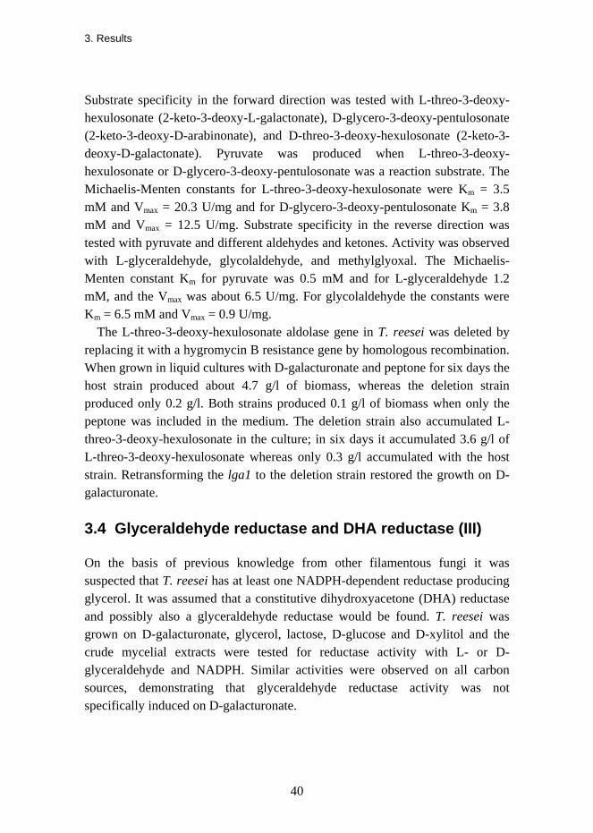
Substrate specificity in the forward direction was tested with L-threo-3-deoxy-hexulosonate (2-keto-3-deoxy-L-galactonate), D-glycero-3-deoxy-pentulosonate (2-keto-3-deoxy-D-arabinonate), and D-threo-3-deoxy-hexulosonate (2-keto-3-deoxy-D-galactonate). Pyruvate was produced when L-threo-3-deoxy-hexulosonate or D-glycero-3-deoxy-pentulosonate was a reaction substrate. The Michaelis-Menten constants for L-threo-3-deoxy-hexulosonate were Km = 3.5 mM and Vmax = 20.3 U/mg and for D-glycero-3-deoxy-pentulosonate Km = 3.8 mM and Vmax = 12.5 U/mg. Substrate specificity in the reverse direction was tested with pyruvate and different aldehydes and ketones. Activity was observed with L-glyceraldehyde, glycolaldehyde, and methylglyoxal. The Michaelis-Menten constant Km for pyruvate was 0.5 mM and for L-glyceraldehyde 1.2 mM, and the Vmax was about 6.5 U/mg. For glycolaldehyde the constants were Km = 6.5 mM and Vmax = 0.9 U/mg.
The L-threo-3-deoxy-hexulosonate aldolase gene in T. reesei was deleted by replacing it with a hygromycin B resistance gene by homologous recombination. When grown in liquid cultures with D-galacturonate and peptone for six days the host strain produced about 4.7 g/l of biomass, whereas the deletion strain produced only 0.2 g/l. Both strains produced 0.1 g/l of biomass when only the peptone was included in the medium. The deletion strain also accumulated L-threo-3-deoxy-hexulosonate in the culture; in six days it accumulated 3.6 g/l of L-threo-3-deoxy-hexulosonate whereas only 0.3 g/l accumulated with the host strain. Retransforming the lga1 to the deletion strain restored the growth on D-galacturonate.
3.4 Glyceraldehyde reductase and DHA reductase (III)
On the basis of previous knowledge from other filamentous fungi it was suspected that T. reesei has at least one NADPH-dependent reductase producing glycerol. It was assumed that a constitutive dihydroxyacetone (DHA) reductase and possibly also a glyceraldehyde reductase would be found. T. reesei was grown on D-galacturonate, glycerol, lactose, D-glucose and D-xylitol and the crude mycelial extracts were tested for reductase activity with L- or D-glyceraldehyde and NADPH. Similar activities were observed on all carbon sources, demonstrating that glyceraldehyde reductase activity was not specifically induced on D-galacturonate.

3. Results
41
3.4.1 Cloning the glyceraldehyde reductase and DHA reductase
In order to find glycerol dehydrogenase genes in the T. reesei genome, the partial amino acid sequences of A. niger NADP+-dependent glycerol dehydrogenase (Norbeck and Blomberg, 1997) were compared to the translated T. reesei genome sequence. Two potential open reading frames were identified. The start and the stop codons were also predicted by comparing other glycerol dehydrogenase sequences to the T. reesei genomic DNA. One open reading frame was cloned from T. reesei cDNA and called gld1 and the other from genomic DNA, since it did not appear to have any introns, and was called gld2. When the cloned gld1 sequence was compared to the corresponding genomic DNA, three introns were detected. The open reading frame that codes for a 331 amino acid protein was deposited in NCBI GenBank with the accession number DQ422037. In the T. reesei genome database v2.0 it has a protein ID 120911. The open reading frame of gld2 that codes for a 318 amino acid protein was deposited in the NCBI GenBank with accession number DQ422038. In the T. reesei genome database v2.0 it has a protein ID 122778. Both cloned genes were expressed in S. cerevisiae under a constitutive promoter in a multicopy plasmid. Activities were measured in the crude cell extracts. The gld1-expressing strain showed activity when NADPH and DL-glyceraldehyde were used as substrates. No activity was detected in the other direction with NADP+ and glycerol. The gld2-expressing strain did show activity with NADP+ and glycerol or with NADPH and DHA, but very low activity with D- and L-glyceraldehyde.
3.4.2 Characterisation of the glyceraldehyde reductase and DHA reductase
An N-terminal polyhistidine tag was added to the GLD1 and GLD2 and the enzymes were produced in S. cerevisiae. The tags did not interfere with the activities and therefore the purified tagged enzymes were used in kinetic characterisations. Both enzymes were specific for NADPH as a cofactor. The GLD1 catalysed reduction of D-glyceraldehyde and L-glyceraldehyde to glycerol. The Michaelis-Menten constant Km for both was about 0.9 mM and for NADPH about 40 µM. The enzyme also had significant activity with glyoxal, methylglyoxal and diacetyl but no activity in the other direction with glycerol and NADP+. The purified GLD2 reduced DHA and oxidised glycerol. The Michaelis-Menten constant Km for DHA was 1 mM and for NADPH 50 µM. In the oxidative direction the Km for glycerol was 350 mM and for NADP+ 110 µM.
4. Discussion
42
4. Discussion The four new enzymes of the previously unknown D-galacturonate pathway are discussed below separately and also together as a pathway. Some possible biotechnological applications of this newly discovered pathway are also presented.
4.1 The fungal D-galacturonate pathway
It is common that in a pathway one or two of the reactions are irreversible (Nielsen, 1997). An irreversible reaction creates a force which pushes metabolites only in one direction along the pathway and also enables an organism to control the flux. The second and the fourth enzyme in the fungal D-galacturonate pathway, the L-galactonate dehydratase and the glyceraldehyde reductase, catalysed an irreversible reaction. The other two reactions, catalysed by the D-galacturonate reductase and by the L-threo-3-deoxy-hexulosonate aldolase, were reversible.
While we were studying the fungal catabolism of D-galacturonate in Trichoderma reesei, another research group was studying this fungal pathway in A. niger. Their approach was a transcriptome analysis of A. niger to identify genes that were induced specifically on the carbon sources D-galacturonate, polygalacturonic acid or sugar beet pectin. They grew A. niger first on D-fructose, transferred the mycelia to media containing D-galacturonate, collected samples after 2, 4, 8 and 24 hours and used the RNA isolated from these samples in the transcriptome analysis. In addition to several genes involved in pectin degradation and potentially in D-galacturonate transport they found four hypothetical genes that formed the D-galacturonate catabolic pathway in A. niger. These four genes were predicted to code for D-galacturonate reductase

4. Discussion
43
(GAAA), L-galactonate dehydratase (GAAB), 2-keto-3-deoxy-L-galactonate aldolase (GAAC) and glyceraldehyde reductase (GAAD) as we had reported earlier for T. reesei. They also noticed that a similar D-galacturonate pathway is strictly conserved in genomes of pectin-degrading filamentous fungi belonging to the subphylum Pezizomycotina. The fungal D-galacturonate pathway gene homologues were not found in any of the sequenced yeast fungi (Martens-Uzunova and Schaap, 2008).
On the whole the D-galacturonate pathways found in T. reesei and A. niger are very similar, but there are also some significant differences. In A. niger transcriptome analysis all the four pathway genes were strongly induced by D-galacturonate (Martens-Uzunova and Schaap, 2008). We observed the enzyme activities of the three first enzymes of the pathway (GAR1, LGD1 and LGA1) in T. reesei only when grown on D-galacturonate and not on other carbon sources, but the activity of the fourth enzyme (GLD1) was not dependent on D-galacturonate. We also studied the gene induction but used Northern hybridization, and of the pathway’s three first genes that were tested only the gar1 showed induction on D-galacturonate. The RNA that we used in the Northern blot was isolated from T. reesei after growing it on D-galacturonate for four days, when the residual D-galacturonate concentration in the culture was typically 6–9 g/l. The enzyme activities were still strong at that time point but it is not clear why the gene induction was not seen at the mRNA level. In the A. niger transcriptome analysis the pathway’s genes were strongly induced after 8 hours from the transfer to D-galacturonate medium, when the residual D-galacturonate concentration was 6.2 g/l (Martens-Uzunova and Schaap, 2008). It should be noted that in the case of the T. reesei gene induction study that the enzyme activity measurements, the D-galacturonate concentration measurements and the Northern blot samples were not prepared from the same cultures. In addition, Northern blot analysis is not the most sensitive method for studying gene transcription; it provides mainly qualitative information of mRNA levels. Some other method, such as real-time qPCR or cDNA microarray analysis, would have been more suitable for studying quantitative transcription of genes of the T. reesei D-galacturonate pathway.
When comparing amino acid sequences of the A. niger GAAB, GAAC and GAAD to T. reesei LGD1, LGA1 and GLD1 it can be seen that the identities are 76%, 60% and 57%, respectively (Table 4). For these three sequences there are no other sequences with higher identities in the two organisms. The high degree of identity indicates that these enzymes probably have the same functions in

4. Discussion
44
both species. By comparison, the enzymes of the glycolytic pathway in T. reesei and A. niger have 60–86% identical amino acid sequences. On the other hand the amino acid sequences of the T. reesei GAR1 and the A. niger GAAA were only 13% identical, even though both have been produced in a heterologous host and have been shown to code for an active D-galacturonate reductase enzyme. However, although the amino acid sequences were not related, the structures and functions of these two enzymes could be similar. The GAR1 was NADPH-dependent. GAAA preferred NADPH as a cofactor, but it could also use NADH. It is possible that two non-homologous genes code for the first enzyme in the T. reesei and Aspergillus pathways. It is conceivable that differing cofactor requirements in these mould fungi might have created an evolutionary pressure to use different D-galacturonate reductases. At the same time, T. reesei has an orthologous gene to A. niger gaaA in its genome, designated gar2, and A. niger has an orthologue to T. reesei gar1. In amino acid sequences the identity between the hypothetical GAR2 and GAAA is 70% and between GAR1 and the hypothetical A. niger counterpart 61% (Table 4). It is not known whether the gar2 in T. reesei or the gar1 orthologue in A. niger is expressed, or whether they code for active enzymes.
Table 4. The identities between amino acid sequences of the T. reesei and the A. niger D-galacturonate pathway enzymes.
T. reesei A. niger Identity D-Galacturonate reductase GAR1 GAR1 ortholog 61% D-Galacturonate reductase GAR2 GAAA 70% GAR1 GAAA 13% L-Galactonate dehydratase LGD1 GAAB 76% L-Threo-3-deoxy-hexulosonate aldolase LGA1 GAAC 60% Glyceraldehyde reductase GLD1 GAAD 57%
We have shown that deleting gar1 in T. reesei almost abolishes growth on D-galacturonate, and therefore the gar2 cannot replace the deleted gar1 (Mojzita et al., 2010). Conversely, it was shown in A. nidulans and A. niger that if the activity of GAAA was lost the mould fungus did not grow on D-galacturonate as the gar1 orthologue could not compensate for the missing activity (Uitzetter et al., 1986; Mojzita et al., 2010). In A. niger only the gaaA was strongly induced on D-galacturonate, whereas the gar1 orthologue was not. Nevertheless, both

4. Discussion
45
genes are conserved not only in T. reesei and in A. niger but also in the other Pezizomycotina. This suggests that both these genes are important for pectin-degrading fungi and could therefore indicate that both are in some way involved in D-galacturonate catabolism. Possibly the capacity to use either one of the two different D-galacturonate reductases gives these filamentous fungi an advantage to adapt to changing growth conditions more efficiently.
In the A. niger genome the gaaA and gaaC share the same promoter region. The arrangement with the bidirectional promoter region between these two genes is a conserved feature in fungi having the D-galacturonate pathway gene homologues. This means that in T. reesei the hypothetical gar2 gene also shares the same promoter region with the lga1. This in turn could indicate that gar2 is also induced on D-galacturonate in T. reesei.
Deleting any of the genes gar1, lgd1 or lga1 in T. reesei abolished almost all growth on D-galacturonate, indicating that there is no alternative D-galacturonate pathway. Although the lgd1-deletion strain did not use D-galacturonate for growth, it removed some of the D-galacturonate from the medium. At the same rate as D-galacturonate disappeared, approximately an equal amount of L-galactonate accumulated in the culture. This was not observed with the wild-type strain. In a similar way, all the D-galacturonate that was removed from the medium by the lga1-deletion strain appeared to be converted to L-threo-3-deoxy-hexulosonate. The concentrations of L-galactonate and L-threo-3-deoxy-hexulosonate were measured only from the growth medium; intracellular concentrations were not measured. However, it is assumed that D-galacturonate is taken up by the fungus and after that the catabolism proceeds inside the mycelia. Probably in the lgd1- and lga1-deletion strains the intracellular concentrations of the accumulated compounds became so high that they leaked out into the growth medium. Surprisingly, the gar1-deletion strain also removed some of the D-galacturonate from the medium, although it did not use it for growth. It was not clear to what compound D-galacturonate was converted (Mojzita et al., 2010). Possibly a double deletion with gar2 would have completely prevented the disappearance of D-galacturonate from the growth medium. The gar1-deletion strain was not tested for D-galacturonate reductase activity. When purifying GAR1 from the wild type T. reesei we observed two fractions that had activity with D-galacturonate and NADPH, but since the activity of one fraction was only about 10% compared to the GAR1 it was not further investigated. It would also be interesting to know whether the gar2-deletion alone would result in an altered phenotype when grown on D-
4. Discussion
46
galacturonate, or whether the expression of gar2 in a heterologous host would result in an active enzyme.
4.2 The D-galacturonate reductase
The purified GAR1 had a molecular mass of 40 kDa as estimated by SDS-PAGE, whether it originated from T. reesei or had been produced as a tagged enzyme in S. cerevisiae. The calculated molecular mass was only 33 940 Da. Glycosylation could explain the difference in mass, although this was not further studied.
The T. reesei GAR1 and the recently cloned and characterised A. niger D-galacturonate reductase GAAA (Martens-Uzunova and Schaap, 2008) had several differences. These D-galacturonate reductases had non-homologous amino acid sequences and different substrate specificities. The GAR1 was NADPH-dependent, whereas the GAAA was shown to use both NADH and NADPH as cofactors. Of the cofactors GAAA had clearly higher affinity for NADPH, as its Km was 0.036 mM whereas that for NADH was 0.326 mM. With the polyhistidine-tagged GAR1 the Km for NADPH was 0.030 mM, which is similar to that of GAAA. Other differences in the kinetic properties were that the GAAA has a much lower Km value for D-galacturonate than GAR1 (0.175 mM and 6 mM, respectively) and also lower Vmax (9 U/mg and 40 U/mg, respectively). Both GAR1 and GAAA had strongest activity with D-galacturonate as a substrate but also reacted with D-glucuronate. DL-Glyceraldehyde was not a substrate for GAAA as it was for GAR1.
There have also been earlier reports of D-galacturonate reductase activity in some other eukaryotes. However, this activity was always connected to L-ascorbic acid synthesis. Some animal enzymes, for example an NADPH-dependent glucuronolactone reductase (EC 1.1.1.20) in rat kidney and an NADPH-dependent aldehyde reductase in chicken kidney, were shown to have activity with D-galacturonate but the corresponding genes were not identified (Hara et al., 1983; Hayashi et al., 1984). In addition to several other substrates they could also react with D-glucuronate and DL-glyceraldehyde, as could the T. reesei GAR1. An NADPH-dependent D-galacturonate reductase was also purified from ripe strawberry fruit (Fragaria x ananassa) and the corresponding gene was identified (Agius et al., 2003). The enzyme was 319 amino acids in size and had 30% identity with GAR1. It catalysed the reaction from D-galacturonate to L-galactonate. Compared to this main reaction it had only about
4. Discussion
47
1% of activity with other substrates, D-glucuronate or L-galactose. Even more recently a D-galacturonate reductase was reported in a unicellular alga Euglena gracilis (Ishikawa et al., 2006). This enzyme had a broad substrate specificity, reacting also with D-galacturonate, D-glucuronate and DL-glyceraldehyde. It was able to use both NADPH and NADH as an electron donor but had about fivefold higher activity with NADPH compared to NADH. On the basis of a short N-terminal sequence published it was seen that this enzyme and T. reesei GAR1 are not closely related.
4.3 The L-galactonate dehydratase
When the T. reesei L-galactonate dehydratase (LGD1) was identified no sequence was available for any enzyme with L-galactonate dehydratase activity. Of previously identified enzymes the LGD1 had highest homology with the D-galactonate dehydratase (EC 4.2.1.6) of E. coli and the mandelate racemase (EC 5.1.2.2) of Pseudomonas putida (Ransom and Gerlt, 1988). Both had 20% identity in amino acid sequences with the LGD1. A D-altronate dehydratase (EC 4.2.1.7.) that is part of the bacterial D-galacturonate pathway in E. coli had only 13% identity in the amino acid sequence compared to LGD1.
An L-fuconate dehydratase (EC 4.2.1.68) was partially purified from the fungus Pullularia (Aureobasidium) pullulans and had high specificity for the substrates L-fuconate and L-galactonate (Veiga and Guimarães, 1991). The Km values were 1.5 mM and 3.3 mM, respectively. An excess of substrate was reported to inhibit the reaction. The enzyme was not active with D-arabinonate as a substrate. The sequence of this L-fuconate dehydratase was not available. Only recently, within the past year, have several bacterial amino acid sequences of L-fuconate dehydratases been published. For example the Xanthomonas campestris L-fuconate dehydratase enzyme (FucD) has 50% identity in amino acids with LGD1. This enzyme was polyhistidine tagged, purified and characterised (Yew et al., 2006). It was a homodimeric enzyme and exhibited activity with L-fuconate, D-arabinonate and L-galactonate as substrates, with Km values of 0.33 mM, 2.1 mM and 6.2 mM, respectively. This was also the order of highest activity, indicating that L-fuconate is probably the physiological substrate. Weaker reactions were observed with L-talonate, D-ribonate, and D-altronate as substrates. The structures of these substrates are presented in Figure 9. After measuring enzyme activity with a considerable number of sugar acid substrates it was concluded that the L-fuconate dehydratase is specific for sugar

4. Discussion
48
acids with a common structure. These sugar acids have the hydroxyl group of the C2 and C5 on the left and those of the C3 and C4 on the right in the Fischer projection. L-Fuconate and L-galactonate both have this structure. D-Arabinonate can also adapt this structure as the hydroxyl group of the C5 is free to rotate about the C4-C5 bond. By contrast, in L-talonate and D-ribonate the hydroxyl group of the C2 and in D-altronate the hydroxyl group of the C5 is on the right and the reaction with these substrates was considerably weaker compared to L-fuconate, D-arabinonate and L-galactonate.
L-Galactonate
COO-
C HHO
C OHH
C OHH
C HHO
CH2OH
COO-
C HHO
C OHH
C OHH
C HHO
H
COO-
C OHH
C OHH
C OHH
C HHO
CH2OH
COO-
C OHH
C OHH
C OHH
C HHO
H
COO-
C HHO
C OHH
C OHH
C OHH
CH2OH
COO-
C HHO
C OHH
C OHH
C HHO
CH3
L-Fuconate D-Arabinonate L-Talonate D-Ribonate D-Altronate
Figure 9. Substrates of the X. campestris FucD.
It is possible that the T. reesei L-galactonate dehydratase has the same stereochemistry requirement for its substrates as the L-fuconate dehydratase. We showed that the LGD1 was able to use L-galactonate and D-arabinonate but could not use D-galactonate, D-gluconate, D-xylonate or L-gulonate as reaction substrates. From this we concluded that the LGD1 catalyses the reaction with sugar acids that have the hydroxyl group of the C2 on the left and of the C3 on the right in the Fischer projection. We did not test other sugar acids with this structure as reaction substrates because they were not commercially available. Our conclusion of the stereospecificity of L-galactonate dehydratase is in agreement with what was shown for L-fuconate dehydratase.
Recently an L-galactonate dehydratase gene was cloned from another filamentous fungus, A. niger and expressed in E. coli (Martens-Uzunova and Schaap, 2008). The corresponding enzyme GAAB and T. reesei LGD1 have 76% identity in amino acid sequence. The purified GAAB had activity with L-galactonate as a substrate, but testing with other sugar acids was not reported. For purification purposes the GAAB was polyhistidine tagged at the N-terminus. We observed that both N- and C-terminal polyhistidine tags severely interfered with the activity of LGD1 and therefore we did not want to use the tagged

4. Discussion
49
enzyme in kinetic measurements. Instead we used the cell extract of S. cerevisiae that was expressing the T. reesei lgd1.
In the determination of Michaelis-Menten constants for both the LGD1 and the GAAB (Martens-Uzunova and Schaap, 2008), there were uncertainties in the obtained activity values. In the activity measurements with LGD1 we did not add bivalent cations to the reaction as we only noticed at a later stage of the study that the enzyme had such a requirement. On the other hand the yeast cell extract itself might have contained the required amount of bivalent cations for maximum enzyme activity, since later we observed that adding MgCl2 to the reaction did not increase the enzyme activity. In fact with 30 mM of MgCl2 in the reaction there was only 49% of the activity left and with 5 mM of MgCl2 86% as compared to the reaction without added MgCl2. We concluded that the loss of activity was caused by the high concentration of chloride ions. For measuring reactions with purified polyhistidine tagged GAAB an addition of 5 mM MgCl2 was justified as there was no cell extract involved. The TBA assay (Buchanan et al., 1999) was used to determine the reaction product of both fungal L-galactonate dehydratases, LGD1 and GAAB (Martens-Uzunova and Schaap, 2008). We noticed that at high substrate concentrations no reaction product was detected, which suggested that the high substrate concentration completely inhibited the enzyme activity. However, product formation was also observed at high substrate concentrations when measured using the DNS assay (Bernfeld, 1955), and it was evident that it was actually the TBA assay that was disturbed by the high substrate concentration. Comparison of the results obtained with these two assaying methods is shown in Figure 10. Because high substrate concentration interfered with the TBA assay, we determined the Km value for the LGD1 based on the DNS assay results. The problem with this non-specific assay was that each individual reducing sugar quantified by this method has its own constant for converting absorbance values to concentrations. We did not perform a calibration for the L-threo-3-deoxy-hexulosonate, the reducing sugar that was measured here, and therefore we could not determine the Vmax for this enzyme. Only later did we realise that the TBA assay could have been used to determine the Michaelis-Menten constants Km and Vmax if the reaction mixtures had simply been diluted before measuring. The group studying the A. niger GAAB reported that the enzyme activity was inhibited by an L-galactonate concentration of 10 mM. They measured that the Km for L-galactonate was 3.4 mM (Martens-Uzunova and Schaap, 2008).
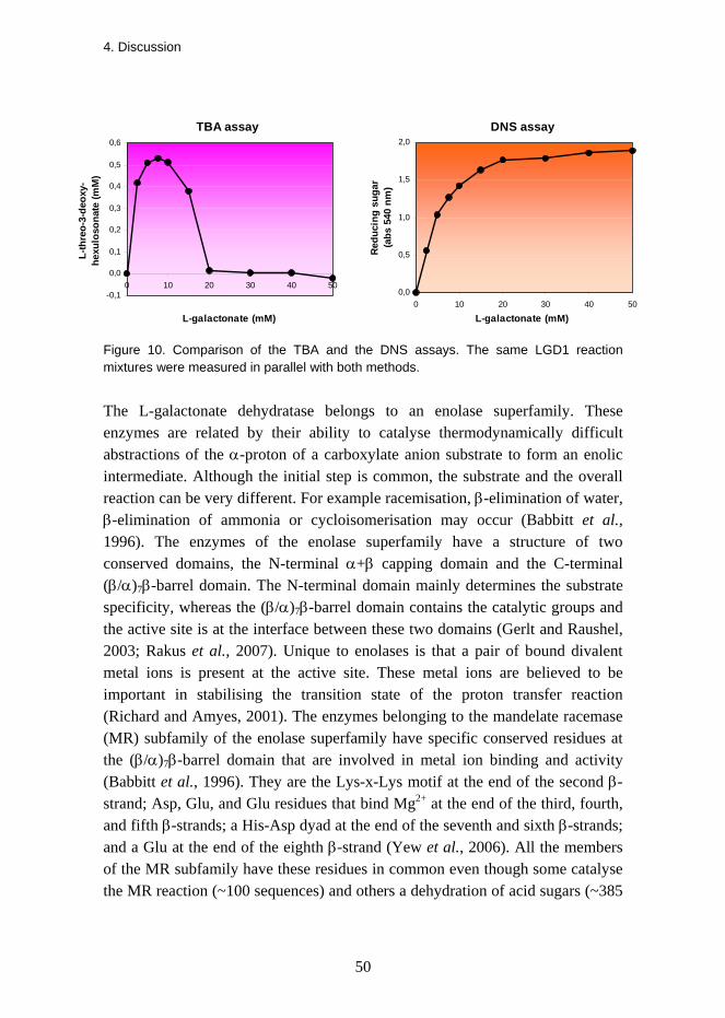
4. Discussion
50
TBA assay
-0,1
0,0
0,1
0,2
0,3
0,4
0,5
0,6
0 10 20 30 40 50
L-galactonate (mM)
L-th
reo-
3-de
oxy-
hexu
loso
nate
(mM
)
DNS assay
0,0
0,5
1,0
1,5
2,0
0 10 20 30 40 50
L-galactonate (mM)
Redu
cing
sug
ar(a
bs 5
40 n
m)
Figure 10. Comparison of the TBA and the DNS assays. The same LGD1 reaction mixtures were measured in parallel with both methods.
The L-galactonate dehydratase belongs to an enolase superfamily. These enzymes are related by their ability to catalyse thermodynamically difficult abstractions of the α-proton of a carboxylate anion substrate to form an enolic intermediate. Although the initial step is common, the substrate and the overall reaction can be very different. For example racemisation, β-elimination of water, β-elimination of ammonia or cycloisomerisation may occur (Babbitt et al., 1996). The enzymes of the enolase superfamily have a structure of two conserved domains, the N-terminal α+β capping domain and the C-terminal (β/α)7β-barrel domain. The N-terminal domain mainly determines the substrate specificity, whereas the (β/α)7β-barrel domain contains the catalytic groups and the active site is at the interface between these two domains (Gerlt and Raushel, 2003; Rakus et al., 2007). Unique to enolases is that a pair of bound divalent metal ions is present at the active site. These metal ions are believed to be important in stabilising the transition state of the proton transfer reaction (Richard and Amyes, 2001). The enzymes belonging to the mandelate racemase (MR) subfamily of the enolase superfamily have specific conserved residues at the (β/α)7β-barrel domain that are involved in metal ion binding and activity (Babbitt et al., 1996). They are the Lys-x-Lys motif at the end of the second β-strand; Asp, Glu, and Glu residues that bind Mg2+ at the end of the third, fourth, and fifth β-strands; a His-Asp dyad at the end of the seventh and sixth β-strands; and a Glu at the end of the eighth β-strand (Yew et al., 2006). All the members of the MR subfamily have these residues in common even though some catalyse the MR reaction (~100 sequences) and others a dehydration of acid sugars (~385
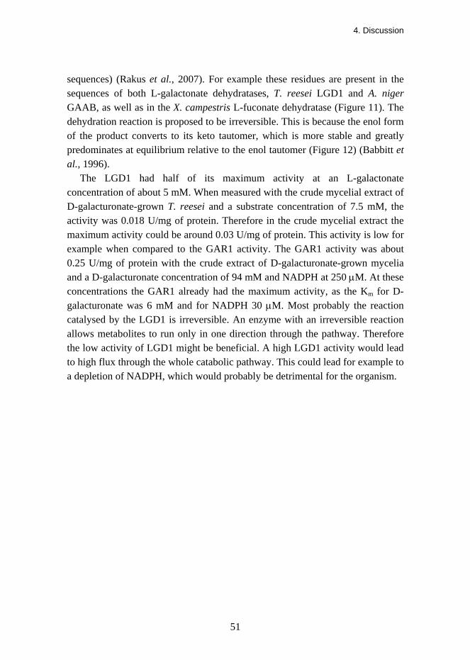
4. Discussion
51
sequences) (Rakus et al., 2007). For example these residues are present in the sequences of both L-galactonate dehydratases, T. reesei LGD1 and A. niger GAAB, as well as in the X. campestris L-fuconate dehydratase (Figure 11). The dehydration reaction is proposed to be irreversible. This is because the enol form of the product converts to its keto tautomer, which is more stable and greatly predominates at equilibrium relative to the enol tautomer (Figure 12) (Babbitt et al., 1996). The LGD1 had half of its maximum activity at an L-galactonate concentration of about 5 mM. When measured with the crude mycelial extract of D-galacturonate-grown T. reesei and a substrate concentration of 7.5 mM, the activity was 0.018 U/mg of protein. Therefore in the crude mycelial extract the maximum activity could be around 0.03 U/mg of protein. This activity is low for example when compared to the GAR1 activity. The GAR1 activity was about 0.25 U/mg of protein with the crude extract of D-galacturonate-grown mycelia and a D-galacturonate concentration of 94 mM and NADPH at 250 μM. At these concentrations the GAR1 already had the maximum activity, as the Km for D-galacturonate was 6 mM and for NADPH 30 μM. Most probably the reaction catalysed by the LGD1 is irreversible. An enzyme with an irreversible reaction allows metabolites to run only in one direction through the pathway. Therefore the low activity of LGD1 might be beneficial. A high LGD1 activity would lead to high flux through the whole catabolic pathway. This could lead for example to a depletion of NADPH, which would probably be detrimental for the organism.
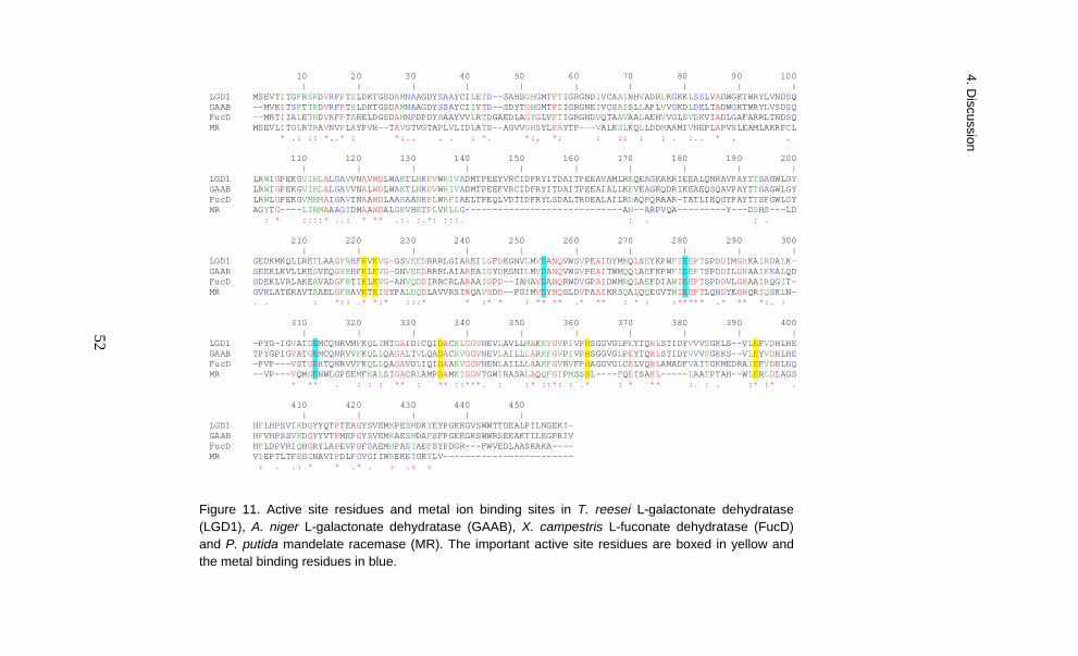
4. Discussion
52
10 20 30 40 50 60 70 80 90 100 | | | | | | | | | |LGD1 MSEVTITGFRSRDVRFPTSLDKTGSDAMNAAGDYSAAYCILETD--SAHSGHGMTFTIGRGNDIVCAAINHVADRLKGKKLSSLVADWGKTWRYLVNDSQGAAB --MVKITSFTTRDVRFPTSLDKTGSDAMNAAGDYSSAYCIIYTD--SDYTGHGMTFTIGRGNEIVCSAISLLAPLVVGKDLDELTADWGKTWRYLVSDSQFucD --MRTIIALETHDVRFPTSRELDGSDAMNPDPDYSAAYVVLRTDGAEDLAGYGLVFTIGRGNDVQTAAVAALAEHVVGLSVDKVIADLGAFARRLTNDSQMR MSEVLITGLRTRAVNVPLAYPVH--TAVGTVGTAPLVLIDLATS--AGVVGHSYLFAYTP---VALKSLKQLLDDMAAMIVNEPLAPVSLEAMLAKRFCL * .: :: *..* : *:.. . . : *. *:. *: : :: : : . :.. * . . 110 120 130 140 150 160 170 180 190 200 | | | | | | | | | |LGD1 LRWIGPEKGVIHLALGAVVNAVWDLWAKTLNKPVWRIVADMTPEEYVRCIDFRYITDAITPEEAVAMLREQEAGKAKRIEEALQNRAVPAYTTSAGWLGYGAAB LRWIGPEKGVIHLALGAVVNALWDLWAKTLNKPVWRIVADMTPEEFVRCIDFRYITDAITPEEAIALLKEVEAGKQDRIKEAEQSQAVPAYTTSAGWLGYFucD LRWLGPEKGVMHMAIGAVINAAWDLAARAANKPLWRFIAELTPEQLVDTIDFRYLSDALTRDEALAILRDAQPQRAAR-TATLIEQGYPAYTTSPGWLGYMR AGYTG----LIRMAAAGIDMAAWDALGKVHETPLVKLLG-----------------------------AN--ARPVQA---------Y---DSHS---LD : * ::::* ..: * ** .:. :.*: :::. : . : . 210 220 230 240 250 260 270 280 290 300 | | | | | | | | | |LGD1 GEDKMKQLLRETLAAGYRHFKVKVG-GSVEEDRRRLGIAREILGFDKGNVLMVDANQVWSVPEAIDYMKQLSEYKPWFIEEPTSPDDIMGHKAIRDALK-GAAB SEEKLKVLLKESVEQGYKHFKLKVG-GNVEEDRRRLAIAREAIGYDKGNILMVDANQVWSVPEAITWMQQLAEFKPWFIEEPTSPDDILGHAAIKKALQDFucD SDEKLVRLAKEAVADGFRTIKLKVG-ANVQDDIRRCRLARAAIGPD--IAMAVDANQRWDVGPAIDWMRQLAEFDIAWIEEPTSPDDVLGHAAIRQGIT-MR GVKLATERAVTAAELGFRAVKTKIGYPALDQDLAVVRSIRQAVGDD--FGIMVDYNQSLDVPAAIKRSQALQQEGVTWIEEPTLQHDYEGHQRIQSKLN- . . : *:: .* *:* :::* * :* * : ** ** .* ** : * : :***** .* ** *:. : 310 320 330 340 350 360 370 380 390 400 | | | | | | | | | |LGD1 -PYG-IGVATGEMCQNRVMFKQLIMTGAIDICQIDACRLGGVNEVLAVLLMAKKYGVPIVPHSGGVGLPEYTQHLSTIDYVVVSGKLS--VLEFVDHLHEGAAB TPYGPIGVATGEMCQNRVVFKQLLQAGALTVLQADACRVGGVNEVLAILLLARKFGVPIVPHSGGVGLPEYTQHLSTIDYVVVSGKKS--VLEYVDHLHEFucD -PVP---VSTGEHTQNRVVFKQLLQAGAVDLIQIDAARVGGVNENLAILLLAAKFGVRVFPHAGGVGLCELVQHLAMADFVAITGKMEDRAIEFVDHLHQMR --VP---VQMGENWLGPEEMFKALSIGACRLAMPDAMKIGGVTGWIRASALAQQFGIPMSSHL----FQEISAHL-----LAATPTAH--WLERLDLAGS * ** . : : : ** : ** ::***. : :* ::*: : .* : * ** :. : . :* :* . 410 420 430 440 450 | | | | | LGD1 HFLHPSVIKDGYYQTPTEAGYSVEMKPESMDKYEYPGKKGVSWWTTDEALPILNGEKI- GAAB HFVHPSSVKDGYYVTPMEPGYSVEMKAESMDAFSFPGEEGKSWWRSEEAKTILEGPRIV FucD HFLDPVRIQHGRYLAPEVPGFSAEMHPASIAEFSYPDGR---FWVEDLAASKAKA---- MR VIEPTLTFEGGNAVIPDLPGVGIIWREKEIGKYLV------------------------ : . .: * * .* . : .: :
Figure 11. Active site residues and metal ion binding sites in T. reesei L-galactonate dehydratase (LGD1), A. niger L-galactonate dehydratase (GAAB), X. campestris L-fuconate dehydratase (FucD) and P. putida mandelate racemase (MR). The important active site residues are boxed in yellow and the metal binding residues in blue.
4. Discussion
52
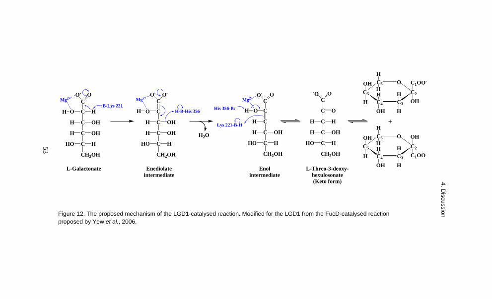
4. Discussion
53
L-Threo-3-deoxy-hexulosonate (Keto form)
L-Galactonate
C HO
C OHH
C OHH
C HHO
CH2OH
C
CO
C OHH
C OHH
C HHO
CH2OH
CO
CH
C OHH
C HHO
CH2OH
H
C
C
C HH
C OHH
C HHO
CH2OH
O:B-Lys 221
H-B-His 356
H2O
His 356-B:
Lys 221-B-H
C6
C5
C4 C3
C2
OOH
H
OH
C1OO-H
HOH
H
H
H
C6
C5
C4 C3
C2
OOH
H
C1OO-
OHH
HOH
H
H
H
+
Enol intermediate
-O OC
O- OC
O- O O-O-
Mg2+
HH
Mg2+ Mg2+
Enediolate intermediate
Figure 12. The proposed mechanism of the LGD1-catalysed reaction. Modified for the LGD1 from the FucD-catalysed reaction proposed by Yew et al., 2006.
53
4. Discussion

4. Discussion
54
4.4 The L-threo-3-deoxy-hexulosonate aldolase
The L-threo-3-deoxy-hexulosonate aldolase (LGA1) is a putative member of the dihydrodipicolinate synthase (DHDPS) family (also called N-acetylneuraminate lyase family) that comprises several pyruvate-dependent class I aldolases. The enzymes of the class I aldolase family share a common TIM (β/α)8 barrel structure but catalyse diverse reactions in different pathways. They all have a conserved lysine residue at the active site that stabilises the reaction intermediate via Schiff-base formation (Lys174 in LGA1). In addition several other highly conserved residues have been found in the enzymes of the DHDPS family (Barbosa et al., 2000). Such residues are also present in the LGA1 sequence, including the GxxGE motif (from Gly49 toGlu53) that is associated with binding the α-keto acid carboxylate moiety of the reaction substrate and the salt bridge forming Arg/Glu residues (Arg287 and Glu61) that link the C-terminal three-helical cluster with the (β/α)8 barrel. The DHDPS family enzymes have a common structure in which the N-terminal TIM (β/α)8 barrel is followed by a C-terminal domain of three α-helices. This structure is also predicted for the LGA1.
The LGA1 showed stereospecificity, as there was activity with L-threo-3-deoxy-hexulosonate and D-glycero-3-deoxy-pentulosonate but not with D-threo-3-deoxy-hexulosonate. This suggested that the reaction substrate must have the hydroxyl group of the C4 on the right in the Fischer projection. In addition the hydroxyl group of the C5 probably has to be on the left, as it is in L-threo-3-deoxy-hexulosonate. The D-glycero-3-deoxy-pentulosonate can also adopt this structure because the single bond at C4-C5 is free to rotate about the bond axis. The D-threo-3-deoxy-hexulosonate that was not a substrate has the opposite configuration at the C4 and C5 compared to the L-form. The structures of these compounds are compared in Figure 13. The LGA1 was also stereospecific in the reverse direction. With pyruvate only those aldehydes reacted that had a hydroxyl group at the C2 and on the left in the Fischer projection. L-Glyceraldehyde was a substrate, whereas D-glyceraldehyde was not. The other accepted substrates glycolaldehyde and methylglyoxal can rotate the hydroxyl group of the C2 on either side. There was no activity with acetaldehyde, which does not have a C2 hydroxyl group.
4. Discussion
55
L-Threo-3-deoxy-hexulosonate
COO-
C
C HH
C OHH
C HHO
CH2OH
D-Glycero-3-deoxy-pentulosonate
COO-
C
C HH
C OHH
C HHO
H
D-Threo-3-deoxy-hexulosonate
COO-
C
C HH
C HHO
C OHH
CH2OH
L-Glyceraldehyde
CHO
C HHO
CH2OH
CHO
C HHO
H
CHO
CHO
CH2
CHO
C OHH
CH2OH
CHO
C HH
H
Glycolaldehyde
Methylglyoxal
D-Glyceraldehyde Acetaldehyde
CHO
CO
CH3
A)
B)
OOO
Figure 13. A) L-Threo-3-deoxy-hexulosonate and D-glycero-3-deoxy-pentulosonate were substrates in the forward reaction of the LGA1. D-Threo-3-deoxy-hexulosonate was not a reaction substrate. B) L-Glyceraldehyde, glycolaldehyde and methylglyoxal were substrates in the reverse reaction with pyruvate. D-glyceraldehyde and acetaldehyde were not reaction substrates.
A similar enzyme to the T. reesei LGA1, a 2-keto-3-deoxy-L-rhamnonate (L-erythro-3,6-dideoxy-hexulosonate) aldolase of P. stipitis, cleaves 2-keto-3-deoxy-L-rhamnonate to pyruvate and L-lactaldehyde (Twerdochlib et al., 1994). In equilibrium the reaction mixture consisted of about 90% of the substrate 2-keto-3-deoxy-L-rhamnonate and 10% of the products pyruvate and L-lactaldehyde. It could be assumed that the LGA1 also has its equilibrium on the side of the substrate. We observed that the L-threo-3-deoxy-hexulosonate accumulated to a concentration of about 1.5 mM (0.3 g/l) in the culture when T. reesei was grown from four to six days on D-galacturonate. The Km for L-threo-3-deoxy-hexulosonate was 3.5 mM. In the D-galacturonate pathway there is an irreversible reaction both before and after the aldolase reaction. The L-galactonate dehydratase produces L-threo-3-deoxy-hexulosonate, the substrate for the aldolase, while the glyceraldehyde reductase removes L-glyceraldehyde,

4. Discussion
56
the reaction product of the aldolase reaction. It appears that the LGA1 can operate close to the equilibrium but the constant production of its substrate and the removal of its product drive the metabolism in the forward direction.
The T. reesei wild type strain and the lga1-deletion strain were both cultivated in liquid medium containing D-galacturonate. The dry masses of the produced mycelia were compared. We also measured the amount of L-threo-3-deoxy-hexulosonate in the media. Both results were reported separately as grams per litre. However, it was not taken into account that there was a lot more of the wild type mycelia in the culture compared to the lga1-deletion strain that hardly grew on D-galacturonate. Even so, the lga1-deletion strain produced significantly more L-threo-3-deoxy-hexulosonate into the growth medium than the wild type strain. The amounts of L-threo-3-deoxy-hexulosonate produced with respect to the dry masses are presented in Table 5.
Table 5. Production of L-threo-3-deoxy-hexulosonate (grams per gram of mycelial dry mass).
Strain 2 days 4 days 6 days Wild type 0.09 0.08 0.06 lga1-deletion 3.9 11 19
The concentration of L-threo-3-deoxy-hexulosonate in the culture of the wild type strain did not increase along with the amount of mycelium. It appears that the flow of metabolites through the D-galacturonate pathway increased when more L-threo-3-deoxy-hexulosonate was produced. Therefore the L-threo-3-deoxy-hexulosonate concentration in the culture was relatively constant throughout the six-day experiment. The opposite was true for the lga1-deletion strain. A small amount of mycelium was produced in the beginning of the experiment from the peptone in the medium. The D-galacturonate could not be used for growth because the produced L-threo-3-deoxy-hexulosonate was not metabolised further. There was no increase in the mycelial dry mass after two days, but the concentration of L-threo-3-deoxy-hexulosonate increased at an almost constant rate. The mycelium corresponding to one gram of dry mass produced about 3.7 grams (18.5 mmol) of L-threo-3-deoxy-hexulosonate per day between days two and six in the experiment. L-Threo-3-deoxy-hexulosonate is produced in the irreversible reaction catalysed by L-galactonate dehydratase. The accumulation of L-threo-3-deoxy-hexulosonate did not appear to inhibit the

4. Discussion
57
preceding reactions, because the production rate did not slow down. The L-threo-3-deoxy-hexulosonate concentration did not increase when all the enzymes of the pathway were present (wild type), which indicates that it is the L-galactonate dehydratase reaction that sets the limit for the flow rate. From this it could be concluded that in vivo in the conditions of this experiment the flow of metabolites through the D-galacturonate pathway was about 18.5 mmol/day/gram of mycelial dry mass.
4.5 The glyceraldehyde reductase and DHA reductase
There are two kinds of NADP+-dependent glycerol dehydrogenases. Those classified as EC 1.1.1.72 catalyse conversion of glycerol and NADP+ to glyceraldehyde, NADPH and H+. The others are classified as EC 1.1.1.156 and catalyse the reaction of glycerol and NADP+ to DHA, NADPH and H+. In many cases a high pH was required to detect reactions in vitro in this direction (Viswanath-Reddy et al., 1978; Schuurink et al., 1990; Witteveen et al., 1990). The reason for this is that hydrogen ions are produced in the dehydrogenase reaction. At high pH they are neutralised to water and do not accumulate in the reaction mixture, which would hinder the reaction (Asnis and Brodie, 1953). Under physiological pH conditions the reaction equilibrium was strongly on the side of glycerol production. Therefore the enzymes are discussed here as glyceraldehyde reductases and DHA reductases, respectively. The enzymes are rather unspecific and in general both types can catalyse a reaction with glyceraldehyde and DHA. There is, however, a preference for one of these substrates over the other.
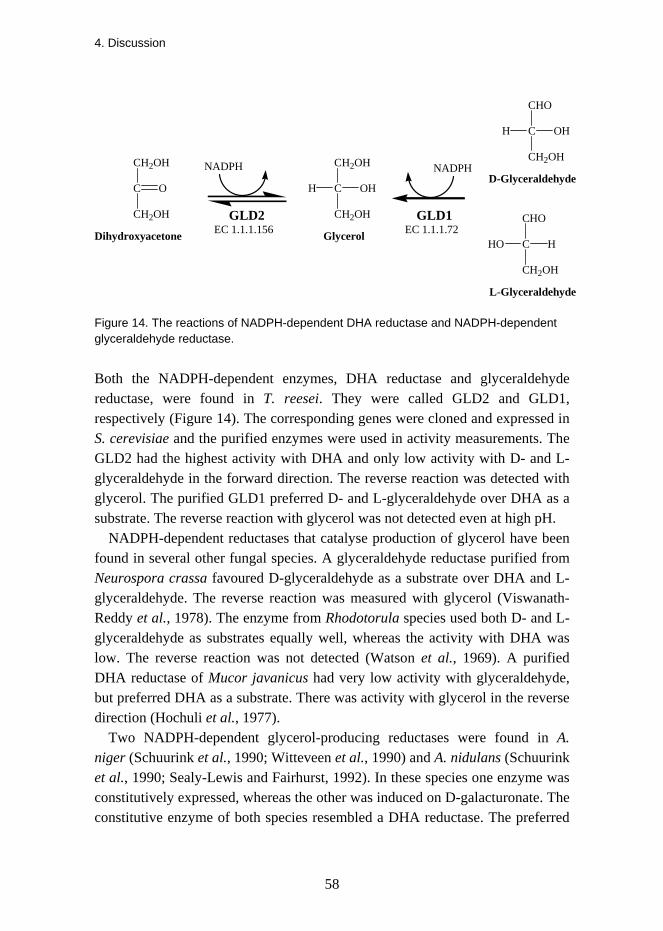
4. Discussion
58
L-Glyceraldehyde
CHO
C HHO
CH2OH
CHO
C OHH
CH2OH
D-GlyceraldehydeCH2OH
C OHH
CH2OH
CH2OH
C
CH2OH
GlycerolDihydroxyacetone
O
NADPH NADPH
GLD1GLD2EC 1.1.1.156 EC 1.1.1.72
Figure 14. The reactions of NADPH-dependent DHA reductase and NADPH-dependent glyceraldehyde reductase.
Both the NADPH-dependent enzymes, DHA reductase and glyceraldehyde reductase, were found in T. reesei. They were called GLD2 and GLD1, respectively (Figure 14). The corresponding genes were cloned and expressed in S. cerevisiae and the purified enzymes were used in activity measurements. The GLD2 had the highest activity with DHA and only low activity with D- and L-glyceraldehyde in the forward direction. The reverse reaction was detected with glycerol. The purified GLD1 preferred D- and L-glyceraldehyde over DHA as a substrate. The reverse reaction with glycerol was not detected even at high pH.
NADPH-dependent reductases that catalyse production of glycerol have been found in several other fungal species. A glyceraldehyde reductase purified from Neurospora crassa favoured D-glyceraldehyde as a substrate over DHA and L-glyceraldehyde. The reverse reaction was measured with glycerol (Viswanath-Reddy et al., 1978). The enzyme from Rhodotorula species used both D- and L-glyceraldehyde as substrates equally well, whereas the activity with DHA was low. The reverse reaction was not detected (Watson et al., 1969). A purified DHA reductase of Mucor javanicus had very low activity with glyceraldehyde, but preferred DHA as a substrate. There was activity with glycerol in the reverse direction (Hochuli et al., 1977).
Two NADPH-dependent glycerol-producing reductases were found in A. niger (Schuurink et al., 1990; Witteveen et al., 1990) and A. nidulans (Schuurink et al., 1990; Sealy-Lewis and Fairhurst, 1992). In these species one enzyme was constitutively expressed, whereas the other was induced on D-galacturonate. The constitutive enzyme of both species resembled a DHA reductase. The preferred

4. Discussion
59
substrate was DHA, whereas the activity with D-glyceraldehyde was about 10% of that with DHA, and there was no reaction with L-glyceraldehyde. The enzymes had activity with glycerol in the reverse direction (Schuurink et al., 1990). The D-galacturonate-induced enzyme was a glyceraldehyde reductase, as was shown with the A. niger enzyme. It had high activity with D-glyceraldehyde (Witteveen et al., 1990). The corresponding enzyme of A. nidulans was also proposed to be involved in D-glyceraldehyde catabolism, but activity was shown only in the reverse direction with glycerol (Sealy-Lewis and Fairhurst, 1992). The difference between these Aspergillus species was that when A. nidulans was grown on D-galacturonate it produced an additional unspecific NAD+-dependent alcohol dehydrogenase that reduced D-glyceraldehyde to glycerol (Hondmann et al., 1991).
Based on its substrate preference the T. reesei GLD2 belonged to the group of NADPH-dependent DHA reductases. The same conclusion was made from its amino acid sequence. There are few sequences available from similar enzymes in other filamentous fungi. The T. reesei GLD2 amino acid sequence was 86% identical with the Trichoderma atroviride Gld1 (Seidl et al., 2004) and 71% identical with the A. nidulans GldB (de Vries et al., 2003). In T. reesei and T. atroviride there were no introns in the corresponding gene, whereas the A. nidulans gene contained one intron. The NADPH-dependent alkyl 4-halo-3-oxobutyrate reductase (KER) of the filamentous fungus Penicillium citrinum did not show activity with glycerol and NADP+ (Asako et al., 2005). However, the amino acid sequence of KER was 67% identical with GLD2.
One role of the NADPH-dependent DHA reductase in filamentous fungi is in glycerol production. In A. nidulans two pathways have been shown to operate simultaneously at high osmolarity. In the main route dihydroxyacetone phosphate (DHAP) is dephosphorylated to DHA that is then reduced to glycerol (Redkar et al., 1995). In the other pathway DHAP is reduced to glycerol-3-phosphate that is then dephosphorylated to glycerol (Fillinger et al., 2001). Another proposed role for DHA reductase was that it rapidly converts the toxic DHA into glycerol. The enzyme apparently has high physiological importance, as it was present at high concentrations under different growth conditions (Schuurink et al., 1990).
Sequences of NADPH-dependent glyceraldehyde reductases of filamentous fungi were not known before the T. reesei GLD1. However, S. cerevisiae has NADPH-dependent reductases Ypr1p and Gcy1p that preferred DL-glyceraldehyde over DHA as a substrate (Petrash et al., 2001; Ford and Ellis,

4. Discussion
60
2002). Their amino acid sequences have been published (Norbeck and Blomberg, 1997) and both are 37% identical with the T. reesei GLD1 (40% and 39% with GLD2, respectively). By comparison, the identity of GLD1 with the above-mentioned A. nidulans GldB, P. citrinum KER and T. atroviride Gld1 is 41%, 40% and 37%, respectively. It was observed in the transcriptome analysis of A. niger that gaaD that was the closest homologue in this species to the T. reesei gld1 gene that was specifically induced on D-galacturonate (Martens-Uzunova and Schaap, 2008). The corresponding enzymes are 57% identical. However, the enzyme activity of GAAD has not been reported.
The main role of the NADPH-dependent glyceraldehyde reductase is in the catabolism of glyceraldehyde. D-Glyceraldehyde is produced in the non-phosphorylated pathway for D-gluconate (Elzainy et al., 1973) and D-galactonate catabolism (Elshafei and Abdel-Fatah, 2001). L-Glyceraldehyde is generated in the D-galacturonate pathway. All these three pathways involve a dehydratase and an aldolase that sequentially convert the sugar acid to the corresponding 2-keto-3-deoxy sugar acid and then to pyruvate and glyceraldehyde.
It could be assumed that the T. reesei GLD2 is constitutively expressed and that the GLD1 is induced when grown on D-galacturonate. However, in contrast to the glyceraldehyde reductases of A. niger (Witteveen et al., 1990) and A. nidulans (Sealy-Lewis and Fairhurst, 1992), the GLD1 activity was not specifically induced on D-galacturonate. The GLD1 activity was tested after growth on D-galacturonate, glycerol, lactose, D-glucose and D-xylitol and similar activity was observed with L- and D-glyceraldehyde in all the tested crude mycelial extracts. This could indicate that the GLD1 has a more general role in T. reesei than to reduce L-glyceraldehyde produced in the D-galacturonate pathway.
T. reesei converts D-galacturonate to pyruvate and glycerol. Pyruvate is an intermediate in several central metabolic pathways. For example, pyruvate can be further converted into carbohydrates, fatty acids, the amino acid alanine or energy. T. reesei is also able to utilise glycerol. Most probably the main route for glycerol catabolism in T. reesei proceeds via glycerol kinase and a mitochondrial FAD-dependent glycerol-3-phosphate dehydrogenase, as in A. nidulans and in most other eukaryotic organisms when growing on glycerol (Hondmann et al., 1991). The product DHAP is an intermediate of the glycolytic pathway, as can be seen in Figure 15. In another possible pathway glycerol is first converted to DHA by a reverse reaction of an NADPH-dependent DHA reductase and subsequently to DHAP by a DHA kinase. In A. oryzae this is the preferred catabolic glycerol pathway (Salazar et al., 2009). This route produces NADPH
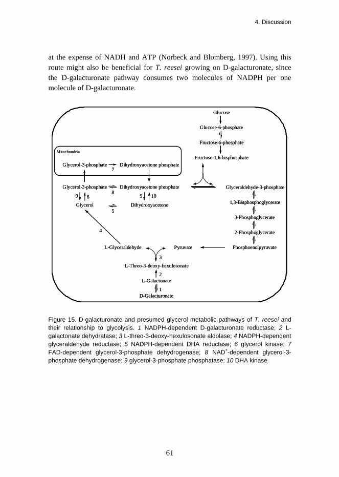
4. Discussion
61
at the expense of NADH and ATP (Norbeck and Blomberg, 1997). Using this route might also be beneficial for T. reesei growing on D-galacturonate, since the D-galacturonate pathway consumes two molecules of NADPH per one molecule of D-galacturonate.
Glucose
Glucose-6-phosphate
Fructose-6-phosphate
Fructose-1,6-bisphosphate
Glyceraldehyde-3-phosphateDihydroxyacetone phosphate
1,3-Bisphosphoglycerate
3-Phosphoglycerate
2-Phosphoglycerate
PhosphoenolpyruvatePyruvate
Glycerol Dihydroxyacetone
Glycerol-3-phosphate
D-Galacturonate
L-Galactonate
L-Threo-3-deoxy-hexulosonate
L-Glyceraldehyde
1
2
3
4
6
7
5
9
Dihydroxyacetone phosphateGlycerol-3-phosphate
89 10
Mitochondria
Figure 15. D-galacturonate and presumed glycerol metabolic pathways of T. reesei and their relationship to glycolysis. 1 NADPH-dependent D-galacturonate reductase; 2 L-galactonate dehydratase; 3 L-threo-3-deoxy-hexulosonate aldolase; 4 NADPH-dependent glyceraldehyde reductase; 5 NADPH-dependent DHA reductase; 6 glycerol kinase; 7 FAD-dependent glycerol-3-phosphate dehydrogenase; 8 NAD+-dependent glycerol-3-phosphate dehydrogenase; 9 glycerol-3-phosphate phosphatase; 10 DHA kinase.
4. Discussion
62
4.6 Biotechnological applications of the D-galacturonate pathway
The production of citrus, apple or sugar beet juice generates processing residues that contain large amounts of pectin and other carbohydrate polysaccharides. Several million dry tons of these residues are produced globally every year, which causes disposal problems. Currently, these carbohydrate-rich residues are mainly used as cattle feed, but their feed value is low because of the low protein content (Grohmann and Bothast, 1994). Furthermore, for transporting and to prevent spoilage the residues must be dried and pelletised. In the case of sugar beet this can require up to 30–40% of the total energy of the processing (Doran et al., 2000). This kind of a carbohydrate-rich by-product could be a potential raw material for microbial conversion to more valuable products.
In many plant materials complexes of lignin and hemicellulose protect cellulose fibres from enzymatic hydrolysis. In citrus, apple and sugar beet processing residues the lignin content is low and the monomeric sugars can easily be released by treatment with cellulolytic and pectinolytic enzymes (Grohmann and Bothast, 1994). After extraction of sucrose from sugar beet, 70–75% of the dry mass of the remaining pulp is still carbohydrates, mainly D-glucose (21.1%), D-galacturonate (21.1%), L-arabinose (20.9%) and five other sugars (10% altogether) (Micard et al., 1996).
Two fungi, T. reesei and A. niger, have been metabolically engineered to convert D-galacturonate to galactarate. In both fungi the gene coding for the D-galacturonate reductase, the gar1 in T. reesei and the gaaA in A. niger, was first deleted in order to prevent them from catabolising D-galacturonate. Then a gene from A. tumefaciens that codes for an NAD+-dependent D-galacturonate dehydrogenase (EC 1.1.1.203) was introduced to both strains. The enzyme catalyses the conversion of D-galacturonate to galactarate. The T. reesei strain produced galactarate at a high yield. The A. niger strain was apparently able to metabolise galactarate, which resulted in a lower yield. Galactarate has applications for example in the food, cosmetics and pharmaceutical industries. It might also be useful in synthesising polymers (Mojzita et al., 2010).
The T. reesei strain in which the lgd1, the gene coding for the L-galactonate dehydratase, was deleted was unable to grow on D-galacturonate but converted it to L-galactonate. L-Galactonate could be useful as an acidifying agent in the food industry and could also have applications in the cosmetics industry. The L-galactonate dehydratase enzyme (LGD1) could be used to produce L-threo-3-
4. Discussion
63
deoxy-hexulosonate from L-galactonate or D-glycero-3-deoxy-pentulosonate from D-arabinonate. The reaction catalysed by the LGD1 is irreversible and the substrate would be completely converted to the dehydrated product. L-Threo-3-deoxy-hexulosonate could also be produced from D-galacturonate with a T. reesei strain in which lga1, the gene coding for L-threo-3-deoxy-hexulosonate aldolase, was deleted. This strain did not grow on D-galacturonate but converted it to L-threo-3-deoxy-hexulosonate.

5. Conclusions and future prospects
64
5. Conclusions and future prospects Previously it was not known how eukaryotes catabolise D-galacturonate, although there was evidence that such a pathway exists and that it is different from the prokaryotic D-galacturonate pathways. In this study the first eukaryotic catabolic D-galacturonate pathway was identified in the filamentous fungus Trichoderma reesei. This novel pathway consisted of four enzymes. These four new fungal enzymes were NADPH-dependent D-galacturonate reductase, L-galactonate dehydratase, L-threo-3-deoxy-hexulosonate aldolase and NADPH-dependent glyceraldehyde reductase. In the first step D-galacturonate reductase used NADPH as a cofactor to convert D-galacturonate to L-galactonate. This was a reversible reaction. In the second step L-galactonate dehydratase removed a water molecule from L-galactonate and this irreversible reaction produced L-threo-3-deoxy-hexulosonate. In the third step L-threo-3-deoxy-hexulosonate aldolase cut the carbon chain of L-threo-3-deoxy-hexulosonate in the middle, which produced pyruvate and L-glyceraldehyde. This enzyme reaction also operated in the reverse direction. In the fourth step L-glyceraldehyde was converted to glycerol by an NADPH-dependent glyceraldehyde reductase. The backward reaction was not detected with this enzyme.
The first three enzyme activities were present in the crude mycelial extract only when T. reesei was grown on D-galacturonate, which indicated that these enzyme activities were involved in D-galacturonate utilisation. When the corresponding gene of any of these three enzymes was deleted in T. reesei the resulting strain was no longer able to grow with D-galacturonate as the only carbon source. This showed that these enzymes were essential for D-galacturonate catabolism and that there were no alternative pathways in T. reesei. The same set of enzymes also operated in Aspergillus niger in D-galacturonate catabolism, and there is genetic evidence that the same pathway is
5. Conclusions and future prospects
65
also conserved in other plant cell wall-degrading fungi in the subphylum Pezizomycotina (Martens-Uzunova and Schaap, 2008).
For a long time there has been a strong interest worldwide in producing ethanol efficiently from plant biomass for use as a renewable transport fuel. Many different approaches have been developed and tested to find the best ways for ethanol production. The catabolic D-galacturonate pathways have also been seen to have some potential in this respect. It has been suggested that D-galacturonate could be converted to ethanol by S. cerevisiae after equipping it with a heterologous D-galacturonate pathway along with a D-galacturonate transporter (van Maris et al., 2006). However, if the fungal D-galacturonate pathway were to be used, one of the main problems with this hypothetical yeast strain might be its increased need for NADPH, because two enzymes of the fungal pathway were NADPH-dependent reductases.
Converting the various components of different biomaterials to more valuable products has become currently one of the main goals in biotechnology. Filamentous fungi are important tools in biotechnology. Basic research on their physiology and enzymology, as performed in this thesis work, creates a basis from which the various biotechnological applications arise. It has already been shown that the different deletion strains constructed during this work can be used to convert D-galacturonate to L-galactonate or to L-threo-3-deoxy-hexulosonate and with additional metabolic engineering to galactarate (Mojzita et al., 2010). In addition, all the four new genes of the fungal D-galacturonate pathway could be cloned and expressed in Saccharomyces cerevisiae as functional enzymes. Discovering new enzymes always extends the possibilities to develop new biotechnological applications.

66
References Agius, F., González-Lamothe, R., Caballero, J.L., Muñoz-Blanco, J., Botella, M.A. and Valpuesta, V. (2003). Engineering increased vitamin C levels in plants by overexpression of a D-galacturonic acid reductase. Nat. Biotechnol. 21, 177–181.
Asako, H., Wakita, R., Matsumura, K., Shimizu, M., Sakai, J. and Itoh, N. (2005). Purification and cDNA cloning of NADPH-dependent aldoketoreductase, involved in asymmetric reduction of methyl 4-bromo-3-oxobutyrate, from Penicillium citrinum IFO4631. Appl. Environ. Microbiol. 71, 1101–1104.
Ashwell, G., Wahba, A.J. and Hickman, J. (1958). A new pathway of uronic acid metabolism. Biochim. Biophys. Acta 30, 186–187.
Ashwell, G., Wahba, A.J. and Hickman, J. (1960). Uronic acid metabolism in bacteria. I. Purification and properties of uronic acid isomerase in Escherichia coli. J. Biol. Chem. 235, 1559–1565.
Asnis, R.E. and Brodie, A.F. (1953). A glycerol dehydrogenase from Escherichia coli. J. Biol. Chem. 203, 153–159.
Babbitt, P.C., Hasson, M.S., Wedekind, J.E., Palmer, D.R.J., Barrett, W.C., Reed, G.H., Rayment, I., Ringe, D., Kenyon, G.L. and Gerlt, J.A. (1996). The enolase superfamily: A general strategy for enzyme-catalyzed abstraction of the α-protons of carboxylic acids. Biochemistry 35, 16489–16501.
Barbosa, J.A.R.G., Smith, B.J., DeGori, R., Ooi, H.C., Marcuccio, S.M., Campi, E.M., Jackson, W.R., Brossmer, R., Sommer, M. and Lawrence, M.C. (2000). Active site modulation in the N-acetylneuraminate lyase sub-family as revealed by the structure of the inhibitor-complexed Haemophilus influenzae enzyme. J. Mol. Biol. 303, 405–421.
Bateman, D.F., Kosuge, T. and Kilgore, W.W. (1970). Purification and properties of uronate dehydrogenase from Pseudomonas syringae. Arch. Biochem. Biophys. 136, 97–105.
Bernfeld, P. (1955) Amylases, α and β. In Methods in Enzymology, Vol. 1. Colovick, S.P. and Kaplan, N.O. (eds.). New York: Academic Press. Pp. 149–158.
Blanco, P., Sieiro, C. and Villa, T.G. (1999). Production of pectic enzymes in yeasts. FEMS Microbiol. Lett. 175, 1–9.
Brownleader, M.D., Jackson, P., Mobasheri, A., Pantelides, A.T., Sumar, S., Trevan, M. and Dey, P.M. (1999). Molecular aspects of cell wall modifications during fruit ripening. Crit. Rev. Food Sci. Nutr. 39, 149–164.

67
Buchanan, C.L., Connaris, H., Danson, M.J., Reeve, C.D. and Hough, D.W. (1999). An extremely thermostable aldolase from Sulfolobus solfataricus with specificity for non-phosphorylated substrates. Biochem. J. 343, 563–570.
Carpita, N.C. and Gibeaut, D.M. (1993). Structural models of primary cell walls in flowering plants: consistency of molecular structure with the physical properties of the walls during growth. Plant J. 3, 1–30.
Chang, Y.F. and Feingold, D.S. (1970). D-Glucaric acid and galactaric acid catabolism by Agrobacterium tumefaciens. J. Bacteriol. 102, 85–96.
Chang, Y.F. and Feingold, D.S. (1969). Hexuronic acid dehydrogenase of Agrobacterium tumefaciens. J. Bacteriol. 99, 667–673.
Cynkin, M.A. and Ashwell, G. (1960). Uronic acid metabolism in bacteria. IV. Purification and properties of 2-keto-3-deoxy-D-gluconokinase in Escherichia coli. J. Biol. Chem. 235, 1576–1579.
Dagley, S. and Trudgill, P.W. (1965). The metabolism of galactarate, D-glucarate and various pentoses by species of Pseudomonas. Biochem. J. 95, 48–58.
Dahms, A.S. and Anderson, R.L. (1969). 2-Keto-3-deoxy-L-arabonate aldolase and its role in a new pathway of L-arabinose degradation. Biochem. Biophys. Res. Commun. 36, 809–814.
Davey, M.W., Gilot, C., Persiau, G., Ostergaard, J., Han, Y., Bauw, G.C. and Van Montagu, M.C. (1999). Ascorbate biosynthesis in Arabidopsis cell suspension culture. Plant Physiol. 121, 535–543.
de Vries, R.P., Flitter, S.J., van de Vondervoort, P.J.I., Chaveroche, M.K., Fontaine, T., Fillinger, S., Ruijter, G.J.G., d'Enfert, C. and Visser, J. (2003). Glycerol dehydrogenase, encoded by gldB is essential for osmotolerance in Aspergillus nidulans. Mol. Microbiol. 49, 131–141.
Doran, J.B., Cripe, J., Sutton, M. and Foster, B. (2000). Fermentations of pectin-rich biomass with recombinant bacteria to produce fuel ethanol. Appl. Biochem. Biotechnol. 84–86, 141–152.
Elshafei, A.M. and Abdel-Fatah, O.M. (2001). Evidence for a non-phosphorylated route of galactose breakdown in cell-free extracts of Aspergillus niger. Enzyme Microb. Technol. 29, 76–83.
Elshafei, A.M. and Abdel-Fatah, O.M. (1991). Nonphosphorolytic pathway for D-galactonate catabolism in Aspergillus terreus. Enzyme Microb. Technol. 13, 930–934.

68
Elzainy, T.A., Hassan, M.M. and Allam, A.M. (1973). New pathway for nonphosphorylated degradation of gluconate by Aspergillus niger. J. Bacteriol. 114, 457–459.
Fillinger, S., Ruijter, G., Tamás, M.J., Visser, J., Thevelein, J.M. and d'Enfert, C. (2001). Molecular and physiological characterization of the NAD-dependent glycerol 3-phosphate dehydrogenase in the filamentous fungus Aspergillus nidulans. Mol. Microbiol. 39, 145–157.
Ford, G. and Ellis, E.M. (2002). Characterization of Ypr1p from Saccharomyces cerevisiae as a 2-methylbutyraldehyde reductase. Yeast 19, 1087–1096.
Gamauf, C., Metz, B. and Seiboth, B. (2007) Degradation of plant cell wall polymers by Fungi. In Environmental and Microbial Relationships, Vol. 4. Kubicek, C.P. and Druzhinina, I.S. (eds.). Springer Berlin Heidelberg. Pp. 325–340.
Gerlt, J.A. and Raushel, F.M. (2003). Evolution of function in (β/α)8-barrel enzymes. Curr. Opin. Chem. Biol. 7, 252–264.
Grant, G.T., Morris, E.R., Rees, D.A., Smith, P.J.C. and Thom, D. (1973). Biological interactions between polysaccharides and divalent cations: The egg-box model. FEBS Lett. 32, 195–198.
Grohmann, K. and Bothast, R.J. (1994) Pectin-rich residues generated by processing of citrus fruits, apples, and sugar beets: enzymatic hydrolysis and biological conversion to value-added products. In Enzymatic Conversion of Biomass for Fuels Production. Himmel, M.E., Baker, J.O. and Overend, R.P. (eds.). ACS Symposium Series 566. Pp. 372–390.
Hara, A., Deyashiki, Y., Nakayama, T. and Sawada, H. (1983). Isolation and characterization of multiforms of aldehyde reductase in chicken kidney. Eur. J. Biochem. 133, 207–214.
Hayashi, S., Watanabe, M. and Kimura, A. (1984). Enzymatic determination of free glucuronic acid with glucuronolactone reductase. I. Isolation and purification of glucuronolactone reductase from rat kidney. J. Biochem. 95, 223–232.
Hickman, J. and Ashwell, G. (1960). Uronic acid metabolism in bacteria. II. Purification and properties of D-altronic acid and D-mannonic acid dehydrogenases in Escherichia coli. J. Biol. Chem. 235, 1566–1570.
Hochuli, E., Taylor, K.E. and Dutler, H. (1977). Dihydroxyacetone reductase from Mucor javanicus. 2. Identification of the physiological substrate and reactivity towards related compounds. Eur. J. Biochem. 75, 433–439.

69
Hondmann, D.H.A., Busink, R., Witteveen, C.F.B. and Visser, J. (1991). Glycerol catabolism in Aspergillus nidulans. J. Gen. Microbiol. 137, 629–636.
Hubbard, B.K., Koch, M., Palmer, D.R.J., Babbitt, P.C. and Gerlt, J.A. (1998). Evolution of enzymatic activities in the enolase superfamily: characterization of the (D)-glucarate/galactarate catabolic pathway in Escherichia coli. Biochemistry 37, 14369-14375.
Hugouvieux-Cotte-Pattat, N. and Robert-Baudouy, J. (1987). Hexuronate catabolism in Erwinia chrysanthemi. J. Bacteriol. 169, 1223–1231.
Ishikawa, T., Masumoto, I., Iwasa, N., Nishikawa, H., Sawa, Y., Shibata, H., Nakamura, A., Yabuta, Y. and Shigeoka, S. (2006). Functional characterization of D-galacturonic acid reductase, a key enzyme of the ascorbate biosynthesis pathway, from Euglena gracilis. Biosci. Biotechnol. Biochem. 70, 2720–2726.
Jayani, R.S., Saxena, S. and Gupta, R. (2005). Microbial pectinolytic enzymes: a review. Process Biochemistry 40, 2931–2944.
Kessler, G., Neufeld, E.F., Feingold, D.S. and Hassid, W.Z. (1961). Metabolism of D-glucuronic acid and D-galacturonic acid by Phaseolus aureus seedlings. J. Biol. Chem. 236, 308–312.
Kilgore, W.W. and Starr, M.P. (1959a). Uronate oxidation by phytopathogenic Pseudomonads. Nature 183, 1412–1413.
Kilgore, W.W. and Starr, M.P. (1959b). Catabolism of galacturonic and glucuronic acids by Erwinia carotovora. J. Biol. Chem. 234, 2227–2235.
Kuhls, K., Lieckfeldt, E., Samuels, G.J., Kovacs, W., Meyer, W., Petrini, O., Gams, W., Börner, T. and Kubicek, C.P. (1996). Molecular evidence that the asexual industrial fungus Trichoderma reesei is a clonal derivative of the ascomycete Hypocrea jecorina. Proc. Natl. Acad. Sci. USA. 93. Pp. 7755–7760.
Loewus, F.A. and Kelly, S. (1961). The metabolism of D-galacturonic acid and its methyl ester in the detached ripening strawberry. Arch. Biochem. Biophys. 95, 483–493.
Mandels, M. and Weber, J. (1969). The production of cellulases. In Cellulases and their applications. Vol. 95. Hajny, G.J. and Reese, E.T. (eds.). American Chemical Society. Pp. 391–414.
Martens-Uzunova, E., Schaap, P., van den Berg, J. and Benen, J. (2005). Cross species gene discovery using microarray analysis allows for the identification of D-galacturonic acid utilization pathway in Aspergillus niger and Aspergillus nidulans. Fungal Genetics Newsletter 52, p. 93.
70
Martens-Uzunova, E.S. and Schaap, P.J. (2008). An evolutionary conserved D-galacturonic acid metabolic pathway operates across filamentous fungi capable of pectin degradation. Fungal Genet. Biol. 45, 1449–1457.
Martinez, D., Berka, R.M., Henrissat B., Saloheimo, M., Arvas, M., Baker, S.E., Chapman, J., Chertkov, O., Coutinho, P.M., Cullen, D., Danchin, E.G.J., Grigoriev, I.V., Harris, P., Jackson, M., Kubicek, C.P., Han, C.S., Ho, I., Larrondo, L.F., de Leon, A.L., Magnuson, J.K., Merino, S., Misra, M., Nelson, B., Putnam, N., Robbertse, B., Salamov, A.A., Schmoll, M., Terry, A., Thayer, N., Westerholm-Parvinen, A., Schoch, C.L., Yao, J., Barabote, R., Nelson, M.A., Detter, C., Bruce, D., Kuske, C.R., Xie, G., Richardson, P., Rokhsar, D.S., Lucas, S.M., Rubin, E.M., Dunn-Coleman, N., Ward, M. and Brettin, T.S. (2008). Genome sequencing and analysis of the biomass-degrading fungus Trichoderma reesei (syn. Hypocrea jecorina). Nat. Biotechnol. 26, 553–560.
Medina-Escobar, N., Cárdenas, J., Moyano, E., Caballero, J.L. and Muñoz-Blanco, J. (1997). Cloning, molecular characterization and expression pattern of a strawberry ripening-specific cDNA with sequence homology to pectate lyase from higher plants. Plant Mol. Biol. 34, 867–877.
Meloche, H.P. and Wood, W.A. (1964). Crystallization and characteristics of 2-keto-3-deoxy-6-phosphogluconic aldolase. J. Biol. Chem. 239, 3515–3518.
Micard, V., Renard, C.M.G.C. and Thibault, J.F. (1996). Enzymatic saccharification of sugar-beet pulp. Enzyme Microb. Technol. 19, 162–170.
Mohnen, D. (2008). Pectin structure and biosynthesis. Curr. Opin. Plant Biol. 11, 266–277.
Mojzita, D., Wiebe, M., Hilditch, S., Boer, H., Penttilä, M. and Richard, P. (2010). Metabolic engineering of fungal strains for conversion of D-galacturonate to meso-galactarate. Appl. Environ. Microbiol. 76, 169–175.
Nevalainen, H., Suominen, P. and Taimisto, K. (1994). On the safety of Trichoderma reesei. J. Biotechnol. 37, 193–200.
Nielsen, J. (1997). Metabolic control analysis of biochemical pathways based on a thermokinetic description of reaction rates. Biochem. J. 321, 133–138.
Norbeck, J. and Blomberg, A. (1997). Metabolic and regulatory changes associated with growth of Saccharomyces cerevisiae in 1.4 M NaCl – Evidence for osmotic induction of glycerol dissimilation via the dihydroxyacetone pathway. J. Biol. Chem. 272, 5544–5554.
Ogawa, K., Nakajima-Kambe, T., Nakahara, T. and Kokufuta, E. (2002). Coimmobilization of gluconolactonase with glucose oxidase for improvement in kinetic property of enzymatically induced volume collapse in ionic gels. Biomacromolecules 3, 625–631.

71
Pérez, S., Rodríguez-Carvajal, M.A. and Doco, T. (2003). A complex plant cell wall polysaccharide: rhamnogalacturonan II. A structure in quest of a function. Biochimie 85, 109–121.
Petrash, J.M., Murthy, B.S.N., Young, M., Morris, K., Rikimaru, L., Griest, T.A. and Harter, T. (2001). Functional genomic studies of aldo-keto reductases. Chem. Biol. Interact. 130, 673–683.
Prasanna, V., Prabha, T.N. and Tharanathan, R.N. (2007). Fruit ripening phenomena – An overview. Crit. Rev. Food Sci. Nutr. 47, 1–19.
Preiss, J. and Ashwell, G. (1963a). Polygalacturonic acid metabolism in bacteria. I. Enzymatic formation of 4-deoxy-L-threo-5-hexoseulose uronic acid. J. Biol. Chem. 238, 1571–1576.
Preiss, J. and Ashwell, G. (1963b). Polygalacturonic acid metabolism in bacteria. II. Formation and metabolism of 3-deoxy-D-glycero-2,5-hexodiulosonic acid. J. Biol. Chem. 238, 1577–1583.
Rakus, J.F., Fedorov, A.A., Fedorov, E.V., Glasner, M.E., Vick, J.E., Babbitt, P.C., Almo, S.C. and Gerlt, J.A. (2007). Evolution of enzymatic activities in the enolase superfamily: D-mannonate dehydratase from Novosphingobium aromaticivorans. Biochemistry 46, 12896–12908.
Ralet, M.C., Dronnet, V., Buchholt, H.C. and Thibault, J.F. (2001). Enzymatically and chemically de-esterified lime pectins: characterisation, polyelectrolyte behaviour and calcium binding properties. Carbohydr. Res. 336, 117–125.
Ransom, S.C. and Gerlt, J.A. (1988). Cloning, DNA sequence analysis, and expression in Escherichia coli of the gene for mandelate racemase from Pseudomonas putida. Biochemistry 27, 540–545.
Redkar, R.J., Locy, R.D. and Singh, N.K. (1995). Biosynthetic pathways of glycerol accumulation under salt strass in Aspergillus nidulans. Exp. Mycol. 19, 241–246.
Reese, E.T. (1976). History of the cellulase program at the U.S. army Natick Development Center. Biotechnol. Bioeng. Symp. Pp. 9–20.
Reese, E.T., Levinson, H.S., Downing, M.H. and White, W.L. (1950). Quartermaster culture collection. Farlowia 4, 45–86.
Renard, C.M.G.C., Crépeau, M.J. and Thibault, J.F. (1995). Structure of the repeating units in the rhamnogalacturonic backbone of apple, beet and citrus pectins. Carbohydr. Res. 275, 155–165.

72
Richard, J.P. and Amyes, T.L. (2001). Proton transfer at carbon. Curr. Opin. Chem. Biol. 5, 626–633.
Ried, J.L. and Collmer, A. (1986). Comparison of pectic enzymes produced by Erwinia chrysanthemi, Erwinia carotovora subsp. carotovora, and Erwinia carotovora subsp. atroseptica. Appl. Environ. Microbiol. 52, 305–310.
Riov, J. (1975). Metabolism of uronic acids in plant tissues: partial purification and properties of uronic acid oxidase from citrus leaves. Plant Physiol. 55, 602–606.
Salazar, M., Vongsangnak, W., Panagiotou, G., Andersen, M.R. and Nielsen, J. (2009). Uncovering transcriptional regulation of glycerol metabolism in Aspergilli through genome-wide gene expression data analysis. Mol. Genet. Genomics 282, 571–586.
Schuurink, R., Busink, R., Hondmann, D.H.A., Witteveen, C.F.B. and Visser, J. (1990). Purification and properties of NADP+-dependent glycerol dehydrogenases from Aspergillus nidulans and A. niger. J. Gen. Microbiol. 136, 1043–1050.
Sealy-Lewis, H.M. and Fairhurst, V. (1992). An NADP+-dependent glycerol dehydrogenase in Aspergillus nidulans is inducible by D-galacturonate. Curr. Genet. 22, 293–296.
Seidl, V., Seibel, C., Kubicek, C.P. and Schmoll, M. (2009). Sexual development in the industrial workhorse Trichoderma reesei. Proc. Natl. Acad. Sci. USA. 106, 13909–13914.
Seidl, V., Seiboth, B., Karaffa, L. and Kubicek, C.P. (2004). The fungal STRE-element-binding protein Seb1 is involved but not essential for glycerol dehydrogenase (gld1) gene expression and glycerol accumulation in Trichoderma atroviride during osmotic stress. Fungal Genet. Biol. 41, 1132–1140.
Siebers, B. and Schönheit, P. (2005). Unusual pathways and enzymes of central carbohydrate metabolism in Archaea. Curr. Opin. Microbiol. 8, 695–705.
Smiley, J.D. and Ashwell, G. (1960). Uronic acid metabolism in bacteria. III. Purification and properties of D-altronic acid and D-mannonic acid dehydrases in Escherichia coli. J. Biol. Chem. 235, 1571–1575.
Soares, M.M.C.N., da Silva, R. and Gomes, E. (1999). Screening of bacterial strains for pectinolytic activity: characterization of the polygalacturonase produced by Bacillus sp. Rev. Microbiol. 30, 299–303.
Tom, G.D., Viswanath-Reddy, M. and Howe, H.B. Jr. (1978). Effect of carbon source on enzymes involved in glycerol metabolism in Neurospora crassa. Arch. Microbiol. 117, 259–263.
73
Twerdochlib, A.L., Pedrosa, F.O., Funayama, S. and Rigo, L.U. (1994). L-Rhamnose metabolism in Pichia stipitis and Debaryomyces polymorphus . Can. J. Microbiol. 40, 896–902.
Uitzetter, J.H.A.A., Bos, C.J. and Visser, J. (1986). Characterization of Aspergillus nidulans mutants in carbon metabolism isolated after D-galacturonate enrichment. J. Gen. Microbiol. 132, 1167–1172.
Uitzetter, J.H.A.A., Bos, C.J. and Visser, J. (1982). Growth characteristics of Aspergillus nidulans mutants defective in carbohydrate metabolism. Antonie van Leeuwenhoek 48, 219–227.
Ul Hassan, M. and Lehninger, A.L. (1956). Enzymatic formation of ascorbic acid in rat liver extracts. J. Biol. Chem. 223, 123–138.
van Maris, A.J.A., Abbott, D.A., Bellissimi, E., van den Brink, J., Kuyper, M., Luttik, M.A.H., Wisselink, H.W., Scheffers, W.A., van Dijken, J.P. and Pronk, J.T. (2006). Alcoholic fermentation of carbon sources in biomass hydrolysates by Saccharomyces cerevisiae: current status. Antonie van Leeuwenhoek 90, 391–418.
Veiga, L.A. and Guimarães, M.F. (1991). L-Fuconate dehydratase: purification and properties of the enzyme from Pullularia pullulans. Arq. Biol. Tecnol. 34, 537–553.
Vidal, S., Doco, T., Williams, P., Pellerin, P., York, W.S., O’Neill, M.A., Glushka, J., Darvill, A.G. and Albersheim, P. (2000). Structural characterization of the pectic polysaccharide rhamnogalacturonan II: evidence for the backbone location of the aceric acid-containing oligoglycosyl side chain. Carbohydr. Res. 326, 277–294.
Viswanath-Reddy, M., Pyle, J.E. and Howe Jr., H.B. (1978). Purification and properties of NADP+-linked glycerol dehydrogenase from Neurospora crassa. J. Gen. Microbiol. 107, 289-296.
Wagner, G. and Hollmann, S. (1976). Uronic acid dehydrogenase from Pseudomonas syringae. Purification and properties. Eur. J. Biochem. 61, 589–596.
Watanabe, S., Kodaki, T. and Makino, K. (2006a). Cloning, expression, and characterization of bacterial L-arabinose 1-dehydrogenase involved in an alternative pathway of L-arabinose metabolism. J. Biol. Chem. 281, 2612–2623.
Watanabe, S., Kodaki, T. and Makino, K. (2006b). A novel α-ketoglutaric semialdehyde dehydrogenase. Evolutionary insight into an alternative pathway of bacterial L-arabinose metabolism. J. Biol. Chem. 281, 28876–28888.
Watanabe, S., Shimada, N., Tajima, K., Kodaki, T. and Makino, K. (2006c). Identification and characterization of L-arabonate dehydratase, L-2-keto-3-deoxyarabonate

74
dehydratase, and L-arabinolactonase involved in an alternative pathway of L-arabinose metabolism. Novel evolutionary insight into sugar metabolism. J. Biol. Chem. 281, 33521–33536.
Watanabe, S., Piyanart, S. and Makino, K. (2008a). Metabolic fate of L-lactaldehyde derived from an alternative L-rhamnose pathway. FEBS J. 275, 5139–5149.
Watanabe, S., Saimura, M. and Makino, K. (2008b). Eukaryotic and bacterial gene clusters related to an alternative pathway of nonphosphorylated L-rhamnose metabolism. J. Biol. Chem. 283, 20372–20382.
Watson, J.A., Hayashi, J.A., Schuytema, E. and Doughty, C.C. (1969). Identification of reduced nicotinamide adenine dinucleotide phosphate-dependent aldehyde reductase in a Rhodotorula strain. J. Bacteriol. 100, 110–116.
Wheeler, G.L., Jones, M.A. and Smirnoff, N. (1998). The biosynthetic pathway of vitamin C in higher plants. Nature 393, 365–369.
Willats, W.G.T., McCartney, L., Mackie, W., and Knox, J.P. (2001a). Pectin: cell biology and prospects for functional analysis. Plant Mol. Biol. 47, 9–27.
Willats, W.G.T., Orfila, C., Limberg, G., Buchholt, H.C., van Alebeek, G.J.W.M., Voragen, A.G.J., Marcus, S.E., Christensen, T.M.I.E., Mikkelsen, J.D., Murray, B.S. and Knox, J.P. (2001b). Modulation of the degree and pattern of methyl-esterification of pectic homogalacturonan in plant cell walls. Implications for pectin methyl esterase action, matrix properties, and cell adhesion. J. Biol. Chem. 276, 19404–19413.
Witteveen, C.F.B., van de Vondervoort, P., Dijkema, C., Swart, K. and Visser, J. (1990). Characterization of a glycerol kinase mutant of Aspergillus niger. J. Gen. Microbiol. 136, 1299–1305.
Yew, W.S., Fedorov, A.A., Fedorov, E.V., Rakus, J.F., Pierce, R.W., Almo, S.C. and Gerlt, J.A. (2006). Evolution of enzymatic activities in the enolase superfamily: L-fuconate dehydratase from Xanthomonas campestris. Biochemistry 45, 14582–14597.
Zajic, J.E. (1959). Hexuronic dehydrogenase of Agrobacterium tumefaciens. J. Bacteriol. 78, 734–735.
Zandleven, J., Beldman, G., Bosveld, M., Benen, J. and Voragen, A. (2005). Mode of action of xylogalacturonan hydrolase towards xylogalacturonan and xylogalacturonan oligosaccharides. Biochem. J. 387, 719–725.
Publication I is not included in the PDF version. Please order the printed version to get the complete publication (http://www.vtt.fi/publications/index.jsp).
Publication II
L-galactonate dehydratase is part of the fungal path for D-galacturonic
acid catabolism
In: Molecular Microbiology 2006. Vol. 61, pp. 1060–1068.
©2006 Blackwell Publishing Ltd. Reprinted with permission from the publisher.


L-galactonate dehydratase is part of the fungal path forD-galacturonic acid catabolism
Satu Kuorelahti,1 Paula Jouhten,2 Hannu Maaheimo,2
Merja Penttilä1 and Peter Richard1*1VTT Biotechnology, Espoo, Finland.2NMR-laboratory and Structural Biology and BiophysicsProgram, VTT Biotechnology, Helsinki, Finland.
Summary
An L-galactonate dehydratase and the correspondinggene were identified from the mould Hypocrea jeco-rina (Trichoderma reesei). This novel enzyme con-verts L-galactonate to L-threo-3-deoxy-hexulosonate(2-keto-3-deoxy-L-galactonate). The enzyme is part ofthe fungal pathway for D-galacturonic acid catabo-lism, a pathway which is only partly known. It isthe second enzyme of this pathway after theD-galacturonic acid reductase. L-galactonate dehy-dratase activity is present in H. jecorina cells grownon D-galacturonic acid but absent when other carbonsources are used for growth. A deletion of theL-galactonate dehydratase gene in H. jecorina resultsin a strain with no growth on D-galacturonic acid. Theactive enzyme was produced in the heterologous hostSaccharomyces cerevisiae and characterized. Itexhibited activity with L-galactonate and D-arabonatewhere the hydroxyl group of the C2 is in L- and thehydroxyl group of the C3 is in D-configuration inthe Fischer projection. However, it did not exhibitactivity with D-galactonate, D-gluconate, L-gulonate orD-xylonate where the hydroxyl groups of the C2 andC3 are in different configuration.
Introduction
D-galacturonic acid is the major component of pectinand consequently an important carbon source for micro-organisms living on decaying plant material. Abacterial catabolic pathway has been described whilea eukaryotic pathway has remained unknown. Thebacterial pathway consists of five enzymes convertingD-galacturonic acid (D-galacturonate) to pyruvate and D-glyceraldehyde-3-phosphate. The intermediate metabo-lites are D-tagaturonate, D-altronate, D-erythro-3-deoxy-hexulosonate and D-erythro-3-deoxy-hexulosonate-6-
phosphate. The enzymes are uronate isomerase,D-tagaturonate reductase, altronate dehydratase,2-dehydro-3-deoxy-D-gluconate kinase and 2-dehydro-3-deoxy-D-gluconate-6-phosphate aldolase respectively(Ashwell et al., 1960; Cynkin and Ashwell, 1960; Hickmanand Ashwell, 1960; Smiley and Ashwell, 1960; Melocheand Wood, 1964). There are no reports about geneswhich are similar to the genes of the bacterialD-galacturonic acid pathway in the sequenced genomesof eukaryotic microorganisms. There are also no reportsdescribing these enzyme activities present in bacteria inyeasts or moulds. However, a similar pathway must existin eukaryotic microorganisms as many species of yeastand mould can utilize and grow on D-galacturonic acid.This suggests that there is a eukaryotic pathway for thecatabolism of D-galacturonic acid which is different fromthe bacterial pathway.
We have previously presented that the first step in thefungal pathway is an NADPH-specific D-galacturonic acidreductase generating L-galactonate. This enzyme activitywas induced in the filamentous fungus Hypocrea jecorina(Trichoderma reesei) when grown on D-galacturonic acidand the activity was absent when grown on other carbonsources (Kuorelahti et al., 2005). Information about howL-galactonate is further catabolized is lacking.
There are only a few other studies on D-galacturonicacid catabolism in fungal microorganisms. Uitzetter et al.(1986) mutagenized the filamentous fungus Aspergillusnidulans and found that mutants lacking pyruvate dehy-drogenase or pyruvate carboxylase activity were unableto grow on D-galacturonic acid, whereas a pyruvatekinase mutant was able to grow on this carbon source.This was interpreted that D-galacturonic acid is convertedto pyruvate but not through phosphoenolpyruvate. It wassuggested that in A. nidulans D-galacturonic acid iscatabolized via a non-phosphorylating pathway throughglyceraldehyde and pyruvate (Visser et al., 1988; Hond-mann et al., 1991), and that D-glyceraldehyde is an inter-mediate (Uitzetter et al., 1986). The non-phosphorylatingpath was supported by a microarray analysis of genestranscribed in Aspergillus niger grown on D-galacturonicacid. Here genes similar to aldoketo reductase, racemaseand aldolase were identified as coexpressed genes(Martens-Uzunova et al., 2005). It was further suggestedthat D-galacturonic acid is metabolized through glycerolbecause a glycerol kinase mutant had reduced growth on
Accepted 19 June, 2006. *For correspondence. E-mail [email protected]; Tel. (+358) 20 7227190; Fax (+358) 20 7227071.
Molecular Microbiology (2006) 61(4), 1060–1068 doi:10.1111/j.1365-2958.2006.05294.x
© 2006 The AuthorsJournal compilation © 2006 Blackwell Publishing Ltd
II/1
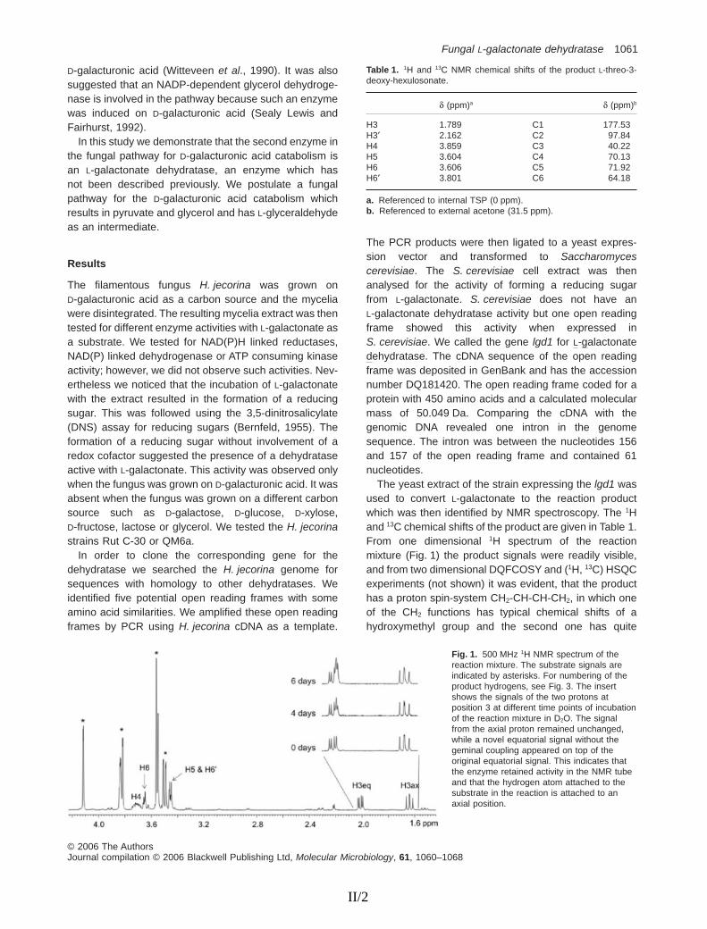
D-galacturonic acid (Witteveen et al., 1990). It was alsosuggested that an NADP-dependent glycerol dehydroge-nase is involved in the pathway because such an enzymewas induced on D-galacturonic acid (Sealy Lewis andFairhurst, 1992).
In this study we demonstrate that the second enzyme inthe fungal pathway for D-galacturonic acid catabolism isan L-galactonate dehydratase, an enzyme which hasnot been described previously. We postulate a fungalpathway for the D-galacturonic acid catabolism whichresults in pyruvate and glycerol and has L-glyceraldehydeas an intermediate.
Results
The filamentous fungus H. jecorina was grown onD-galacturonic acid as a carbon source and the myceliawere disintegrated. The resulting mycelia extract was thentested for different enzyme activities with L-galactonate asa substrate. We tested for NAD(P)H linked reductases,NAD(P) linked dehydrogenase or ATP consuming kinaseactivity; however, we did not observe such activities. Nev-ertheless we noticed that the incubation of L-galactonatewith the extract resulted in the formation of a reducingsugar. This was followed using the 3,5-dinitrosalicylate(DNS) assay for reducing sugars (Bernfeld, 1955). Theformation of a reducing sugar without involvement of aredox cofactor suggested the presence of a dehydrataseactive with L-galactonate. This activity was observed onlywhen the fungus was grown on D-galacturonic acid. It wasabsent when the fungus was grown on a different carbonsource such as D-galactose, D-glucose, D-xylose,D-fructose, lactose or glycerol. We tested the H. jecorinastrains Rut C-30 or QM6a.
In order to clone the corresponding gene for thedehydratase we searched the H. jecorina genome forsequences with homology to other dehydratases. Weidentified five potential open reading frames with someamino acid similarities. We amplified these open readingframes by PCR using H. jecorina cDNA as a template.
The PCR products were then ligated to a yeast expres-sion vector and transformed to Saccharomycescerevisiae. The S. cerevisiae cell extract was thenanalysed for the activity of forming a reducing sugarfrom L-galactonate. S. cerevisiae does not have anL-galactonate dehydratase activity but one open readingframe showed this activity when expressed inS. cerevisiae. We called the gene lgd1 for L-galactonatedehydratase. The cDNA sequence of the open readingframe was deposited in GenBank and has the accessionnumber DQ181420. The open reading frame coded for aprotein with 450 amino acids and a calculated molecularmass of 50.049 Da. Comparing the cDNA with thegenomic DNA revealed one intron in the genomesequence. The intron was between the nucleotides 156and 157 of the open reading frame and contained 61nucleotides.
The yeast extract of the strain expressing the lgd1 wasused to convert L-galactonate to the reaction productwhich was then identified by NMR spectroscopy. The 1Hand 13C chemical shifts of the product are given in Table 1.From one dimensional 1H spectrum of the reactionmixture (Fig. 1) the product signals were readily visible,and from two dimensional DQFCOSY and (1H, 13C) HSQCexperiments (not shown) it was evident, that the producthas a proton spin-system CH2-CH-CH-CH2, in which oneof the CH2 functions has typical chemical shifts of ahydroxymethyl group and the second one has quite
Table 1. 1H and 13C NMR chemical shifts of the product L-threo-3-deoxy-hexulosonate.
d (ppm)a d (ppm)b
H3 1.789 C1 177.53H3′ 2.162 C2 97.84H4 3.859 C3 40.22H5 3.604 C4 70.13H6 3.606 C5 71.92H6′ 3.801 C6 64.18
a. Referenced to internal TSP (0 ppm).b. Referenced to external acetone (31.5 ppm).
Fig. 1. 500 MHz 1H NMR spectrum of thereaction mixture. The substrate signals areindicated by asterisks. For numbering of theproduct hydrogens, see Fig. 3. The insertshows the signals of the two protons atposition 3 at different time points of incubationof the reaction mixture in D2O. The signalfrom the axial proton remained unchanged,while a novel equatorial signal without thegeminal coupling appeared on top of theoriginal equatorial signal. This indicates thatthe enzyme retained activity in the NMR tubeand that the hydrogen atom attached to thesubstrate in the reaction is attached to anaxial position.
Fungal L-galactonate dehydratase 1061
© 2006 The AuthorsJournal compilation © 2006 Blackwell Publishing Ltd, Molecular Microbiology, 61, 1060–1068
II/2
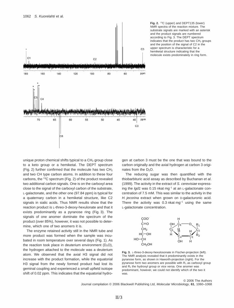
unique proton chemical shifts typical to a CH2 group closeto a keto group or a hemiketal. The DEPT spectrum(Fig. 2) further confirmed that the molecule has two CH2
and two CH type carbon atoms. In addition to these fourcarbons, the 13C spectrum (Fig. 2) of the product revealedtwo additional carbon signals. One is on the carboxyl areaclose to the signal of the carboxyl carbon of the substrate,L-galactonate, and the other one (97.84 ppm) is typical fora quaternary carbon in a hemiketal structure, like C2signals in sialic acids. Thus NMR results show that thereaction product is L-threo-3-deoxy-hexulonate and that itexists predominantly as a pyranose ring (Fig. 3). Thesignals of one anomer dominate the spectrum of theproduct (over 85%), however, it was not possible to deter-mine, which one of two anomers it is.
The enzyme retained activity still in the NMR tube andmore product was formed when the sample was incu-bated in room temperature over several days (Fig. 1). Asthe reaction took place in deuterium environment (D2O),the hydrogen attached to the molecule was a deuteriumatom. We observed that the axial H3 signal did notincrease with the product formation, while the equatorialH3 signal from the newly formed product had lost itsgeminal coupling and experienced a small upfield isotopeshift of 0.02 ppm. This indicates that the equatorial hydro-
gen at carbon 3 must be the one that was bound to thecarbon originally and the axial hydrogen at carbon 3 origi-nates from the D2O.
The reducing sugar was then quantified with thethiobarbituric acid assay as described by Buchanan et al.(1999). The activity in the extract of S. cerevisiae express-ing the lgd1 was 0.15 nkat mg-1 at an L-galactonate con-centration of 7.5 mM. This was similar to the activity in theH. jecorina extract when grown on D-galacturonic acid.There the activity was 0.3 nkat mg-1 using the sameL-galactonate concentration.
Fig. 2. 13C (upper) and DEPT135 (lower)NMR spectra of the reaction mixture. Thesubstrate signals are marked with an asteriskand the product signals are numberedaccording to Fig. 3. The DEPT spectrumindicates that the product has two CH2 groupsand the position of the signal of C2 in theupper spectrum is characteristic for ahemiketal structure indicating that themolecule exists predominately in ring form.
COO-
C
CH2
HC
CH
CH2OH
O
OH
HO
C6
C5
C4 C3
C2
OOH
H
R1
R2
H
HOH
H
H
H
Fig. 3. L-threo-3-deoxy-hexulosonate in Fischer-projection (left).The NMR analysis revealed that it predominantly exists in thepyranose form, as shown in Haworth-projection (right). For thepyranose form two anomers are possible with R1 as carboxyl groupand R2 the hydroxyl group or vice versa. One anomer waspredominant, however, we could not identify which of the two itwas.
1062 S. Kuorelahti et al.
© 2006 The AuthorsJournal compilation © 2006 Blackwell Publishing Ltd, Molecular Microbiology, 61, 1060–1068
II/3

The LGD1 protein was tagged with a histidine tagand produced in S. cerevisiae. In one construct theC-terminus of the protein was tagged, in another theN-terminal end of the protein. The C-terminal taggingresulted in an inactive protein while the N-terminal taggingresulted in a protein with reduced activity. The activityfound in the crude extract of the N-terminally taggedprotein was about five times lower than the non-taggedprotein.
As the histidine tagged LGD1 protein was either inac-tive or had reduced activity we used the crude cell extractof the yeast strain expressing the lgd1 to analyse thekinetics of the L-galactonate dehydratase. To study thespecificity of the enzyme we used different sugar acidsand tested the activity of the enzyme to convert the sugaracid to a reducing sugar using the DNS assay. Activitywas observed with the sugar acids L-galactonate andD-arabonate. The activity with D-arabonate was about50% of the activity with L-galactonate under the conditionsas specified in the Experimental procedures. Activity wasonly observed in the extract of the strain expressing thelgd1. No activity was observed in the control strain. WhenD-galactonate, D-gluconate, D-xylonate or L-gulonate wasused as substrates no activity was observed in the strainexpressing the lgd1 or in the control strain.
For the substrate L-galactonate we estimated a Km
value of about 5 mM. This was measured at pH 7.0 withyeast extract containing the LGD1 enzyme at a final con-centration of 1 g l-1 of extracted protein and following theproduction of L-threo-3-deoxy-hexulosonate using thethiobarbituric acid assay as described by Buchanan et al.(1999) or measuring the production of a reducing sugarusing the DNS assay. The control strain with the emptyplasmid showed no activity. The same Km forL-galactonate was found when a H. jecorina extract wasanalysed. Using the yeast extract, an incubation of24 h led to a complete conversion when the initialL-galactonate was 20 mM or lower, i.e. the L-threo-3-deoxy-hexulosonate concentration was equal to theinitial L-galactonate concentration (data not shown). AtL-galactonate concentrations above 20 mM the thiobarbi-turic acid assay gave values for the product formationwhich were lower that with the DNS assay. This wasinterpreted as an interference of the L-galactonic acid withthe thiobarbituric acid assay.
The enzyme has an essential requirement for bivalentcations. In the presence of 5 mM EDTA we observed acomplete loss of activity.
The transcription of the lgd1 gene on different carbonsources was examined by a Northern blot analysis. Thecarbon sources D-galacturonate, D-glucose, glycerol,lactose and D-xylose were tested. The gene was tran-scribed on all these carbon sources. The transcriptionlevels of the lgd1 gene were normalized with the transcrip-
tion levels of actin. The ratio of these two transcriptionlevels, L-galactonic acid dehydratase over actin was thesame on D-galacturonic acid, glycerol and lactose. It wasthree times higher on D-glucose and D-xylose.
To check if the lgd1 gene is essential for theD-galacturonic acid metabolism we deleted the gene in aRut C-30 strain of H. jecorina. For that purpose weconstructed a deletion cassette which contained thehygromycin B phosphotransferase gene (hph), which wastransformed and selected for hygromycin B resistance. APCR of these strains showed that only 10% had thedeletion cassette in the intended locus. These strains werefurther analysed by Southern hybridization to verify thatonly one copy of the deletion cassette had integrated intothe correct position. In the Southern hybridizations the lgd1and the hph were probed. The lgd1 gene was only seen inthe control strain while the hph gene which replaced thelgd1 gene was only in the deletion strain (not shown). Thedeletion strain (VTT D-05369) had the same growth rate onlactose as the host strain Rut C-30 on plate or in liquidculture but only residual growth on D-galacturonic acidwhile the host strain grew normal. When the lgd1 gene wastransformed back to the deletion strain the growth onD-galacturonic acid was restored. The growth rate of all thestrains including the wild-type strain QM6a was comparedin liquid medium containing 20 g l-1D-galacturonic acid and0.5 g l-1 proteose peptone or only 0.5 g l-1 proteosepeptone as a carbon source. The residual growth of thedeletion strain is due to growth on proteose peptonebecause all the strains produced the same amount ofbiomass also on proteose peptone only (Table 2).
Discussion
In this manuscript we describe the identification of a dehy-dratase that is active on the sugar acids L-galactonate andD-arabonate. These two sugar acids have in common thatthe hydroxyl groups of the C2 and the C3 in the Fischerprojection are in L- and D-configuration respectively. Othersugar acids with such a configuration were not testedbecause they were not commercially available. The dehy-dratase was neither active with sugar acids where thehydroxyl groups of C2 were in D- and C3 in L-configurationas in D-galactonate, D-gluconate and D-xylonate, nor witha sugar acid with the hydroxyl groups of C2 and C3 inD-configuration as in L-gulonate.
Dehydratases and their corresponding genes, active onsugar acids with the hydroxyl groups C2 in D- and C3 inL-configuration have been described previously. A dehy-dratase active with D-xylonate and L-arabonate wasdescribed by Niu et al. (2003) and a dehydratase activewith D-gluconate in the non-phosphorylated Entner–Doudoroff pathway was described in Sulfolobus solfatari-cus by Buchanan et al. (1999).
Fungal L-galactonate dehydratase 1063
© 2006 The AuthorsJournal compilation © 2006 Blackwell Publishing Ltd, Molecular Microbiology, 61, 1060–1068
II/4

A dehydratase which is similar to the enzyme describedin this manuscript is the D-altronate dehydratase E.C.4.2.1.7 (Smiley and Ashwell, 1960). D-altronate has thehydroxyl groups of C2 and C3 in L- and D-configurationlike L-galactonate. The enzyme is part of the bacterialD-galacturonic acid pathway and the purified D-altronatedehydratase from Escherichia coli has been character-ized (Smiley and Ashwell, 1960; Dreyer, 1987). A genecoding for the D-altronate dehydratase had been inferredby electronic annotation (GenBank accession numberAAC76126). A CLUSTALW alignment of the L-galactonatedehydratase with D-altronate dehydratase revealed a 13%identity in the amino acid sequences. The low degreeof homology suggests that L-galactonate dehydrataseand D-altronate dehydratase belong to different proteinfamilies.
The homology of L-galactonate dehydratase with anno-tated enzymes was highest with the D-galactonate dehy-dratase (E.C. 4.2.1.6) of E. coli and the mandelateracemase (E.C. 5.1.2.2) of Pseudomonas putida(Ransom et al., 1988). Their Swiss-Protein accessioncodes are Q6BF17 and P11444 respectively. Both theseenzymes have only 20% identity in amino acid sequenceswith L-galactonate dehydratase in a CLUSTALW alignment.Nevertheless, the conserved residues such as the activesite and the metal binding residues of mandelate race-mase subgroup of enolase superfamily suggested byBabbitt et al. (1996) are present in the sequence ofL-galactonate dehydratase. These conserved residueswere not found in the D-altronate dehydratase of E. coli.The reaction of each enzyme in the enolase superfamily isinitiated by an abstraction of the a-proton of a carboxylicacid to form an enolic intermediate.
The reaction product of the L-galactonate dehydratasewas identified as a reducing sugar. Reducing sugars arecommonly measured with the DNS assay. In order todefine the reaction product it was analysed by NMR. Togenerate a sufficient amount of reaction productL-galactonate was incubated in the yeast extract of thestrain expressing the L-galactonate dehydratase gene. Inthis extract the reaction product did not react further,
which facilitated the NMR analysis. In the H. jecorinamycelia extract the reaction product was degraded,making the NMR analysis more difficult. The NMR analy-sis showed that erythro- or threo-3-deoxy-hexulosonatewas formed. Knowing the substrate of the dehydratasereaction we concluded that it was L-threo-3-deoxy-hexulosonate. The NMR analysis also revealed that it ispredominantly in the pyranose form (Fig. 3). For the pyra-nose form two anomers are possible; the carboxyl groupin R1 and the hydroxyl group in R2 or vice versa (Fig. 3).The NMR suggested that one anomer is predominant butit did not allow determining which of the two anomers it is.The NMR analysis revealed also that the axial hydrogenat the carbon 3 is the hydrogen that was added during thereaction. However, as there are two possible chair con-formations of the pyranose ring it remains unclear whichof the two protons is in axial position.
Knowing that the reaction product is L-threo-3-deoxy-hexulosonate enabled us to measure its concentration.An assay where the 2-keto-3-deoxy sugar acid gives acolour reaction with thiobarbituric acid allowed the quan-tification (Buchanan et al., 1999). The H. jecorina extractproduced L-threo-3-deoxy-hexulosonate with a rate of0.3 nkat mg-1 of extracted protein; in the extract of yeastexpressing the lgd1 the rate was 0.15 nkat mg-1. Theenzyme activities of the same order of magnitude indi-cate that the heterologous expression in yeast results inan active enzyme. The accumulation of the reactionproduct in the H. jecorina extract indicated that at leastin the extract it is metabolized only slowly. In the yeastextract L-galactonate was completely converted toL-threo-3-deoxy-hexulosonate demonstrating that theenergetic equilibrium is on the side of the reactionproduct. The irreversibility of the D-galactonate dehy-dratase reaction was described by Donald et al. (1979).The reaction results in an enolic intermediate that under-goes a spontaneous ketonization. The keto tautomer ofthe product is greatly favoured relative to the enol tau-tomer. It was also shown that the D-galactonate dehy-dratase is unable to enolize the product 2-keto-3-deoxy-D-galactonate.
Table 2. Growth of the Igd1 deletion strain and control in liquid medium.
H. jecorina strainLactose + peptonedry mass (g l-1)
D-galacturonic acid + peptonedry mass (g l-1)
Peptonedry mass (g l-1)
QM6a 0.7 ± 0.1 3.4 ± 1.3 0.1 ± 0.0RutC30 4.2 ± 0.1 2.4 ± 0.7 0.1 ± 0.0RutC30-Dlgd1 A 3.3 ± 1.0 0.1 ± 0.0 0.1 ± 0.0RutC30-Dlgd1 B 3.9 ± 0.8 0.1 ± 0.0 0.1 ± 0.0RutC30-Dlgd1-lgd1 AA 4.2 ± 0.1 2.5 ± 0.6 0.1 ± 0.0RutC30-Dlgd1-lgd1 AB 4.1 ± 0.7 3.2 ± 0.5 0.1 ± 0.0
Dry mass (g l-1) after 5 days of cultivation on 0.05% peptone and 2% of another carbon source if indicated. Values are averages of triplicates. Twoparallel deletion strains were studied (A and B) and also two parallel retransformed strains (AA and AB) that were constructed by transforming lgd1into the deletion strain A.
1064 S. Kuorelahti et al.
© 2006 The AuthorsJournal compilation © 2006 Blackwell Publishing Ltd, Molecular Microbiology, 61, 1060–1068
II/5

The L-galactonate dehydratase is part of a eukaryoticpathway for the catabolism of D-galacturonic acid. Thefirst enzyme in this pathway is the D-galacturonic acidreductase converting D-galacturonic acid to L-galactonate(Kuorelahti et al., 2005), and the L-galactonate dehy-dratase described in this manuscript the second. Thereare several observations supporting this assumption. Firstof all the LGD1 protein was active with L-galactonate.Second, the activity was observed in H. jecorina extractonly when mycelia were grown on D-galacturonic acid.The activity was not observed when mycelia were grownon other carbon sources. This is already an indicationthat the L-galactonate dehydratase is related to theD-galacturonic acid catabolic pathway. Another indicationis that we did not find any other enzyme activities withL-galactonate such as reductase, dehydrogenase orkinase activities; however, this is not a proof that theseactivities do not exist. A deletion of the lgd1 gene inH. jecorina gives evidence that the L-galactonate dehy-dratase is part of the D-galacturonic acid pathwaybecause in the resulting strain growth on D-galacturonicacid is abolished (Table 2).
In the eukaryotic pathway for D-galacturonic acidcatabolism the product of this second enzyme, L-threo-3-deoxy-hexulosonate, has to be further catabolized. Thispart of the pathway is still unknown. A hypotheticalpathway is shown in Fig. 4. In this hypothetical pathwayL-threo-3-deoxy-hexulosonate is converted to pyruvateand L-glyceraldehyde by the action of an aldolase.L-glyceraldehyde is, unlike pyruvate, a metabolite which isnot part of any known metabolic pathway. We hypothesizethat L-glyceraldehyde is converted to glycerol. An NADP-dependent glycerol dehydrogenase is induced in filamen-tous fungi grown on D-galacturonate (Sealy Lewis andFairhurst, 1992). Such an enzyme might convertL-glyceraldehyde to glycerol. Glycerol is then metabolizedthrough glycerol-3-phosphate and dihydroxyacetone
phosphate as described for the mould A. nidulans (Hond-mann et al., 1991).
The catabolic path for D-galacturonic acid in fungi isdifferent from the corresponding pathway in bacteria.For E. coli a pathway was described consisting of fiveenzymes converting D-galacturonic acid to pyruvic acidand D-glyceraldehyde-3-phosphate (Ashwell et al., 1960;Cynkin and Ashwell, 1960; Hickman and Ashwell, 1960;Smiley and Ashwell, 1960; Meloche and Wood, 1964).The enzymes of this pathway are uronate isomerase,NADH-utilizing D-tagaturonate reductase, altronatedehydratase, D-erythro-3-deoxy-D-hexulosonate kinaseand D-erythro-3-deoxy-D-hexulosonate-6-phosphatealdolase. We suggest that in the fungal path only fourenzymes are required, NADPH-utilizing D-galacturonatereductase, L-galactonate dehydratase, L-threo-3-deoxy-hexulosonate aldolase and a glycerol dehydrogenasethat can convert L-glyceraldehyde in the reverse reaction(Fig. 4). The differences between bacteria and fungiare that in fungi NADPH is the required cofactor, nophosphorylated intermediates are involved and noisomerases are used.
That bacteria and fungal microorganisms use differentcatabolic pathways is not unique for D-galacturonic acid.Also other abundant carbon sources such as D-xylose andL-arabinose are catabolized differently. In bacteriaD-xylose is isomerized to D-xylulose before it is phospho-rylated to D-xylulose 5-phosphate (Lawlis et al., 1984),while in fungi the conversion from D-xylose to D-xylulose isa two-step process involving two redox reactions (Wanget al., 1980). Also the L-arabinose catabolic pathway isdistinctly different in bacteria and fungi (Lee et al., 1986;Richard et al., 2001; 2002). Common in these examples isthat the bacterial pathways have isomerase activitieswhich is absent in their fungal counterparts.
When comparing the sequence of the open readingframe with other sequences deposited in GenBank in a
Fig. 4. Hypothetical pathway for the catabolism of D-galacturonic acid in eukaryotic microorganisms. The metabolites are represented inFischer-projection. In this projection the C1 of the D-galacturonic acid is the reduced C6 of the L-galactonate. The first two enzymes of thispathway, the D-galacturonic acid reductase and the L-galactonate dehydratase have been identified. The L-galactonate dehydratase is thesubject of this communication. The remaining two enzymes, the L-threo-3-deoxy-hexulosonate aldolase and L-glyceraldehyde reductase arehypothetical.
Fungal L-galactonate dehydratase 1065
© 2006 The AuthorsJournal compilation © 2006 Blackwell Publishing Ltd, Molecular Microbiology, 61, 1060–1068
II/6

BLAST search, we identified hypothetical proteins with highhomology in other filamentous fungi such as Neurosporacrassa and A. nidulans. This suggests that L-galactonatedehydratase activity is not a unique feature of H. jecorina,but common in filamentous fungi. We also found hypo-thetical proteins with high homology (more than 50% iden-tities in the amino acid sequences) from bacteria andhigher eukaryotes indicating that the L-galactonate dehy-dratase might also be present in organisms other thanfilamentous fungi.
Experimental procedures
Strains, growth conditions and protein extracts
The E. coli strain DH5a was used in all the cloning proce-dures except the deletion cassette construction whereTOP10 Electrocomp Cells (Invitrogen) were used. Bacteriawere grown in Luria–Bertani medium with ampicillin at 37°C.The S. cerevisiae strain CEN.PK2-1D (VW-1B) was the hostfor the heterologous expression. It was grown in syntheticmedium lacking uracil when required for selection at 30°C.We used the H. jecorina (T. reesei) strains Rut C-30 orQM6a. The strains were grown in a liquid medium containing20 g l-1D-galacturonic acid/sodium D-galacturonate (pH 7.0)or another carbon source when specified, 0.5 g l-1 proteosepeptone, 15 g l-1 KH2PO4, 5 g l-1 (NH4)2SO4, 0.6 g l-1
MgSO4·7H2O, 0.6 g l-1 CaCl2·2H2O and trace elements(Mandels and Weber, 1969) at 28°C. The agar plates for themould contained also 1 ml l-1 Triton X-100 and 20 g l-1 granu-lated Difco Agar. To make protein extracts of H. jecorina orS. cerevisiae about 100 mg of fresh mycelia or cells weremixed with 300 ml of glass beads (diameter 0.4 mm) and400 ml of buffer [5 mM sodium phosphate pH 7.0 and Com-plete, EDTA-free protease inhibitor (Roche)] and disinte-grated in a Mini-Bead Beater (Biospec Products) for threetimes for 30 s. The mixture was then centrifuged in an Eppen-dorf microcentrifuge at full speed for 25 min at 4°C and thesupernatant used for the analysis. For fungal protein extractsupernatant a second centrifugation of 10 min was needed.The protein content of the extract was estimated using theBio-Rad protein assay and BSA as a standard. To assay theL-galactonate dehydratase activity L-galactonate was mixedwith the protein extract and formation of reducing sugarsdetected using the DNS assay for reducing sugars followinga standard protocol (Bernfeld, 1955). L-galactonate wasderived from L-galactonic acid-g-lactone as described previ-ously (Kuorelahti et al., 2005).
Cloning of the L-galactonate dehydratase and Northernblot analysis
The H. jecorina genome at JGI database (http://gsphere.lanl.gov/trire1/trire1.home.html) was screened for genes withhomology to D-galactonate dehydratases (E.C. 4.2.1.6). Thisenzyme catalyses reaction of D-galactonate to 2-dehydro-3-deoxy-D-galactonate and water and has a role inD-galactonate catabolism in bacteria (De Ley and Doudoroff,1957). PCR primers containing BamHI restriction sites were
designed to amplify the open reading frames and PCR wasrun using a cDNA library as a template (Margolles-Clarket al., 1996). The PCR product was ligated to a pCR2.1-TOPO vector (Invitrogen). From the resulting vector theBamHI fragment was released and ligated to the yeastexpression vector p2159. This expression vector was a mul-ticopy yeast expression vector with the constitutive TPI1promoter. It was derived from the pYX212 plasmid (R and DSystems) by digesting it with EcoRI and XhoI to remove theATG and HA-tag from the multiple cloning site and introduc-ing a BamHI restriction site to the cloning site by inserting aEcoRI and SalI cut fragment from the pUC19 plasmid (Nor-rander et al., 1983). The resulting vectors were then trans-formed to the S. cerevisiae strain CEN.PK2-1D (VW-1B). Theresulting S. cerevisiae strains were then disintegrated by vor-texing with glass beads and the yeast extract analysedfor L-galactonate dehydratase activity by monitoring theproduction of a reducing sugar. Using the primer5′-GGATCCACCATGTCTGAAGTCACCAT-3′ in sense andthe primer 5′-GGATCCTCAGATCTTCTCTCCGTTCA-3′ inantisense direction resulted in an active L-galactonate dehy-dratase after expression in S. cerevisiae. The gene wascalled lgd1. To generate a histidine-tagged L-galactonatedehydratase with six histidines at either the N-terminal or theC-terminal end of the protein, primers containing the codingsequences for the histidines were used. For the N-terminaltag an ATG followed by the histidine coding sequence wasintroduced before the starting ATG-codon of lgd1 and for theC-terminal tag the histidine coding sequence was introducedbefore the stop codon.
For the Northern blot analysis the H. jecorina strain QM6awas grown on different carbon sources. The mycelium wascollected by filtration, frozen in liquid nitrogen and groundwith a pestle and mortar. Total RNA was isolated with theTrizol reagent kit (Invitrogen); 15 mg of RNA was glyoxylated,separated in a 1% agarose gel and transferred to Hybond Nfilter (Amersham). The Northern blot was performed usingstandard procedures. As a probe we used a 750 bp PCRfragment form the 5′ end of the open reading frame of thelgd1. To normalize the signals a PCR fragment of the gene foractin (act1) was also used as a probe. The intensities of thesignals were measured using the Typhoon 8600 instrument(Amersham).
Quantification of the reaction product and testing thespecificity of the L-galactonate dehydratase
For the quantification of the reaction product and the Km
measurement different concentrations of L-galactonate(pH 7.0) were mixed with the yeast extract of the strainexpressing the lgd1 and incubated at 28°C. The reactionmixture contained 10 mM sodium phosphate buffer pH 7.0 or10 mM Tris-HCl, pH 7.0. The buffer had no effect on theactivity. The pH did not change after the addition ofL-galactonate. As a control the extract of the yeast strain withthe empty p2159 plasmid was treated in a similar way. Thereaction product was identified as a 2-keto-3-deoxy sugaracid in a chemical assay using thiobarbituric acid and quan-tified as described by Buchanan et al. (1999). In this colori-metric assay the absorbance was read at 549 nm and anabsorbance coefficient of 67.8 ¥ 103 M-1 cm-1 was used.
1066 S. Kuorelahti et al.
© 2006 The AuthorsJournal compilation © 2006 Blackwell Publishing Ltd, Molecular Microbiology, 61, 1060–1068
II/7

To test the specificity of the enzyme, the yeast extractprepared as described before was mixed at a final con-centration of 1 g l-1 of extracted protein with 10 mM sugaracids D-gluconate, D-arabonate, D-xylonate, L-gulonate,D-galactonate and L-galactonate in 10 mM sodium phosphatebuffer pH 7.0. After incubating 4 h at 28°C the formation of areducing sugar was verified with the DNS assay as describedabove.
NMR analysis of the reaction product
L-galactonate at a concentration of 110 mM was incubatedwith the yeast extract of the strain expressing the lgd1 asdescribed above. The reaction mixture was then analysed byNMR after different time intervals. The reaction product wasidentified by comparing with the NMR spectrum of pureL-galactonate.
The NMR experiments were carried out at 23°C on aVarian Inova spectrometer operating on a proton frequency of500 MHz. The spectral widths of the 1D 1H and 13C spectrawere 5000 Hz and 30 675 Hz respectively. In DQFCOSY andTOCSY experiments, the spectral width was 3400 Hz andmatrices of 1024 ¥ 128 complex data points were acquired.The spinlock time in the TOCSY was 80 ms. In HSQC thespectral widths in 1H and 13C dimensions were 1654 Hz and10 000 Hz respectively, and a matrix of 1024 ¥ 256 complexdata points was acquired. All 2D data matrices were zero-filled once in F1 and a cosine bell weighting functionwas applied in both dimensions prior to the Fouriertransformation.
Deletion of the L-galactonate dehydratase in H. jecorina
For the deletion of the lgd1 gene in H. jecorina a deletioncassette was constructed. For the deletion cassette 1.5 kb ofthe genomic DNA sequence from both sides of theL-galactonate dehydratase gene were cloned and ligated tothe pBluekan7-1.NotI plasmid (obtained from P. J. Punt, TNONutrition and Food Research, the Netherlands). The plasmidcontains an expression cassette for hygromycin resistanceconsisting of the A. nidulans gpdA promoter and trpC termi-nator and the E. coli hph. The part upstream the lgd1 wascloned using the primers 5′-GAGCTCAAGCTTCCACGCAGTTGCTACTTCTA-3′ and 5′-GAGCTCTGGTTATTTGGCAGAGCGAC-3′ introducing SacI and HindIII restriction sites.The SacI fragment was ligated to the SacI cloning site of thepBluekan7-1.NotI. The part downstream of the lgd1 wascloned with the primers 5′-ACTAGTGGGGCAAAGTTGGACATGAT-3′ and 5′-ACTAGTAAGCTTGCAATACCTGGACCAAGCTA-3′ introducing SpeI and HindIII restriction sites. TheSpeI fragment was ligated to the SpeI site of the pBluekan7-1.NotI. In the resulting vector it was checked that the orien-tation of the two DNA fragments relative to each other wasnot changed, the gene for hygromycin B resistance wasplaced in between them and HindIII digestion could releasethe deletion cassette. The deletion cassette released byHindIII digestion was transformed to the H. jecorina Rut C-30strain as described previously (Penttilä et al., 1987) andselected for hygromycin B resistance (Mach et al., 1994).Strains where the deletion cassette had integrated into the
lgd1 locus were identified by PCR. Primers used were fromthe gpdA promoter sequence of the deletion cassette andfrom the genomic DNA sequence 1.6 kb downstream fromlgd1 gene. Southern analysis was then used to verify thatthese transformants contained only one copy of the hph geneand that this copy replaced the coding sequence of the lgd1gene. The Southern hybridization was performed using stan-dard procedures (Sambrook et al., 1989). DNA was isolatedfrom the Rut C-30 and the strain with the lgd1 deletion andthe DNA was digested with the restriction enzymes EcoRIand HindIII. The Hybond N filter was then probed with a800 bp fragment of the hph gene and a 750 bp fragment ofthe lgd1. The resulting deletion strain was called VTTD-05369.
The lgd1 gene was transformed back into the H. jecorinalgd1 deletion strain to verify that there had not been anyother changes in the genome in addition to the lgd1 dele-tion. An expression plasmid pAN52-1NotI (obtained fromP.J. Punt, TNO Nutrition and Food Research, the Nether-lands) was modified by PCR so that NcoI restriction sitewas removed and restriction sites for BamHI, SacI, SpeI,EcoRV, ClaI and ApaI were included in between the A. nidu-lans gpdA promoter and trpC terminator. The H. jecorinalgd1 gene was cut from TOPO-vector with BamHI andligated into the BamHI site of the modified pAN52-1NotIplasmid. The resulting plasmid was cotransformed into thelgd1 deletion strain with a selection plasmid pTOC202 thathas an acetamidase enzyme encoding A. nidulans amdSgene as a marker. Transformants were selected for aceta-mide resistance. The retransformation of the lgd1 gene wasverified by PCR using oligos from the lgd1 gene and fromthe gpdA promoter and was also confirmed by growingthe strains on plate with D-galacturonic acid and 10 mMacetamide.
Acknowledgements
This work was supported by the Maj and Tor NesslingFoundation. P.R. is an Academy Research Fellow of theAcademy of Finland. We thank Dr Ann Westerholm-Parvinenfor helping us with the transformation of H. jecorina cells.
References
Ashwell, G., Wahba, A.J., and Hickman, J. (1960) Uronic acidmetabolism in bacteria. I. Purification and properties ofuronic acid isomerase in Escherichia coli. J Biol Chem 235:1559–1565.
Babbitt, P.C., Hasson, M.S., Wedekind, J.E., Palmer, D.R.,Barrett, W.C., Reed, G.H., et al. (1996) The enolase super-family: a general strategy for enzyme-catalyzed abstractionof the alpha-protons of carboxylic acids. Biochemistry 35:16489–16501.
Bernfeld, P. (1955) Amylases, a and b. In Methods in Enzy-mology, Vol. 1. Colovick, S.P., and Kaplan, N.O. (eds).New York: Academic Press, pp. 149–158.
Buchanan, C.L., Connaris, H., Danson, M.J., Reeve, C.D.,and Hough, D.W. (1999) An extremely thermostable aldo-lase from Sulfolobus solfataricus with specificity for non-phosphorylated substrates. Biochem J 343: 563–570.
Fungal L-galactonate dehydratase 1067
© 2006 The AuthorsJournal compilation © 2006 Blackwell Publishing Ltd, Molecular Microbiology, 61, 1060–1068
II/8

Cynkin, M.A., and Ashwell, G. (1960) Uronic acid metabolismin bacteria. IV. Purification and properties of 2-keto-3-deoxy-D-gluconokinase in Escherichia coli. J Biol Chem235: 1576–1579.
De Ley, J., and Doudoroff, M. (1957) The metabolism ofD-galactose in Pseudomonas saccharophila. J Biol Chem227: 745–757.
Donald, A., Sibley, D., Lyons, D.E., and Dahms, A.S. (1979)D-Galactonate dehydrase. Purification and properties.J Biol Chem 254: 2132–2137.
Dreyer, J.L. (1987) The role of iron in the activation of man-nonic and altronic acid hydratases, two Fe-requiring hydro-lyases. Eur J Biochem 166: 623–630.
Hickman, J., and Ashwell, G. (1960) Uronic acid metabolismin bacteria. II. Purification and properties of D-altronic acidand D-mannonic acid dehydrogenases in Escherichia coli.J Biol Chem 235: 1566–1570.
Hondmann, D.H., Busink, R., Witteveen, C.F., and Visser, J.(1991) Glycerol catabolism in Aspergillus nidulans. J GenMicrobiol 137: 629–636.
Kuorelahti, S., Kalkkinen, N., Penttilä, M., Londesborough, J.,and Richard, P. (2005) Identification in the mold Hypocreajecorina of the first fungal D-galacturonic acid reductase.Biochemistry 44: 11234–11240.
Lawlis, V.B., Dennis, M.S., Chen, E.Y., Smith, D.H., andHenner, D.J. (1984) Cloning and sequencing of the xyloseisomerase and xylulose kinase genes of Escherichia coli.Appl Environ Microbiol 47: 15–21.
Lee, N., Gielow, W., Martin, R., Hamilton, E., and Fowler, A.(1986) The organization of the araBAD operon of Escheri-chia coli. Gene 47: 231–244.
Mach, R.L., Schindler, M., and Kubicek, C.P. (1994) Trans-formation of Trichoderma reesei based on hygromycin Bresistance using homologous expression signals. CurrGenet 25: 567–570.
Mandels, M., and Weber, J. (1969) The production ofcellulases. In Cellulases and Their Applications. Advancesin Chemistry Series No. 95. Hajny, G.J., and Reese, E.T.(eds). Washington, DC: American Chemical Society, pp.391–414.
Margolles-Clark, E., Tenkanen, M., Nakari-Setälä, T., andPenttilä, M. (1996) Cloning of genes encoding alpha-L-arabinofuranosidase and beta-xylosidase from Tricho-derma reesei by expression in Saccharomyces cerevisiae.Appl Environ Microbiol 62: 3840–3846.
Martens-Uzunova, E., Schaap, P., van den Berg, J., andBenen, J. (2005) Cross species gene discovery usingmicroarray analysis allows for the identification ofD-galacturonic acid pathway in Aspergillus niger andAspergillus nidulans. Fungal Genet Newsl 52 (Suppl.): 93.
Meloche, H.P., and Wood, W.A. (1964) Crystallizationand characteristics of 2-keto-3-deoxy-6-phosphogluconicaldolase. J Biol Chem 239: 3515–3518.
Niu, W., Molefe, M.N., and Frost, J.W. (2003) Microbial syn-thesis of the energetic material precursor 1,2,4-butanetriol.J Am Chem Soc 125: 12998–12999.
Norrander, J., Kempe, T., and Messing, J. (1983) Construc-tion of improved M13 vectors using oligodeoxynucleotide-directed mutagenesis. Gene 26: 101–106.
Penttilä, M., Nevalainen, H., Rättö, M., Salminen, E., andKnowles, J. (1987) A versatile transformation system forthe cellulolytic filamentous fungus Trichoderma reesei.Gene 61: 155–164.
Ransom, S.C., Gerlt, J.A., Powers, V.M., and Kenyon, G.L.(1988) Cloning, DNA sequence analysis, and expression inEscherichia coli of the gene for mandelate racemase fromPseudomonas putida. Biochemistry 27: 540–545.
Richard, P., Londesborough, J., Putkonen, M., Kalkkinen, N.,and Penttilä, M. (2001) Cloning and expression of a fungalL-arabinitol 4-dehydrogenase gene. J Biol Chem 276:40631–40637.
Richard, P., Putkonen, M., Väänänen, R., Londesborough, J.,and Penttilä, M. (2002) The missing link in the fungalL-arabinose catabolic pathway, identification of theL-xylulose reductase gene. Biochemistry 41: 6432–6437.
Sambrook, J., Fritsch, E.F., and Maniatis, T. (1989) Molecu-lar Cloning: A Laboratory Manual. Cold Spring Harbor, NY:Cold Spring Harbor Laboratory Press.
Sealy-Lewis, H.M., and Fairhurst, V. (1992) An NADP+-dependent glycerol dehydrogenase in Aspergillus nidulansis inducible by D-galacturonate. Curr Genet 22: 293–296.
Smiley, J.D., and Ashwell, G. (1960) Uronic acid metabolismin bacteria. III. Purification and properties of D-altronic acidand D-mannonic acid dehydrases in Escherichia coli. J BiolChem 235: 1571–1575.
Uitzetter, J.H., Bos, C.J., and Visser, J. (1986) Characteriza-tion of Aspergillus nidulans mutants in carbon metabolismisolated after D-galacturonate enrichment. J Gen Microbiol132: 1167–1172.
Visser, J., Van Rooijen, R., Dijkema, C., Swart, K., and Sealy-Lewis, H.M. (1988) Glycerol uptake mutants of the hyphalfungus Aspergillus nidulans. J Gen Microbiol 134: 655–659.
Wang, P.Y., Shopsis, C., and Schneider, H. (1980) Fermen-tation of a pentose by yeasts. Biochem Biophys ResCommun 94: 248–254.
Witteveen, C.F., van de Vondervoort, P., Dijkema, C., Swart,K., and Visser, J. (1990) Characterization of a glycerolkinase mutant of Aspergillus niger. J Gen Microbiol 136:1299–1305.
1068 S. Kuorelahti et al.
© 2006 The AuthorsJournal compilation © 2006 Blackwell Publishing Ltd, Molecular Microbiology, 61, 1060–1068
II/9

Publication III
Enzymes for the NADPH-dependent reduction of dihydroxyacetone and
D-glyceraldehyde and L-glyceraldehyde in the mould
Hypocrea jecorina
In: FEBS Journal 2006. Vol. 273, pp. 4229–4235.
© 2006 Federation of European Biochemical Societies. Reprinted with permission from the publisher.


Enzymes for the NADPH-dependent reduction ofdihydroxyacetone and D-glyceraldehyde andL-glyceraldehyde in the mould Hypocrea jecorinaJanis Liepins1,2, Satu Kuorelahti1, Merja Penttila1 and Peter Richard1
1 VTT Biotechnology, Espoo, Finland
2 University of Latvia, Institute of Microbiology and Biotechnology, Riga, Latvia
Dihydroxyacetone (DHA), d-glyceraldehyde and
l-glyceraldehyde can be reduced using NADPH as a
cofactor to form glycerol and NADP. Enzymes cataly-
sing this reaction are generally called NADP:glycerol
dehydrogenases. NADP:glycerol dehydrogenase activ-
ity is common in moulds and filamentous fungi.
Enzymes from different species of filamentous fungi
have been purified and characterized. The enzymes
purified from Aspergillus niger [1] and Aspergillus nidu-
lans [2] catalyse the reversible reaction from glycerol
and NADP to DHA and NADPH. For the A. niger
enzyme, an equilibrium constant of 3.1–4.6 · 10)12m
was estimated for the reaction:
Glycerolþ NADPÐ DHA þ NADPHþHþ
A glycerol dehydrogenase with slightly different prop-
erties was described in Neurospora crassa, where
d-glyceraldehyde was the preferred substrate over
DHA in the reductive reaction. This enzyme was also
reversible, i.e. it showed activity with glycerol and
NADP [3]. The purified glycerol dehydrogenases from
A. nidulans and A. niger also showed low activity with
d-glyceraldehyde; however, DHA was the preferred
substrate [2]. The A. niger enzyme was commercially
available as a partly purified preparation, and partial
amino acid sequences were available [4].
Keywords
dihydroxyacetone; glycerol dehydrogenase;
Hypocrea jecorina; L-glyceraldehyde; NADP-
specific glycerol dehydrogenase
Correspondence
P. Richard, VTT, Tietotie 2, Espoo,
PO Box 1000, 02044 VTT, Finland
Fax: +358 20 722 7071
Tel: +358 20 722 7190
E-mail: [email protected]
(Received 4 May 2006, revised 7 July 2006,
accepted 17 July 2006)
doi:10.1111/j.1742-4658.2006.05423.x
The mould Hypocrea jecorina (Trichoderma reesei) has two genes coding
for enzymes with high similarity to the NADP-dependent glycerol dehy-
drogenase. These genes, called gld1 and gld2, were cloned and expressed in
a heterologous host. The encoded proteins were purified and their kinetic
properties characterized. GLD1 catalyses the conversion of d-glyceralde-
hyde and l-glyceraldehyde to glycerol, whereas GLD2 catalyses the con-
version of dihydroxyacetone to glycerol. Both enzymes are specific for
NADPH as a cofactor. The properties of GLD2 are similar to those of the
previously described NADP-dependent glycerol-2-dehydrogenases
(EC 1.1.1.156) purified from different mould species. It is a reversible
enzyme active with dihydroxyacetone or glycerol as substrates. GLD1
resembles EC 1.1.1.72. It is also specific for NADPH as a cofactor but has
otherwise completely different properties. GLD1 reduces d-glyceraldehyde
and l-glyceraldehyde with similar affinities for the two substrates and sim-
ilar maximal rates. The activity in the oxidizing reaction with glycerol as
substrate was under our detection limit. Although the role of GLD2 is to
facilitate glycerol formation under osmotic stress conditions, we hypothes-
ize that GLD1 is active in pathways for sugar acid catabolism such as
d-galacturonate catabolism.
Abbreviations
DHA, dihydroxyacetone; DHAP, dihydroxyacetone phosphate.
FEBS Journal 273 (2006) 4229–4235 ª 2006 The Authors Journal compilation ª 2006 FEBS 4229
III/1

Glycerol dehydrogenases have different functions
in filamentous fungi. One role is to form part of the
biosynthetic pathway for glycerol production. In this
pathway, dihydroxyacetone phosphate (DHAP) is
dephosphorylated to DHA and then reduced to gly-
cerol by an NADP-dependent glycerol dehydrogenase
[5]. This is different from the situation in yeast. Yeast
lacks the enzyme activity to dephosphorylate DHAP
[6]. Instead, DHAP is first reduced to glycerol 3-phos-
phate, which is then dephosphorylated to form gly-
cerol. Glycerol dehydrogenase activities, however, have
been reported in different yeast species [6]. In filamen-
tous fungi, the NADP-dependent glycerol dehydro-
genase was also suggested to be functional in the
catabolism of DHA [2].
Another function of a glycerol dehydrogenase is to
reduce glyceraldehyde. d-Glyceraldehyde is generated
in the nonphosphorylated pathway for d-gluconate [7]
or d-galactonate catabolism [8]. l-Glyceraldehyde was
suggested to be generated in the catabolic pathway
for d-galacturonate (Kuorelahti et al., unpublished
results). In these pathways, the sugar acids d-gluco-
nate, d-galactonate and l-galactonate (in the d-galac-
turonate pathway) are converted by a dehydratase to
the corresponding 2-keto-3-deoxy sugar acid, which is
then split by an aldolase to form pyruvate and d-glyc-
eraldehyde or pyruvate and l-glyceraldehyde.
A glycerol dehydrogenase is probably not part of
the path for glycerol catabolism. A glycerol dehydroge-
nase mutant of A. nidulans was not affected in growth
on glycerol [9]. Glycerol is catabolized in filamentous
fungi through glycerol kinase and a mitochondrial gly-
cerol 3-phosphate dehydrogenase, as in yeast [10].
Aspergillus nidulans probably has more than one gly-
cerol dehydrogenase; one constitutive and one indu-
cible on d-galacturonate [11].
A gene for a glycerol dehydrogenase, gldB, was iden-
tified in A. nidulans. This gene was shown to be effect-
ive for osmotolerance; a gldB disruptant did not
produce glycerol, and the mutant had lost osmotoler-
ance and showed no glycerol dehydrogenase activity
[9]. A homologue of gldB, gld1, was identified in
Trichoderma atroviride. Here, the glycerol dehydro-
genase activity of the mycelial extract correlated with
the transcription level of gld1 [12].
In this study, we identified two open reading frames
with high homology to previously described glycerol
dehydrogenases in the genome of the filamentous fun-
gus Hypocrea jecorina (Trichoderma reesei). These open
reading frames were expressed in the yeast Saccharo-
myces cerevisiae, and the enzymes were purified and
characterized. We show that one enzyme catalyses the
reduction of d-glyceraldehyde and l-glyceraldehyde to
glycerol, whereas the other reduces DHA. This is the
first report on heterologous expression combined with
kinetic characterizations of NADP-dependent glycerol
dehydrogenases from mould.
Results
Partial amino acid sequences of an NADP-dependent
glycerol dehydrogenase from A. niger had been des-
cribed previously [4]. We used these sequences to find
homologies in the translated H. jecorina genome
sequence. We identified two potential genes in the gen-
ome sequence that had, after translation, homologies
to the partial amino acid sequences of the A. niger
enzyme. Comparing the nucleotide sequence with
sequences of other dehydrogenases enabled us to pre-
dict the start and the stop codons and to design prim-
ers to amplify the open reading frames using PCR.
For the first potential glycerol dehydrogenase gene, we
predicted introns in the genomic DNA. For that rea-
son, we amplified the open reading frame from cDNA.
For the second of the two potential glycerol dehydro-
genases, we predicted no introns and therefore ampli-
fied the open reading frame from the genomic DNA.
We called the genes gld1 and gld2, respectively.
Comparison of the cDNA of gld1 with the genome
sequence revealed that the genomic DNA indeed con-
tained three introns. The intron sequences started after
nucleotides 327, 510 and 916 of the open reading
frame and contained 70, 69 and 62 nucleotides,
respectively. The sequence of the open reading frame
codes for a protein with 331 amino acids and a calcu-
lated molecular mass of 36 232 Da. The sequence is
deposited at GenBank and has the accession number
DQ422037.
The open reading frame of gld2 coded for a protein
with 318 amino acids and a calculated molecular mass
of 35 663 Da. The open reading frame for gld2 is
deposited at GenBank and has the accession number
DQ422038.
The gld1 and gld2 genes were expressed in a heterol-
ogous host, the yeast S. cerevisiae, under a strong and
constitutive promoter. The control strain contained the
empty expression vector. The cells were then disinte-
grated and the crude extract was analysed. S. cerevisiae
is a suitable expression system because it does not have
endogenous NADP:glycerol dehydrogenase activity.
gld1
The expression of gld1 in S. cerevisiae did not result in
glycerol dehydrogenase activity; that is, in the assay
with glycerol and NADP as substrates, no activity was
NADP-glycerol dehydrogenases in mould J. Liepins et al.
4230 FEBS Journal 273 (2006) 4229–4235 ª 2006 The Authors Journal compilation ª 2006 FEBS
III/2

detected. Even at an alkaline pH of 9.5, the activity
was below our detection limit, which was about
0.1 nkatÆmg)1. Also, the control strain did not show
such activity. However, in the reverse or reductive
direction, we observed activity with NADPH and
dl-glyceraldehyde. The reductive activity in the crude
extract was estimated as 2 nkat per mg of extracted
protein. In the control strain carrying the empty plas-
mid, this activity was below 0.1 nkatÆmg)1. The activ-
ity with NADPH and dl-glyceraldehyde in an extract
of H. jecorina was about 3 nkatÆmg)1. The GLD1 pro-
tein was tagged with a histidine tag at the N-terminal
end by adding the coding sequence for six histidines to
the end of the open reading frame, and then expressed
in S. cerevisiae. The tagged protein had a similar activ-
ity in the crude extract as the nontagged protein, indi-
cating that the tag did not affect the protein activity.
The tagged protein was then purified and further ana-
lysed.
The purified GLD1 showed activity with dl-glycer-
aldehyde and NADPH as a cofactor. It had a very
much reduced activity with DHA (Table 1). No activ-
ity was observed with NADH as a cofactor. Other
aldehydes were tested with NADPH and the results
are summarized in Table 1. We found activity with
glyoxal (ethane-1,2-dione), methylglyoxal (pyruvalde-
hyde) and diacetyl (2,3-butanedione), but no activity
with C5 or C6 sugars. We tested d-glyceraldehyde and
l-glyceraldehyde individually and observed similar
activities; the activity with l-glyceraldehyde was only
slightly lower. For d-glyceraldehyde and l-glyceralde-
hyde, we also observed similar Michaelis–Menten con-
stants of about 0.9 mm (Table 1); the Michaelis–
Menten constant for NADPH was about 40 lm. As
with the crude extract, we did not observe oxidative
activity with glycerol and NADP. Also, with other C4
and C5 polyols no activity with NADP as a cofactor
was observed. We tested erythritol, ribitol, xylitol and
dl-arabinitol at a concentration of 50 mm.
gld2
The expression of gld2 in S. cerevisiae resulted in gly-
cerol dehydrogenase activity. In the assay with glycerol
and NADP as substrates, we found an activity of
0.5 nkatÆmg)1 in the crude extract. Activity was also
observed in the reverse direction. With DHA and
NADPH, the activity was 15 nkatÆmg)1. GLD2 was
tagged with a histidine tag at the N-terminus, in the
same way as GLD1, to facilitate enzyme purification.
The tagged protein had a similar activity in crude yeast
extract as the untagged protein, indicating that the tag
was not interfering with the protein activity. The
tagged protein was purified and then used for further
analysis.
In the reductive reaction, the Michaelis–Menten con-
stant Km for DHA was 1 mm, and the Km for
NADPH was 50 lm. The Vmax was estimated at
2400 nkat per mg of purified protein. In the oxidative
reaction, the Km for glycerol was 350 mm and the Km
for NADP was 110 lm. The Vmax was about 1200 nkatÆmg)1. In the reductive reaction, very low activity was
observed with d-glyceraldehyde and l-glyceraldehyde
(Table 1). Lower activities were also observed with
methylglyoxal and diacetyl. In the oxidative reaction,
the enzyme was active with glycerol and to a lower
Table 1. The specificities and kinetic properties of the histidine-tagged and purified GLD1 and GLD2. The reductive assay conditions were
10 mM sodium phosphate (pH 7.0) and 0.4 mM NADPH. The oxidative assay conditions were 200 mM Tris ⁄ HCl (pH 9.5) and 1 mM NADP.
The activities are given in nkat per mg of protein and in kcat (in parentheses). The enzyme efficacy, Vmax ⁄ Km, is given in s)1ÆM)1. ND, no
activity detected.
Vmax (nkatÆmg)1Æs)1) Km (mM) Vmax ⁄ Km (s)1ÆM)1)
GLD1 GLD2 GLD1 GLD2 GLD1 GLD2
Dihydroxyacetone 30 (1.4) 2400 (86) 5.8 1 240 0.086
L-Glyceraldehyde 140 (5.0) 500 (18) 0.9 8 5500 2250
D-Glyceraldehyde 150 (5.5) 210 (7.5) 0.9 96 6100 78
Diacetyl 330 (12) 2500 (88) 0.9 13 13 6800
Glyoxal 375 (14) 260 (9.2) 2.4 30 5800 310
Methylglyoxal 410 (15) 3300 (120) 0.4 37 500 3600
Acetoin 300 (11) 480 (21) 122 113 90 185
D-Ribose 160 (5.8) ND 122 ND 48
D-Xylose 450 (16) ND 334 ND 48
D-Glucose 190 (6.8) ND 470 ND 14
Glycerol ND 1200 (56) ND 350 160
J. Liepins et al. NADP-glycerol dehydrogenases in mould
FEBS Journal 273 (2006) 4229–4235 ª 2006 The Authors Journal compilation ª 2006 FEBS 4231
III/3

extent with erythritol. Low activities were also
observed with C5 and C6 sugar alcohols (Table 1).
The enzyme was, like GLD1, specific for the cofactor
couple NADP ⁄NAPDH.
Discussion
There have been several reports about NADP-depend-
ent glycerol dehydrogenases in mould. The previously
purified enzymes showed activity with glycerol and
NADP in the oxidizing direction and activities with
DHA or d-glyceraldehyde and NADPH in the redu-
cing direction. According to the International Union
of Biochemistry and Molecular Biology (IUBMB),
there are two kinds of NADP-dependent glycerol
dehydrogenase. One is an enzyme with the systematic
name glycerol:NADP+ oxidoreductase (EC 1.1.1.72)
that facilitates the reaction of glycerol and NADP to
form d-glyceraldehyde and NADPH; the other is a
glycerol:NADP+ 2-oxidoreductase (EC 1.1.1.156) that
facilitates the reaction of glycerol and NADP to form
DHA and NADPH. The enzymes purified from
A. niger and A. nidulans fall into the category
EC 1.1.1.156, because they are mainly active with
DHA, as shown by 90% smaller activity with d-glycer-
aldehyde and no activity with l-glyceraldehyde [2].
The glycerol dehydrogenase purified from N. crassa [3]
is in the category EC 1.1.1.72, because this enzyme has
the highest activity with d-glyceraldehyde.
There are also indications that mould can contain
more than one NADP:glycerol dehydrogenase. In
A. nidulans, it was shown that upon induction by
d-galacturonic acid, a second NADP:glycerol dehy-
drogenase was induced [11]. Also in A. niger, the pro-
duction of a d-glyceraldehyde-specific enzyme was
induced by d-galacturonic acid [13].
All these observations harmonize with our finding
that the H. jecorina genome has two genes coding for
enzymes that are similar to NADP:glycerol dehydro-
genases. Accordingly, we cloned these two open
reading frames, expressed them in S. cerevisiae and
confirmed that active enzymes were expressed. The his-
tidine-tagged proteins were then purified and used for
kinetic analysis (Fig. 1).
The gld2 gene had the highest homology to gldB of
A. nidulans and gld1 of T. atroviride [12]. GLD2 had
the highest activity with DHA and only low activity
with d-glyceraldehyde and l-glyceraldehyde. It is con-
sequently a glycerol:NADP+ 2-oxidoreductase with
the number EC 1.1.1.156. The properties of GLD2 are
similar to those of the enzymes purified from A. niger
[1] and A. nidulans [2]; that is, the enzyme catalyses
the reversible reduction of DHA to glycerol using
NADPH as a cofactor and has only low activity with
d-glyceraldehyde or l-glyceraldehyde. The function of
gld2 is probably in glycerol synthesis, similar to gldB
in A. nidulans.
The gld1 gene showed highest homology to an aldo-
ketoreductase from Penicillium citrinum [14] in a blast
search, not considering hypothetical proteins. The kin-
etic properties of GLD1 were also distinctly different
from those of GLD2. GLD1 had the highest activity
with d-glyceraldehyde and only low activity with
DHA. Thus the enzyme should be called gly-
cerol:NADP+ oxidoreductase, with the number
EC 1.1.1.72. The kinetic properties of GLD1 showed
some similarity to those of the glycerol dehydrogenase
purified from N. crassa [3]. The N. crassa enzyme also
had the highest activity with d-glyceraldehyde and
lower activity with DHA. However, GLD1 had several
properties that were different from those of the
N. crassa enzyme. GLD1 had a lower activity with
DHA and higher activity with l-glyceraldehyde.
Another significant difference is that the N. crassa
enzyme is reversible, i.e. shows activity with glycerol
Fig. 1. SDS ⁄ PAGE of the histidine-tagged and purified GLD1 and
GLD2 proteins. GLD1 is in lane B and GLD2 in lane C. Lane A con-
tains the molecular mass markers with masses 107, 81, 48.7, 33.8,
27 and 20.7 kDa (from top to bottom).
NADP-glycerol dehydrogenases in mould J. Liepins et al.
4232 FEBS Journal 273 (2006) 4229–4235 ª 2006 The Authors Journal compilation ª 2006 FEBS
III/4
and NADP; however, it is not clear whether glyceral-
dehyde or DHA is formed. With GLD1, the activity
with glycerol and NADP was below our detection
limits.
A possible interpretation of this difference in the
reversibility of the two enzymes is that the N. crassa
enzyme is converting glycerol to DHA, whereas GLD1
is converting glycerol to glyceraldehyde. The formation
of glyceraldehyde is energetically less favourable than
the formation of DHA, and is not observed for this
reason. Another possible explanation is that the oxida-
tion of glycerol by GLD1 is allosterically inhibited.
We have made a clustalw alignment of GLD1 and
GLD2 of H. jecorina together with some homologous
proteins for which the protein sequences have been
published and some of the kinetic properties have been
described. GLD1 showed highest homology to the
P. citrinum KER [14], the S. cerevisiae YPR1 [15] and
the S. cerevisiae GCY1 [16]. From their kinetic pro-
perties, all these proteins can be categorized as
EC 1.1.1.72. Another group of proteins that showed a
high degree of homology were the H. jecorina GLD2,
the A. nidulans GLDB [9] and the T. atroviride GLD1
[12]. These three proteins can be categorized as
EC 1.1.1.156 according to their kinetic properties. The
high degree of homology within these two groups of
proteins might be used to predict the enzyme class of
yet uncharacterized proteins.
Because GLD1 had the highest activity with d-glyc-
eraldehyde and similar activity with l-glyceraldehyde,
we would assume that the role of this enzyme is to
convert d-glyceraldehyde and l-glyceraldehyde to gly-
cerol. d-Glyceraldehyde is an intermediate in the cata-
bolic path for d-gluconate [7] and d-galactonate [8].
l-Glyceraldehyde is an intermediate in the catabolic
path for d-galacturonate [17,18].
A glycerol dehydrogenase has been described previ-
ously to be induced by d-galacturonate in the mould
A. nidulans [11]. It would be reasonable to assume that
this induced enzyme also has a role in d-galacturonate
catabolism. This additional glycerol dehydrogenase in
A. nidulans was observed when the mycelial extract
was separated by native polyacrylamide gel electro-
phoresis, and enzyme activities with NADP and gly-
cerol as substrates were visualized by Zymogram
staining; that is, only enzymes that had activity with
glycerol and NADP were visualized. As GLD1 is not
active with glycerol and NADP, it must be different
from the enzyme induced by d-galacturonate.
As GLD1 or any enzyme reducing l-glyceraldehyde
has a clear function in d-galacturonate catabolism, we
tested whether such an enzyme activity is induced. For
that purpose, we grew mycelia on different carbon
sources including d-galacturonate, and tested the crude
mycelial extracts for activity with l-glyceraldehyde or
d-glyceraldehyde and NADPH. We observed similar
activities on all carbon sources, suggesting that GLD1
is not induced by d-galacturonic acid (data not shown).
NADP-dependent glycerol dehydrogenase activity
has also been reported in yeast. From the fission yeast
Schizosaccharomyces pombe, a glycerol:NADP 2-oxido-
reductase was purified. This enzyme was reversible and
had a 100-fold higher activity with DHA than with
dl-glyceraldehyde. The active enzyme complex consis-
ted of two different subunits with masses of 25 and
30 kDa [19]. The corresponding genes have not been
identified. In this context, it is interesting to note that
S. pombe also has an NAD-dependent glycerol dehy-
drogenase, a glycerol:NAD 2-oxidoreductase [20], an
enzyme that has not been reported in mould.
In S. cerevisiae, NADP:glycerol dehydrogenase act-
ivies have not been described to the best of our know-
ledge. However, it was suggested that the GCY1 of
S. cerevisiae codes for such an enzyme, because the
amino acid sequence had homologies to the purified
enzyme from A. niger [4]. The Ypr1p of S. cerevisiae
had a high degree of homology to Gcy1p but less to
the purified enzyme from A. niger. YPR1 was expressed
in E. coli and the enzyme catalytic properties were
studied. The enzyme used NADPH to reduce dl-glycer-
aldehyde and had about 100-fold lower activity with
DHA. Ypr1p also showed activity in the oxidative
direction with glycerol and NADP. However, this activ-
ity was about 4000 times lower than in the reducing
direction with dl-glyceraldehyde and NADPH [15].
In this article we have shown that the same mould
species can contain two distinctly different glycerol
dehydrogenases, one for DHA (EC 1.1.1.156) and
one for d-glyceraldehyde and l-glyceraldehyde
(EC 1.1.1.72). This seems to be a common feature in
different moulds, as other mould species such as
N. crassa and A. nidulans contain genes with high
homology to both gld1 and gld2. Although the two
genes have a high degree of homology, the differences
in sequence are sufficient to predict the specificity.
Experimental procedures
Cloning and expression of the open reading
frames for gld1 and gld2
The gld1 gene was cloned from a cDNA library of the
H. jecorina strain Rut C-30 [21] by PCR. The following
primers, introducing an EcoRI restriction site, were used:
5¢-gaattcaacatgtcttccggaaggac-3¢ and 5¢-gaattcttacagcttgatgacagcag-3¢. The PCR product was cloned in a TOPO vector
J. Liepins et al. NADP-glycerol dehydrogenases in mould
FEBS Journal 273 (2006) 4229–4235 ª 2006 The Authors Journal compilation ª 2006 FEBS 4233
III/5
(Invitrogen, Carlsbad, CA, USA), and an EcoRI fragment
of about 1 kb isolated. This fragment was then ligated to
the EcoRI site of the p2159 vector, a vector with TPI1 pro-
moter and URA3 selection marker derived from the
pYX212 [17], and the orientation of the open reading frame
in the expression vector was checked. The S. cerevisiae
strain CEN.PK2-1B was then transformed with the expres-
sion vector and grown on selective medium. As a control,
the same strain was transformed with the empty vector
p2159.
The gld2 gene was cloned by PCR using genomic DNA
derived from the H. jecorina strain QM6a as a template.
The following primers were used: 5¢-gaattcagaatggcctccaagacgta-3¢ and 5¢-gaattcttattcctcctctggccaaa-3¢. The
PCR product was cloned, similar to gld1, first in a TOPO
vector and then in the expression vector p2159. The S. cere-
visiae strain CEN.PK2-1B was then transformed with the
expression vector.
The gld1 and gld2 genes were also expressed with N-ter-
minal or C-terminal histidine tags. For that purpose, a cod-
ing sequence for six histidines was introduced by PCR
either at the N-terminus, after the ATG, or at the C-termi-
nus before the stop codon. The expression of these histi-
dine-tagged proteins was done as described above.
Strains, growth conditions and cell extracts
The E. coli strain DH5a was used in the cloning proce-
dures. It was grown in LB medium with ampicillin at
37 �C. The S. cerevisiae strain CEN.PK2-1D (VW-1B) was
the host for the heterologous expression. It was grown in
synthetic medium lacking uracil when required for selection
at 30 �C. The H. jecorina (T. reesei) strain was Rut C-30 or
QM6a. Hypocrea jecorina was grown in liquid medium con-
taining 2 gÆL)1 proteose peptone, 15 gÆL)1 KH2PO4, 5 gÆL)1
(NH4)2SO4, 0.6 gÆL)1 MgSO4.7H2O, 0.6 gÆL)1 CaCl2.2H2O,
trace elements [22], and 20 gÆL)1 of the main carbon source,
as specified, at 28 �C. To make mycelial or cell extracts of
H. jecorina or S. cerevisiae, about 100 lg of fresh mycelia
or cells were mixed with 300 lL of glass beads (diameter
0.4 mm) and 400 lL of buffer [5 mm sodium phosphate,
pH 7.0, and complete, EDTA-free protease inhibitor
(Roche, Basel, Switzerland)] and disintegrated in a Mini-
Bead Beater (Biospec Products, Bartlesville, OK, USA)
three times for 30 s. The mixture was then centrifuged in
an Eppendorf microcentrifuge at full speed for 25 min, and
the supernatant used for the analysis. The protein content
of the extract was estimated using the Bio-Rad protein
assay, and c-globulin was used as a standard.
Enzyme purification and assays
To purify the histidine-tagged proteins, the S. cerevisiae
cells expressing the tagged constructs were grown and
a cell extract was obtained as described before. The
histidine-tagged protein was purified with a nickel ⁄nitrilotri-acetic acid column (Qiagen, Hilden, Germany) according to
the manufacturer’s instructions. The glycerol dehydrogenase
activity was measured in a buffer containing 200 mm
Tris ⁄HCl (pH 9.5), 1 mm NADP and purified enzyme. The
reaction was started by adding glycerol. When analysing
cell extracts, we used a final glycerol concentration of
10 mm and a pH of 8.0. The reductase activity was meas-
ured in a buffer containing 10 mm sodium phosphate
(pH 7.0) and 400 lm NADPH, which was supplemented
with the cell extract or the purified enzyme. The reaction
was started by adding DHA, d-glyceraldehyde, l-glyceral-
dehyde or any of the other substrates, and the reaction fol-
lowed spectrophotometrically by monitoring the NADPH
at 340 nm. When the Michaelis–Menten constants were
measured, all substrates were first mixed and the reaction
was then started by adding the purified enzyme. All assays
were performed at 30 �C in a Cobas Mira automated
analyser (Roche). l-Glyceraldehyde was synthesized from
l-gulono-1,4-lactone as described previously [23,24].
Acknowledgements
JL was supported by travel grant from CIMO, a FEBS
short-term fellowship and the European Social Foun-
dation. SK was supported by the Maj and Tor Nes-
sling Foundation and PR was an Academy Research
Fellow of the Academy of Finland.
References
1 Baliga BS, Bhatnagar GM & Jagannathan V (1964)
Nicotinamide adenine dinucleotide phosphate-specific
glycerol dehydrogenase of Aspergillus niger. Ind J Bio-
chem 1, 86–92.
2 Schuurink R, Busink R, Hondmann DH, Witteveen CF
& Visser J (1990) Purification and properties of
NADP+-dependent glycerol dehydrogenases from
Aspergillus nidulans and A. niger. J Gen Microbiol 136,
1043–1050.
3 Viswanath-Reddy M, Pyle JE & Howe HB (1978) Puri-
fication and properties of NADP-linked glycerol dehy-
drogenase from Neurospora crassa. J Gen Microbiol 107,
289–296.
4 Norbeck J & Blomberg A (1997) Metabolic and regula-
tory changes associated with growth of S. cerevisiae in
1.4 M NaCl. Evidence for osmotic induction of glycerol
dissimilation via the dihydroxyacetone pathway. J Biol
Chem 272, 5544–5554.
5 Fillinger S, Ruijter G, Tamas MJ, Visser J, Thevelein
JM & d’Enfert C (2001) Molecular and physiological
characterization of the NAD-dependent glycerol 3-phos-
phate dehydrogenase in the filamentous fungus Aspergil-
lus nidulans. Mol Microbiol 39, 145–157.
NADP-glycerol dehydrogenases in mould J. Liepins et al.
4234 FEBS Journal 273 (2006) 4229–4235 ª 2006 The Authors Journal compilation ª 2006 FEBS
III/6
6 Prior BA & Hohmann S (1997) Glycerol production
and osmoregulation. In Yeast Sugar Metabolism (Zim-
mermann FK & Entian K-D, eds), pp. 313–337. Tech-
nomic Publishing Co., Inc., Lancaster, PA, USA.
7 Elzainy TA, Hassan MM & Allam AM (1973) New
pathway for nonphosphorylated degradation of gluco-
nate by Aspergillus niger. J Bacteriol 114, 457–459.
8 Elshafei AM & Abdel-Fatah OM (2001) Evidence for a
non-phosphorylated route of galactose breakdown in
cell-free extracts of Aspergillus niger. Enzyme Microb
Technol 29, 76–83.
9 de Vries RP, Flitter SJ, van de Vondervoort PJ, Chaver-
oche MK, Fontaine T, Fillinger S, Ruijter GJ, d’Enfert
C & Visser J (2003) Glycerol dehydrogenase, encoded
by gldB is essential for osmotolerance in Aspergillus
nidulans. Mol Microbiol 49, 131–141.
10 Hondmann DH, Busink R, Witteveen CF & Visser J
(1991) Glycerol catabolism in Aspergillus nidulans.
J Gen Microbiol 137, 629–636.
11 Sealy-Lewis HM & Fairhurst V (1992) An NADP+-
dependent glycerol dehydrogenase in Aspergillus nidulans
is inducible by d-galacturonate. Curr Genet 22, 293–296.
12 Seidl V, Seiboth B, Karaffa L & Kubicek CP (2004)
The fungal STRE-element-binding protein Seb1 is
involved but not essential for glycerol dehydrogenase
(gld1) gene expression and glycerol accumulation in Tri-
choderma atroviride during osmotic stress. Fungal Genet
Biol 41, 1132–1140.
13 Witteveen CF, van de Vondervoort P, Dijkema C,
Swart K & Visser J (1990) Characterization of a gly-
cerol kinase mutant of Aspergillus niger. J Gen Micro-
biol 136, 1299–1305.
14 Asako H, Wakita R, Matsumura K, Shimizu M, Sakai
J & Itoh N (2005) Purification and cDNA cloning of
NADPH-dependent aldoketoreductase, involved in
asymmetric reduction of methyl 4-bromo-3-oxobutyrate,
from Penicillium citrinum IFO4631. Appl Environ Micro-
biol 71, 1101–1104.
15 Ford G & Ellis EM (2002) Characterization of Ypr1p
from Saccharomyces cerevisiae as a 2-methylbutyralde-
hyde reductase. Yeast 19, 1087–1096.
16 Traff KL, Jonsson LJ & Hahn-Hagerdal B (2002) Puta-
tive xylose and arabinose reductases in Saccharomyces
cerevisiae. Yeast 19, 1233–1241.
17 Kuorelahti S, Kalkkinen N, Penttila M, Londesborough
J & Richard P (2005) Identification in the mold Hypo-
crea jecorina of the first fungal d-galacturonic acid
reductase. Biochemistry 44, 11234–11240.
18 Kuorelahti S, Jouhten P, Maaheimo H, Penttila M &
Richard P (2006) l-Galactonate dehydratase is part of
the fungal path for d-galacturonic acid catabolism. Mol
Microbiol 61, 1060–1068.
19 Marshall JH, Kong YC, Sloan J & May JW (1989) Pur-
ification and properties of glycerol:NADP+ 2-oxido-
reductase from Schizosaccharomyces pombe. J Gen
Microbiol 135, 697–701.
20 Marshall JH, May J & Sloan J (1985) Purification and
properties of glycerol:NAD+ 2-oxidoreductase (glycerol
dehydrogenase) from Schizosaccharomyces pombe. J Gen
Microbiol 131, 1581–1588.
21 Margolles-Clark E, Tenkanen M, Nakari-Setala T &
Penttila M (1996) Cloning of genes encoding alpha-l-
arabinofuranosidase and beta-xylosidase from Tricho-
derma reesei by expression in Saccharomyces cerevisiae.
Appl Environ Microbiol 62, 3840–3846.
22 Mandels M & Weber J (1969) The production of cellu-
lases. In Cellulases and Their Applications (Hajny GJ &
Reese ET, eds), pp. 391–414. American Chemical Soci-
ety, Washington, DC.
23 Hubschwerlen C (1986) A convenient synthesis of L-(S)-
glyceraldehyde acetonide from l-ascorbic acid. Synth
Commun 11, 962–964.
24 Lamble HJ, Danson MJ, Hough DW & Bull SD (2005)
Engineering stereocontrol into an aldolase-catalysed
reaction. Chem Commun (Camb), doi: 10.1039/b413255f.
J. Liepins et al. NADP-glycerol dehydrogenases in mould
FEBS Journal 273 (2006) 4229–4235 ª 2006 The Authors Journal compilation ª 2006 FEBS 4235
III/7


Publication IV
The missing link in the fungal D-galacturonate pathway:
Identification of of the L-threo- 3-deoxy-hexulosonate aldolase
In: The Journal of Biological Chemistry 2007. Vol. 282, No. 36, pp. 26195–26201.
©2007 The American Society for Biochemistry and Molecular Biology.
Reprinted with permission from the publisher.

The Missing Link in the Fungal D-Galacturonate PathwayIDENTIFICATION OF THE L-THREO-3-DEOXY-HEXULOSONATE ALDOLASE*
Received for publication, May 29, 2007, and in revised form, July 3, 2007 Published, JBC Papers in Press, July 3, 2007, DOI 10.1074/jbc.M704401200
Satu Hilditch‡, Suvi Berghall‡, Nisse Kalkkinen§, Merja Penttila‡, and Peter Richard‡1
From the ‡VTT Technical Research Centre of Finland, Tietotie 2, Espoo, 02044 VTT, Finland and the §Institute of Biotechnology,University of Helsinki, 00014 Helsinki, Finland
The fungal path for the catabolism of D-galacturonate is onlypartially known. It is however distinctly different to the well-known bacterial path. The known elements of the fungal pathare D-galacturonate reductase converting D-galacturonate toL-galactonate and L-galactonate dehydratase converting L-galac-tonate to L-threo-3-deoxy-hexulosonate (2-keto-3-deoxy-L-ga-lactonate). Here we describe themissing link in this pathway, analdolase converting L-threo-3-deoxy-hexulosonate to pyruvateand L-glyceraldehyde. Fungal enzymes converting L-glyceralde-hyde to glycerol have been described previously. The L-threo-3-deoxy-hexulosonate aldolase activity was induced in the moldHypocrea jecorina (Trichoderma reesei) during growth on D-ga-lacturonate. The enzyme was purified from this mold and a par-tial amino acid sequence obtained. This sequence was then usedto identify the corresponding gene from theH. jecorina genome.The deletion of the gene resulted in a strain unable to grow onD-galacturonate and accumulating L-threo-3-deoxy-hexu-losonate. The open reading frame was cloned from cDNA andfunctionally expressed in the yeast Saccharomyces cerevisiae. Ahistidine-tagged proteinwas expressed, purified, and character-ized. The enzyme catalyzed reaction was reversible. WithL-threo-3-deoxy-hexulosonate as substrate the Km was 3.5 mM
and with pyruvate and L-glyceraldehyde theKmwere 0.5 and 1.2mM, respectively.
D-Galacturonate is an important carbon source for microor-ganisms living on decaying plant material because D-galactur-onate is the principal component of pectin. It is also of rele-vance in biotechnology when cheap raw materials such aspectin-richmaterials like citrus peel or sugar beet pulp are to beexploited. However, knowledge about the microbial pathwaysfor D-galacturonate catabolism is rather limited.Abacterial cat-abolic pathway has been described previously, whereas a fungalpath is only partly known. The bacterial pathway consists of fiveenzymes converting D-galacturonate to pyruvate and D-glycer-aldehyde-3-phosphate. The intermediate metabolites are D-ta-
gaturonate, D-altronate, D-erythro-3-deoxy-hexulosonate, andD-erythro-3-deoxy-hexulosonate-6-phosphate. The enzymesin this path are uronate isomerase (EC 5.3.1.12) (1), NADH-utilizing D-tagaturonate reductase (EC 1.1.1.5) (2), altronatedehydratase (EC 4.2.1.7) (3), 2-dehydro-3-deoxy-D-gluconatekinase (EC 2.7.1.45) (4), and 2-dehydro-3-deoxy-D-gluconate-6-phosphate aldolase (EC 4.1.2.14) (5). The fungal path is dis-tinctly different. The first two steps of this pathway have beendescribed. In the first step D-galacturonate is reduced to L-ga-lactonate by an NADPH-coupled D-galacturonate reductase(6). In Hypocrea jecorina (Trichoderma reesei) this enzymeactivity was induced when grown on D-galacturonate butthe activity was absent during growth on other carbon sources.The D-galacturonate reductase gene gar1 is identified and thepurified protein characterized (6). In the second step L-galacto-nate is converted to L-threo-3-deoxy-hexulosonate (2-keto-3-deoxy-L-galactonate) by a dehydratase (7). Also this activity wasinduced in H. jecorina when D-galacturonate was the carbonsource for growth. The L-galactonate dehydratase gene lgd1was identified and the corresponding enzyme characterized. Adeletion in the lgd1 gene resulted in a H. jecorina strain unableto grow on D-galacturonate (7). It remained unclear how theL-threo-3-deoxy-hexulosonate was further metabolized,whether it was phosphorylated by a kinase as in the bacterialpath and then e.g. split by an aldolase to result in pyruvate andL-glyceraldehyde-3-phosphate, or whether an aldolase wasactive on the non-phosphorylated 2-keto-3-deoxy sugar acid,or whether still another enzyme was involved. In a previouswork we hypothesized that the L-threo-3-deoxy-hexulosonateis split into pyruvate and L-glyceraldehyde by aldolase (7),although such an activity has never been shown. In a subse-quent reaction the L-glyceraldehyde could then be convertedby a reductase to glycerol. Such a reductase that would useNADPH as a cofactor has been described previously forH. jecorina (8). The missing link in this pathway is anL-threo-3-deoxy-hexulosonate aldolase that would convertL-threo-3-deoxy-hexulosonate to pyruvate and L-glyceralde-hyde. It was earlier suggested that D-galacturonate is metab-olized through a non-phosphorylating pathway in the moldAspergillus nidulans (9, 10). However it was speculated thatD-glyceraldehyde is an intermediate (11). Further supportfor such a non-phosphorylating pathway comes from amicroarray analysis of genes transcribed in Aspergillus nigergrown on D-galacturonate where, among others, genes withhomologies to aldoketo reductase, racemase, and aldolaseare identified, although the gene sequences are not available(12).
* This work was supported by the Maj and Tor Nessling Foundation andthrough an Academy Research Fellowship from the Academy of Finland(to P. R.). The costs of publication of this article were defrayed in part by thepayment of page charges. This article must therefore be hereby marked“advertisement” in accordance with 18 U.S.C. Section 1734 solely to indi-cate this fact.
The nucleotide sequence(s) reported in this paper has been submitted to the Gen-BankTM/EBI Data Bank with accession number(s) EF203091.
1 To whom correspondence should be addressed: VTT P. O. Box 1000, Tietotie2, Espoo, 02044 VTT, Finland. Tel.: 358-20-722-7190; Fax: 358-20-722-7071;E-mail: [email protected].
THE JOURNAL OF BIOLOGICAL CHEMISTRY VOL. 282, NO. 36, pp. 26195–26201, September 7, 2007© 2007 by The American Society for Biochemistry and Molecular Biology, Inc. Printed in the U.S.A.
SEPTEMBER 7, 2007 • VOLUME 282 • NUMBER 36 JOURNAL OF BIOLOGICAL CHEMISTRY 26195
at VT
T on M
arch 17, 2008 w
ww
.jbc.orgD
ownloaded from
IV/1
EXPERIMENTAL PROCEDURES
Enzyme Activity Measurements—The aldolase activity in theforward direction was measured in a coupled assay with lactatedehydrogenase. The buffer contained 10 mM sodium phos-phate, pH 7.0, 0.2 mM NADH, and lactate dehydrogenase (4units/ml; Roche). With the fungal and yeast crude extracts thefinal protein concentration in the reaction was 0.5 mg/ml andwith the purified histidine-tagged protein 1.1 �g/ml. The reac-tionwas started by adding L-threo-3-deoxy-hexulosonate to thefinal concentration of 10 mM. When the Michaelis-Mentenconstant wasmeasured this concentrations was varied between20�M and 40mM. The reactionwasmonitored by following theNADH absorbance at 340 nm. The measurements were per-formed in a Cobas Mira automated analyzer (Roche) at a tem-perature of 30 °C. L-Threo-3-deoxy-hexulosonate was synthe-sized from L-galactonate as described earlier (7).
In the reverse direction the enzyme activity was assessed bymeasuring the production of L-threo-3-deoxy-hexulosonateusing the thiobarbituric acid method as described earlier (13).The buffer contained 10 mM sodium phosphate, pH 7.0, 40 mM
pyruvate, and 16 mM glyceraldehyde, and the reaction wasstarted by adding crude protein extract to the final concentra-tion of 0.5 mg/ml. When the affinities for the aldehyde weremeasured the pyruvate concentrationwas 60mM, andwhen theaffinity for pyruvate was measured the L-glyceraldehyde con-centrationwas 24mM, and the purified histidine-tagged proteinwas used at concentration 1.1 �g/ml. The reactionmixture wasincubated for 1 and 1.5 h at room temperature and formation ofthe 2-keto-3-deoxy acid quantified as described (13). L-Glycer-aldehyde was synthesized from L-gulono-1,4-lactone asdescribed previously (14, 15). The activity is given in units,which is the activity that forms 1 �mol of product per minute.Enzyme Purification, Identification of the Gene, and Cloning
of the Open Reading Frame—The mold H. jecorina (T. reesei)strain VTT-D-80133 was grown in a medium containing D-ga-lacturonic acid/sodium D-galacturonate 20 g/liter (pH 7.0), 0.5g/liter proteose peptone, 15 g/liter KH2PO4, 5 g/liter(NH4)2SO4, 0.6 g/liter MgSO4�7H2O, 0.6 g/liter CaCl2�2H2O,and trace elements (16). The mycelia were lysed by vortexingwith glass beads, and the resulting protein extract was desaltedby gel filtration using a PD10 column (GE Healthcare) asdescribed (6). The protein extract in a buffer containing 5 mM
sodium phosphate, pH 7.0, was then bound to a DEAE column(GEHealthcare). 33mg of proteinwas bound to 10ml of DEAE.The protein was eluted in 2.5-ml fractions with a salt gradientfrom 0 to 200 mM NaCl in the same buffer. The fractions weretested for L-threo-3-deoxy-hexulosonate aldolase activity in theforward direction as described above. The active fractions wereidentified and separated on an SDS-PAGE. A 37-kDa proteinband was identified as the aldolase. The protein band was thencut out from the gel and the protein alkylated in-gel withiodoacetamide and digested with trypsin. The peptides result-ing from trypsin cleavage were extracted, and the quality of thegenerated peptide mass fingerprint was first determined bymatrix-assisted laser desorption ionization time-of-flight(MALDI-TOF) mass spectrometry. Partial sequences of indi-vidual peptides were then determined by liquid chromatogra-
phy/electrospray tandem mass spectrometry (LC-ESI-MS/MS). The details of that procedure have been describedelsewhere (17).The obtained peptide sequences were then used to identify
the corresponding gene in the H. jecorina genome. The openreading frame was amplified by PCR usingH. jecorina cDNA asa template, using the DyNAzymeTM EXT DNA Polymerase(Finnzymes) and introducing two BamHI restriction sites. Thefollowing primers were used: 5�-GGATCCACCATGGCCCC-CCCATCCCTA-3� and 5�-GGATCCCTACAAAGTCTTCT-CAATCTC-3�. The PCR product was then cloned in a TOPOvector (Invitrogen) and sequenced.Expression of the Aldolase Gene—From the TOPO vector
described above the 1-kb BamHI fragment was released andligated to a yeast expression vector. The expression vector wasa multicopy expression vector for Saccharomyces cerevisiaecontaining URA3 for selection and the constitutive TPI pro-moter. The plasmid was derived from the pYX212 plasmid(R&D Systems); it was modified to remove the ATG and thehemagglutinin tag from the multiple cloning sites and to intro-duce a BamHI restriction site in themultiple cloning sites. Afterthe correct orientation was verified the vector was transformedto the S. cerevisiae strain CEN.PK2–1B and grown in selectivemedium. The L-threo-3-deoxy-hexulosonate aldolase with ahistidine tag at the C- or N-terminal end was produced in asimilarmanner as described above except that that the histidinetag was introduced by PCR. To introduce the C-terminal histi-dine tag the primers 5�-GGATCCACCATGGCCCCCCCAT-CCCTA-3� for the sense direction and 5�-GGATCCCTAGTG-GTGATGATGGTGATGCAAAGTCTTCTCAATCTCC-3�for the antisense direction were used. To introduce the N-ter-minal histidine tag the primers 5�-GGATCCACCATGCATC-ACCATCATCACCACATGGCCCCCCCATCCCTAC-3� forthe sense and 5�-GGATCCCTACAAAGTCTTCTCAAT-CTC-3� for the antisense direction were used.Characterization of the Enzyme Activity—The S. cerevisiae
strains expressing the aldolase with or without histidine tagwere disintegrated by vortexing with glass beads and the yeastextract analyzed for L-threo-3-deoxy-hexulosonate aldolaseactivity in the forward direction as described above. In a controlexperiment a similar strain but with an empty plasmid wasused. The N-terminally histidine-tagged L-threo-3-deoxy-hexulosonate aldolase was purified using a nickel-nitrilotriace-tic acid-agarose column (Qiagene) according to the instruc-tions of the manufacturer. The purified enzyme was used toestimate the Michaelis-Menten constants.Deletion of theAldolaseGene inH. jecorina—For the deletion
of the aldolase gene a deletion cassette was constructed. For thedeletion cassette 1.5 kb areas from both sides of the L-threo-3-deoxy-hexulosonate aldolase gene were cloned and ligated tothe pBluekan7-1.NotI plasmid (obtained from P.J. Punt, TNONutrition and Food Research, The Netherlands). The partupstream of the lga1was cloned using the oligos 5�-GAGCTC-GATATCAGGAATTGAGGGGCCATTTG-3� and 5�-GAG-CTCGGCTGTTGTGATGGAAGGAT-3� introducing SacIand EcoRV restriction sites. The SacI fragment was ligated tothe SacI cloning site of the pBluekan7-1.NotI. The part down-stream of the lga1was cloned with the oligos 5�-ACTAGTGA-
L-Threo-3-deoxy-hexulosonate Aldolase
26196 JOURNAL OF BIOLOGICAL CHEMISTRY VOLUME 282 • NUMBER 36 • SEPTEMBER 7, 2007
at VT
T on M
arch 17, 2008 w
ww
.jbc.orgD
ownloaded from
IV/2
TATCAGGCACTGGATCGTGTCATT-3� and 5�-ACTAGT-GTAGGCATTAGGTACGTAG-3� introducing SpeI andEcoRV restriction sites. The SpeI fragment was ligated to theSpeI site of the pBluekan7-1.NotI. The two DNA fragmentswere ligated to the pBluekan7-1.NotI vector in such a way thatthe orientation of the twoDNA fragments relative to each otherwas not changed, and EcoRV could be used to release the dele-tion cassette. In this construct a gene for hygromycin B resist-ance was placed between the two fragments. The deletion cas-sette comprising of the two DNA fragments and thehygromycin B resistance gene was then released by EcoRVdigestion, transformed to the H. jecorina VTT-D-80133 strainand selected for hygromycin B resistance using standard proto-cols (18). The resulting strain was called VTT-D-80133 �lga1.Retransformation of the Aldolase Gene to H. jecorina—The
aldolase gene was transformed back into the H. jecorina strainwith the deletion in the aldolase gene to show that no otherchanges in the genome other than the aldolase gene deletioncaused the observed phenotypes. An expression plasmidpAN52-1.Not1 (obtained from P.J. Punt, TNO Nutrition andFood Research, The Netherlands) was modified so that theNcoI restriction site was removed and restriction sites forBamHI, SacI, SpeI, EcoRV, ClaI, and ApaI were includedbetween the A. nidulans gpdA promoter and trpC terminator.The aldolase open reading frame was released from the TOPOvector described above and ligated to the BamHI site of themodified pAN52–1.NotI plasmid. After the correct orientationof the open reading frame was confirmed the plasmid wascotransformed with the selection plasmid pTOC202, whichcontained the acetamidase-encoding amdS gene from A. nidu-lans as a selection marker. Transformants were selected foracetamide resistance. The retransformation of the aldolasegene was confirmed by PCR using oligos from the aldolase geneand from the gpdA promoter. The resulting strain was theVTT-D-80133 �lga1 lga1.Growth on D-Galacturonate—The H. jecorina strains VTT-
D-80133, VTT-D-80133 �lga1, and VTT-D-80133 �lga1 lga1were grown in a D-galacturonic acid/D-galacturonate-contain-ing medium as described above. 250-ml shake flasks with 50mlof medium were inoculated with 1 � 107 spores and incubatedon a shaker at 28 °C, and the contents of two parallel flasks werefiltered after 2, 4, and 6 days. The biomass was dried overnightat 110 °C for dry mass. The supernatants were analyzed by highperformance anion exchange chromatography with pulsedamperometric detection (HPAEC-PAD) as described earlier(19).
RESULTS
L-Threo-3-deoxy-hexulosonate Aldolase Activity—The moldH. jecorina was grown with D-galacturonate, D-glucose, D-xy-lose, lactose, or glycerol as the carbon source. Themycelia werethen disintegrated by vortexing with glass beads and centri-fuged to obtain a crude cytosolic cell extract. This crude extractwas then tested for enzyme activities. In the forward directionwith L-threo-3-deoxy-hexulosonate as a substrate we observedpyruvate production (0.16 units/mg) when the mycelia weregrown on D-galacturonate, but significantly less (0.03 units/mg)when grown on other carbon sources. The samewas seen in the
reverse direction with L-glyceraldehyde and pyruvate as sub-strates. The reverse reaction was significantly stronger withL-glyceraldehyde (0.20 units/mg) as a substrate than withD-glyceraldehyde (0.01 units/mg). The reverse reactionwas alsoinhibited by D-glyceraldehyde because D,L-glyceraldehyde wasnearly as poor a substrate as D-glyceraldehyde (0.02 units/mg).Partial Purification of the L-Threo-3-deoxy-hexulosonate
Aldolase and Cloning of the Open Reading Frame—For purifi-cation of the L-threo-3-deoxy-hexulosonate aldolase the for-ward reaction with L-threo-3-deoxy-hexulosonate as a sub-strate was used to detect the enzyme. The desalted crudeextract containing 33 mg of protein and 0.2-units/mg proteinactivity was bound to a DEAE column and eluted with a saltgradient. The protein with L-threo-3-deoxy-hexulosonatealdolase activity was eluted at aNaCl concentration between 60and 90 mM as a single activity peak. The fraction with highestactivity contained 0.1 mg/ml protein and had an activity of 4.5units/mg of protein. The active fractionswere analyzed by SDS-PAGE, and a 37-kDa protein was observed to coincide with theactivity. This protein band was then cut out from the gel andprocessed to obtain partial amino acid sequences. Because thegenome sequence of H. jecorina is known, these partial aminoacid sequences could then be used to identify the correspond-ing gene sequence. The following four peptide sequenceswere obtained: GLVVMGSDGEAVHCTR, GFQSVPVLFGAT-EGS, AAGAAAALVLPPSYYR, and QTYYGYGGYPR. Thesesequences were encoded by a single gene of unknown function.We called this gene lga1 for 2-keto-3-deoxy-L-galactonatealdolase. We then cloned the open reading frame by PCR usingcDNA as a template. Comparing the cDNA clone with thegenomic DNA showed that there were no introns. The openreading frame consists of 945 nucleotides, and the sequence hasbeen deposited at GenBankTM with the accession numberEF203091. The open reading frame coded for a proteinwith 315amino acids and a calculated molecular mass of 33,351 g/mol.Expression in S. cerevisiae—Theopen reading framewas then
transferred to a yeast expression vector, which was a multicopyvector with a strong constitutive promoter.When transformedto a yeast strain this vector caused the expression of an activeL-threo-3-deoxy-hexulosonate aldolase. In a crude yeast extractthe activity was 0.05 units/mg of protein as measured in theforward direction, whereas in the control strain no activitycould be detected.To facilitate the purification we introduced a histidine tag to
the N terminus or to the C terminus of the protein. The histi-dine tags were introduced by adding additional nucleotidesequence by PCR to the 3�-end, just before the stop codon or tothe start of the open reading frame, just after the start codon.Both constructs were then expressed with the same vector inthe same yeast strain. When testing the two modified proteinsin the crude extract of S. cerevisiae we observed that the C-ter-minal tag had an activity, which was about 70% reduced whencompared with the non-tagged protein. The protein with theN-terminal histidine tag however had the same activity as thenon-tagged. TheN-terminally tagged protein was then purifiedand used for more detailed enzymatic characterization.Characterization of the Purified Protein—The histidine-
tagged L-threo-3-deoxy-hexulosonate aldolase was purified
L-Threo-3-deoxy-hexulosonate Aldolase
SEPTEMBER 7, 2007 • VOLUME 282 • NUMBER 36 JOURNAL OF BIOLOGICAL CHEMISTRY 26197
at VT
T on M
arch 17, 2008 w
ww
.jbc.orgD
ownloaded from
IV/3
from the crude yeast extract using a nickel-nitrilotriacetic acidcolumn. This resulted in a protein preparation, which gave asingle band in an SDS-PAGE.To test the specificity of the enzyme we tried as substrates
D-glycero-3-deoxy-pentulosonate (2-keto-3-deoxy-D-arabonate),D-threo-3-deoxy-hexulosonate (2-keto-3-deoxy-D-galactonate),and L-threo-3-deoxy-hexulosonate (2-keto-3-deoxy-L-galacto-nate). The production of pyruvate was measured with a coupledenzymatic assay. Activity was observed with D-glycero-3-deoxy-pentulosonate and L-threo-3-deoxy-hexulosonate, and thekinetic constants were then estimated for each of the com-pounds. For the L-threo-3-deoxy-hexulosonate we found aVmax of 20.3 units/mg of protein corresponding to a turnovernumber of 11.3 s�1 and a Km of 3.5 mM, for the D-glycero-3-deoxy-pentulosonate the Vmax and Km were 12.5 units/mg ofprotein (6.4 s�1) and 3.8 mM, respectively (Fig. 1).
In the reverse direction we tested the activity with 50 mMpyruvate and various aldehydes and ketones at 16 mM. To ana-lyze the product formation we used the thiobarbituric acidmethod (13). We observed activity with L-glyceraldehyde, gly-colaldehyde, and methylglyoxal. With the other substratestested the activity was close to the detection limit, which wasabout 3% of the activity with L-glyceraldehyde. We tested thealdehydes formaldehyde, acetaldehyde, D-glyceraldehyde andglyoxal, and the ketones dihydroxyacetone, diacetyl, and ace-tone (Table 1).The Michaelis-Menten constants for pyruvate and L-glycer-
aldehyde were estimated. The Km values were 0.5 mM for pyru-vate and 1.2 mM for L-glyceraldehyde, the Vmax was about 6.5units/mg of purified protein which corresponds to a turnovernumber of 3.6 s�1 (Fig. 2, A and B). For glycolaldehyde a Km of6.5 mM and Vmax of 0.9 units/mg were estimated (not shown).
To test whether the lga1was essential for the growth of D-ga-lacturonate we deleted the gene inH. jecorina. A control strainwhere the lga1 was expressed in the lga1-deletion strain wasalso generated. In this control strain the lga1was not under the
natural promoter but under the gpdA from A. nidulans. Thesethree strains were tested for growth on D-galacturonate, glyc-erol, and in medium without additional carbon source. Theresult is summarized in Fig. 3. On glycerol all three strains grewto about the same biomass. On D-galacturonate the strain with
FIGURE 1. Kinetics of the purified histidine-tagged protein. The activitywas assessed by measuring pyruvate in a coupled assay with lactated dehy-drogenase. As substrates L-threo-3-deoxy-hexulosonate (open squares) andD-glycero-3-deoxy-pentulosonate (open triangles) were tested. The curves arecalculated for Michaelis-Menten kinetics using the constants Km 3.5 mM andVmax 20.3 units/mg for L-threo-3-deoxy-hexulosonate and Km 3.8 mM and Vmax12.5 units/mg for D-glycero-3-deoxy-pentulosonate.
TABLE 1Activity with pyruvate and various aldehydes and ketonesThe pyruvate concentration was 50 mM, the concentration of the cosubstrate 16mM. The purified aldolase was incubated with the two substrates, and the productformation was tested using the thiobarbituric acid assay. (b.d. stands for belowdetection limit.)
Substrate Activityunits/mg
L-Glyceraldehyde 9.8D-Glyceraldehyde b.d.Glycolaldehyde 0.5Methylglyoxal 0.6Formaldehyde b.d.Acetaldehyde b.d.Glyoxal b.d.Acetone b.d.Diacetyl b.d.Dihydroxyacetone b.d.
FIGURE 2. Kinetics of the purified histidine-tagged protein in reversedirection. The activity was assessed by measuring L-threo-3-deoxy-hexu-losonate and D-glycero-3-deoxy-pentulosonate with the thiobarbituric acidassay. A, the L-glyceraldehyde concentration is 24 mM. B, the pyruvate con-centration is 60 mM. The curves are calculated for Michaelis-Menten kineticsusing the constants Km 0.5 mM and Vmax 6.2 units/mg for pyruvate and Km 1.2mM and Vmax 6.6 units/mg for L-glyceraldehyde.
L-Threo-3-deoxy-hexulosonate Aldolase
26198 JOURNAL OF BIOLOGICAL CHEMISTRY VOLUME 282 • NUMBER 36 • SEPTEMBER 7, 2007
at VT
T on M
arch 17, 2008 w
ww
.jbc.orgD
ownloaded from
IV/4

lga1-deletion (�lga1) did not grow, whereas the control strain(lga1) regained the ability to grow on D-galacturonate. Thegrowth ratewas slightly reduced, whichmight be because of theheterologous promoter that was used. There was also somegrowth when a medium with no additional carbon source wasused. This residual growth is because of the proteose peptone,which is always present in the medium.In the lga1-deletion strain (�lga1) L-threo-3-deoxy-hexu-
losonate was accumulating. The growthmedia was analyzed byHPLC for D-galacturonate, L-galactonate and L-threo-3-deoxy-hexulosonate. During the 6 days of growth the D-galacturonatewas consumed completely by the parent strain, whereas only asmall fraction was consumed by the lga1-deletion strain. L-Ga-lactonate accumulated with both strains to about 0.3 g/literduring the same period. L-Threo-3-deoxy-hexulosonate accu-mulated mainly in the deletion strain. It accumulated with anearly constant rate to 3.6 g/liter after 6 days, whereas with theparent strain 0.3 g/liter were not exceeded.
DISCUSSION
Here we describe the identification of the missing link in thefungal D-galacturonate pathway. In previous communicationswe described the enzymes and the corresponding genes of D-ga-lacturonate reductase and L-galactonate dehydratase convert-ing D-galacturonate to L-galactonate and L-galactonate toL-threo-3-deoxy-hexulosonate (2-keto-3-deoxy-L-galactonate),respectively (6, 7). We also identified an enzyme and corre-sponding gene for an L-glyceraldehyde reductase (8). Themiss-ing link is an L-threo-3-deoxy-hexulosonate aldolase. Such anenzyme has not been described previously.The L-threo-3-deoxy-hexulosonate aldolase activity is
induced when D-galacturonate is the carbon source for growthbut is absent on other carbon sources. This is already an indi-cation that the L-threo-3-deoxy-hexulosonate aldolase isrelated to the D-galacturonate pathway. Upon deletion of thealdolase gene the growth on D-galacturonate is prevented pro-viding evidence that the enzyme is part of the pathway. It alsoshows that it is part of the main catabolic path, i.e. there are noalternative routes. This is further supported by the observation
that L-threo-3-deoxy-hexulosonate accumulated in the pres-ence of D-galacturonate in the lga1-deletion strain (seeTable 2).The protein has similarities with the protein family of dihy-
drodipicolinate synthases. These are so called class I aldolaseswith a lysine in the active site, which is also involved in thecatalytic reaction. In the LGA1 protein the correspondinglysine is in position 174. The protein also showed homologies tothe protein family of 2-keto-3-deoxy-gluconate aldolases.These proteins are found in archae and are part of the non-phosphorylated Entner-Doudoroff pathway facilitating thealdol cleavage of 2-keto-3-deoxy-gluconate to pyruvate andD-glyceraldehyde. The protein does, however, not show signif-icant sequence similarities to microbial 2-keto-3-deoxy-phos-phogluconate aldolases. In a BLAST search the LGA1 proteinhadhighest homologywith hypothetical proteins from filamen-tous fungi such as those with the protein identifiers XP385304,XP957838, AAZ79448, XP754430, BAE57893, and XP660463.These proteins have between 60 and 75% identities in the aminoacid sequence. They also have in common the followingsequence around the catalytic lysine:HXNIXGTK*FTCXXTG-KLTRVA. The catalytic lysine is marked with an asterisk. It islikely that these proteins catalyze the same reaction as theLGA1 protein.After the lga1 gene coding for L-threo-3-deoxy-hexulosonate
aldolase was identified. We expressed it in the heterologoushost S. cerevisiae. This resulted in an active enzyme. The tag-ging of the N terminus, unlike the tagging of the C terminus,had no detrimental effect on the enzyme activity so that wechose this construct to analyze the kinetics.To test the specificity in the forward direction we used L- and
D-threo-3-deoxy-hexulosonate. We found activity with L- butnot with the D-form. The two compounds differ in their stereospecificity in C5 and C6. We also tested D-glycero-3-deoxy-pentulosonate. This compound is similar to the L-threo-3-de-oxy-hexulosonate except that it lacksC6 (Fig. 4). The D-glycero-
FIGURE 4. Fischer projection of the substrates tested for activity with theLGA1 protein. The L-threo-3-deoxy-hexulosonate (2-keto-3-deoxy-L-galac-tonate) and D-glycero-3-deoxy-pentulosonate (2-keto-3-deoxy-D-arabonate)were substrates.
TABLE 2Accumulation of L-galactonate and L-threo-3-deoxy-hexulosonate inthe growth medium for the parent strain (wt) and for thelga1-deletion strain (�lga1)The concentrations are given in g/l.
0 days 2 days 4 days 6 daysL-Galactonate (wt) 0.0 0.1 0.4 0.3L-Galactonate (�lga1) 0.0 0.1 0.2 0.3L-Threo-3-deoxy-hexulosonate (wt) 0.0 0.1 0.3 0.3L-Threo-3-deoxy-hexulosonate (�lga1) 0.0 0.8 2.3 3.6
FIGURE 3. Growth of the lga1-deletion strain and controls in liquidmedium. The dry mass of three strains, parent strain (wt), lga1-deletion strain(�lga1), and the control strain where the lga1 was retransformed to the dele-tion strain (lga1), were grown for 2, 4, and 6 days. The medium was supple-mented with D-galacturonate or glycerol or was not supplemented with anadditional carbon source.
L-Threo-3-deoxy-hexulosonate Aldolase
SEPTEMBER 7, 2007 • VOLUME 282 • NUMBER 36 JOURNAL OF BIOLOGICAL CHEMISTRY 26199
at VT
T on M
arch 17, 2008 w
ww
.jbc.orgD
ownloaded from
IV/5
3-deoxy-pentulosonate showed activity, however, with a lowermaximal velocity. This suggests that the aldolase recognizes thestereo specificity of the substrate only until the C4. This is sup-ported by the activities found in reverse direction. Only thealdehydes L-glyceraldehyde, glycolaldehyde, andmethylglyoxalshowed activity together with pyruvate. The lack of activitywith acetaldehyde suggests that the hydroxyl group on C5 isrecognized and essential for the activity.The enzyme catalyzed reaction is reversible. In the forward
direction with L-threo-3-deoxy-hexulosonate as a substrate weestimated a Vmax of 20 units/mg or 11.3 s�1. In the reversedirection with pyruvate and L-glyceraldehyde as substratesthe Vmax was 6.5 units/mg or 3.6 s�1. We did not estimate theequilibrium concentrations. However, for a similar enzyme, theL-erythro-3,6-dideoxy-hexulosonate (2-keto-3-deoxy-L-rham-nonate) aldolase, the equilibrium was on the substrate site. Inequilibrium the substrate had about 10-fold higher concentra-tion than pyruvate and the aldehyde (20). The Km in forwarddirection was 3.5 mM. This together with the assumption thatfor the LGA1 reaction the equilibrium is also on the site of thesubstrate would suggest that L-threo-3-deoxy-hexulosonatemight accumulate. We indeed found an accumulation of up to0.3 g/liter corresponding to 1.5 mM in the growth medium forthe strain without modifications. Other reactions in the path-way, such as the reactions before and after the aldolase, werenot reversible. The L-galactonate dehydratase was shown toconvert L-galactonate quantitatively (7), and also the glyceral-dehyde reductase showed activity only with aldehyde and notwith glycerol as substrate. It seems that the aldolase operatesclose to equilibrium, and that the catabolism through this path-way is driven by the enzymes before and after the aldolase(Fig. 5).The catabolic path for D-galacturonate metabolism has,
except for the first step, similarities to the “non-phosphorylatedEntner-Doudoroff pathway”. In this pathway, D-gluconate,derived from D-glucose, is converted to D-erythro-3-deoxy-hexulosonate (2-keto-3-deoxy-gluconate) which is subse-quently split to pyruvate and D-glyceraldehyde. This pathway isdescribed in a hyperthermophilic archeon (13). Similar bacte-rial pathways have been described for D-xylose (21) and L-arab-inose (22). Common is that the 2-keto-3-deoxy-pentulosonateis split by an aldolase to pyruvate and an aldehyde.
There are also a few examples for such pathways ineukaryotes. In Aspergilli similar pathways for D-gluconate (23)and D-galactonate (24) were described in which an aldolasesplits the 2-keto-3-deoxy-gluconate or -galactonate to pyruvateand D-glyceraldehyde. Also a yeast catabolic path for L-rham-nose is described where the 2-keto-3-deoxy-L-rhamnonate issplit to pyruvate and L-lactaldehyde by an aldolase (20). In theeukaryotic pathways these aldolase activities were describedbut not the corresponding genes. The identification of theL-threo-3-deoxy-hexulosonate aldolase gene might help toidentify the other aldolases.
Acknowledgments—We thank Gunilla Ronnholm for determinationof the partial peptide sequences and John Londesborough for criticalreading of the manuscript and helpful suggestions.
REFERENCES1. Ashwell, G., Wahba, A. J., and Hickman, J. (1960) J. Biol. Chem. 235,
1559–15652. Hickman, J., and Ashwell, G. (1960) J. Biol. Chem. 235, 1566–15703. Smiley, J. D., and Ashwell, G. (1960) J. Biol. Chem. 235, 1571–15754. Cynkin, M. A., and Ashwell, G. (1960) J. Biol. Chem. 235, 1576–15795. Meloche, H. P., and Wood, W. A. (1964) J. Biol. Chem. 239,
3515–35186. Kuorelahti, S., Kalkkinen,N., Penttila,M., Londesborough, J., and Richard,
P. (2005) Biochemistry 44, 11234–112407. Kuorelahti, S., Jouhten, P., Maaheimo, H., Penttila, M., and Richard, P.
(2006)Mol. Microbiol. 61, 1060–10688. Liepins, J., Kuorelahti, S., Penttila, M., and Richard, P. (2006) FEBS J. 273,
4229–42359. Visser, J., Van Rooijen, R., Dijkema, C., Swart, K., and Sealy-Lewis, H. M.
(1988) J. Gen. Microbiol. 134, 655–65910. Hondmann, D. H., Busink, R.,Witteveen, C. F., and Visser, J. (1991) J. Gen.
Microbiol. 137, 629–63611. Uitzetter, J. H., Bos, C. J., and Visser, J. (1986) J. Gen. Microbiol. 132,
1167–117212. Martens-Uzunova, E., Schaap, P., van den Berg, J., and Benen, J. (2005) in
XXIII Fungal Genetics Conference, Vol. 52, page 93, Fungal GeneticsNewsletter, Asilomar, California
13. Buchanan, C. L., Connaris, H., Danson, M. J., Reeve, C. D., and Hough,D. W. (1999) Biochem. J. 343, 563–570
14. Hubschwerlen, C. (1986) Synth. Commun. 11, 962–96415. Lamble, H. J., Danson, M. J., Hough, D. W., and Bull, S. D. (2005) Chem.
Commun. (Camb.) 1, 124–12616. Mandels, M., and Weber, J. (1969) in Cellulases and Their Applications
FIGURE 5. Fungal pathway for D-galacturonate catabolism. The metabolites are represented in Fischer projection. In this projection the C6 of D-galactur-onate becomes the C1 of L-galactonate and the following metabolites. The D-galacturonate reductase and the glyceraldehyde reductase use NADPH as acofactor. The D-galacturonate reductase and the L-threo-3-deoxy-hexulosonate aldolase catalyzed reactions were reversible, whereas for the L-galactonatedehydratase and the glyceraldehyde reductase the reverse reaction was not detected.
L-Threo-3-deoxy-hexulosonate Aldolase
26200 JOURNAL OF BIOLOGICAL CHEMISTRY VOLUME 282 • NUMBER 36 • SEPTEMBER 7, 2007
at VT
T on M
arch 17, 2008 w
ww
.jbc.orgD
ownloaded from
IV/6

(Hajny, G. J., and Reese, E. T., eds) Vol. 95, pp. 391–414, American Chem-ical Society, Washington, D. C.
17. Poutanen, M., Salusjarvi, L., Ruohonen, L., Penttila, M., and Kalkkinen, N.(2001) Rapid Commun. Mass Spectrom. 15, 1685–1692
18. Penttila, M., Nevalainen, H., Ratto, M., Salminen, E., and Knowles, J.(1987) Gene 61, 155–164
19. Tenkanen, M., and Siika-aho, M. (2000) J. Biotechnol. 78, 149–16120. Twerdochlib, A. L., Pedrosa, F. O., Funayama, S., and Rigo, L. U. (1994)
Can. J. Microbiol. 40, 896–90221. Dahms, A. S. (1974) Biochem. Biophys. Res. Commun. 60, 1433–143922. Dahms, A. S., andAnderson, R. L. (1969) Biochem. Biophys. Res. Commun.
36, 809–81423. Elzainy, T. A., Hassan, M. M., and Allam, A. M. (1973) J. Bacteriol. 114,
457–45924. Elshafei, A. M., and Abdel-Fatah, O. M. (2001) Enzyme Microb. Technol.
29, 76–83
L-Threo-3-deoxy-hexulosonate Aldolase
SEPTEMBER 7, 2007 • VOLUME 282 • NUMBER 36 JOURNAL OF BIOLOGICAL CHEMISTRY 26201
at VT
T on M
arch 17, 2008 w
ww
.jbc.orgD
ownloaded from
IV/7

Series title, number and report code of publication
VTT Publications 739 VTT-PUBS-739
Author(s) Satu Hilditch Title
Identification of the fungal catabolic D-galacturonate pathway
Abstract Pectin is a natural polymer consisting mainly of D-galacturonic acid monomers. Microorganisms living on decaying plant material can use D-galacturonic acid for growth. Although bacterial pathways for D-galacturonate catabolism had been described previously, no eukaryotic pathway for D-galacturonate catabolism was known at the beginning of this work. The aim of this work was to identify such a pathway.
In this thesis the pathway for D-galacturonate catabolism was identified in the filamentous fungus Trichoderma reesei. The pathway consisted of four enzymes: NADPH-dependent D-galacturonate reductase (GAR1), L-galactonate dehydratase (LGD1), L-threo-3-deoxy-hexulosonate aldolase (LGA1) and NADPH-dependent glyceraldehyde reductase (GLD1). In this pathway D-galacturonate was converted to pyruvate and glycerol via L-galactonate, L-threo-3-deoxy-hexulosonate and L-glyceraldehyde.
The enzyme activities of GAR1, LGD1 and LGA1 were present in crude mycelial extract only when T. reesei was grown on D-galacturonate. The activity of GLD1 was equally present on all the tested carbon sources. The corresponding genes were identified either by purifying and sequencing the enzyme or by expressing genes with homology to other similar enzymes in a heterologous host and testing the activities. The new genes that were identified were expressed in Saccharomyces cerevisiae and resulted in active enzymes. The GAR1, LGA1 and GLD1 were also produced in S. cerevisiae as active enzymes with a polyhistidine-tag, and purified and characterised. GAR1 and LGA1 catalysed reversible reactions, whereas only the forward reactions were observed for LGD1 and GLD1. When gar1, lgd1 or lga1 was deleted in T. reesei the deletion strain was unable to grow with D-galacturonate as the only carbon source, demonstrating that all the corresponding enzymes were essential for D-galacturonate catabolism and that no alternative D-galacturonate pathway exists in T. reesei.
A challenge for biotechnology is to convert cheap raw materials to useful and more valuable products. Filamentous fungi are especially useful for the conversion of pectin, since they are efficient producers of pectinases. Identification of the fungal D-galacturonate pathway is of fundamental importance for the utilisation of pectin and its conversion to useful products. ISBN 978-951-38-7398-1 (soft back ed.) 978-951-38-7399-8 (URL: http://www.vtt.fi/publications/index.jsp)
Series title and ISSN Project number
VTT Publications 1235-0621 (soft back ed.) 1455-0849 (URL: http://www.vtt.fi/publications/index.jsp)
Date Language Pages June 2010 English 74 p. + app. 38 p.
Keywords Publisher Trichoderma reesei, filamentous fungus, D-galacturonate, catabolic pathway, D-galacturonate reductase, L-galactonate dehydratase, L-threo-3-deoxy-hexulosonate aldolase, glyceraldehyde reductase, enzyme activity
VTT Technical Research Centre of Finland P.O. Box 1000, FI-02044 VTT, Finland Phone internat. +358 20 722 4520 Fax +358 20 722 4374


VTT CREATES BUSINESS FROM TECHNOLOGY�Technology�and�market�foresight�•�Strategic�research�•�Product�and�service�development�•�IPR�and�licensing�•�Assessments,�testing,�inspection,�certification�•�Technology�and�innovation�management�•�Technology�partnership
Satu Hilditch
Identification of the fungal catabolic D-galacturonate pathway
Many microorganisms that live on decaying plant material can use D-galacturonate for growth. Eukaryotic catabolic D-galacturonate pathway was not known before. In this thesis work the pathway for D-galacturonate catabolism was identified in the filamentous fungus Trichoderma reesei. The pathway consisted of four enzymes: NADPH- dependent D-galacturonate reductase (GAR1), L-galactonate dehydratase (LGD1), L-threo-3-deoxy-hexulosonate aldolase (LGA1) and NADPH-dependent glycer-aldehyde reductase (GLD1). In this pathway D-galacturonate was converted to pyruvate and glycerol via L-galactonate, L-threo-3-deoxy-hexulosonate and L- glyceraldehyde. The enzyme activities of GAR1, LGD1 and LGA1 were present in crude mycelial extract only when T. reesei was grown on D-galacturonate. The corresponding genes were identified and cloned. They were functionally expressed in Saccharomyces cerevisiae, and the enzymes were characterised. GAR1 and LGA1 catalysed reversible reactions, whereas only the forward reactions were observed for LGD1 and GLD1. When gar1, lgd1 or lga1 was deleted in T. reesei the deletion strain was unable to grow with D-galacturonate as the only carbon source, demonstrating that all the corresponding enzymes were essential for D-galacturonate catabolism and that no alternative D-galacturonate pathway exists in T. reesei.
Dissertation VTT PUBLICATIONS 739
•�•�•��VTT�PUB
LICA
TION
S�739��IDEN
TIFICA
TION
�OF�TH
E�FUN
GA
L�CA
TAB
OLIC
�D-G
ALA
CTU
RO
NA
TE�PATH
WAY�
ISBN 978-951-38-7398-1 (soft back ed.) ISBN 978-951-38-7399-8 (URL: http://www.vtt.fi/publications/index.jsp)ISSN 1235-0621 (soft back ed.) ISSN 1455-0849 (URL: http://www.vtt.fi/publications/index.jsp)