Embed Size (px)
Citation preview
Dental/Maxillofacial
Identification of the difficult airwayian calder
Abstractnearly all patients who are seriously difficult to manage are easily identi-
fied because they have grossly obvious abnormalities. conversely, it is
difficult to identify the few normal-looking patients that are difficult to
manage.
Keywords airway; difficult; intubation; tracheal
What is a ‘difficult airway’?
An essential function of an anaesthetist is to ensure that oxy-genation occurs, which means that the airway must be at least partially patent. A difficult airway is therefore one in which the standard airway maintenance manoeuvres (head tilt, jaw thrust, recovery position, face mask, laryngeal mask airway (LMA), tracheal tube, tracheostomy) are hard or impossible to imple-ment. An airway that is difficult to maintain using one method may be easily controlled with another.
Causes of difficulty
Unwanted reflex activities (excess salivation, breath holding, laryngospasm) provide the most frequent problems (Table 1). Stiffness, deformity, or swelling of the joints and tissues of the cranio-cervical junction (CCJ), temporo-mandibular joint (TMJ), face, mouth, pharynx, cervical spine, glottis or trachea cause dif-ficulty. It is tedious to list all the described causes; ‘pathological sieves’ can be applied, such as congenital or acquired, traumatic, neoplastic, inflammatory, endocrine and iatrogenic, or ‘in the lumen’, ‘in the wall’, ‘outside the wall’. Increasing age is strongly associated with difficulty because the range of motion of the CCJ, cervical spine and TMJ decreases with age.
Additional risk factors include heart or lung disease, which may increase the likelihood of hypoxaemia. Constraints and stress may be imposed on the anaesthetist by factors such as an impossible venous access, a patient with a full stomach, and uncooperative patients.
Types of difficulty
Difficult direct laryngoscopy - Cormack and Lehane grade three (epiglottis only) occurs in about 2–5% of general surgical
Ian Calder, FRCA, is Consultant Anaesthetist at the National Hospital for
Neurology and Neurosurgery, Queen Square, London.
anaeStHeSia anD intenSiVe caRe MeDicine 9:8 348
patients. Grade four (no laryngeal structure visible) is rare. The outdated term ‘anterior larynx’ is still sometimes used, but has no value.
Difficult intubation is rare (the term difficult intubation is often erroneously used to denote difficult laryngoscopy).
Difficult mask ventilation is also rare. A four-grade classifi-cation has been proposed, where grade 3 involves two anaesthe-tists (one holding the mask and the other squeezing the bag) and grade 4 is impossible.1 Kheterpal et al. encountered 37 grade 4 patients in 23,000 attempts. All but one patient was successfully intubated after direct laryngoscopy.1
Can’t ventilate, can’t intubate (CVCI) – it is extremely rare to encounter CVCI in a patient who does not have any obvious air-way abnormalities. The most important avoidable cause of CVCI is difficulty with face-mask ventilation arising after repeated failed attempts at intubation.
Difficult tracheotomy/ostomy is another important subset of difficult airways.
Mortality or brain damage due to airway problems
The most important causes of avoidable disasters are unrecog-nized oesophageal intubation, mediastinitis due to perforation of pharyngeal mucosa, and CVCI due to repeated attempts at int-ubation. In other words, it is very unusual for unexpected airway difficulty to be the actual cause of death, but a complication of the management of the case may result in tragedy.
Predicting difficultyOne sometimes hears or reads that anaesthetists ‘should not use techniques liable to cause apnoea when difficulty with the air-way is predicted’. This may be sensible advice when there are grossly obvious abnormalities, but needs qualification because ‘bedside’ methods of prediction are seriously inaccurate. What constitutes a ‘prediction’ of difficulty is an uncertain issue. Bed-side prediction of airway difficulty in apparently normal patients is of limited value. Nevertheless, a minimum examination of dental health, interdental distance and mandibular protrusion
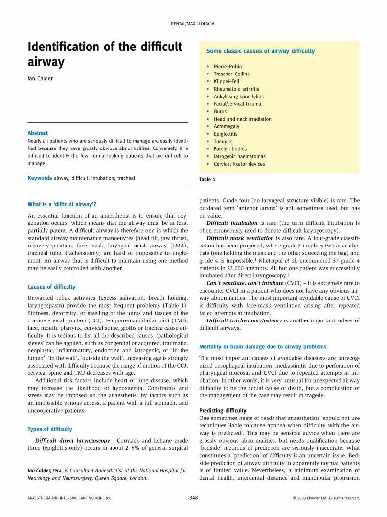
Some classic causes of airway difficulty
• Pierre–Robin
• treacher–collins
• Klippel–feil
• Rheumatoid arthritis
• ankylosing spondylitis
• facial/cervical trauma
• Burns
• Head and neck irradiation
• acromegaly
• epiglottitis
• tumours
• foreign bodies
• iatrogenic haematomas
• cervical fixator devices
Table 1
© 2008 elsevier ltd. all rights reserved.
Dental/Maxillofacial
should be routine. It would be indefensible to find that a patient had an interdental distance of only 2 cm after induction.
When possible, a history, examination, and special investiga-tions approach is appropriate (it may not be possible in emergency situations when patients are obtunded or unable to cooperate).
History: a history of difficulty or disease associated with diffi-culty should prompt appropriate examination, investigation and the assembly of suitable staff and equipment.
Symptoms: one of the principal difficulties in predicting air-way problems under anaesthesia is that in most unexpected cases there are no symptoms. The symptoms associated with obstructed sleep apnoea (OSA) syndrome should be sought in suspected cases. Anaesthetists should be aware of the symptom-atology of impending airway obstruction (Table 2).
Examination: the first consideration is whether a seal can be obtained with a face mask. If this is impossible then apnoea could be disastrous if an LMA or tracheal intubation fail. A history of snoring or OSA, beards, obesity, age greater than 55 years, poor Mallampati grade, thyro-mental distance less than 6 cm, and poor mandibular protrusion are all associated with difficult face-mask ventilation, but these factors, even in combination have poor predictive value.1 If there is serious doubt about the practicality of face-mask ventilation because of a poor seal or inability to maintain the airway, then awake intubation must be considered.
The presence of large quantities of blood, pus or mucus is significant because fibre-optic endoscopy will be impeded.
Specific areas of examinationDental health should be examined and patients warned of
possible damage to loose, restored or diseased teeth.Mouth opening – the lower limit of normality for interdental
distance is 3.7 cm in young people. Assessment is by ‘eye’, but measurement should be performed when restriction is detected. Mandibular protrusion is a vital part of normal mouth opening (and airway management) and should be assessed by asking the patient to place the lower incisors in front of the upper incisors.
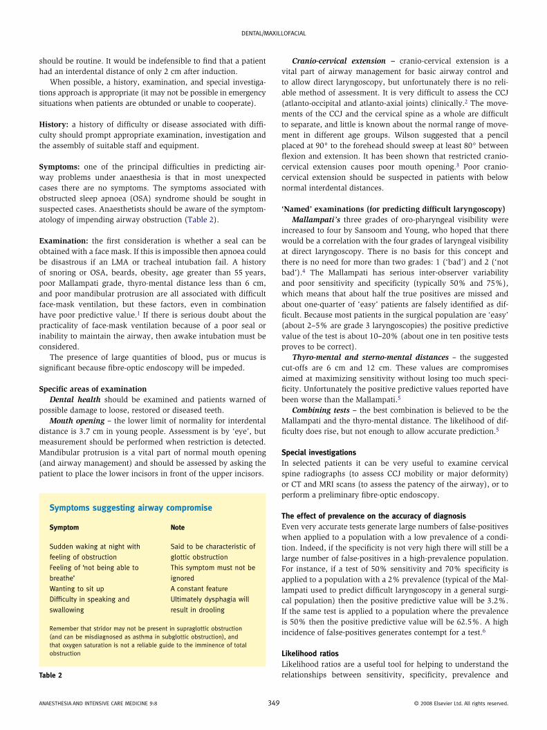
Symptoms suggesting airway compromise
Symptom Note
Sudden waking at night with
feeling of obstruction
Said to be characteristic of
glottic obstruction
feeling of ‘not being able to
breathe’
this symptom must not be
ignored
Wanting to sit up a constant feature
Difficulty in speaking and
swallowing
Ultimately dysphagia will
result in drooling
Remember that stridor may not be present in supraglottic obstruction (and can be misdiagnosed as asthma in subglottic obstruction), and that oxygen saturation is not a reliable guide to the imminence of total obstruction
Table 2
anaeStHeSia anD intenSiVe caRe MeDicine 9:8 34
Cranio-cervical extension – cranio-cervical extension is a vital part of airway management for basic airway control and to allow direct laryngoscopy, but unfortunately there is no reli-able method of assessment. It is very difficult to assess the CCJ (atlanto-occipital and atlanto-axial joints) clinically.2 The move-ments of the CCJ and the cervical spine as a whole are difficult to separate, and little is known about the normal range of move-ment in different age groups. Wilson suggested that a pencil placed at 90° to the forehead should sweep at least 80° between flexion and extension. It has been shown that restricted cranio- cervical extension causes poor mouth opening.3 Poor cranio-cervical extension should be suspected in patients with below normal interdental distances.
‘Named’ examinations (for predicting difficult laryngoscopy)Mallampati’s three grades of oro-pharyngeal visibility were
increased to four by Sansoom and Young, who hoped that there would be a correlation with the four grades of laryngeal visibility at direct laryngoscopy. There is no basis for this concept and there is no need for more than two grades: 1 (‘bad’) and 2 (‘not bad’).4 The Mallampati has serious inter-observer variability and poor sensitivity and specificity (typically 50% and 75%), which means that about half the true positives are missed and about one-quarter of ‘easy’ patients are falsely identified as dif-ficult. Because most patients in the surgical population are ‘easy’ (about 2–5% are grade 3 laryngoscopies) the positive predictive value of the test is about 10–20% (about one in ten positive tests proves to be correct).
Thyro-mental and sterno-mental distances – the suggested cut-offs are 6 cm and 12 cm. These values are compromises aimed at maximizing sensitivity without losing too much speci-ficity. Unfortunately the positive predictive values reported have been worse than the Mallampati.5
Combining tests – the best combination is believed to be the Mallampati and the thyro-mental distance. The likelihood of dif-ficulty does rise, but not enough to allow accurate prediction.5
Special investigationsIn selected patients it can be very useful to examine cervical spine radiographs (to assess CCJ mobility or major deformity) or CT and MRI scans (to assess the patency of the airway), or to perform a preliminary fibre-optic endoscopy.
The effect of prevalence on the accuracy of diagnosisEven very accurate tests generate large numbers of false-positives when applied to a population with a low prevalence of a condi-tion. Indeed, if the specificity is not very high there will still be a large number of false-positives in a high-prevalence population. For instance, if a test of 50% sensitivity and 70% specificity is applied to a population with a 2% prevalence (typical of the Mal-lampati used to predict difficult laryngoscopy in a general surgi-cal population) then the positive predictive value will be 3.2%. If the same test is applied to a population where the prevalence is 50% then the positive predictive value will be 62.5%. A high incidence of false-positives generates contempt for a test.6
Likelihood ratiosLikelihood ratios are a useful tool for helping to understand the relationships between sensitivity, specificity, prevalence and
9 © 2008 elsevier ltd. all rights reserved.
Dental/Maxillofacial
positive predictive value.7 A likelihood ratio is calculated by dividing the true positive rate of a test by the false-positive rate (sensitivity by 1-specificity). It indicates roughly how many more times likely it is that a patient with a positive test result has the problem or disease. Multiplying the prevalence of the disease in the study population by the likelihood ratio gives the positive predictive value (the proportion of positive tests that are cor-rect). The Mallampati has been found to have a likelihood ratio of about three.5
Serious morbidity and mortality and prediction of airway difficultyUnexpected difficulty with face-mask ventilation or direct laryn-goscopy is a feature of life for anaesthetists. Anaesthetists must keep in mind that mortality associated with airway management is most often due to loss of airway after repeated attempts at intubation, infection after perforation of the pharyngeal mucosa, and oesophageal intubation.
Summary• No special training or aptitude is required to recognize most patients with seriously difficult airways.• It is difficult to identify the rare, relatively normal-looking patients who present important degrees of difficulty during anaesthesia.• Serious upper-airway obstruction can be present without stridor or desaturation.
anaeStHeSia anD intenSiVe caRe MeDicine 9:8 3
• Mortality and serious morbidity as a result of unexpected dif-ficulty with airway management is avoidable in many cases by avoiding multiple fruitless attempts to intubate, being alert to the risks of oesophageal intubation and perforation of the mucosa. ◆
RefeReNCeS
1 Kheterpal S, Han R, tremper KK, et al. incidence and predictors of
difficult and impossible mask ventilation. Anesthesiology 2006;
105: 885–91.
2 Urakami Y, takennaka i, nakamura M, et al. the reliability of
the Bellhouse test for evaluating extension capacity of the
occipitoatlantoaxial complex. Anesth Analg 2002; 95: 1437–41.
3 calder i, Picard J, chapman M, o’Sullevan c, crockard Ha. Mouth
opening – a new angle. Anesthesiology 2003; 99: 799–801.
4 calder i. acromegaly, the Mallampati and difficult intubation.
Anesthesiology 2001; 94: 1148–9.
5 Shiga t, Wajima Z, inoue t, Sakamoto a. Predicting difficult
intubation in apparently normal patients: a meta-analysis of bedside
screening test performance. Anesthesiology 2005; 103: 429–37.
6 Yentis SM. Predicting difficult intubation – worthwhile exercise or
pointless ritual? Anaesthesia 2002; 57: 105–9.
7 calder i. Difficult airways: causation and prediction. in: calder i,
Pearce a, eds. core topics in airway management. cambridge:
cambridge University Press, 2005.
50 © 2008 elsevier ltd. all rights reserved.