Embed Size (px)
Citation preview
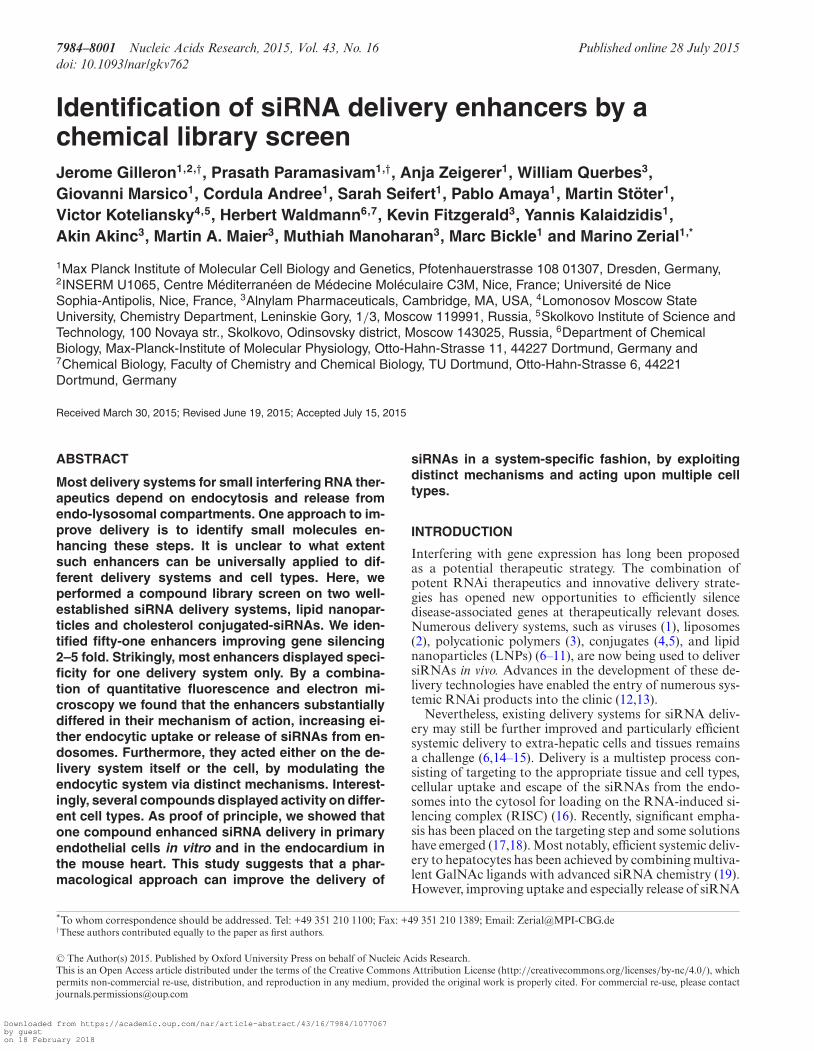
7984–8001 Nucleic Acids Research, 2015, Vol. 43, No. 16 Published online 28 July 2015doi: 10.1093/nar/gkv762
Identification of siRNA delivery enhancers by achemical library screenJerome Gilleron1,2,†, Prasath Paramasivam1,†, Anja Zeigerer1, William Querbes3,Giovanni Marsico1, Cordula Andree1, Sarah Seifert1, Pablo Amaya1, Martin Stoter1,Victor Koteliansky4,5, Herbert Waldmann6,7, Kevin Fitzgerald3, Yannis Kalaidzidis1,Akin Akinc3, Martin A. Maier3, Muthiah Manoharan3, Marc Bickle1 and Marino Zerial1,*
1Max Planck Institute of Molecular Cell Biology and Genetics, Pfotenhauerstrasse 108 01307, Dresden, Germany,2INSERM U1065, Centre Mediterraneen de Medecine Moleculaire C3M, Nice, France; Universite de NiceSophia-Antipolis, Nice, France, 3Alnylam Pharmaceuticals, Cambridge, MA, USA, 4Lomonosov Moscow StateUniversity, Chemistry Department, Leninskie Gory, 1/3, Moscow 119991, Russia, 5Skolkovo Institute of Science andTechnology, 100 Novaya str., Skolkovo, Odinsovsky district, Moscow 143025, Russia, 6Department of ChemicalBiology, Max-Planck-Institute of Molecular Physiology, Otto-Hahn-Strasse 11, 44227 Dortmund, Germany and7Chemical Biology, Faculty of Chemistry and Chemical Biology, TU Dortmund, Otto-Hahn-Strasse 6, 44221Dortmund, Germany
Received March 30, 2015; Revised June 19, 2015; Accepted July 15, 2015
ABSTRACT
Most delivery systems for small interfering RNA ther-apeutics depend on endocytosis and release fromendo-lysosomal compartments. One approach to im-prove delivery is to identify small molecules en-hancing these steps. It is unclear to what extentsuch enhancers can be universally applied to dif-ferent delivery systems and cell types. Here, weperformed a compound library screen on two well-established siRNA delivery systems, lipid nanopar-ticles and cholesterol conjugated-siRNAs. We iden-tified fifty-one enhancers improving gene silencing2–5 fold. Strikingly, most enhancers displayed speci-ficity for one delivery system only. By a combina-tion of quantitative fluorescence and electron mi-croscopy we found that the enhancers substantiallydiffered in their mechanism of action, increasing ei-ther endocytic uptake or release of siRNAs from en-dosomes. Furthermore, they acted either on the de-livery system itself or the cell, by modulating theendocytic system via distinct mechanisms. Interest-ingly, several compounds displayed activity on differ-ent cell types. As proof of principle, we showed thatone compound enhanced siRNA delivery in primaryendothelial cells in vitro and in the endocardium inthe mouse heart. This study suggests that a phar-macological approach can improve the delivery of
siRNAs in a system-specific fashion, by exploitingdistinct mechanisms and acting upon multiple celltypes.
INTRODUCTION
Interfering with gene expression has long been proposedas a potential therapeutic strategy. The combination ofpotent RNAi therapeutics and innovative delivery strate-gies has opened new opportunities to efficiently silencedisease-associated genes at therapeutically relevant doses.Numerous delivery systems, such as viruses (1), liposomes(2), polycationic polymers (3), conjugates (4,5), and lipidnanoparticles (LNPs) (6–11), are now being used to deliversiRNAs in vivo. Advances in the development of these de-livery technologies have enabled the entry of numerous sys-temic RNAi products into the clinic (12,13).
Nevertheless, existing delivery systems for siRNA deliv-ery may still be further improved and particularly efficientsystemic delivery to extra-hepatic cells and tissues remainsa challenge (6,14–15). Delivery is a multistep process con-sisting of targeting to the appropriate tissue and cell types,cellular uptake and escape of the siRNAs from the endo-somes into the cytosol for loading on the RNA-induced si-lencing complex (RISC) (16). Recently, significant empha-sis has been placed on the targeting step and some solutionshave emerged (17,18). Most notably, efficient systemic deliv-ery to hepatocytes has been achieved by combining multiva-lent GalNAc ligands with advanced siRNA chemistry (19).However, improving uptake and especially release of siRNA
*To whom correspondence should be addressed. Tel: +49 351 210 1100; Fax: +49 351 210 1389; Email: [email protected]†These authors contributed equally to the paper as first authors.
C© The Author(s) 2015. Published by Oxford University Press on behalf of Nucleic Acids Research.This is an Open Access article distributed under the terms of the Creative Commons Attribution License (http://creativecommons.org/licenses/by-nc/4.0/), whichpermits non-commercial re-use, distribution, and reproduction in any medium, provided the original work is properly cited. For commercial re-use, please [email protected]
Downloaded from https://academic.oup.com/nar/article-abstract/43/16/7984/1077067by gueston 18 February 2018
Nucleic Acids Research, 2015, Vol. 43, No. 16 7985
from unproductive intracellular compartments remains achallenge for many other tissues and cell types (6,15).
Recently, high throughput screening strategies have beenapplied to improve the composition (20–22) and physico-chemical properties (23) of siRNA delivery systems. Thisapproach has also been used to rapidly define the optimalconditions for efficient transfection (24). An alternative ap-proach is to identify chemical compounds that enhance theefficiency of existing siRNA delivery systems (25,26). How-ever, to what extent such approach is a viable strategy re-mains to be determined. First, some chemical compoundscould improve delivery of oligonucleotides by interferingwith endocytic uptake, endosomal acidification and pro-gression of cargo along the degradative pathway, as in thecase of choloroquine or bafilomycin (27,28). This would in-crease the residence time of siRNAs in early endosomesleading to a higher probability of escape before degradationin late endosomes and lysosomes. However, such enhancersmay not have sufficient potency and also induce high celltoxicity given the essential function of the endocytic sys-tem in cell homeostasis, signaling and metabolism (29–31).Second, identifying compounds that enhance the escape ofoligonucleotides from endosomes remains a challenge. Thisis because, with the exception of single molecule detection,the fluorescence microscopy methods do not have the ade-quate sensitivity and resolution to detect the few hundredsof molecules in the cytosol that are necessary for gene si-lencing (6,32–33). Several approaches have been used to cir-cumvent such a limit in the sensitivity of detection suchas the use of high doses of fluorescently labeled oligonu-cleotides (34), or unspecific markers like fluorescently la-beled Dextran (26) or the colocalization with endosomalmarkers (26,35–37). However, these indirect approaches donot faithfully reveal the true state of siRNA escape fromendosomes into the cytosol within the therapeutic concen-tration range. Therefore, more quantitative and higher res-olution methods are necessary to assess the mode of actionof oligonucleotide delivery enhancers under physiologicalconditions. Third, for compounds acting upon the endo-cytic system, it is unclear whether they can be active acrossmultiple delivery systems or exhibit system-specificity and,fourth, whether they can enhance delivery in multiple celltypes or rather display a narrow range of cell specificity.
Here, we screened a small molecule library aimed toimprove the efficiency of gene silencing of two siRNAdelivery systems, LNPs and cholesterol-conjugated siR-NAs (Chol-siRNAs). Interestingly, comparison of the com-pounds identified from the two screens indicated that themajority were specific for either delivery system. By ap-plying a combination of high resolution fluorescence mi-croscopy and electron microscopy we found that the com-pounds have different modes of action, either acting uponthe delivery system itself or upon the cellular machineryto either enhance uptake or increase endosomal escape. Fi-nally, the compounds were also effective in physiologicallyrelevant cell types, including cells that usually are refractoryto delivery such as primary fibroblasts and hepatocytes invitro, and endothelial cells in vivo.
MATERIALS AND METHODS
Animals
All animal studies were conducted in accordance with Ger-man animal welfare legislation and in strict pathogen-freeconditions in the animal facility of the Max Planck In-stitute of Molecular Cell Biology and Genetics, Dresden,Germany. Protocols were approved by the Institutional An-imal Welfare Officer (Tierschutzbeauftragter), and neces-sary licenses were obtained from the regional Ethical Com-mission for Animal Experimentation of Dresden, Germany(Tierversuchskommission, Landesdirektion Dresden). Allprocedures used in animal studies conducted at AlnylamPharmaceuticals, Cambridge, U.S.A. were approved by theInstitutional Animal Care and Use Committee and wereconsistent with local, state and federal regulations as ap-plicable.
siRNA modification and formulation into lipid nanoparticles
The siRNAs used in this study target GFP (eGFP plasmid,Clonetech). The procedure used to produce LNP-siRNA,LNP-siRNA-alexa647 and LNP-siRNA-gold were exten-sively described previously (6).
Cholesterol conjugates were made as described previ-ously (38).
Cell culture and cell lines
GFP-HeLa cells (39) were cultured in DMEM media com-plemented with 10% FBS and 1% penicillin-streptomycin at37◦C and 5% CO2. Primary human fibroblasts (GM00041),obtained from Coriell Institute, were cultured and in-fected with Rab5-GFP as previously described (40). Pri-mary mouse hepatocytes and endothelial cells were ob-tained from GFP-lifeact transgenic mice (41), following pre-viously described isolation and culture protocols (42,43).When required the cells were seeded on 24 (for electron mi-croscopy analysis) or 96 (for fluorescence microscopy anal-ysis) well plates.
High throughput screening
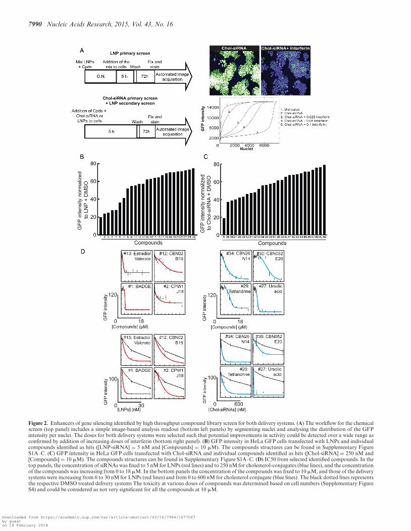
GFP-HeLa cells were seeded in 96 or 384 well plates. Cellswere transfected with a mixture of LNP-siRNA (5 nM)pre-incubated for 16 h with the compounds (10 �M) orDMSO (MOCK). The library contains 45 567 diverse com-pounds with a subset of kinase inhibitors (75 compounds),FDA approved drugs (∼1000 compounds), pure naturalcompounds (∼400 compounds) and compounds selectedon drug-like criteria (∼44 000 compounds). After 5h, thecells were washed and incubated with fresh media and fixedwith PFA 4% 72 h after transfection. Nuclei were stainedwith DAPI and the cells were imaged (at least 25 fields perconditions) with a Perkin Elmer Operetta automated mi-croscope (TDS, MPI-CBG, Dresden) and analyzed withAcapella and MotionTracking software (44). Similar pro-cedures were applied for Cholesterol conjugated-siRNA(250 nM) except that the compounds (10 �M) or DMSO(MOCK) were not pre-incubated but freshly added to the
Downloaded from https://academic.oup.com/nar/article-abstract/43/16/7984/1077067by gueston 18 February 2018
7986 Nucleic Acids Research, 2015, Vol. 43, No. 16
cells. All transfections (LNPs and Chol-siRNA) were per-formed in serum containing media to mimic blood flow con-ditions. The chemical structures of the enhancers are shownin the Supplementary Figure S1A–C.
We determined the mean GFP intensity within the seg-mented nuclei instead of the total GFP intensity per fieldto exclude false positives of GFP reduction caused by vari-ations in cell number (either due to toxicity or decreasedproliferation). The toxicity of the compounds based on cellnumber is shown in Supplementary Figure S2. We also useddefined thresholds to select the enhancers. Compounds wereconsidered as enhancers when they improved GFP silencingby at least 20%. In addition, compounds that reduced cellnumber by more than 35% were considered as toxic and,thus, excluded from the rest of the analysis. Few compoundsthat improved the Chol-siRNA silencing efficiently (#26,#29, #30, #41), but with a toxicity value slightly above thethreshold, were retained. The rationale was that varyingtheir concentration allows finding a window where they areactive but non-toxic.
Importantly, to control for non-specific silencing, in ad-dition to un-treated (UT) and DMSO treated conditions,we verified that the compounds did not decrease the GFPintensity when incubated alone (i.e. without the delivery sys-tem) with the GFP-expressing cells. All compounds show-ing a reduction in GFP mean intensity within the segmentednuclei without addition of LNPs or Chol-siRNAs were ex-cluded from the hit list. We also tested whether the en-hancers could transfect naked (non-formulated) siRNAs, toidentify potential transfection reagents. We identified twocompounds that have this property.
Knock-down assay
HeLa GFP cells, Rab5-GFP human primary fibroblasts andGFP–lifeact primary mouse hepatocytes were transfectedwith LNP-siRNA formulation preincubated or not withthe compounds (following similar procedures as in the pri-mary screen). After 72h, the cells were fixed with PFA 4%(pH 7.2 in phosphate buffer) for 20 min at room tempera-ture. After washing, nuclei were labeled with Dapi and cy-tosol with SytoBlue. Acquisition and analysis of images (atleast 25 fields per conditions) were done on an Arrayscan-VTI with TwisterII automated wide field microscope (TDS,MPI-CBG, Dresden).
Uptake assay
For the in vitro uptake assay, cells were transfected eitherwith LNP-siRNA-alexa647 or with cholesterol conjugated-siRNA-alexa647 treated or not with the compounds. Then,cells were fixed and stained as for the knock-down assay.Images were acquired on a Perkin Elmer Opera automatedconfocal microscope (TDS, MPI-CBG, Dresden) and ana-lyzed on MotionTracking software (http://motiontracking.mpi-cbg.de) as previously described (6).
To determine the endocytic pathway used by LNPs orChol-siRNAs to enter the cell, we performed a depletionof key endocytic machinery as previously described (6).
For the in vivo uptake assay, LNP-siRNA-alexa647,treated or not with BADGE, were injected in the heart
cavity of sacrificed mice. Then the hearts were collected,washed extensively in PBS and fixed with PFA 4% overnightat 4◦C. Tissues were sliced on cryostat after OCT embed-ding and nuclei were stained with Dapi. Then, sections weremounted with mowiol and coverslip designed for high reso-lution observation. Images (at least 15 fields per conditions)were acquired on an Olympus Fluoview 1000 laser scanningconfocal microscope (light microscopy facility, MPI-CBG,Dresden) equipped with an Olympus UPlanSApo 60x 1.35Oil immersion objective. Images were analyzed on Motion-Tracking.
Determination of the mechanism of action
Two pilot screens were performed either by pre-incubatingthe compounds with the delivery systems overnight priorto adding them to the cells (pre-incubation condition), orby adding the compounds together with the delivery systemdirectly to the cells (direct incubation condition). The pilotscreens revealed that the pre-incubation condition increasedthe number of hits for LNPs but not for Chol-siRNAs.Therefore, we performed the full primary screen under thepre-incubation condition for LNPs and under the direct in-cubation condition for Chol-siRNAs. Since, all the identi-fied enhancers for LNPs exert their effect with an overnightpre-incubation, a secondary screen was performed to deter-mine which compounds are able to improve silencing underdirect incubation condition.
From these two screens, we were able to distinguish com-pounds that improved GFP down-regulation by acting mostprobably on the LNPs from those that were not. In ad-dition, we determined the compounds that act on the up-take or on the siRNA release. For this, we analyzed the up-take of alexa647-labeled siRNAs (incorporated in LNPs orcholesterol-conjugated) under pre-incubation (compoundsthat act on delivery systems) or direct incubation condi-tion (compounds that act on cells). Compounds that signif-icantly increased the amount of siRNA-alexa647 were con-sidered as acting on uptake. Compounds that did not affector reduce the amount of intracellular siRNA were consid-ered as acting on siRNA endosomal release.
Electron microscopy
Morphological experiments were analyzed in a blind fash-ion using a code that was not broken until the quantitationwas completed.
For electron microscopy analysis, HeLa cells were trans-fected with LNP-siRNA-gold and fixed with 2.5% glu-taraldehyde (in phosphate buffer) overnight. Then, cellswere post-fixed in ferrocyanide reduced osmium as pre-viously described (45). Cells were dehydrated in increas-ing bath of ethanol for 10 min, infiltrates with mixtureof ethanol and epon (3:1 and 1:3) and pure epon for 1h.After epon polymerization overnight at 60◦C, the 24 wellplates were broken and pieces of epon were glued on eponsticks. 70–50 nm sections were then cut and stained withuranyl acetate and lead citrate following classical proce-dure. Supermontages of 100 images were randomly col-lected at 11000x magnification on a Tecnai 12 TEM micro-scope (FEI) (electron microscopy facility, MPI-CBG, Dres-den) and the stitching of the images was achieved by using
Downloaded from https://academic.oup.com/nar/article-abstract/43/16/7984/1077067by gueston 18 February 2018
Nucleic Acids Research, 2015, Vol. 43, No. 16 7987
the open access software Blendmont (Boulder Laboratory,University of Colorado, USA).
To quantify the total uptake as well as the ratio of struc-tures labeled versus unlabeled in a reliable manner, a stere-ological approach based on randomly distributed crosseswas applied allowing relative loading index calculation andnormalization of the number of structures counted (46). Toquantify the ratio of siRNA escape from endosomes, we de-veloped a plugin for automatically counting the total num-ber of gold particles per montage. Images were processedby performing morphological bottom-hat filtering on thegrayscale input image (47). The structuring element used forthis was a circle of a radius bigger than the object of inter-est (radius 4). Following this, we performed image equal-ization to the interval [0;1] and thresholding with a thresh-old set at 0.3. The binarized images were then analyzed bythe watershed transform to split contiguous gold particles.A last post-processing step was performed to remove un-certain gold particles (particles having the average intensityvalue less than 5 standard deviations of the median inten-sity value in the whole image). Then, for a set of images, thenumber of particles were automatically counted and manu-ally counted with an error rate determined to be <1% con-firming that our procedure succeeded in correctly identify-ing gold particles. Finally, the procedure was applied to de-termine the total number of gold particles in the images. Inaddition, the number of gold particles was counted man-ually within the cytoplasm based on morphological recog-nition. For this analysis, we selected three pieces of eponcontaining cells incubated for 6 h with LNP-siRNA-gold.For each piece, we cut several sections that were collectedon eight grids. Among these eight grids covering a largeportion of the cells, we randomly picked three grids. Fivesuper-montaging, at random places but in areas containingcells, were made for each grid. The super-montaging cov-ered approximately two cells. In each experiment we ana-lyzed approximately 45 super-montaging corresponding toabout 90 cells per condition. We analyzed three indepen-dent experiments, amounting to ∼270 cells per condition.For each condition we counted automatically at least 100000 siRNA-gold particles.
Quantitative multiparametrics image analysis
Quantitative multi-parametric image analysis was per-formed in two sequential rounds of calculations. In the firstround, aiming at the identification of fluorescent vesicles,the image intensity was fitted by a sum of powered Loren-zian functions (48). The coefficients of those functions werethen used to describe the features of individual objects (e.g.intracellular position relative to the nucleus, size, intensity,total vesicular intensity, etc.). Additionally, nuclei and cellswere identified by a pipeline involving several operationsfrom morphological image analysis (47). Briefly, nuclei werefound by a maximum-entropy based local thresholding andcells by a region growing algorithm based on the water-shed transform. In the second round, a set of statistics wasextracted from the distributions of the endosome parame-ters measured in the first round. Statistical filters based onthe mean intensity of the fitted object were then applied toremove the background and the unspecific staining (using
control image with secondary antibody alone). This set ofvalues, that quantitatively describes the fluorescence infor-mation of every channel in the image, has been used forcomparing the different conditions as previously described(44).
Co-localization analysis was performed by assessing thepercentage of overlapping objects. Object ‘A’ in channel ‘1’is considered to co-localize to object ‘B’ in channel ‘2’ ifthe integral intensity profile of A overlaps to the one of Bmore than a user-defined percentage threshold, here set to40%. Co-localization was calculated both by number (per-centage of LNP vesicles that are positive for LAMP) and byintensity volume (percentage of LNP amount in the LAMP-positive compartment). The described approach is morepowerful than classical correlation and pixel co-occurrenceanalyses, since it allows us to (i) discriminate between back-ground and foreground (object) fluorescence and (ii) inter-pret the results in terms of percentage of structures that arelocalized to objects in another channel of interest.
Statistics
Data were expressed as the mean ± standard error ofthe mean (SEM). Statistical analysis was determined usingANOVA test followed by Student T-test test. The two-tailedPvalue were added within the figure or the figure legends.
RESULTS
LNPs and cholesterol conjugate siRNA delivery systems havedifferent uptake mechanisms
We aimed at improving the efficiency of two well-establishedsiRNA delivery systems, lipid nanoparticles (LNPs) andcholesterol-conjugated siRNAs (Chol-siRNAs) (9,49–51)by performing a high throughput screen to discover smallmolecule delivery enhancers. Given that these two deliverysystems differ fundamentally in composition, size and mor-phology, we hypothesized that their mechanism of actionmay differ significantly. Therefore, we first investigated theirmechanism of uptake and endocytosis.
The siRNAs were labeled with Alexa Fluor 647 and for-mulated in LNPs (6) or conjugated to cholesterol. Inter-nalization in HeLa cells could be visualized (Figure 1A)at concentrations sufficient for efficient silencing of tar-geted genes, in vitro (Figure 1B) as previously shown in vivo(38,42). Chol-siRNAs uptake behaved like free cholesterol(52), yielding both diffuse staining and a punctate pattern.In contrast, siRNAs encapsulated in LNPs only displayeda punctate pattern (Figure 1A). Such difference in prop-erties may entail different mechanisms of association withthe plasma membrane and cellular uptake. To test this, wedepleted various regulatory components of the endocyticmachinery and analyzed the effects on uptake. The inter-nalization of cholesterol is thought to be mainly mediatedby LDL receptor endocytosis upon interaction with serumlipoproteins (53,54). Consistent with this, we found thatthe uptake of Chol-siRNAs required mainly components ofclathrin-mediated endocytosis (CME) (Figure 1C), in con-trast to LNP which enter via both CME and macropinocy-tosis (6). Moreover, the uptake kinetics of Chol-siRNA and
Downloaded from https://academic.oup.com/nar/article-abstract/43/16/7984/1077067by gueston 18 February 2018

7988 Nucleic Acids Research, 2015, Vol. 43, No. 16
Figure 1. LNPs and cholesterol conjugates differ in uptake mechanism. (A) Images of HeLa cells after incubation with LNP-siRNA-alexa647 (top panels)or cholesterol conjugated-siRNA (Chol-siRNA)-alexa647 (bottom panels) for 5 h and 1 h, respectively. (B) Quantification of the percentage of GFPdownregulation in HeLa GFP-expressing cells and primary mouse hepatocytes expressing Lifeact-GFP exposed to 40 nM of LNP-siRNA and 1 �M ofChol-siRNA (n = 3, mean ± SEM). (C) Chol-siRNAs-alexa647 uptake in HeLa cells after silencing of dynamin (DNM1L), clathrin light chain (CLTC),Caveolin (CAV1), CDC42 (for clathrin-independent endocytosis)) and RAC1 (Macropinocytosis). Mean ± SEM, n = 3, P-value relative to control. (D)Uptake kinetics of LNP-siRNA-alexa647 (40 nM, red curve) compared to Chol-siRNA-alexa647 (250 nM, green curve).
Downloaded from https://academic.oup.com/nar/article-abstract/43/16/7984/1077067by gueston 18 February 2018

Nucleic Acids Research, 2015, Vol. 43, No. 16 7989
LNPs were markedly different. Chol-siRNA was internal-ized quickly and followed a Gaussian curve (Figure 1D).The decrease in intracellular Chol-siRNA over time can beexplained by depletion of LDL receptor on the cell sur-face or increase in efflux as previously observed for choles-terol (55,56). The increased intracellular Chol-siRNA uponCAV1 knock down (Figure 1C) may be explained by the roleof caveolin in the cholesterol efflux as recently shown (57).This process that avoids an excess of Chol-siRNA within thecell could have important impact on the silencing efficiencyof this particular conjugate. In contrast, the uptake of LNPswas delayed in time and followed an exponential curve (Fig-ure 1D), as shown previously (6). Altogether, these data in-dicate that the two siRNA delivery systems differ in the waythey enter the endocytic pathway. Therefore, we rationalizedthat separate screens would need to be carried out for thetwo systems, as unique compounds may emerge for each.
A high throughput chemical screen allows identification ofcompounds that improve siRNA delivery
To identify chemical compounds that improve siRNA de-livery, we screened both delivery systems with the same li-brary of compounds. We performed our primary screenin HeLa cells, a cell line easy to culture in a screenableformat and which exhibits the key general features of themammalian endocytic pathway. The cells constitutively ex-press green fluorescent protein (GFP) for direct readout ofRNAi-mediated silencing by quantitative fluorescence mi-croscopy. To exclude chemicals that may affect GFP transla-tion or nuclear localization, the compounds were also testedalone, i.e. in the absence of siRNA. In a second step, we val-idated the results by performing a secondary screen in pri-mary cells, fibroblasts and hepatocytes, to assess the cross-activity between cell types and species. Our primary assayconsisted of GFP-expressing HeLa cells transfected withsuboptimal doses of either LNPs or Chol-siRNAs with ananti-GFP siRNA. We optimized the assay to obtain reduc-tion in GFP expression of only about 20% as measured byquantitative fluorescence microscopy. As a pre-requisite forthe screen, we verified that under the same conditions silenc-ing could be boosted to higher than 80% by adding transfec-tion reagent (see GFP-positive versus -negative cells in Fig-ure 2A). This demonstrated that the amount of siRNA perse was not limiting, but rather cellular uptake and/or escapefrom the endolysosomal system was suboptimal. Therefore,the experimental conditions used were appropriate to biasthe screen toward compounds, which could improve thesespecific steps.
We initially performed two pilot screens on a small setof compounds, with or without overnight pre-incubationof the compounds with the delivery system prior to addingthe mixtures to the cells. The pilot screens revealed thatan overnight pre-incubation increased the number of hitsfor LNPs but not for Chol-siRNAs. Therefore, such a pre-incubation step was incorporated in the LNP screen, but notin the Chol-siRNA screen. Under these conditions, all theidentified LNP enhancers will work with an overnight pre-incubation with the LNPs, whereas those improving Chol-siRNAs silencing will work upon direct contact with thecells. The scheme of the assay protocol used in the screen
is shown in Figure 2A. After 5 h of incubating the cells withthe compound (10 �M) and siRNA mixtures, the mediumwas replaced by fresh medium, and the cells were furtherincubated for 72 h. Thereafter, cells were fixed, nuclei werestained with Hoechst and the GFP expression was quanti-fied on an automated fluorescence microscope. In order toexclude toxic compounds, enhancers were considered whenthe mean GFP intensity per cell decreased by > 20% butthe number of cells (nuclei) decreased by <35%. Of the 45567 compounds tested in both delivery systems, 25 and 28compounds (see the structures provided on SupplementaryFigure S1A–C) improved the silencing activity of LNPs andChol-siRNAs, respectively (Figure 2B, C). Silencing effi-ciency reached up to 80% without (or with very limited) tox-icity for the cells (Supplementary Figure S2A, B). Strikingly,only two compounds (#5 and #18) were found to improvesilencing in both delivery systems. These compounds werealso able to induce silencing with non-modified, commer-cially available siRNAs (Supplementary Figure S3), sug-gesting that they may act as general transfection reagents.Importantly, all other identified hits reduced GFP expres-sion only in conjunction with their respective delivery sys-tems. We examined the dose response of the hit compoundsand determined their IC50 to be in the low micromolar range(Figure 2D), with very limited or no toxicity up to 10 �M(Supplementary Figure S4). Therefore, the screen identifiedchemical compounds that improve siRNA delivery specif-ically for each of the LNPs and Chol-siRNA delivery sys-tems.
Compounds identified in a high throughput chemical screenefficiently improve siRNA delivery in primary cells
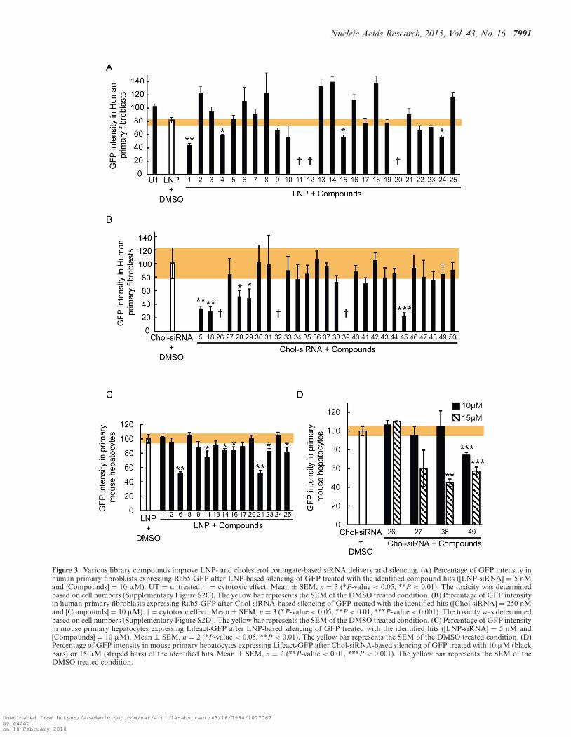
We previously demonstrated that different cell types differin their siRNA delivery mechanism (6). Can the enhancersidentified in the screen improve delivery in different celltypes? To test this, we performed a screen in human primaryfibroblasts and mouse primary hepatocytes. First, we testedall compounds identified with their respective siRNA de-livery systems in the fibroblasts. We chose these particularcells because they internalize LNPs inefficiently and, conse-quently, are also poorly transfected (6). This screen revealedthat 18% of the LNP hits (Figure 3A; #1, #4, #15 and #24)and 21% of the Chol-siRNA hits (Figure 3B; #5, #18, #28,#29 and # 47) identified in HeLa cells improved the effi-ciency of GFP silencing by 1.5- to 4-fold also in those cells.Some compounds (3 LNP and 3 Chol-siRNA hits; #11,#12, #20, #26, #32 and #39), however, demonstrated toxi-city in the primary fibroblasts (Supplementary Figure S2C,D).
Unlike fibroblasts, hepatocytes internalize LNPs effi-ciently. Therefore, we tested the compounds in mouse pri-mary hepatocytes to determine whether they can enhancethe silencing also in these cells. However, since these cellsare not well suited for high throughput screening, we testeda random sample of the full set of hits, consisting of 15 hitsfrom the LNP screen and 4 from the Chol-siRNA screen.Seven compounds (46%) from the LNP screen and 1 com-pound (25%) from the Chol-siRNA screen improved the si-lencing efficiency (Figure 3C, D; #6, #11, #14, #16, #21,#23, #25 and #49). However, increasing the concentration
Downloaded from https://academic.oup.com/nar/article-abstract/43/16/7984/1077067by gueston 18 February 2018
7990 Nucleic Acids Research, 2015, Vol. 43, No. 16
Figure 2. Enhancers of gene silencing identified by high throughput compound library screen for both delivery systems. (A) The workflow for the chemicalscreen (top panel) includes a simple image-based analysis readout (bottom left panels) by segmenting nuclei and analysing the distribution of the GFPintensity per nuclei. The doses for both delivery systems were selected such that potential improvements in activity could be detected over a wide range asconfirmed by addition of increasing doses of interferin (bottom right panel). (B) GFP intensity in HeLa GFP cells transfected with LNPs and individualcompounds identified as hits ([LNP-siRNA] = 5 nM and [Compounds] = 10 �M). The compounds structures can be found in Supplementary FigureS1A–C. (C) GFP intensity in HeLa GFP cells transfected with Chol-siRNA and individual compounds identified as hits ([Chol-siRNA] = 250 nM and[Compounds] = 10 �M). The compounds structures can be found in Supplementary Figure S1A–C. (D) IC50 from selected identified compounds. In thetop panels, the concentration of siRNAs was fixed to 5 nM for LNPs (red lines) and to 250 nM for cholesterol-conjugates (blue lines), and the concentrationof the compounds was increasing from 0 to 18 �M. In the bottom panels the concentration of the compounds was fixed to 10 �M, and those of the deliverysystems were increasing from 0 to 30 nM for LNPs (red lines) and from 0 to 600 nM for cholesterol conjugate (blue lines). The black dotted lines representsthe respective DMSO treated delivery systems The toxicity at various doses of compounds was determined based on cell numbers (Supplementary FigureS4) and could be considered as not very significant for all the compounds at 10 �M.
Downloaded from https://academic.oup.com/nar/article-abstract/43/16/7984/1077067by gueston 18 February 2018
Nucleic Acids Research, 2015, Vol. 43, No. 16 7991
Figure 3. Various library compounds improve LNP- and cholesterol conjugate-based siRNA delivery and silencing. (A) Percentage of GFP intensity inhuman primary fibroblasts expressing Rab5-GFP after LNP-based silencing of GFP treated with the identified compound hits ([LNP-siRNA] = 5 nMand [Compounds] = 10 �M). UT = untreated, † = cytotoxic effect. Mean ± SEM, n = 3 (*P-value < 0.05, **P < 0.01). The toxicity was determinedbased on cell numbers (Supplementary Figure S2C). The yellow bar represents the SEM of the DMSO treated condition. (B) Percentage of GFP intensityin human primary fibroblasts expressing Rab5-GFP after Chol-siRNA-based silencing of GFP treated with the identified hits ([Chol-siRNA] = 250 nMand [Compounds] = 10 �M). † = cytotoxic effect. Mean ± SEM, n = 3 (*P-value < 0.05, **P < 0.01, ***P-value < 0.001). The toxicity was determinedbased on cell numbers (Supplementary Figure S2D). The yellow bar represents the SEM of the DMSO treated condition. (C) Percentage of GFP intensityin mouse primary hepatocytes expressing Lifeact-GFP after LNP-based silencing of GFP treated with the identified hits ([LNP-siRNA] = 5 nM and[Compounds] = 10 �M). Mean ± SEM, n = 2 (*P-value < 0.05, **P < 0.01). The yellow bar represents the SEM of the DMSO treated condition. (D)Percentage of GFP intensity in mouse primary hepatocytes expressing Lifeact-GFP after Chol-siRNA-based silencing of GFP treated with 10 �M (blackbars) or 15 �M (striped bars) of the identified hits. Mean ± SEM, n = 2 (**P-value < 0.01, ***P < 0.001). The yellow bar represents the SEM of theDMSO treated condition.
Downloaded from https://academic.oup.com/nar/article-abstract/43/16/7984/1077067by gueston 18 February 2018

7992 Nucleic Acids Research, 2015, Vol. 43, No. 16
from 10 �M (used in the screen) to 15 �M improved theKD efficiency in hepatocytes, as shown for compounds #27,#36 and #49, suggesting that more compounds could be ac-tive at higher doses. Note that we did not observe hepato-cyte toxicity at these doses (Supplementary Figure S2E, F).This analysis revealed that the compounds that improve si-lencing in fibroblasts are not efficient in hepatocytes and,conversely, those active in hepatocytes are inefficient in fi-broblasts.
Altogether, our results demonstrate that some of the hitsidentified in HeLa cells were also active across different celltypes, including primary cells with very different origin andcharacteristics.
Compounds can improve uptake or endosomal escape
The finding that there was little overlap between com-pounds acting on fibroblasts and hepatocytes may reflecta different mechanism of action of the compounds. Sincethe LNPs and Chol-siRNAs were directly added to thecells, the hits could either increase the uptake or improvethe release of the siRNAs from intracellular compart-ments. To discriminate between the two scenarios, weanalyzed the impact of the hits on the uptake of LNP-siRNA-alexa647 or Chol-siRNA-alexa647 in HeLa cells(Figure 4A). We found that 56% and 64% of the com-pounds were able to strongly increase the uptake of LNPsand Chol-siRNA, respectively (Figure 4B). This increaseis exemplified by the higher intensity of fluorescent siR-NAs in endosomes delivered via LNPs in cells incubatedwith BADGE (#1, Bisphenol A diglycidyl ether; 2,2′-[(1-methylethylidene)bis(4,1-phenyleneoxymethylene)]bis-oxirane) and via Chol-siRNAs in cells treated withcompound #29 (Tetrandrine), compared with control(Figure 4A). Interestingly, 11 compounds out of 25 forthe LNPs and 5 out of 14 for the Chol-siRNAs did notsignificantly increase cellular uptake (Figure 4B), as shownfor the compound designated as #2 (CPW1-J18) and #35(Lomatin) (Figure 4A). These compounds in all likelihoodimprove delivery by enhancing the intracellular releaseof the siRNA from endo-lysosomal compartments (seebelow). The uptake of LNP-siRNAs or Chol-siRNAs isa multi-step process that requires binding to the plasmamembrane and/or to a specific receptor (LDLR for LNPs(6,8)) followed by internalization. Similarly, the releaseof siRNAs from endosomes requires the unpacking ofthe siRNAs from the particles before they can cross theendosomal membrane to reach the cytosol. The identifiedenhancers may act on one or several of these steps toimprove silencing.
To gain insights into the mode of action of the LNPenhancers, we tested whether they can improve silencingupon immediate contact with the cells as compared to anovernight pre-incubation. To this end, the silencing en-hancers were either pre-incubated with the cells or wereadded to the cells concomitantly with the delivery systems.The rationale behind this experiment is that if a compoundwere acting primarily on the cell, we would expect its silenc-ing enhancer effect to occur also when added to the cellsprior to adding the siRNAs, whereas an action on the deliv-ery platform would depend on a prior pre-incubation with
the siRNAs. By this criterion, 28% of the enhancer com-pounds were active only when pre-incubated with the LNPs(Figure 4B; black bars) whereas the remaining 72% were ac-tive upon direct incubation with the cells (Figure 4B; graybars). Interestingly, almost all compounds that improved si-lencing activity presumably by facilitating endo-lysosomalescape were active upon direct incubation with the cells,suggesting that they act upon the cellular machinery. Wefound that the enhancers that act only when pre-incubatedwith the LNPs are more active in increasing the uptake thanthose acting upon direct incubation with the cells (Figure4B; black bars versus gray bars), suggesting that they maydirectly impact on the LNPs and facilitate their endocytosis.
To explore the mechanism whereby the compounds en-hance the siRNA activity, we focused on two of the most ac-tive compounds, BADGE and CPW1-J18 impacting LNP-mediated delivery. We used the analytical electron mi-croscopy methodology previously developed (6) to quantifythe uptake and escape of LNPs encapsulating gold-labeledsiRNA (Figure 4C). Among the LNP hits, BADGE andCPW1-J18 had the strongest positive effect of GFP down-regulation in their respective categories, e.g. improving up-take or endosomal release (Figure 4B and SupplementaryFigure S5). This assay was limited to LNPs, since cova-lent attachment of gold particles to Chol-siRNA can be ex-pected to substantially impact uptake and trafficking.
By using this approach, we first confirmed that BADGEincreased the uptake of LNPs as indicated by the 15-foldincrease in the amount of siRNA-gold internalized by thecells (Figure 4D). Moreover, the ratio of siRNA-gold in thecytosol versus the total amount internalized was not in-creased (Figure 4E). These results suggest that BADGE actsexclusively on the uptake rather than on the release of siR-NAs from endosomes. In contrast, CPW1-J18 was found toslightly reduce the total amount of siRNAs-gold internal-ized (Figure 4D) while increasing the ratio of siRNA-gold inthe cytosol versus the total amount internalized by ∼5-fold(Figure 4E). These results suggest that CPW1-J18 improvesthe release of siRNAs from endosomes. Overall, the totalamount of cytosolic siRNA-gold was significantly more el-evated with BADGE and CPW1-J18 treatment comparedto DMSO (Figure 4F), as evidenced by the EM images (ar-rows in Figure 4C). Our results suggest that the compoundsidentified act via two different mechanisms, improving ei-ther uptake or endosomal escape.
Endosomal escape of siRNA can be enhanced via differentmechanisms
We can envisage various mechanisms whereby the chemi-cal compounds can facilitate endosomal escape. One pos-sibility is that they prevent lysosomal acidification simi-lar to choloroquine or bafilomycin and/or progression ofcargo along the degradative pathway (28). Interestingly,hydroxychloroquine and bafilomycin were not hits in thescreen, neither for LNPs nor Chol-siRNA. This discrepencycould be explained either by the fact that the compoundconcentrations in the screens were different than those re-ported (58,59), or that they also inhibit endocytosis (60–61,6). An alternative possibility is that some compounds lo-
Downloaded from https://academic.oup.com/nar/article-abstract/43/16/7984/1077067by gueston 18 February 2018

Nucleic Acids Research, 2015, Vol. 43, No. 16 7993
Figure 4. Mechanism of action of the compounds on LNP- and cholesterol conjugate-based siRNA delivery and silencing. (A) Images illustrating theuptake of LNP-siRNA-alexa647 (40 nM, 5 h incubation, left panel) and Chol-siRNA-alexa647 (250 nM, 5h incubation, right panel) treated with DMSO(top panels) or with the compounds (10 �M) that increased the uptake (bottom panels: BADGE and Tetrandrine) or not (middle panels: CPW1-J18 andLomatin) in HeLa cells. (B) Quantitative analysis of LNP-siRNA-alexa647 (40 nM, 5 h incubation, top panel) and Chol-siRNA-alexa647 (250 nM, 5hincubation, bottom panel) uptake in cells treated with DMSO or compounds (10 �M). Compounds acting on the delivery systems are represented withblack bars and those acting on the cells with gray bars. The compounds structure can be found in Supplementary Figure S1A–C. (C) LNP-siRNA-golddetected in HeLa cells in vitro, by EM ([LNP = 40 nM]; [compounds] = 10 �M). siRNA-gold were detected in the cytosol (arrows) or within severalendocytic compartments. Magnified images (insets) permit appreciation of the cytosolic localization of siRNA-gold. (D) Automated quantification of thetotal number of siRNA-gold (representing the uptake) found per �m2 of cell section. Mean ± SEM, n = 3 (*P-value < 0.05, ***P < 0.001). (E) Semi-automated quantification of the ratio between the number of cytosolic siRNA-gold and the total number of siRNA-gold internalized (representing thepercentage of siRNA escape). Mean ± SEM, n = 3 (*P-value < 0.05). (F) Semi-automated quantification of the number of cytosolic siRNA-gold per �m2
of cells. Mean ± SEM, n = 3 (*P-value < 0.05, ***P-value < 0.001).
Downloaded from https://academic.oup.com/nar/article-abstract/43/16/7984/1077067by gueston 18 February 2018

7994 Nucleic Acids Research, 2015, Vol. 43, No. 16
cally destabilize the endosomal membrane thus increasingleakage of content from the lumen (62,63).
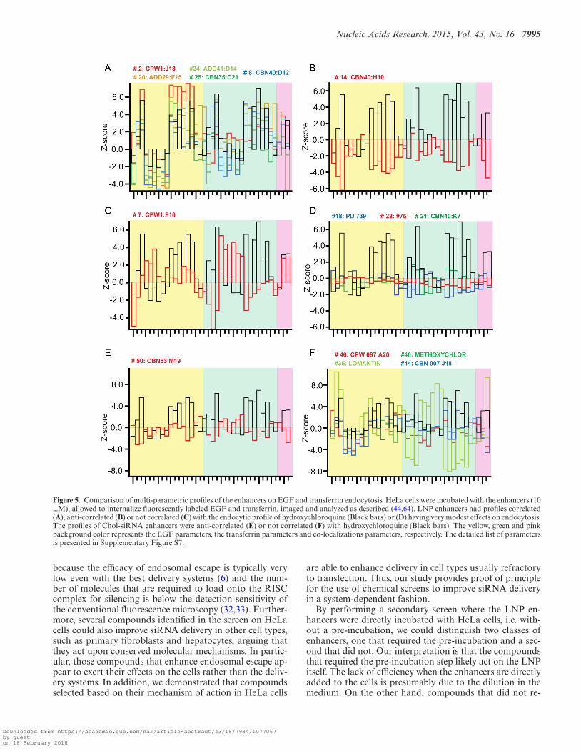
To explore the effects of the compounds on the endoso-mal system, we performed a focused screen on the candidatecompounds for endosomal escape at the same concentra-tion as in the RNAi enhancement primary screen (10 �M)using an image-based assay that quantitatively measures theuptake of EGF and transferrin, as described (64). Quantita-tive multi-parametric image analysis (QMPIA) was appliedfor a phenotypic description of the effect of each compoundon the endosomal network (endosome number, size, intra-cellular position, etc.) (44). We then compared the multi-parametric profiles across the compounds and with respectto hydroxychloroquine, because this compound has a char-acteristic phenotypic signature of blocked endosome acidi-fication and maturation. The compounds had very differenteffects on the endosomal system (Figure 5). For LNP en-hancers, the phenotypic profiles of the compounds could begrouped in four distinct categories. In the first category, theprofiles correlated with that of hydroxychloroquine (Fig-ure 5A), suggesting that these compounds have a similarinhibitory effect on endosome acidification and/or matu-ration. In contrast, the second category was anti-correlated(Figure 5B), ruling out such an inhibitory effect. The third,which contains only compounds #7, was neither corre-lated nor anti-correlated with the hydroxychloroquine pro-file (Figure 5C), suggesting that it affects endocytic traffick-ing but by a mechanism distinct from endosome matura-tion. The fourth category is very interesting because noneof the compounds in this group were found to have a signif-icant impact on cargo endocytosis and the endosomal pa-rameters (Figure 5D). These enhancers therefore do not ap-pear to have a strong influence on endosomal transport andare candidates for improving escape through destabilizationof the endosomal membrane.
For the Chol-siRNA enhancers, we could distinguishonly two categories, none of which was correlated withhydroxychloroquine. The first category was anti-correlated(Figure 5E) and the second had no correlation with the pro-file of hydroxychloroquine, suggesting again an action thatis distinct from a block of acidification and/or endosomalmaturation. Among these compounds, #35 (Lomatin), wasparticularly interesting because of its uncoupling betweenthe effect on EGF and transferrin endocytosis (Figure 5F;light green bars). This uncoupling suggests that compound#35 act specifically on the endocytic/recycling pathway.
Altogether, these results demonstrate that the com-pounds have very distinct effects on the endosomal systemand suggest that they enhance endosomal escape by differ-ent mechanisms.
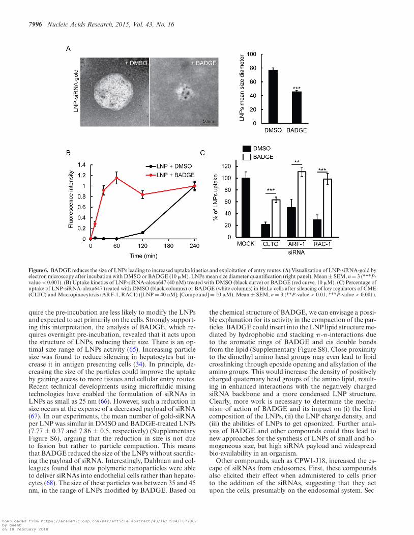
BADGE treatment increases cell delivery and kineticsthrough LNP size reduction
To determine the mechanism of action underlying the en-hanced uptake, we focused on BADGE (compound #1)since it had the strongest effect on LNP uptake. By EM neg-ative staining analysis of LNPs treated with BADGE, wefound that this compound had an effect on the nanopar-ticles themselves (Figure 6A), consistent with the predic-tions from the incubation analysis (Figure 4B). Indeed,
this enhancer was capable of reducing the size of LNPsby ∼2-fold (Figure 6A). This reduction in size was associ-ated with a dramatic acceleration of uptake kinetics (Figure6B). Therefore, the activity of BADGE could result fromthe facilitation of the entry mechanism used by LNPs. Wetested this hypothesis by interfering with different endocyticroutes. Strikingly, we found that the uptake of LNPs ex-posed to BADGE is much less sensitive to the knock-downof clathrin, ARF-1 and RAC-1 compared to the control(Figure 6C), suggesting that the smaller LNPs are capturedthrough a broader set of endocytic mechanisms.
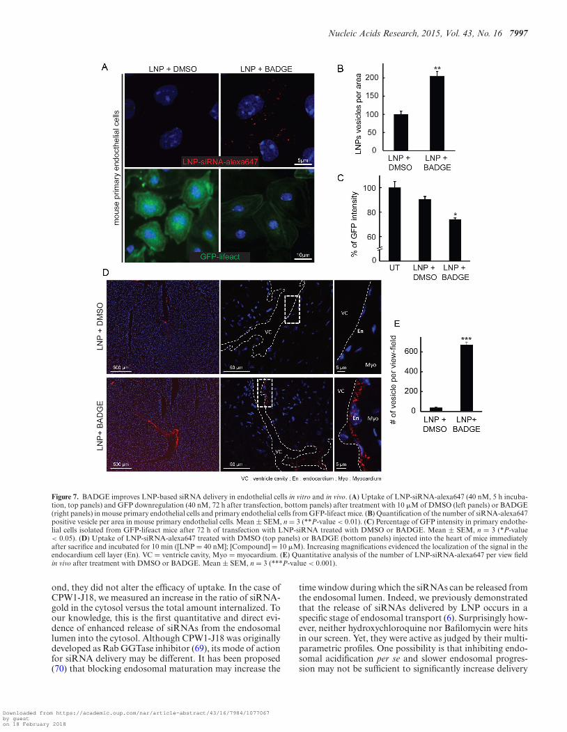
The finding that BADGE modifies the physicochemicalproperties of LNPs to improve uptake both in HeLa cellsand primary fibroblasts in vitro, prompted us to test whetherit can also enhance the delivery of siRNAs in other celltypes in vitro and within a tissue. We focused on endothe-lial cells, which are important in a number of diseases anddifficult to transfect with siRNAs. First, we looked at theeffect of BADGE treatment on LNP-based siRNA deliv-ery in primary endothelial cells (Figure 7A). We found thatBADGE-treated LNPs were delivered twice as efficiently ascontrol LNPs in these cells (Figure 7A, B). Consistently,this increase in uptake was accompanied by a similar (∼2-fold) increase in silencing activity, as determined by analyz-ing the GFP intensity in primary endothelial cells isolatedfrom GFP-lifeact transgenic mice (Figure 7A bottom paneland Figure 7C). Second, to test whether BADGE could im-prove delivery to the endothelium in a whole tissue, we in-jected BADGE-treated LNPs into the heart of mice. LNPspre-treated with BADGE were strongly captured by the en-docardium cell layer as evidenced by the numerous fluores-cent vesicles (Figure 7D). The quantification of the numberof vesicles loaded with LNP-siRNA-alexa647 revealed thatthe treatment with BADGE increased the uptake in the en-dothelial cell in vivo by ∼14-fold (Figure 7E).
Altogether, our results demonstrate that performing ascreen of chemical compounds in HeLa cells allowed theidentification of small molecules that efficiently improve thesilencing in several cell types having different properties andfunctions such as skin fibroblasts, hepatocytes and endothe-lial cells.
DISCUSSION
In this study, we performed a chemical library screen andidentified compounds that improve the activity of siRNAsdelivered to cells via LNPs and Chol-siRNAs. An inter-esting result was that we found fundamental differences inthe cellular routes used by the two delivery systems, andmost compounds identified acted upon one delivery sys-tem but not the other. Furthermore, using a combination ofquantitative fluorescence light and electron microscopy weidentified compounds that act on two distinct bottlenecksof siRNA delivery (6), the uptake of siRNAs by cells andtheir release from endosomes into the cytosol. We wouldlike to emphasize that without these methods it is impos-sible to accurately determine the intracellular localizationand fraction of siRNAs that could escape from the lumenof the endosomes into the cytosol without overloading cellswith siRNA doses beyond the therapeutic range or usingunspecific and biased ‘escape’ markers (26,34–37). This is
Downloaded from https://academic.oup.com/nar/article-abstract/43/16/7984/1077067by gueston 18 February 2018
Nucleic Acids Research, 2015, Vol. 43, No. 16 7995
Figure 5. Comparison of multi-parametric profiles of the enhancers on EGF and transferrin endocytosis. HeLa cells were incubated with the enhancers (10�M), allowed to internalize fluorescently labeled EGF and transferrin, imaged and analyzed as described (44,64). LNP enhancers had profiles correlated(A), anti-correlated (B) or not correlated (C) with the endocytic profile of hydroxychloroquine (Black bars) or (D) having very modest effects on endocytosis.The profiles of Chol-siRNA enhancers were anti-correlated (E) or not correlated (F) with hydroxychloroquine (Black bars). The yellow, green and pinkbackground color represents the EGF parameters, the transferrin parameters and co-localizations parameters, respectively. The detailed list of parametersis presented in Supplementary Figure S7.
because the efficacy of endosomal escape is typically verylow even with the best delivery systems (6) and the num-ber of molecules that are required to load onto the RISCcomplex for silencing is below the detection sensitivity ofthe conventional fluorescence microscopy (32,33). Further-more, several compounds identified in the screen on HeLacells could also improve siRNA delivery in other cell types,such as primary fibroblasts and hepatocytes, arguing thatthey act upon conserved molecular mechanisms. In partic-ular, those compounds that enhance endosomal escape ap-pear to exert their effects on the cells rather than the deliv-ery systems. In addition, we demonstrated that compoundsselected based on their mechanism of action in HeLa cells
are able to enhance delivery in cell types usually refractoryto transfection. Thus, our study provides proof of principlefor the use of chemical screens to improve siRNA deliveryin a system-dependent fashion.
By performing a secondary screen where the LNP en-hancers were directly incubated with HeLa cells, i.e. with-out a pre-incubation, we could distinguish two classes ofenhancers, one that required the pre-incubation and a sec-ond that did not. Our interpretation is that the compoundsthat required the pre-incubation step likely act on the LNPitself. The lack of efficiency when the enhancers are directlyadded to the cells is presumably due to the dilution in themedium. On the other hand, compounds that did not re-
Downloaded from https://academic.oup.com/nar/article-abstract/43/16/7984/1077067by gueston 18 February 2018
7996 Nucleic Acids Research, 2015, Vol. 43, No. 16
Figure 6. BADGE reduces the size of LNPs leading to increased uptake kinetics and exploitation of entry routes. (A) Visualization of LNP-siRNA-gold byelectron microscopy after incubation with DMSO or BADGE (10 �M). LNPs mean size diameter quantification (right panel). Mean ± SEM, n = 3 (***P-value < 0.001). (B) Uptake kinetics of LNP-siRNA-alexa647 (40 nM) treated with DMSO (black curve) or BADGE (red curve, 10 �M). (C) Percentage ofuptake of LNP-siRNA-alexa647 treated with DMSO (black columns) or BADGE (white columns) in HeLa cells after silencing of key regulators of CME(CLTC) and Macropinocytosis (ARF-1, RAC1) ([LNP = 40 nM]; [Compound] = 10 �M). Mean ± SEM, n = 3 (**P-value < 0.01, ***P-value < 0.001).
quire the pre-incubation are less likely to modify the LNPsand expected to act primarily on the cells. Strongly support-ing this interpretation, the analysis of BADGE, which re-quires overnight pre-incubation, revealed that it acts uponthe structure of LNPs, reducing their size. There is an op-timal size range of LNPs activity (65). Increasing particlesize was found to reduce silencing in hepatocytes but in-crease it in antigen presenting cells (34). In principle, de-creasing the size of the particles could improve the uptakeby gaining access to more tissues and cellular entry routes.Recent technical developments using microfluidic mixingtechnologies have enabled the formulation of siRNAs inLNPs as small as 25 nm (66). However, such a reduction insize occurs at the expense of a decreased payload of siRNA(67). In our experiments, the mean number of gold-siRNAper LNP was similar in DMSO and BADGE-treated LNPs(7.77 ± 0.37 and 7.86 ± 0.5, respectively) (SupplementaryFigure S6), arguing that the reduction in size is not dueto fission but rather to particle compaction. This meansthat BADGE reduced the size of the LNPs without sacrific-ing the payload of siRNA. Interestingly, Dahlman and col-leagues found that new polymeric nanoparticles were ableto deliver siRNAs into endothelial cells rather than hepato-cytes (68). The size of these particles was between 35 and 45nm, in the range of LNPs modified by BADGE. Based on
the chemical structure of BADGE, we can envisage a possi-ble explanation for its activity in the compaction of the par-ticles. BADGE could insert into the LNP lipid structure me-diated by hydrophobic and stacking �-�-interactions dueto the aromatic rings of BADGE and cis double bondsfrom the lipid (Supplementary Figure S8). Close proximityto the dimethyl amino head groups may even lead to lipidcrosslinking through epoxide opening and alkylation of theamino groups. This would increase the density of positivelycharged quaternary head groups of the amino lipid, result-ing in enhanced interactions with the negatively chargedsiRNA backbone and a more condensed LNP structure.Clearly, more work is necessary to determine the mecha-nism of action of BADGE and its impact on (i) the lipidcomposition of the LNPs, (ii) the LNP charge density, and(iii) the abilities of LNPs to get opsonized. Further anal-ysis of BADGE and other compounds could thus lead tonew approaches for the synthesis of LNPs of small and ho-mogeneous size, but high siRNA payload and widespreadbio-availability in an organism.
Other compounds, such as CPW1-J18, increased the es-cape of siRNAs from endosomes. First, these compoundsalso elicited their effect when administered to cells priorto the addition of the siRNAs, suggesting that they actupon the cells, presumably on the endosomal system. Sec-
Downloaded from https://academic.oup.com/nar/article-abstract/43/16/7984/1077067by gueston 18 February 2018
Nucleic Acids Research, 2015, Vol. 43, No. 16 7997
Figure 7. BADGE improves LNP-based siRNA delivery in endothelial cells in vitro and in vivo. (A) Uptake of LNP-siRNA-alexa647 (40 nM, 5 h incuba-tion, top panels) and GFP downregulation (40 nM, 72 h after transfection, bottom panels) after treatment with 10 �M of DMSO (left panels) or BADGE(right panels) in mouse primary endothelial cells and primary endothelial cells from GFP-lifeact mice. (B) Quantification of the number of siRNA-alexa647positive vesicle per area in mouse primary endothelial cells. Mean ± SEM, n = 3 (**P-value < 0.01). (C) Percentage of GFP intensity in primary endothe-lial cells isolated from GFP-lifeact mice after 72 h of transfection with LNP-siRNA treated with DMSO or BADGE. Mean ± SEM, n = 3 (*P-value< 0.05). (D) Uptake of LNP-siRNA-alexa647 treated with DMSO (top panels) or BADGE (bottom panels) injected into the heart of mice immediatelyafter sacrifice and incubated for 10 min ([LNP = 40 nM]; [Compound] = 10 �M). Increasing magnifications evidenced the localization of the signal in theendocardium cell layer (En). VC = ventricle cavity, Myo = myocardium. (E) Quantitative analysis of the number of LNP-siRNA-alexa647 per view fieldin vivo after treatment with DMSO or BADGE. Mean ± SEM, n = 3 (***P-value < 0.001).
ond, they did not alter the efficacy of uptake. In the case ofCPW1-J18, we measured an increase in the ratio of siRNA-gold in the cytosol versus the total amount internalized. Toour knowledge, this is the first quantitative and direct evi-dence of enhanced release of siRNAs from the endosomallumen into the cytosol. Although CPW1-J18 was originallydeveloped as Rab GGTase inhibitor (69), its mode of actionfor siRNA delivery may be different. It has been proposed(70) that blocking endosomal maturation may increase the
time window during which the siRNAs can be released fromthe endosomal lumen. Indeed, we previously demonstratedthat the release of siRNAs delivered by LNP occurs in aspecific stage of endosomal transport (6). Surprisingly how-ever, neither hydroxychloroquine nor Bafilomycin were hitsin our screen. Yet, they were active as judged by their multi-parametric profiles. One possibility is that inhibiting endo-somal acidification per se and slower endosomal progres-sion may not be sufficient to significantly increase delivery
Downloaded from https://academic.oup.com/nar/article-abstract/43/16/7984/1077067by gueston 18 February 2018
7998 Nucleic Acids Research, 2015, Vol. 43, No. 16
of the siRNAs by the particular systems used and under thestringency of our experimental conditions (low siRNA con-centrations, compounds concentration, incubation, thresh-old of detection, etc). Another possibility is that chloro-quine and bafilomycin improve siRNA release but concomi-tantly decrease in LNP uptake, as they are known to inhibitreceptor mediated endocytosis (6,60–61). In contrast, theenhancers we identified do not significantly reduce LNP up-take as compared to DMSO (see Figure 4B). Undoubtedly,a preferred strategy would be to enhance escape of siRNAsfrom endosomes.
Analysis of the effect of the compounds on the endocyticsystem and on different cell types provided some importantclues on their respective mechanisms of action. Using hy-droxychloroquine as reference compound that alters endo-somal maturation we could profile the compounds with re-spect to this mechanism. We found that several compoundshad a phenotypic profile similar to hydroxychloroquine. Onthis basis, one could interpret that they may indeed alterendosomal acidification and/or maturation. However, thefinding that such compounds were not active on all celltypes tested speaks against this interpretation. For exam-ple, the activity of CPW1-J18 in fibroblast is modest (Fig-ure 3), arguing that its mechanism of action may still dif-fer from that of hydroxychloroquine, which is a lysoso-motropic agent active on a wide spectrum of cells. There-fore, increasing the residence time in the endosomes maynot be sufficient to significantly enhance the release of siR-NAs into the cytosol and CPW1-J18 and similarly activecompounds may have some additional effect. In addition,most compounds had a very different profile from hydroxy-chloroquine. Among those compounds, #35 (Lomatin) ap-peared to effect specifically endocytic/recycling cargo (as vi-sualized by the specific alteration on Tf but not EGF traf-ficking). However, for LNPs none of the enhancers hada similar profile to #35. Strikingly, a group of enhancersdid not have detectable effects on the endosomal system.These compounds are candidates for destabilizing endoso-mal membranes, as it has been proposed for larger cationicand amphipathic cell-permeating peptides (71,72), or for fu-sion with the limiting membrane as shown for intra-luminalvesicles (73). Altogether, our results suggest that enhancersmay facilitate the escape of siRNAs from endosomes by avariety of mechanisms.
Interestingly, the vast majority of compounds active onone delivery system were not active on the other. This sup-ports the idea that the compounds do not share a commonmechanism but enhance siRNA delivery depending on thesystem. In contrast, enhancers within a delivery system didshow an appreciable level of activity between different cells,but depending on the cell type. For example, 60% of thecompounds (both for LNPs and Chol-siRNAs) increaseduptake in fibroblasts, cells for which uptake is typically in-efficient. In contrast, only 20% of the compounds increaseduptake in hepatocytes, which are normally proficient in in-ternalization. Therefore, the knowledge of the mode of ac-tion of the compounds may help predicting which cell typemay be predisposed to the specific effect of a given com-pound on delivery.
The endocytic pathway is largely conserved between cells.Although most compounds identified in the screen im-
proved delivery for only one delivery system, it is possiblethat the principle behind their mechanism of action may beapplicable to multiple delivery systems of the same class andshared between different cell types. The compounds thatimproved the particular LNPs used here may also improvenanoparticles of different chemical composition. Likewise,those that improved the Chol-siRNAs might have similareffects on other siRNA-conjugate types. Nevertheless, sev-eral of the compounds identified were active on differentprimary cells but others were not. This suggests that thereis considerable cell-type specificity for the enhancers in con-junction with the delivery system.
Future work will be required to determine the precisemode of action of the identified enhancers. Our results ar-gue that more than one mechanism can be exploited andpose a number of questions. First, by which mechanismcompounds like BADGE are able to reduce the size of theLNPs without reducing the siRNA payload? Second, whichare the molecular targets of the compounds improving up-take? Third, by which mechanisms can siRNAs escape fromendosomes? Clearly, the pharmacological approach needsfurther development to improve delivery across multiple de-livery systems and cell types for therapeutic applications.However, there is another important aspect of this approachthat may have more far reaching implications. It providesthe possibility to learn from the mechanism of action of thecompounds and establish common principles for improv-ing the uptake and escape from endosomes. One possibleapproach is to directly measure the escape of siRNAs fromthe lumen of endosomes in an in vitro assay in the presenceof the compounds (63,74). The assay could shed light on themembrane integrity (74), dependence on membrane fusionand transport. Understanding these mechanisms in moredetail may provide the means of using a rational approacheither to improve the existing or to design a new generationof delivery systems.
SUPPLEMENTARY DATA
Supplementary Data are available at NAR Online.
ACKNOWLEDGEMENTS
We acknowledge I. Patten, T. Galvez, E. Perini and K. Dia-mantara for discussions and comments on the manuscript.We thank J-M. Verbavatz and J. Peychl respectively forthe management of the Electron Microscopy Facility andthe Light Microscopy Facility. We acknowledge A. Palfor Rab5-GFP primary human fibroblasts preparation. Wethank W. John and A. Muench-Wuttke from the Biomed-ical Service Facility for mouse care and injections. We arethanking Russian Scientific Fund (grant 14–34–00017) forfinancial support.Author contributions: M.Z. conceived and directed theproject. M.Z., J.G., P.P., A.Z., W.Q., A.A., M.A.M., M.B.,C.A., K.F. and Y.K. designed the experiments. M.A.M. syn-thesized the siRNA-alexa647 and the siRNA-gold conju-gates. A.A. formulated LNP. C.A. under the supervision ofM.B. performed the High throughput screening. J.G., P.P.and C.A. performed the mode of action validation in HeLaand human fibroblasts. M.S. and C.A. performed the auto-mated image acquisitions with the OPERA. G.M. under the
Downloaded from https://academic.oup.com/nar/article-abstract/43/16/7984/1077067by gueston 18 February 2018

Nucleic Acids Research, 2015, Vol. 43, No. 16 7999
supervision of Y.K. provided the quantitative multipara-metrics image analysis and the statistics. S.S. under the su-pervision of J.G. performed mouse intra-cardiac injectionof LNPs. J.G. performed the sections, staining and imagingof the heart. S.S. and P.A. under the supervision of A.Z.performed primary hepatocytes and primary endothelialcells preparation. J.G. developed the quantitative electronmicroscopy. G.M. developed the software for automatedcounting of gold number on electron microscopy images.M.Z. and J.G. wrote the manuscript and M.B, M.A.M.,M.M., W.Q. and A.A. edited it.
FUNDING
Max Planck Society (MPG); DFG and Alnylam Phar-maceuticals; EMBO [ALTF298-2009 to J.G.]. Funding foropen access charge: Max Planck Society (MPG).Conflict of interest statement. W.Q., K.F., A.A., M.A.M.and M.M. are Alnylam Pharmaceuticals employees; M.Z.received funding from and was a consultant with AlnylamPharmaceuticals.
REFERENCES1. Sliva,K. and Schnierle,B.S. (2010) Selective gene silencing by viral
delivery of short hairpin RNA. Virol. J., 7, 248–258.2. Buyens,K., De Smedt,S.C., Braeckmans,K., Demeester,J., Peeters,L.,
van Grunsven,L.A., de Mollerat du Jeu,X., Sawant,R., Torchilin,V.,Farkasova,K. et al. (2012) Liposome based systems for systemicsiRNA delivery: stability in blood sets the requirements for optimalcarrier design. J. Control. Release, 158, 362–370.
3. Castillo,B., Bromberg,L., Lopez,X., Badillo,V., GonzalezFeliciano,J.A., Gonzalez,C.I., Hatton,T.A. and Barletta,G. (2012)Intracellular Delivery of siRNA by Polycationic SuperparamagneticNanoparticles. J. Drug Deliv., 2012, 218940–218951.
4. Lopez-Davila,V., Seifalian,A.M. and Loizidou,M. (2012) Organicnanocarriers for cancer drug delivery. Curr. Opin. Pharmacol., 12,414–419.
5. Nair,J., Willoughby,J., Chan,A., Charisse,K., Alam,R.M., Wang,Q.,Hoekstra,M., Kandasamy,P., Kel’in,A.V., Milstein,S. et al. (2014)Multivalent N-Acetylgalactosamine-Conjugated siRNA Localizes inHepatocytes and Elicits Robust RNAi-mediated Gene Silencing. J.Am. Chem. Soc., 136, 16958–16961.
6. Gilleron,J., Querbes,W., Zeigerer,A., Borodovsky,A., Marsico,G.,Schubert,U., Manygoats,K., Seifert,S., Andree,C., Stoter,M. et al.(2013) Image-based analysis of lipid nanoparticle-mediated siRNAdelivery, intracellular trafficking and endosomal escape. Nat.Biotechnol., 31, 638–646.
7. Akinc,A., Goldberg,M., Qin,J., Dorkin,J.R., Gamba-Vitalo,C.,Maier,M., Jayaprakash,K.N., Jayaraman,M., Rajeev,K.G.,Manoharan,M. et al. (2009) Development of Lipidoid-siRNAFormulations for Systemic Delivery to the Liver. Mol. Ther., 17,872–879.
8. Akinc,A., Querbes,W., De,S., Qin,J., Frank-Kamenetsky,M.,Jayaprakash,K.N., Jayaraman,M., Rajeev,K.G., Cantley,W.L.,Dorkin,J.R. et al. (2010) Targeted delivery of RNAi therapeutics withendogenous and exogenous ligand-based mechanisms. Mol. Ther., 18,1357–1364.
9. Jayaraman,M., Ansell,S.M., Mui,B.L., Tam,Y.K., Chen,J., Du,X.,Butler,D., Eltepu,L., Matsuda,S., Narayanannair,J.K. et al. (2012)Maximizing the potency of siRNA lipid nanoparticles for hepaticgene silencing in vivo. Angew. Chem. Int. Ed. Engl., 51, 8529–8533.
10. Love,K.T., Mahon,K.P., Levins,C.G., Whitehead,K.A., Querbes,W.,Dorkin,J.R., Qin,J., Cantley,W., Qin,L.L., Racie,T. et al. (2010)Lipid-like materials for low-dose, in vivo gene silencing. Proc. Nat.Acad. Sci. U.S.A., 107, 1864–1869.
11. Zimmermann,T.S., Lee,A.C., Akinc,A., Bramlage,B., Bumcrot,D.,Fedoruk,M.N., Harborth,J., Heyes,J.A., Jeffs,L.B., John,M. et al.(2006) RNAi-mediated gene silencing in non-human primates.Nature, 441, 111–114.
12. Kanasty,R., Dorkin,J.R., Vegas,A. and Anderson,D. (2013) Deliverymaterials for siRNA therapeutics. Nat. Mater., 12, 967–977.
13. Coelho,T., Adams,D., Silva,A., Lozeron,P., Hawkins,P.N., Mant,T.,Perez,J., Chiesa,J., Warrington,S., Tranter,E. et al. (2013) Safety andEfficacy of RNAi Therapy for Transthyretin Amyloidosis. N. Engl. J.Med., 369, 819–829.
14. Pei,Y., Hancock,P.J., Zhang,H., Bartz,R., Cherrin,C., Innocent,N.,Pomerantz,C.J., Seitzer,J., Koser,M.L., Abrams,M.T. et al. (2010)Quantitative evaluation of siRNA delivery in vivo. RNA, 16,2553–2563.
15. Xu,Y., Ou,M., Keough,E., Roberts,J., Koeplinger,K., Lyman,M.,Fauty,S., Carlini,E., Stern,M., Zhang,R. et al. (2014) Quantitation ofphysiological and biochemical barriers to siRNA liver delivery vialipid nanoparticle platform. Mol. Pharm., 11, 1424–1434.
16. Akinc,A. and Battaglia,G. (2013) Exploiting endocytosis fornanomedicines. Cold Spring Harb. Perspect. Biol., 5, a016980.
17. Lee,S.K., Siefert,A., Beloor,J., Fahmy,T.M. and Kumar,P. (2012)Cell-specific siRNA delivery by peptides and antibodies. MethodsEnzymol., 502, 91–122.
18. Nielsen,C., Kjems,J., Sorensen,K.R., Engelholm,L.H. andBehrendt,N. (2014) Advances in targeted delivery of small interferingRNA using simple bioconjugates. Expert Opin. Drug. Deliv., 11,791–822.
19. Prakash,T.P., Graham,M.J., Yu,J., Carty,R., Low,A., Chappell,A.,Schmidt,K., Zhao,C., Aghajan,M., Murray,H.F. et al. (2014)Targeted delivery of antisense oligonucleotides to hepatocytes usingtriantennary N-acetyl galactosamine improves potency 10-fold inmice. Nucleic Acids Res., 42, 8796–8807.
20. Hibbitts,A., Lieggi,N., McCabe,O., Thomas,W., Barlow,J., O’Brien,F.and Cryan,S.A. (2011) Screening of siRNA nanoparticles for deliveryto airway epithelial cells using high-content analysis. Ther. Deliv., 2,987–999.
21. Li,L., Wang,F., Wu,Y., Davidson,G. and Levkin,P.A. (2013)Combinatorial synthesis and high-throughput screening of alkylamines for nonviral gene delivery. Bioconjug. Chem., 24, 1543–1551.
22. Zugates,G.T., Anderson,D.G. and Langer,R. (2013) High-throughputmethods for screening polymeric transfection reagents. Cold SpringHarb. Protoc., 11, 1–14.
23. Wang,A., Marinakos,S.M., Badireddy,A.R., Powers,C.M. andHouck,K.A. (2013) Characterization of physicochemical propertiesof nanomaterials and their immediate environments inhigh-throughput screening of nanomaterial biological activity. WileyInterdiscip. Rev. Nanomed. Nanobiotechnol., 5, 430–448.
24. Nabzdyk,C.S., Chun,M., Pradhan,L. and Logerfo,F.W. (2011) Highthroughput RNAi assay optimization using adherent cell cytometry.J. Transl. Med., 9, 48–56.
25. Ming,X., Carver,K., Fisher,M., Noel,R., Cintrat,J.C., Gillet,D.,Barbier,J., Cao,C., Bauman,J. and Juliano,R.L. (2013) The smallmolecule Retro-1 enhances the pharmacological actions of antisenseand splice switching oligonucleotides. Nucleic Acids Res., 41,3673–3687.
26. Yang,B., Ming,X., Cao,C., Laing,B., Yuan,A., Porter,M.A.,Hull-Ryde,E.A., Maddry,J., Suto,M., Janzen,W.P. et al. (2015)High-throughput screening identifies small molecules that enhancethe pharmacological effects of oligonucleotides. Nucleic Acids Res.,43, 1987–1996.
27. Cotten,M., Langle-Rouault,F., Kirlappos,H., Wagner,E.,Mechtler,K., Zenke,M., Beug,H. and Birnstiel,M.L. (1990)Transferrin-polycation-mediated introduction of DNA into humanleukemic cells: stimulation by agents that affect the survival oftransfected DNA or modulate transferrin receptor levels. Proc. Natl.Acad. Sci. U.S.A., 87, 4033–4037.
28. Bhattarai,S.R., Muthuswamy,E., Wani,A., Brichacek,M.,Castaneda,A.L., Brock,S.L. and Oupicky,D. (2010) Enhanced geneand siRNA delivery by polycation-modified mesoporous silicananoparticles loaded with chloroquine. Pharm. Res., 27, 2556–2568.
29. Settembre,C., Fraldi,A., Medina,D.L. and Ballabio,A. (2013) Signalsfrom the lysosome: a control centre for cellular clearance and energymetabolism. Nat. Rev. Mol. Cell Biol., 14, 283–296.
30. Sorkin,A. and von Zastrow,M. (2009) Endocytosis and signalling:intertwining molecular networks. Nat. Rev. Mol. Cell Biol.10,609–622.
Downloaded from https://academic.oup.com/nar/article-abstract/43/16/7984/1077067by gueston 18 February 2018
8000 Nucleic Acids Research, 2015, Vol. 43, No. 16
31. Saftig,P. and Klumperman,J. (2009) Lysosome biogenesis andlysosomal membrane proteins: trafficking meets function. Nat. Rev.Mol. Cell Biol., 10, 623–635.
32. Wei,J., Jones,J., Kang,J., Card,A., Krimm,M., Hancock,P., Pei,Y.,Ason,B., Payson,E., Dubinina,N. et al. (2011) RNA-inducedsilencing complex-bound small interfering RNA is a determinant ofRNA interference-mediated gene silencing in mice. Mol. Pharmacol.,79, 953–963.
33. Landesman,Y., Svrzikapa,N., Cognetta,A. 3rd, Zhang,X.,Bettencourt,B.R., Kuchimanchi,S., Dufault,K., Shaikh,S., Gioia,M.,Akinc,A. et al. (2010) In vivo quantification of formulated andchemically modified small interfering RNA by heating-in-Tritonquantitative reverse transcription polymerase chain reaction (HITqRT-PCR). Silence, 1, 16–29.
34. Basha,G., Novobrantseva,T.I., Rosin,N., Tam,Y.Y., Hafez,I.M.,Wong,M.K., Sugo,T., Ruda,V.M., Qin,J., Klebanov,B. et al. (2011)Influence of Cationic Lipid Composition on Gene SilencingProperties of Lipid Nanoparticle Formulations of siRNA inAntigen-Presenting Cells. Mol. Ther., 19, 2186–2200.
35. Tamura,A., Oishi,M. and Nagasaki,Y. (2009) Enhanced cytoplasmicdelivery of siRNA using a stabilized polyion complex based onPEGylated nanogels with a cross-linked polyamine structure.Biomacromolecules, 10, 1818–1827.
36. Akita,H., Kogure,K., Moriguchi,R., Nakamura,Y., Higashi,T.,Nakamura,T., Serada,S., Fujimoto,M., Naka,T., Futaki,S. et al.(2010) Nanoparticles for ex vivo siRNA delivery to dendritic cells forcancer vaccines: programmed endosomal escape and dissociation. J.Control. Release, 143, 311–317.
37. Sakurai,Y., Hatakeyama,H., Sato,Y., Akita,H., Takayama,K.,Kobayashi,S., Futaki,S. and Harashima,H. (2011) Endosomal escapeand the knockdown efficiency of liposomal-siRNA by the fusogenicpeptide shGALA. Biomaterials, 32, 5733–5742.
38. Soutschek,J., Akinc,A., Bramlage,B., Charisse,K., Constien,R.,Donoghue,M., Elbashir,S., Geick,A., Hadwiger,P., Harborth,J. et al.(2004) Therapeutic silencing of an endogenous gene by systemicadministration of modified siRNAs. Nature, 432, 173–178.
39. Bramsen,J.B., Laursen,M.B., Nielsen,A.F., Hansen,T.B., Bus,C.,Langkjaer,N., Babu,B.R., Hojland,T., Abramov,M., Van Aerschot,A.et al. (2009) A large-scale chemical modification screen identifiesdesign rules to generate siRNAs with high activity, high stability andlow toxicity. Nucleic Acids Res., 37, 2867–2881.
40. Pal,A., Severin,F., Lommer,B., Shevchenko,A. and Zerial,M. (2006)Huntingtin-HAP40 complex is a novel Rab5 effector that regulatesearly endosome motility and is up-regulated in Huntington’s disease.J. Cell Biol., 172, 605–618.
41. Riedl,J., Flynn,K.C., Raducanu,A., Gartner,F., Beck,G., Bosl,M.,Bradke,F., Massberg,S., Aszodi,A., Sixt,M. et al. (2010) Lifeact micefor studying F-actin dynamics. Nat. Methods, 7, 168–169.
42. Zeigerer,A., Gilleron,J., Bogorad,R.L., Marsico,G., Nonaka,H.,Seifert,S., Epstein-Barash,H., Kuchimanchi,S., Peng,C.G.,Ruda,V.M. et al. (2012) Rab5 is necessary for the biogenesis of theendolysosomal system in vivo. Nature, 485, 465–470.
43. Limmer,A., Ohl,J., Kurts,C., Ljunggren,H.G., Reiss,Y.,Groettrup,M., Momburg,F., Arnold,B. and Knolle,P.A. (2000)Efficient presentation of exogenous antigen by liver endothelial cellsto CD8+ T cells results in antigen-specific T-cell tolerance. Nat.Med., 6, 1348–1354.
44. Collinet,C., Stoter,M., Bradshaw,C.R., Samusik,N., Rink,J.C.,Kenski,D., Habermann,B., Buchholz,F., Henschel,R., Mueller,M.S.et al. (2010) Systems survey of endocytosis by multiparametric imageanalysis. Nature, 464, 243–249.
45. Karnovsky,M.J. (1971) Proceedings of the Eleventh Annual Meeting ofthe American Society for Cell Biology. 11th Am Soc Cell Biol NewOrleans, Vol. 284, p. 146.
46. Lucocq,J.M. (2007) Efficient quantitative morphological phenotypingof genetically altered organisms using stereology. Transgenic Res., 16,133–145.
47. Soille,P. (1999) Morphological Image Analysis: Principles andApplications. Springer-Verlag Telos, London.
48. Rink,J., Ghigo,E., Kalaidzidis,Y. and Zerial,M. (2005) Rabconversion as a mechanism of progression from early to lateendosomes. Cell, 122, 735–749.
49. Wolfrum,C., Shi,S., Jayaprakash,K.N., Jayaraman,M., Wang,G.,Pandey,R.K., Rajeev,K.G., Nakayama,T., Charrise,K.,
Ndungo,E.M. et al. (2007) Mechanisms and optimization of in vivodelivery of lipophilic siRNAs. Nat. Biotechnol., 25, 1149–1157.
50. Frank-Kamenetsky,M., Grefhorst,A., Anderson,N.N., Racie,T.S.,Bramlage,B., Akinc,A., Butler,D., Charisse,K., Dorkin,R., Fan,Y.et al. (2008) Therapeutic RNAi targeting PCSK9 acutely lowersplasma cholesterol in rodents and LDL cholesterol in nonhumanprimates. Proc. Natl. Acad. Sci. U.S.A., 105, 11915–11920.
51. Semple,S.C., Akinc,A., Chen,J., Sandhu,A.P., Mui,B.L., Cho,C.K.,Sah,D.W., Stebbing,D., Crosley,E.J., Yaworski,E. et al. (2010)Rational design of cationic lipids for siRNA delivery. Nat.Biotechnol., 28, 172–176.
52. Mukherjee,S., Zha,X., Tabas,I. and Maxfield,F.R. (1998) Cholesteroldistribution in living cells: fluorescence imaging usingdehydroergosterol as a fluorescent cholesterol analog. Biophys. J., 75,1915–1925.
53. Ikonen,E. (2008) Cellular cholesterol trafficking andcompartmentalization. Nat. Rev. Mol. Cell Biol., 9, 125–138.
54. Brown,M.S. and Goldstein,J.L. (1986) A receptor-mediated pathwayfor cholesterol homeostasis. Science, 232, 34–47.
55. Brown,M.S. and Goldstein,J.L. (1997) The SREBP pathway:regulation of cholesterol metabolism by proteolysis of amembrane-bound transcription factor. Cell, 89, 331–340.
56. Maxfield,F.R. and Tabas,I. (2005) Role of cholesterol and lipidorganization in disease. Nature, 438, 612–621.
57. Chen,Y.H., Lin,W.W., Liu,C.S., Hsu,L.S., Lin,Y.M. and Su,S.L.(2014) Caveolin-1 provides palliation for adverse hepatic reactions inhypercholesterolemic rabbits. PLoS One, 9, e71862.
58. Simeoni,F., Morris,M.C., Heitz,F. and Divita,G. (2003) Insight intothe mechanism of the peptide-based gene delivery system MPG:implications for delivery of siRNA into mammalian cells. NucleicAcids Res., 31, 2717–2724.
59. Luthman,H. and Magnusson,G. (1983) High efficiency polyomaDNA transfection of chloroquine treated cells. Nucleic Acids Res., 11,1295–1308.
60. Pack,D.W., Hoffman,A.S., Pun,S. and Stayton,P.S. (2005) Design anddevelopment of polymers for gene delivery. Nature reviews. Drugdiscovery, 4, 581–593.
61. Tietze,C., Schlesinger,P. and Stahl,P. (1980) Chloroquine andammonium ion inhibit receptor-mediated endocytosis ofmannose-glycoconjugates by macrophages: apparent inhibition ofreceptor recycling. Biochemical and biophysical researchcommunications, 93, 1–8.
62. Starai,V.J., Jun,Y. and Wickner,W. (2007) Excess vacuolar SNAREsdrive lysis and Rab bypass fusion. Proc. Natl. Acad. Sci. U.S.A., 104,13551–13558.
63. Ohya,T., Miaczynska,M., Coskun,U., Lommer,B., Runge,A.,Drechsel,D., Kalaidzidis,Y. and Zerial,M. (2009) Reconstitution ofRab- and SNARE-dependent membrane fusion by syntheticendosomes. Nature, 459, 1091–1097.
64. Sundaramurthy,V., Barsacchi,R., Samusik,N., Marsico,G.,Gilleron,J., Kalaidzidis,I., Meyenhofer,F., Bickle,M., Kalaidzidis,Y.and Zerial,M. (2013) Integration of chemical and RNAimultiparametric profiles identifies triggers of intracellularmycobacterial killing. Cell Host Microbe, 13, 129–142.
65. Akinc,A., Goldberg,M., Qin,J., Dorkin,J.R., Gamba-Vitalo,C.,Maier,M., Jayaprakash,K.N., Jayaraman,M., Rajeev,K.G.,Manoharan,M. et al. (2009) Development of Lipidoid-siRNAFormulations for Systemic Delivery to the Liver. Mol. Ther., 17,872–879.
66. Belliveau,N.M., Huft,J., Lin,P.J., Chen,S., Leung,A.K., Leaver,T.J.,Wild,A.W., Lee,J.B., Taylor,R.J., Tam,Y.K. et al. (2012) MicrofluidicSynthesis of Highly Potent Limit-size Lipid Nanoparticles for In VivoDelivery of siRNA. Mol. Ther. Nucleic Acids, 1, e37.
67. Tam,Y.Y., Chen,S. and Cullis,P.R. (2013) Advances in LipidNanoparticles for siRNA Delivery. Pharmaceutics, 5, 498–507.
68. Dahlman,J.E., Barnes,C., Khan,O.F., Thiriot,A., Jhunjunwala,S.,Shaw,T.E., Xing,Y., Sager,H.B., Sahay,G., Speciner,L. et al. (2014) Invivo endothelial siRNA delivery using polymeric nanoparticles withlow molecular weight. Nat Nanotechnol., 9, 648–655.
69. Tan,K.T., Guiu-Rozas,E., Bon,R.S., Guo,Z., Delon,C., Wetzel,S.,Arndt,S., Alexandrov,K., Waldmann,H., Goody,R.S. et al. (2009)Design, synthesis, and characterization of peptide-based rabgeranylgeranyl transferase inhibitors. J. Med. Chem., 52, 8025–8037.
Downloaded from https://academic.oup.com/nar/article-abstract/43/16/7984/1077067by gueston 18 February 2018
Nucleic Acids Research, 2015, Vol. 43, No. 16 8001
70. Ma,D. (2014) Enhancing endosomal escape for nanoparticlemediated siRNA delivery. Nanoscale, 6, 6415–6425.
71. Thoren,P.E., Persson,D., Esbjorner,E.K., Goksor,M., Lincoln,P. andNorden,B. (2004) Membrane binding and translocation ofcell-penetrating peptides. Biochemistry, 43, 3471–3489.
72. El-Andaloussi,S., Holm,T. and Langel,U. (2005) Cell-penetratingpeptides: mechanisms and applications. Curr. Pharm. Des., 11,3597–3611.
73. Bissig,C. and Gruenberg,J. (2014) ALIX and the multivesicularendosome: ALIX in Wonderland. Trends Cell. Biol., 24, 19–25.
74. Bartz,R., Fan,H., Zhang,J., Innocent,N., Cherrin,C., Beck,S.C.,Pei,Y., Momose,A., Jadhav,V., Tellers,D.M. et al. (2011) EffectivesiRNA delivery and target mRNA degradation using an amphipathicpeptide to facilitate pH-dependent endosomal escape. Biochem. J.,435, 475–487.
Downloaded from https://academic.oup.com/nar/article-abstract/43/16/7984/1077067by gueston 18 February 2018