Embed Size (px)
Citation preview

ORIGINAL ARTICLE
Identification of Novel Tumor Suppressor Genes Down-Regulatedin Recurrent Nasopharyngeal Cancer by DNA Microarray
Zhenxiao Huang • Wenfeng Li • Sen Lin • Xiaobi Fang •
Chunhong Zhang • Zhisu Liao
Received: 24 April 2011 / Accepted: 10 November 2011
� Association of Otolaryngologists of India 2011
Abstract The nasopharyngeal cancer is a common cancer
among southern Chinese. In order to better understand
molecular mechanism of recurrent nasopharyngeal cancer
(rNPC), we used DNA microarray to identify down-regu-
lated tumor suppressed genes (TSGs) in rNPC, and bioin-
formatics to analyze their chromosomal localizations and
molecular functions. Eight non-recurrent nasopharyngeal
cancer (nNPC) and six rNPC tissue samples were selected,
and Affymetrix Gene1.0 ST chips were used to construct
the expression profiling of each tissue sample. Identify the
down-regulated TSGs in rNPC by comparing expression
profiling data of two type tissue samples. A total of five
TSGs were identified to be down-regulated in rNPC. These
five TSGs include SERPINF1, TPD52L1, FBLN1,
RASSF6, and S100A2, and Signal Log Ratio were -2.2,
-2.3, -3.5, -3.9 and -6.9 respectively. Chromosomal
localization analysis showed that S100A2, RASSF6,
TPD52L1, SERPINF1, and FBLN1 were located on chro-
mosomes 1q, 4q, 6q, 17p and 22q, respectively. Functional
analysis showed that SERPINF1 and TPD52L1 belonged to
enzyme activity genes, S100A2 and FBLN1 belonged to
calcium ion binding genes, RASSF6 belong to protein
binding genes. Five TSGs likely to be the candidate TSGs
involved in rNPC, and may play important roles in
occurrence of rNPC. Chromosomes 1q, 4q, 6q, 17p and 22q
may be considered as important region for screening TSGs
that may relevant to rNPC. Those genes and chromosomal
region need to be further studied.
Keywords Recurrent nasopharyngeal cancer �Tumor suppressor genes � DNA microarray �Gene expression � Chromosomal localization �Molecular function
Introduction
Nasopharyngeal carcinoma (NPC) is a unique malignancy
that is particularly prevalent among the southern Chinese
but it is rare in Caucasian. It has a significant geographical
distribution, being most common among Asians, particu-
larly the adult population of southern China and south-
eastern Asia (prevalence: 15–50/100,000, 37) [1]. NPC
differs from other squamous cell carcinomas (SCCs) of the
head and neck in that recurrence can occur late. The clin-
ical data showed 15–48% of the NPC patients have locally
persistent, recurrent or new primary tumors in the previ-
ously irradiated volume or its vicinity [2, 3].
However, the mechanism of occurrence and recurrence
of NPC is not fully clear. Environmental, dietary and
genetic factors, as well as Epstein-Barr virus (EBV; human
herpesvirus-4, HHV-4) infection are important etiological
associations [1]. A variety of cellular proliferation path-
ways were up regulated in NPC, such as the Akt pathway,
mitogen-activated protein kinases, Wnt pathway, and epi-
dermal growth factor receptor signaling pathway. Genetic
studies of endemic populations revealed the association of
HLA antigen haplotype with NPC: HLA-2, HLA-B17, and
HLA-Bw26 double the risk of the disease, and genomic
and cytogenetic studies have shown multiple aberrations in
Z. Huang � S. Lin � X. Fang � C. Zhang � Z. Liao (&)
Department of Otolaryngology, The First Affiliated Hospital of
Wenzhou Medical College, 2 Fuxue Road, Wenzhou 32500,
Zhejiang, People’s Republic of China
e-mail: [email protected]
W. Li
Department of Radiotherapy, The First Affiliated Hospital of
Wenzhou Medical College, Wenzhou 32500, Zhejiang, People’s
Republic of China
123
Indian J Otolaryngol Head Neck Surg
DOI 10.1007/s12070-011-0359-7
chromosomes 1, 3, 9, 11, 12, and 14 [3]. Recent studies
showed that higher incidence of loss of heterozygosity on
chromosome 3p, 9p, 11q, and 14q, suggesting that tumor
suppressor genes (TSGs) involved in the development of
NPC [4–7].
In recent years, the gene chip technique, or DNA
microarrays has been widely used to detect the gene
expression differences in human tumor by parallel analysis.
However, few studies have been done on mechanism of
occurrence or genetic alterations of recurrent nasopharyn-
geal carcinoma (rNPC). Our studies by DNA microarrays
find that 102 and 44 differentially expressed genes in pri-
mary nasopharyngeal carcinoma and rNPC, respectively.
Most genes locates on chromosome 1, 3 and 7 (Data
published in China, 2009 and 2010) [8, 9]. In present study,
we used Affymetrix Gene 1.0 ST gene chips to screen
TSGs in rNPC, and bioinformatics to analyze chromosomal
localizations and molecular functions. This will be help-
ful in understanding the pathways involved in mechanism
of occurrence of rNPC at molecular level. In addition,
further studies on rNPC will be summarized in our next
study.
Materials and Methods
Oligonucleotide Microarray Gene Chips
Affymetrix Gene1.0 ST gene chips were used to identify
the down regulated TSGs in rNPC. This array contained
about whole human TSGs from the Unigene GenBank of
NCBI.
Patients and Tissue Specimens
A total of 14 human nasopharyngeal cancer specimens
from patients who treated at our department from 2005 to
2009. Three cases of normal nasopharynx epithelium tissue
specimens were from volunteers for external reference. All
biopsies tissue specimens were immediately cryo-pre-
served in liquid N2 after removal. Eight rNPC patients
were diagnosed local recurrence (nasopharynx only) of
nasopharyngeal carcinoma after first course radiotherapy
and recurrence times C24 months (24–75 months). CT
scans of chest, head, and pelvis were normal, and there
were no palpable lymph nodes in the neck region or any
findings suggesting distant metastases. Six nNPC patients
were initial diagnosis of NPC without recurrence and
metastases. All patients and tumor characteristics are
summarized in Table 1. Clinical stage was determined
according to the UICC TNM and WHO classification. The
protocol was approved by hospital ethics committee, and
consent was obtained from all patients.
Preparation of Messenger RNA
RNA was isolated using Trizol reagent (Invitrogen,
Carlsbad, CA, USA) according to the manufacturer’s
instructions. The quality and concentration of RNA were
determined by electrophoresis and spectrophotometry
(Beckman DU800, Beckman, Fullerton, CA, USA). Before
probe labeling, ribosomal RNA (rRNA) reduction proce-
dure was performed, and RNA linear amplification system
was used in vitro transcription. Briefly, the procedure
included the following steps for linear amplification:
(a) double-stranded cDNA was synthesized with random
hexamers tagged with a T7 promoter sequence and col-
umn-purified, (b) double-stranded cDNA was used as a
template and amplified by T7 RNA polymerase producing
antisense cRNA, (c) antisense RNA was column purified
and (d) second cycle of cDNA was synthesized from
reversed transcription of the cRNA, DNA was labeled by
terminal deoxynucleotidyl transferase (TdT) with the Af-
fymetrix� proprietary DNA labeling reagent.
Microarray Hybridization and Data Analysis
Prepared the eukaryotic hybridization mix, and hybridiz-
ated labeled target onto the Affymetrix Gene1.0 ST gene
chips for 16 h. Each sample was washed and stained with
Wash Buffer A (Non-Stringent Wash Buffer) and Wash
Buffer B (Stringent Wash Buffer), 29 Stain Buffer 10 mg/
ml Goat IgG Stock. The arrays were scanned by the Af-
fymetrix GeneChip Scanner 3000 (Affymetrix, Santa
Clara, CA, USA). The primary signal data were normalized
and corrected by using an internal reference gene (house-
keeping gene). Data were analyzed by Gene Chip Oper-
ating Software (GCOS, Affymetrix, Santa Clara, CA,
USA) and the intensity of fluorescence signals and their
ratios were calculated. Data of fold change were calculated
from the Signal Log Ratio (SLR). Genes with SLR C2 or
B-2 were selected. Changes in genes expression of TSGs
in rNPC were assessed by using Significance Analysis of
Microarray (SAM) software.
Chromosomal Localization and Functional Category
Analysis
Chromosomal localizations for the down-regulated TSGs
in rNPC were obtained from National Center for Biotech-
nology Information (NCBI) Map Viewer (http://www.
ncbi.nlm.nih.gov/mapview/) and European bioinformatics
Institute (EMBL-EBI) genome site http://www.ebi.ac.uk/
Databases/genomes.html). Molecular function analysis
was applied by Gene ontology (GO) site (http://www.
geneontology.org/).
Indian J Otolaryngol Head Neck Surg
123

Results
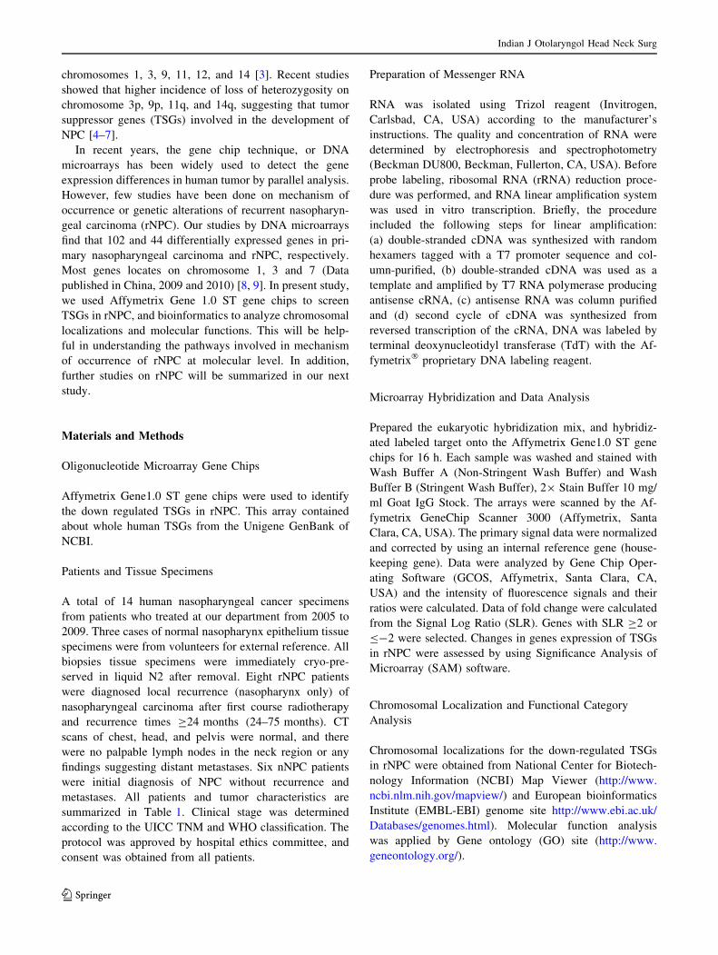
Quality of RNA and RT-PCR
Total RNA of 17 tissue samples were qualified for PCR
and hybridization: A260/A280 C 2.0, 2100 RIN C 7.0,
28S/18S C 0.7. The differential expression of genes iden-
tified by the hybridization method was confirmed by RT-
PCR (Fig. 1).
Hybridization with Gene Chips
rNPC and nNPC tissue samples were scanned respectively
after hybridization with the Affymetrix Gene1.0 ST gene
chips. The results were collected and analyzed by bioin-
formatics. A total of five TSGs were identified to be down-
regulated in rNPC. These five TSGs include SERPINF1,
TPD52L1, FBLN1, RASSF6, and S100A2, and Signal Log
Ratio (SLR) were -2.2, -2.3, -3.5, -3.9 and -6.9
respectively (Fig. 2).
Chromosomal Localization and Functional Categories
of Down-Regulated TSGs
Chromosomal localization analysis showed that analysis
showed that S100A2, RASSF6, TPD52L1, SERPINF1, and
FBLN1 were located on chromosomes 1q, 4q, 6q, 17p and
22q, respectively (Fig. 3). Functional analysis showed that
SERPINF1 and TPD52L1 belonged to enzyme activity
genes, S100A2 FBLN1 belonged to calcium ion binding
genes, and RASSF6 belong to protein binding genes
(Table 2).
Discussion
Recent advances in basic biological research and genomics
have improved our understanding of the molecular basis of
NPC development and progression. As a result, a variety of
molecular tumor markers characterized in the laboratory
have been studied in the clinic for their potential to predict
Table 1 Tumor stage and
recurrent month are list for all
samples
Sample ID WHO histological
diagnosis
TNM Gender Age Recurrent
month
nNPC 1 WHOI T1N0M0 Male 52
nNPC 2 WHOI T1N0M0 Female 52
nNPC 3 WHOI T1N0M0 Male 36
nNPC 4 WHOI T1N0M0 Male 56
nNPC 5 WHOI T1N0M0 Male 53
nNPC 6 WHOI T1N0M0 Male 59
nNPC 7 WHOI T1N0M0 Female 67
nNPC 8 WHOI T1N0M0 Male 39
rNPC 1 rNPC rT1 Male 56 24
rNPC 2 rNPC rT1 Female 68 72
rNPC 3 rNPC rT1 Male 45 28
rNPC 4 rNPC rT1 Male 62 75
rNPC 5 rNPC rT1 Female 61 35
rNPC 6 rNPC rT1 Male 58 48
Fig. 1 Quality of RNA from all
sample: nNPC: No. 1–8;
rNPC:No. 8–6. A260/
A280 C 2.0, 2100 RIN C 7.0,
28S/18S C 0.7
Indian J Otolaryngol Head Neck Surg
123

disease outcome or response to therapy in NPC patients.
Our study is concentrated on patterns of TSGs expression
of nNPC and rNPC, which different from previous research
that focused on genetic aberrations between normal naso-
pharyngeal epithelial/cell lines and NPC. Because of rela-
tively few cases of rNPC (within nasopharynx) without
metastasizes, we selected nNPC only on stage I (T1N0M0)
as control.
To our knowledge, there have been no previous cDNA
microarray studies focused on chromosomal localization of
rNPC. Previous studies revealed that genetic alterations in
NPC were prevalent on 1q, 3p, 4q, 7, 9q, 11, 12q, and 18q
Fig. 2 Signal log ratio (SLR) of
five TSGs in rNPC
Fig. 3 The down-regulated TSGs in rNPC were distributed on chromosomes 1q, 4q, 6q, 17p, 22q
Indian J Otolaryngol Head Neck Surg
123

[7, 10–12]. Additionally, chromosome 3p plays an impor-
tant role in cancergenesis in NPC, and was a putative
nasopharyngeal carcinoma susceptibility locus [6, 13].
TSGs are involved in protecting a cell form one step on
the path to cancer. These genes have a dampening or
repressive effect on the regulation of the cell cycle or
promote apoptosis. Loss or reduction of suppressors may
promote tumorigenesis of normal cell. Inactivation of
tumor suppressors on 3p21.3 and 9p21 was demonstrated to
be an early event during the development of NPC [14–16].
Additionally, chromosome 14 harbors tumor suppressor
genes associated with NPC [17]. Many TSGs have been
reported to relate to NPC, such as BLU, RASSF1A,
TSLC1, PTPRG, P16, and DLEC1. In this study, none of
TSGs were found in chromosome 3p.
Of interest, five candidate TSGs, included SERPINF1,
TPD52L1, RASSF6, FBLN1, S100A2, were found among
these four categories of genes in this study. We believe that
this information may be important when trying to select
candidate genes for the further study of the molecular
mechanism of occurrence of rNPC.
SERPINF1 is a member of the serpin gene family, and
involves in tumor cell proliferation and angiogenesis.
Reduction in SERPINF1 expression levels may lead to
tumor malignancy in VEGF-low ovarian tumors [18].
TPD52L1 is involved in cell proliferation and calcium
signaling; it also interacts with the mitogen-activated pro-
tein kinase 5 (MAP3K5/ASK1) and positively regulates
MAP3K5 and up-regulates the ASK induced apoptosis
[19]. RASSF6 is a member of the RAS association (RA)
domain family of RAS effectors/tumor suppressors that
mediate some of the growth inhibitory properties of RAS.
Allen et al. [20] found RASSF6 is a novel RASSF family
member that demonstrates the properties of a Ras effector
and tumor suppressor. FBLN1 is a secreted glycoprotein
that becomes incorporated into a fibrillar extracellular
matrix, and has been implicated in a role in cellular
transformation and tumor invasion. FBLN1 mediates
platelet adhesion via binding fibrinogen. It enhanced
apoptosis of endothelial cells and tumor cells, and also
inhibits tumor angiogenesis [21]. S100A2 protein encoded
by this gene is a member of the S100 family of proteins
containing 2 EF-hand calcium-binding motifs. These pro-
teins are localized in the cytoplasm and/or nucleus of a
wide range of cells, and involved in the regulation of a
number of cellular processes such as cell cycle progres-
sion and differentiation. Zhang et al. [22] found S100A2
was down-regulated in lymph node metastasis of squa-
mous cell carcinoma of the head and neck (SCCHN),
suggesting that instead of being a putative tumor sup-
pressor, S100A2 may play a role in the metastasis of
SCCHN. Thus it may be inferred that occurrence of rNPC
is likely as a result of inactivation of TSGs. Accordingly,
the development of rNPC may involved in abnormity of
angiogenesis, cell proliferation/apoptosis, calcium signal-
ing, tumor cell metastasis and invasion. Moreover, these
five TSGs found in our study, located on chromosomes
1q, 4q, 6q, 17p, and 22q respectively, needed further
study for confirmation.
Previous researches found genetic alterations in NPC
that paved the way for us for further study in rNPC.
Although the sample sizes of this study were small and our
findings therefore require further validation in larger trials,
such preliminary results may provide important clues to the
understanding of the various gene networks implicated in
rNPC carcinogenesis and may contribute to the selection of
target TSGs for possible molecular diagnosis and therapy
in the future.
Conclusions
We identify five differentially TSGs in rNPC by cDNA
hybridization. These TSGs, including SERPINF1,
TPD52L1, RASSF6, FBLN1, S100A2. They are belonged
to enzyme activity, calcium ion binding and protein bind-
ing genes, and chromosomal localization showed the
majority of genes located on chromosomes 17p, 6q, 22q, 4q
and 1q. We suggest these TSGs may have a contribution to
the mechanism of rNPC. The next study is needed to
investigate roles of these predictor genes and select can-
didate genes in specific region of these four chromosomes
that abnormalities occurred.
Key messages: This research is first to study TSGs in
recurrent nasopharyngeal cancer. Ours result may con-
tribute to the selection of target TSGs for possible molec-
ular diagnosis and therapy in the future, and still warrant
further study.
Table 2 Information and
functional categories of five
down-regulated TSGs
Gene symbol Gene name SLR Genbank no. Molecular function
RASSF6 Ras association domain family member 6 -3.9 BC058835 Protein binding
FBLN1 Fibulin 1 -3.5 AF126110 Calcium ion binding
S100A2 S100 calcium binding protein A2 -6.9 BC002829 Calcium ion binding
SERPINF1 Serpin peptidase inhibitor, clade F member 1 -2.2 M76979 Enzyme activity
TPD52L1 Tumor protein D52-like 1 -2.3 U44427 Enzyme activity
Indian J Otolaryngol Head Neck Surg
123

Acknowledgments This study was funded by Zhejiang Science and
Technology Agency (No.2007C23G2090017), Zhejiang P.R. China.
References
1. Hildesheim A, Levine PH (1993) Etiology of nasopharyngeal
carcinoma: a review. Epidemiol Rev 15:466–485
2. Lengyel E, Baricza K, Somogyi A, Olajos J, Papai Z, Godeny M
et al (2003) Reirradiation of locally recurrent nasopharyngeal
carcinoma. Strahlenther Onkol 179:298–305
3. Zhou X, Cui J, Macias V, Kajdacsy-Balla AA, Ye H, Wang J et al
(2007) The progress on genetic analysis of nasopharyngeal car-
cinoma. Comp Funct Genomics 2007:57513
4. Shao J, Li Y, Wu Q, Liang X, Yu X, Huang L et al (2002) High
frequency loss of heterozygosity on the long arms of chromo-
somes 13 and 14 in nasopharyngeal carcinoma in southern China.
Chin Med J (Engl) 115:571–575
5. Hu LF, Eiriksdottir G, Lebedeva T, Kholodniouk I, Alimov A,
Chen F et al (1996) Loss of heterozygosity on chromosome arm
3p in nasopharyngeal carcinoma. Genes Chromosomes Cancer
17:118–126
6. Xiong W, Zeng ZY, Xia JH, Xia K, Shen SR, Li XL et al (2004)
A susceptibility locus at chromosome 3p21 linked to familial
nasopharyngeal carcinoma. Cancer Res 64:1972–1974
7. Hui ABY, Lo KW, Leung SF, Choi PHK, Fong Y, Lee JCK et al
(1996) Loss of heterozygosity on the long arm of chromosome 11
in nasopharyngeal carcinoma. Cancer Res 56:3225
8. Huang Z, Li W, Du J, Huang Y, Tan Y, Fang W et al (2009)
Research on differentially expressed genes in primary and
relapsed nasopharyngeal carcinoma by DNA microarray.
J Wenzhou Med Coll 39:568–571 (Article in Chinese)
9. Huang Z, Li W, Lin S, Huang Y, Du J, Tan Y et al (2010)
Identification of differentially expressed genes in recurrent
nasopharyngeal carcinoma and analysis of their chromosomal
location. Chin J Otorhinolaryngol Head Neck Surg 45:47–51
(Article in Chinese)
10. Fang Y, Guan X, Guo Y, Sham J, Deng M, Liang Q et al (2001)
Analysis of genetic alterations in primary nasopharyngeal carci-
noma by comparative genomic hybridization. Genes Chromo-
somes Cancer 30:254–260
11. Hui AB, Lo KW, Leung SF, Teo P, Fung MK, To KF et al (1999)
Detection of recurrent chromosomal gains and losses in primary
nasopharyngeal carcinoma by comparative genomic hybridisa-
tion. Int J Cancer 82:498–503
12. Hui AB, Lo KW, Teo PM, To KF, Huang DP (2002) Genome
wide detection of oncogene amplifications in nasopharyngeal
carcinoma by array based comparative genomic hybridization. Int
J Oncol 20:467–473
13. Zeng Z, Zhou Y, Zhang W, Li X, Xiong W, Liu H et al (2006)
Family-based association analysis validates chromosome 3p21 as
a putative nasopharyngeal carcinoma susceptibility locus. Genet
Med 8:156–160
14. Chan AS, To KF, Lo KW, Mak KF, Pak W, Chiu B et al (2000)
High frequency of chromosome 3p deletion in histologically
normal nasopharyngeal epithelia from southern Chinese. Cancer
Res 60:5365–5370
15. Chan AS, To KF, Lo KW, Ding M, Li X, Johnson P et al (2002)
Frequent chromosome 9p losses in histologically normal naso-
pharyngeal epithelia from southern Chinese. Int J Cancer
102:300–303
16. Cheng Y, Lung HL, Wong PS, Hao DC, Man CS, Stanbridge EJ
et al (2004) Chromosome 13q12 region critical for the viability
and growth of nasopharyngeal carcinoma hybrids. Int J Cancer
109:357–362
17. Cheung AK, Lung HL, Ko JM, Cheng Y, Stanbridge EJ, Zaba-
rovsky ER et al (2009) Chromosome 14 transfer and functional
studies identify a candidate tumor suppressor gene, mirror image
polydactyly 1, in nasopharyngeal carcinoma. Proc Natl Acad Sci
USA 106:14478–14483
18. Tsuchiya T, Nakahama K, Asakawa Y, Maemura T, Tanaka M,
Takeda S et al (2009) The reduction in pigment epithelium-
derived factor is a sign of malignancy in ovarian cancer
expressing low-level of vascular endothelial growth factor.
Gynecol Endocrinol 25:104–109
19. Cho S, Ko HM, Kim JM, Lee JA, Park JE, Jang MS et al (2004)
Positive regulation of apoptosis signal-regulating kinase 1 by
hD53L1. J Biol Chem 279:16050–16056
20. Allen NP, Donninger H, Vos MD, Eckfeld K, Hesson L, Gordon
L et al (2007) RASSF6 is a novel member of the RASSF family
of tumor suppressors. Oncogene 26:6203–6211
21. Xie L, Palmsten K, MacDonald B, Kieran MW, Potenta S, Vong
S et al (2008) Basement membrane derived fibulin-1 and fibulin-5
function as angiogenesis inhibitors and suppress tumor growth.
Exp Biol Med (Maywood) 233:155–162
22. Zhang X, Hunt JL, Shin DM, Chen ZG (2007) Down-regulation
of S100A2 in lymph node metastases of head and neck cancer.
Head Neck 29:236–243
Indian J Otolaryngol Head Neck Surg
123