Embed Size (px)
Citation preview
Identification of novel suggestive loci for high-grade myopiain Polish families
Malgorzata Rydzanicz,1 Swapan K. Nath,2 Celi Sun,2 Monika Podfigurna-Musielak,3 Agata Frajdenberg,4,5,6
Malgorzata Mrugacz,7 Daniel Winters,8 Uppala Ratnamala,9 Uppala Radhakrishna,9 Bassem A. Bejjani,10
Marzena Gajecka1,11
1Institute of Human Genetics, Polish Academy of Sciences, Poznan, Poland; 2Arthritis and Immunology Research Program,Oklahoma Medical Research Foundation, Oklahoma City, OK; 3Department of Ophthalmology, Leszno Hospital, Leszno, Poland;4Department of Ophthalmology, Marcinkowski University of Medical Sciences, Poznan, Poland; 5Namsos Hospital, Department ofOphthalmology, Namsos, Norway; 6University Hospital in Linköping, Department of Ophthalmology, Linköping, Sweden;7Department of Pediatric Ophthalmology, Medical University of Bialystok, Bialystok, Poland; 8School of Molecular Biosciences,Washington State University, Spokane, WA; 9Department of Surgery-Transplant, University of Nebraska Medical Center, Omaha,NE; 10Signature Genomic Laboratories, LLC, Spokane, WA; 11Basic Medical Sciences Program, WWAMI, Spokane, WA
Purpose: Myopia is the most common human eye disorder with complex genetic and environmental causes. To date,several myopia loci have been identified in families of different geographic origin. However, no causative gene(s) haveyet been identified. The aim of this study was the characterization of Polish families with high-grade myopia, includinggenetic analysis.Methods: Forty-two multiplex Polish families with non–syndromic high-grade myopia participated in the study. Allfamily members underwent detailed ophthalmic examination and high-grade myopia was defined as ≤-6.0 diopters (D)based on the spherical refractive error. A genome-wide single nucleotide polymorphism (SNP)-based high-density linkagescan was performed using Affymetrix Human SNP Array 6.0 on a selected family (HM-32) with multiple affectedindividuals.Results: Nonparametric linkage analysis identified three novel loci in family HM-32 at chromosome 7p22.1–7p21.1([NPL] 8.26; p=0.006), chromosome 7p12.3–7p11.2 ([NPL] 8.23; p=0.006), and chromosome 12p12.3–12p12.1 ([NPL]8.02; p=0.006), respectively. The effect of linkage disequilibrium on linkage due to dense SNP map was addressed bysystematically pruning SNPs from the linkage panel.Conclusions: Haplotype analysis with informative crossovers in affected individuals defined a 12.2; 10.9; and 9.5 Mbgenomic regions for high-grade myopia spanned between SNP markers rs11977885/rs10950639, rs11770622/rs9719399,and rs4763417/rs10842388 on chromosomes 7p22.1–7p21.1, 7p12.3–7p11.2, and 12p12.3–12p12.1, respectively.
Myopia, also known as shortsightedness, is the mostcommon eye disorder worldwide. In myopic subjects, theimage of distant objects falls in front of the retina, either asthe eye is too long (axial myopia), the cornea is too convex orthe index of refraction of the lens is too high (refractivemyopia) [1]. The myopic eye is generally vulnerable andpersons with ≤-6.0 diopters (D) are more liable to a wide rangeof ocular pathologies. The development of high-grade myopiainvolves anterior-posterior enlargement of the eye, scleralthinning, changes in the diameter of scleral collagen fibrils,and frequent detachment of the retina resulting from stressrelated with axial elongation [2].
The estimated prevalence of high grade myopia is ~2.5to 9.6% in the elderly world population [3,4]. However, itshighest prevalence rates are in Asians, in whom almost 50 to
Correspondence to: Marzena Gajecka, Ph.D., Institute of HumanGenetics, Polish Academy of Sciences, Strzeszynska 32, Poznan,60-479, Poland; Phone: (061) 657-9160; FAX: (061) 823-3235;email: [email protected]
80% of the adult populations are myopic [5-7]. Recentpopulation-based studies suggest that the prevalence isincreasing, specifically in Asian populations [8,9]. Thefrequency of myopia in the Polish population is unknown, andthere is a paucity of data about the epidemiology of high-myopia in Poland. Until the present study, no analysis has yetbeen made on familial high-grade myopia. However, inPoland the main cause of blindness and ~12% childhoodvisual impairment is due to high-grade myopia [10].
Myopia may be of diverse etiology, includingenvironmental and genetic factors [11-17]. However, high-grade myopia is highly heritable and genetic predispositionsseem to be a dominant factor of its development andprogression [18,19]. Families with autosomal dominant,autosomal recessive, and X-linked inheritance of high-grademyopia have been described, though the majority of thereports deal with the autosomal dominant form [20-28].Although there were no obvious phenotypic differencesbetween the affected subjects of families used in thepreviously published linkage analyses, the data were
Molecular Vision 2011; 17:2028-2039 <http://www.molvis.org/molvis/v17/a221>Received 24 March 2011 | Accepted 18 July 2011 | Published 22 July 2011
© 2011 Molecular Vision
2028
inconsistent, suggesting genetic heterogeneity amongpopulations of different geographic origin [20,23]. Severalstudies have demonstrated a significant genetic component inthe familial aggregation of high-grade and/or moderatemyopia. Up to 17 loci on different chromosomal regions havebeen identified [20,22,24-37]. Additionally, increasedincidence of single nucleotide polymorphism (SNP)sassociation between the insulin-like growth factor 1 (IGF-1)gene and high-grade myopia [38], and polymorphisms in thepromoter regions of matrix metalloproteinase (MMP) geneswere reported [39]. Moreover, two recent independentgenome-wide association studies conducted on large cohortsof refractive error patients identified loci at chromosome15q14 and 15q25 and suggest that the genetic variance inrefractive errors is mostly determined by multiple variantswith a low to moderate penetrance [40,41].
In this study we present clinical characteristics of fortytwo Polish families with non-syndromic high-grade myopiaand the results of a high-density SNP-based linkage analysisfor one selected large high-grade myopia family with multipleaffected and normal individuals. Our findings providedevidence of suggestive linkage at three distinct novel loci onchromosome 7p22.1–7p21.3, 7p12.3–7p11.2, and 12p12.3–12p12.1 in the analyzed family.
METHODSRecruitment and clinical evaluation of high-grade myopiafamilies: The study population consisted of 42 multiplex high-grade myopia families from Poland, who were ascertained atthree independent Polish institutions: 1) Department ofOphthalmology, Marcinkowski University of MedicalSciences, in Poznan, 2) Department of PediatricOphthalmology, University of Medical Sciences in Bialystok,and 3) Department of Ophthalmology, Hospital, Leszno. Aconstant clinical evaluation procedure was applied at allclinical sites. Informed consent was obtained from all studysubjects after the possible consequences of participating in thestudy were explained, in accordance with the Declaration ofHelsinki.
All study subjects underwent a detailed ophthalmicevaluation using computer-assisted equipment included: avisual acuity testing, best-corrected visual acuity testing, a slitlamp evaluation, intraocular pressure examination,fundoscopy, axial length determination, keratometry andrefractometry. Biometric axial length (including anterior-chamber depth, lens thickness, and total axial length) wasmeasured using ultrasonography (A, OPTOPOL, Desmin F/H, version 2.06.21). In children ≤15 years old, the refractiveerror was measured with an autorefractor after cycloplegia. Acomplete questionnaire was filled for each subject withclinical and family history.
To minimize misclassification, clear diagnostic criteriawere established for all high-grade myopia study subjects
including spherical refractive error analysis. The subjectswere classified into three groups, 1) Affected individuals withhigh-grade myopia, 2) Individuals with an unknown status and3) Unaffected persons. All affected individuals showed: 1)bilateral axial high-grade myopia, in excess of or equal to−6.0 D (≤-6.0 D) for at least one eye and in excess of or equalto −5.0 D (≤-5.0 D) for the second eye; 2) a history of onsetof myopia at age ≤15 years, and 3) individual with affectedstatus while high-grade myopia was identified in multiplemembers of their family in different generations. Individualswho were classified as unknown were: 1) all children ≤15years unless they fulfill criteria for affected status as specifiedabove, or 2) individuals who have myopia with −6.0 D < X≤ −4.0 D, or 3) individuals, with a refractive error of ≤-6.0 Dfor one eye and a refractive error >-5.0 D for the second eye,or 4) individuals with late age of onset (>15 years). All theremaining were treated as unaffected as neither of them wereclassified as affected nor unknown for the analysis.
For all 42 Polish HM families we have performed theanalysis using microsatellite markers to exclude or confirmlinkage with known high myopia loci (data not shown). In allfamilies previously suggested candidate loci/genes for highmyopia were excluded (data not shown).Statistical analysis in clinical evaluation: Differences inophthalmic parameters obtained for respective groups, as wellas comparison of age were analyzed by the Kruskal–Wallistest [42]. Gender distribution was calculated by χ2 test. Allanalyzed features were compared among groups according tothe scheme: affected versus unaffected, affected versusunknown and unaffected versus unknown. The differencesbetween examined groups were considered significant if thevalue of probability (p) did not exceed 0.05. Axial length inaffected individuals helped reveal whether a patient hadcorrective surgery in the past. Affected subjects whounderwent corrective surgery were not included in analysis ofmean refraction value for high-grade myopes versus non-highly myopic subjects.Genome-wide genotyping in family HM-32: The familyHM-32 was chosen for genome-wide genotyping analysis.The selected pedigree was the largest, multigenerational,representative family with many available family members,including patients with high-grade myopia, as well asunaffected relatives.
A genome-wide SNP-based high-density linkage scanwas performed using the Affymetrix Human SNP array 6.0(Affymetrix Inc., Santa Clara, CA) which features 1.8 milliongenetic markers, including 906,600 SNPs and 946,000 probesfor the detection of copy number variation. The assay wasperformed with 500 ng of genomic DNA, and more than 99%of the SNPs were determined unequivocally for each sample.Scanned images were processed with gene microarraysoftware (Affymetrix) and the data was analyzed (GDAS ver.2, software; GeneChip Data Analysis; Affymetrix).
Molecular Vision 2011; 17:2028-2039 <http://www.molvis.org/molvis/v17/a221> © 2011 Molecular Vision
2029
PEDCHECK [43] was used to identify Mendelianinconsistencies, and the MERLIN [44] program was used todetect double recombination events over short geneticdistances that were probably due to genotyping errors. Afterthe quality control (QC) of the raw genotype data whichdeleted SNPs with missing genotypes and all SNPs at whichall individuals have the ‘BB’ or ‘AA’ genotypes, there werea total 550,441 SNPs left for analysis. The genotypes and themarkers generated from the Affymetrix 6.0 platform were sodense that linkage disequilibrium between many markers willresult in severe biases in linkage calculations. Since weanalyzed one family with 16 samples genotypes available, weused the allele frequencies from HapMap CEU population forthe analysis. To prevent bias from linkage disequilibrium(LD) in linkage calculations, we first created genotype subsetwith 4,417 SNPs for the genome wide linkage scan byselecting one SNP from every 100,000 bases in the QCgenotype data set. While selecting SNPs, the minor allelefrequency (MAF) >1% at each SNP was also a criterion forselection. To maximize the heterozygosity, we alwaysselected the SNPs with high MAF. In a case of identifiedcandidate interval(s) further analysis was performed,selecting one SNP from every two SNPs from candidateinterval(s) for the analysis to eliminate the bias from LD.Moreover, linkage analysis was also done by another way toaccount for LD effect by using genetic distances, selecting oneSNP per 0.5 cM, 1 cM, 1.5 cM, and 2 cM for the linkageanalysis, respectively.
SNP genotype data were imported into the linkage-analysis programs GENEHUNTER [45] and MERLIN [44].In the initial genome scan, evidence of linkage was assessedwith a nonparametric, penetrance-independent, affected-only,and allele-sharing analysis (Z-mean from MERLIN andnonparametric linkage (NPL) from GENEHUNTER). WithMERLIN, one can convert this into a nonparametric logarithm(base 10) of odds (LOD*) score by maximizing the likelihood
with respect to a scalar parameter, δ, that measures the amountof excess sharing of identical-by-descent alleles amongaffected relatives (with δ=0) corresponding to the nullhypothesis of no linkage [46]. We used the Sall scoring functionand the exponential allele-sharing model to generate therelevant linkage statistic. When significant evidence oflinkage was found by exceeding the predetermined threshold(p<0.01), two-point as well as multipoint LOD scoresmaximized over various plausible genetic model parameterswere calculated. For the parametric linkage analysis the bestmodel was estimated as an autosomal dominant mode ofinheritance with reduced penetrance (0.6) and phenocopy rate(0.01) and a disease allele frequency of 0.0001. In addition,for the parametric linkage analysis an affected only analysiswas performed under an autosomal dominant mode ofinheritance allowing for phenocopies. Genetic map distanceswere derived from the Rutgers combined linkage-physicalmap of the human genome [47], either directly or byinterpolation. Haplotypes were reconstructed using bothGENEHUNTER and SIMWALK2 programs [48,49].
RESULTSClinical and demographic characteristics of studied families:The forty-two large Polish pedigrees enrolled in the study, hadfamilies with five-generation (n=10), four-generations (n=7),three-generations (n=27), and two-generations (n=7) with anaverage number of individuals in each generation per familyof 3.5 (Figure 1, Figure 2, and Appendix 1). The mean familysize was 8.3 individuals (range 3–45), with the averageaffected individuals per family of 3.0 (range 2–10), unaffected4.1 (range 1–28) and unknown status 0.8 (range 1–7). Thespecified information was based on individuals whounderwent an ophthalmic examination.
A complete eye examination was performed for 331participated individuals. In accordance with our classificationcriteria, 128 individuals were considered as affected, 171 as
Figure 1. Pedigree of family HM-32 with high myopia. Blackened symbols: individuals with high myopia; unblackened symbols: unaffectedindividuals; symbols with question mark: individuals with unknown disease status. Individuals used in the linkage analysis are numberedunder their symbols in the pedigree.
Molecular Vision 2011; 17:2028-2039 <http://www.molvis.org/molvis/v17/a221> © 2011 Molecular Vision
2030
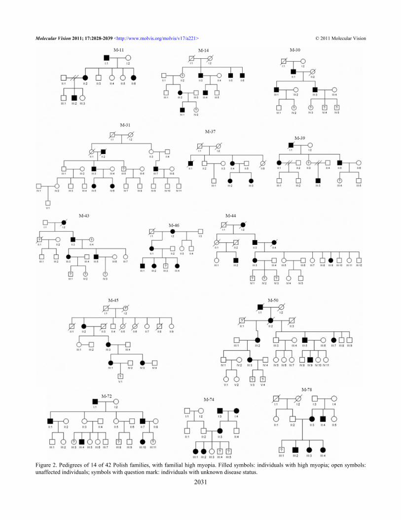
Figure 2. Pedigrees of 14 of 42 Polish families, with familial high myopia. Filled symbols: individuals with high myopia; open symbols:unaffected individuals; symbols with question mark: individuals with unknown disease status.
Molecular Vision 2011; 17:2028-2039 <http://www.molvis.org/molvis/v17/a221> © 2011 Molecular Vision
2031
unaffected, and 32 with unknown status. The characteristicsand details of the ophthalmic examinations in particular studygroups are given in Table 1. Additionally, Appendix 2 showsdetailed clinical findings in family HM-32. Spherical
refractive error alone (without cylindrical refractive error)was enough to classify 128 affected individuals in thiscategory.
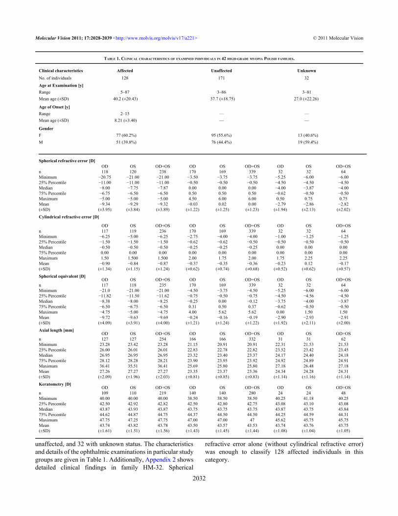
TABLE 1. CLINICAL CHARACTERISTICS OF EXAMINED INDIVIDUALS IN 42 HIGH-GRADE MYOPIA POLISH FAMILIES.
Clinical characteristics Affected Unaffected UnknownNo. of individuals 128 171 32Age at Examination [y]Range 5–87 3–86 3–81Mean age (±SD) 40.2 (±20.43) 37.7 (±18.75) 27.0 (±22.26)
Age of Onset [y]Range 2–15 — —Mean age (±SD) 8.21 (±3.40) — —
GenderF 77 (60.2%) 95 (55.6%) 13 (40.6%)M 51 (39.8%) 76 (44.4%) 19 (59.4%)
Spherical refractive error [D] OD OS OD+OS OD OS OD+OS OD OS OD+OSn 118 120 238 170 169 339 32 32 64Minimum −20.75 −21.00 −21.00 −3.50 −3.75 −3.75 −5.25 −6.00 −6.0025% Percentile −11.00 −11.00 −11.00 −0.50 −0.50 −0.50 −4.50 −4.50 −4.50Median −8.00 −7.75 −7.87 0.00 0.00 0.00 −4.00 −3.87 −4.0075% Percentile −6.75 −6.50 −6.50 0.50 0.50 0.50 −0.62 −0.50 −0.50Maximum −5.00 −5.00 −5.00 4.50 6.00 6.00 0.50 0.75 0.75Mean −9.34 −9.29 −9.32 −0.03 0.02 0.00 −2.79 −2.86 −2.82(±SD) (±3.95) (±3.84) (±3.89) (±1.22) (±1.25) (±1.23) (±1.94) (±2.13) (±2.02)Cylindrical refractive error [D]
OD OS OD+OS OD OS OD+OS OD OS OD+OSn 117 119 236 170 169 339 32 32 64Minimum −6.25 −5.00 −6.25 −2.75 −4.00 −4.00 −1.00 −1.25 −1.2525% Percentile −1.50 −1.50 −1.50 −0.62 −0.62 −0.50 −0.50 −0.50 −0.50Median −0.50 −0.50 −0.50 −0.25 −0.25 −0.25 0.00 0.00 0.0075% Percentile 0.00 0.00 0.00 0.00 0.00 0.00 0.00 0.00 0.00Maximum 1.50 1.500 1.500 2.00 1.75 2.00 1.75 2.25 2.25Mean −0.90 −0.84 −0.87 −0.37 −0.35 −0.36 −0.23 0.12 −0.17(±SD) (±1.34) (±1.15) (±1.24) (±0.62) (±0.74) (±0.68) (±0.52) (±0.62) (±0.57)Spherical equivalent [D] OD OS OD+OS OD OS OD+OS OD OS OD+OSn 117 118 235 170 169 339 32 32 64Minimum −21.0 −21.00 −21.00 −4.50 −3.75 −4.50 −5.25 −6.00 −6.0025% Percentile −11.82 −11.50 −11.62 −0.75 −0.50 −0.75 −4.50 −4.56 −4.50Median −8.38 −8.00 −8.25 −0.25 0.00 −0.12 −3.75 −4.00 −3.8775% Percentile −6.50 −6.75 −6.50 0.31 0.50 0.37 −0.62 −0.50 −0.50Maximum −4.75 −5.00 −4.75 4.00 5.62 5.62 0.00 1.50 1.50Mean −9.72 −9.63 −9.69 −0.24 −0.16 −0.19 −2.90 −2.93 −2.91(±SD) (±4.09) (±3.91) (±4.00) (±1.21) (±1.24) (±1.22) (±1.92) (±2.11) (±2.00)Axial length [mm] OD OS OD+OS OD OS OD+OS OD OS OD+OSn 127 127 254 166 166 332 31 31 62Minimum 23.28 23.42 23.28 21.15 20.91 20.91 22.31 21.53 21.5325% Percentile 26.00 26.01 26.01 22.83 22.78 22.82 23.52 23.42 23.45Median 26.95 26.95 26.95 23.32 23.40 23.37 24.17 24.40 24.1875% Percentile 28.12 28.28 28.21 23.90 23.95 23.92 24.92 24.89 24.91Maximum 36.41 35.51 36.41 25.69 25.80 25.80 27.18 26.48 27.18Mean 27.26 27.27 27.27 23.35 23.37 23.36 24.34 24.28 24.31(±SD) (±2.09) (±1.96) (±2.03) (±0.81) (±0.85) (±0.83) (±1.14) (±1.16) (±1.14)Keratometry [D] OD OS OD+OS OD OS OD+OS OD OS OD+OSn 109 110 219 140 140 280 24 24 48Minimum 40.00 40.00 40.00 38.50 38.50 38.50 40.25 41.18 40.2525% Percentile 42.50 42.92 42.82 42.50 42.80 42.75 43.08 43.10 43.08Median 43.87 43.93 43.87 43.75 43.75 43.75 43.87 43.75 43.8475% Percentile 44.62 44.87 44.75 44.37 44.50 44.50 44.25 44.59 44.31Maximum 47.75 47.25 47.75 47.00 47.00 47 45.62 45.75 45.75Mean 43.74 43.82 43.78 43.50 43.57 43.53 43.74 43.76 43.75(±SD) (±1.61) (±1.51) (±1.56) (±1.43) (±1.45) (±1.44) (±1.08) (±1.04) (±1.05)
Molecular Vision 2011; 17:2028-2039 <http://www.molvis.org/molvis/v17/a221> © 2011 Molecular Vision
2032
There are several individuals in this study with unknowndisease status, which partly due to the involvement of children<15 years. In 50% of cases the unknown disease status is dueto the inclusion of individuals with an average sphericalrefractive error (SPH) ranges between −6.0 D < X ≤ −4.0 D.For example, in families 10, 14, 39 and 75, individuals 10-III:3, 14-II: 2, 39-III: 4 and 75-I: 4 presented SPH as follow:−4.75/-4.75 D, −4.5/-5.25 D, −4.25/-4.25 D and −4.5/-6.0 D,respectively.
Based on medical records and/or self-reports the averageage of onset in myopic subjects was ~8 years (range 2–15).Affected females had slightly earlier onset than affected males(7.78 years, range 2–14 versus 8.88 years, range 2–15,respectively), however the difference was not statistically
significant (p=0.077). Some of the affecteds were found withvarious associated anomalies including glaucoma (11.7%;n=15), cataract (4.7%; n=6), retinal detachment (RD; 5.5%;n=7), and RD in both eyes (n=2). In both unknown andunaffected individuals no other anomalies were identifiedexcept one normal individual with glaucoma (0.6%).
We have found statistically significant differences inspherical refractive error, spherical equivalent refractive error(SE) and axial length (AL) between the studied groups(p<0.001; Appendix 3). In affected subjects the average SPHwas −9.32 D (±3.89), where the median value was −7.87 D,compared with −2.82 D (±2.02, median −4.00 D) for unknownand 0.00 D (±1.23, median 0.00 D) for unaffected individuals.The average spherical equivalent refractive error of the
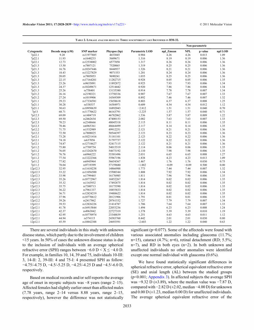
TABLE 2. LINKAGE ANALYSIS RESULTS: THREE SUSCEPTIBILITY LOCI IDENTIFIED IN HM-32.
Non-parametric
Cytogenetic Decode map (cM) SNP marker Physpos (bp) Parametric LOD npl_Zmean NPL p value npl LOD7p22.1 9.20 rs11977885 4653045 0.904 4.26 4.26 0.013 1.097p22.1 11.93 rs1640233 5609671 1.316 8.19 8.19 0.006 1.367p22.1 12.73 rs12538002 6577858 1.317 8.26 8.26 0.006 1.367p21.3 13.50 rs7807121 7520065 1.319 8.25 8.25 0.006 1.367p21.3 16.76 rs10247446 8444937 1.326 8.21 8.21 0.006 1.367p21.3 18.43 rs13227829 9071553 1.201 8.24 8.24 0.006 1.367p21.3 20.05 rs7805053 9690241 1.035 8.25 8.25 0.006 1.367p21.3 22.15 rs17164201 11282715 0.928 8.05 8.05 0.006 1.357p21.3 23.26 rs9655091 11892872 0.925 7.93 7.93 0.006 1.347p21.3 24.37 rs10249671 12514442 0.920 7.86 7.86 0.006 1.347p21.3 25.26 rs758401 13135380 0.914 7.78 7.78 0.007 1.347p21.2 26.16 rs2190321 13750336 0.907 7.67 7.67 0.007 1.337p21.2 27.24 rs1019906 14369309 0.892 7.46 7.46 0.007 1.327p21.1 29.23 rs17336581 15658618 0.803 6.37 6.37 0.008 1.257p21.1 30.28 rs538537 16584971 0.609 4.54 4.54 0.012 1.127p21.1 30.63 rs10950639 16892043 −2.812 1.51 1.51 0.040 0.707p13 68.71 rs11770622 46163791 −2.225 1.57 1.57 0.040 0.71
7p12.3 69.09 rs1404719 46782062 1.536 5.87 5.87 0.009 1.227p12.3 69.50 rs2462634 47400153 2.082 7.63 7.63 0.007 1.337p12.3 70.23 rs2348666 48045318 2.115 8.11 8.11 0.006 1.357p12.3 70.46 rs6965361 48664499 2.117 8.14 8.14 0.006 1.357p12.2 71.75 rs1532989 49912251 2.121 8.21 8.21 0.006 1.367p12.2 72.50 rs7808025 50544397 2.123 8.23 8.23 0.006 1.367p12.1 73.28 rs10231416 51181101 2.123 8.22 8.22 0.006 1.367p12.1 74.18 rs637056 51797737 2.122 8.22 8.22 0.006 1.367p12.1 74.87 rs12718627 52417115 2.122 8.21 8.21 0.006 1.367p11.2 75.80 rs7789754 54015519 2.114 8.06 8.06 0.006 1.357p11.2 76.05 rs13242670 54673192 2.110 7.98 7.98 0.006 1.357p11.2 76.76 rs6966222 55301496 2.031 6.65 6.65 0.008 1.277p11.2 77.54 rs13222366 55967196 1.838 4.23 4.23 0.013 1.097p11.2 77.82 rs6945964 56654367 1.467 1.76 1.76 0.030 0.757p11.2 78.04 rs9719399 57159680 −1.882 −0.09 −0.09 0.500 0.00
12p12.3 32.95 rs11610238 14866988 1.773 7.44 7.44 0.007 1.3212p12.3 33.22 rs11056500 15500344 1.808 7.92 7.92 0.006 1.3412p12.3 33.98 rs1799465 16176985 1.811 7.96 7.96 0.006 1.3512p12.3 35.26 rs10772967 16852267 1.814 8.02 8.02 0.006 1.3512p12.3 35.32 rs1163932 17492875 1.814 8.02 8.02 0.006 1.3512p12.3 35.75 rs7300713 18175590 1.814 8.02 8.02 0.006 1.3512p12.3 36.22 rs7961337 18855833 1.814 8.02 8.02 0.006 1.3512p12.3 36.71 rs12824219 19488622 1.814 8.02 8.02 0.006 1.3512p12.2 37.96 rs10770612 20121906 1.791 8.01 8.01 0.006 1.3512p12.2 39.26 rs2417862 20761532 1.727 7.79 7.79 0.007 1.3412p12.1 39.53 rs12826226 21414787 1.706 7.64 7.64 0.007 1.3312p12.1 41.78 rs16915844 22053236 1.494 6.23 6.23 0.008 1.2412p12.1 42.37 rs4963842 22727319 1.378 5.39 5.39 0.010 1.1912p12.1 42.95 rs10770974 23348839 1.251 4.63 4.63 0.011 1.1212p12.1 44.94 rs574115 24383760 0.442 2.01 2.01 0.030 0.8012p12.1 45.59 rs10842388 24693591 −2.603 1.22 1.22 0.040 0.63
Molecular Vision 2011; 17:2028-2039 <http://www.molvis.org/molvis/v17/a221> © 2011 Molecular Vision
2033
affected was −9.69 D (±4.00, median −8.25 D), where forunknown and unaffected respectively, −2.91 D (±2.00,median −3.87 D) and −0.19 D (±1.22, median −0.12 D). Themean AL (27.27±2.03, median 26.95 mm) of high-grademyopic eyes was significantly higher than observed forunknown (24.31±1.14, median 24.18 mm) and unaffectedindividuals (23.36±0.83, median 23.37 mm; Table 1 andAppendix 3). There was no difference in keratometry amongthe studied groups.
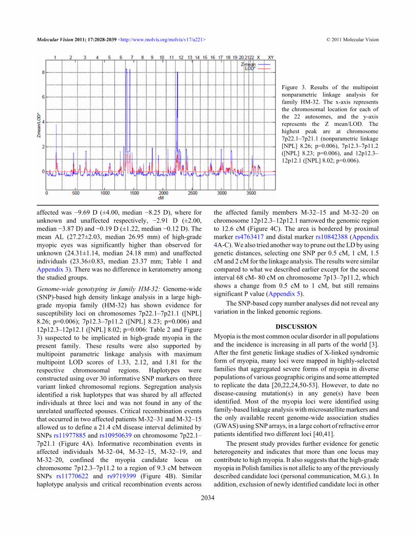
Genome-wide genotyping in family HM-32: Genome-wide(SNP)-based high density linkage analysis in a large high-grade myopia family (HM-32) has shown evidence forsusceptibility loci on chromosomes 7p22.1–7p21.1 ([NPL]8.26; p=0.006); 7p12.3–7p11.2 ([NPL] 8.23; p=0.006) and12p12.3–12p12.1 ([NPL] 8.02; p=0.006: Table 2 and Figure3) suspected to be implicated in high-grade myopia in thepresent family. These results were also supported bymultipoint parametric linkage analysis with maximummultipoint LOD scores of 1.33, 2.12, and 1.81 for therespective chromosomal regions. Haplotypes wereconstructed using over 30 informative SNP markers on threevariant linked chromosomal regions. Segregation analysisidentified a risk haplotypes that was shared by all affectedindividuals at three loci and was not found in any of theunrelated unaffected spouses. Critical recombination eventsthat occurred in two affected patients M-32–31 and M-32–15allowed us to define a 21.4 cM disease interval delimited bySNPs rs11977885 and rs10950639 on chromosome 7p22.1–7p21.1 (Figure 4A). Informative recombination events inaffected individuals M-32–04, M-32–15, M-32–19, andM-32–20, confined the myopia candidate locus onchromosome 7p12.3–7p11.2 to a region of 9.3 cM betweenSNPs rs11770622 and rs9719399 (Figure 4B). Similarhaplotype analysis and critical recombination events across
the affected family members M-32–15 and M-32–20 onchromosome 12p12.3–12p12.1 narrowed the genomic regionto 12.6 cM (Figure 4C). The area is bordered by proximalmarker rs4763417 and distal marker rs10842388 (Appendix4A-C). We also tried another way to prune out the LD by usinggenetic distances, selecting one SNP per 0.5 cM, 1 cM, 1.5cM and 2 cM for the linkage analysis. The results were similarcompared to what we described earlier except for the secondinterval 68 cM- 80 cM on chromosome 7p13–7p11.2, whichshows a change from 0.5 cM to 1 cM, but still remainssignificant P value (Appendix 5).
The SNP-based copy number analyses did not reveal anyvariation in the linked genomic regions.
DISCUSSIONMyopia is the most common ocular disorder in all populationsand the incidence is increasing in all parts of the world [3].After the first genetic linkage studies of X-linked syndromicform of myopia, many loci were mapped in highly-selectedfamilies that aggregated severe forms of myopia in diversepopulations of various geographic origins and some attemptedto replicate the data [20,22,24,50-53]. However, to date nodisease-causing mutation(s) in any gene(s) have beenidentified. Most of the myopia loci were identified usingfamily-based linkage analysis with microsatellite markers andthe only available recent genome-wide association studies(GWAS) using SNP arrays, in a large cohort of refractive errorpatients identified two different loci [40,41].
The present study provides further evidence for geneticheterogeneity and indicates that more than one locus maycontribute to high myopia. It also suggests that the high-grademyopia in Polish families is not allelic to any of the previouslydescribed candidate loci (personal communication, M.G.). Inaddition, exclusion of newly identified candidate loci in other
Figure 3. Results of the multipointnonparametric linkage analysis forfamily HM-32. The x-axis representsthe chromosomal location for each ofthe 22 autosomes, and the y-axisrepresents the Z mean/LOD. Thehighest peak are at chromosome7p22.1–7p21.1 (nonparametric linkage[NPL] 8.26; p=0.006), 7p12.3–7p11.2([NPL] 8.23; p=0.006), and 12p12.3–12p12.1 ([NPL] 8.02; p=0.006).
Molecular Vision 2011; 17:2028-2039 <http://www.molvis.org/molvis/v17/a221> © 2011 Molecular Vision
2034
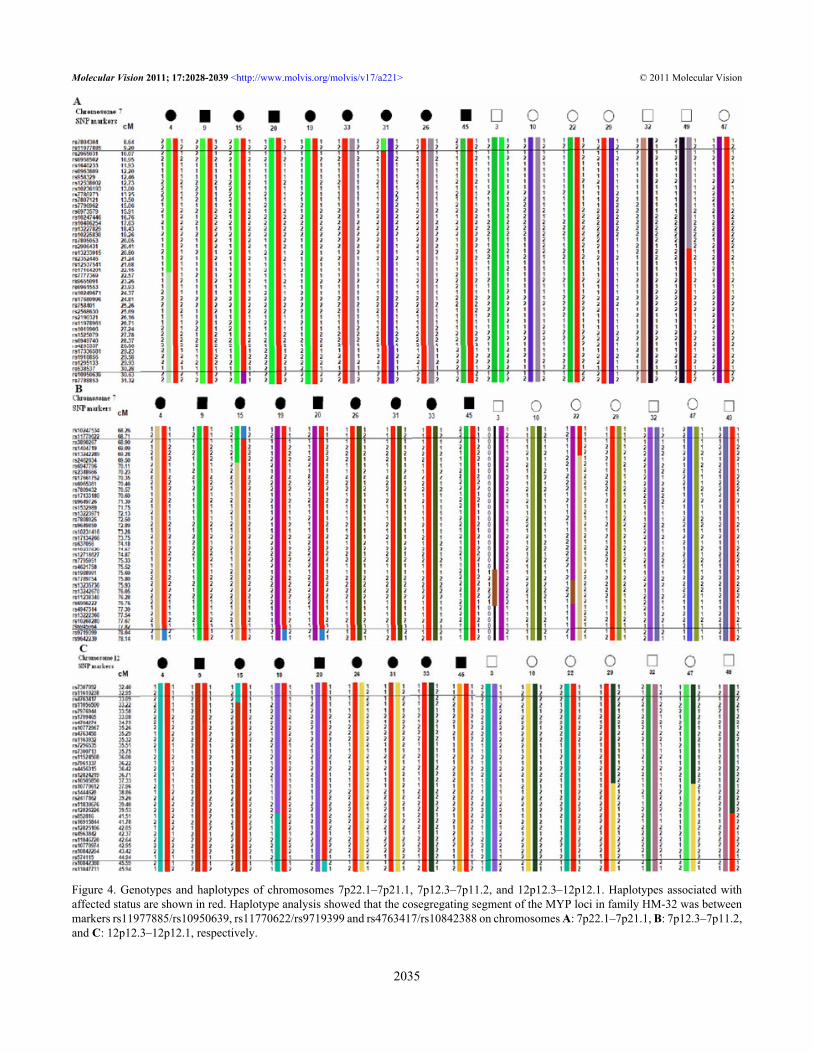
Figure 4. Genotypes and haplotypes of chromosomes 7p22.1–7p21.1, 7p12.3–7p11.2, and 12p12.3–12p12.1. Haplotypes associated withaffected status are shown in red. Haplotype analysis showed that the cosegregating segment of the MYP loci in family HM-32 was betweenmarkers rs11977885/rs10950639, rs11770622/rs9719399 and rs4763417/rs10842388 on chromosomes A: 7p22.1–7p21.1, B: 7p12.3–7p11.2,and C: 12p12.3–12p12.1, respectively.
Molecular Vision 2011; 17:2028-2039 <http://www.molvis.org/molvis/v17/a221> © 2011 Molecular Vision
2035
Polish families indicates possible genetic heterogeneitywithin Polish population signifying that genome-wide linkageanalysis in these families may reveal novel locus/loci for highmyopia. Naiglin et al. [21] also reported genetic heterogeneityin families with high myopia. Earlier linkage for high myopiawas reported on chromosome 7p and 12p; however these locido not overlap with the genomic regions identified in thepresent family HM-32 [51,54,55]. Since we are drawing thelinkage inferences from one large family with a high-densitySNP data, we took proper care for accounting the false positivedue to high LD, and our results were very consistent. At thesame time, we have chosen the SNPs in our initial linkagepanel in a way (MAF>1%) that increased the markerheterozygosity, hence, increased the linkage informationcontent that improves the likelihood of detecting arecombinant event.
The 12.2 Mb candidate interval on chromosome 7p22.1–7p21.1 contains 61 known transcripts. These include genesinvolved in regulation of cell proliferation, growth andextension: β-actin (ACTB [OMIM 102630]), fascin homolog1, actin-bundling protein (FSCN1 [OMIM 602689]), ras-related C3 botulinum toxin substrate 1 (RAC1 [OMIM602048]), as well as in gene expression: zinc finger protein 12(ZNF12 [OMIM 194536]). Further, genes considered in thislocus are: islet cell autoantigen 1 (ICA1 [OMIM 147625]) andcollagen, type XXVIII, alpha 1 (COL28A1 [OMIM 609996]).
The second interesting locus identified at chromosome7p12.3–7p11.2 that maps to a 10.9 Mb region comprises 30known transcripts, including growth factor receptor-boundprotein 10 (GRB10 [OMIM 601523]) and epidermal growthfactor receptor (EGFR [OMIM 131550]). GRB10 encodes agrowth factor receptor-binding protein that interacts withinsulin receptors and insulin-like growth-factor receptors[55]. Based on an animal model, it has been established, thatGRB10 acts as an inhibitor of intracellular signaling pathwaysregulating growth and metabolism. Gene knockouts in miceresults in disproportionate overgrowth of the embryo andplacenta [56]. EGFR and its ligands are cell signalingmolecules involved in diverse cellular functions, includingcell proliferation, differentiation, motility, and survival, andin tissue development [57]. EGFR is the prototypical tyrosinekinase receptor localized to basal and differentiated epitheliain the cornea and is a key regulator for maintaining a healthycornea and promoting regrowth of the wounded cornea [58,59]. Furthermore, Domínguez et al. [60] reported variousEGFR functions in Drosophila eye development.
The locus on chromosome 12p12.3–12p12.1 maps within9.5 Mb region that has 32 known transcripts which areinvolved in cell signaling and proliferation: Rho GDPdissociation inhibitor (GDI) beta (ARHGDIB [OMIM602843]), RAS-like, estrogen-regulated, growth inhibitor(RERG [OMIM 612664]), and epidermal growth factorreceptor pathway substrate 8 (EPS8 [OMIM 600206]),
phosphoinositide-3-kinase, class 2, gamma polypeptide(PIK3C2G [OMIM 609001]).
In the present linkage analysis, all HM-32 familymembers who carried the three disease-related haplotypeswere found with high-grade myopia, indicating that more thanone locus contributes to the high myopia phenotype in thispedigree. It is also possible that one of these linked loci is amajor dominant determinant and that the others are modifiergenomic variants. Therefore, we hypothesize that the high-grade myopia phenotype in this family could be due tomultifactorial inheritance; however, it is difficult to prove thishypothesis until we identify the pathologic mutations. Furtherresearch is needed to understand role of multifactorialinheritance and how high-grade myopia can be prevented and/or treated.
ACKNOWLEDGMENTSWe thank the members of all families for their collaboration.The authors thank Kristen A. Bailey for assistance with datachecking and pedigree analyses. Supported by Ministry ofEducation and Science, Poland, Grant 2 PO5A 095 29.
REFERENCES1. Llorente L, Barbero S, Cano D, Dorronsoro C, Marcos S.
Myopic versus hyperopic eyes: axial length, corneal shapeand optical aberrations. J Vis 2004; 4:288-98. [PMID:15134476]
2. Burton TC. The influence of refractive error and latticedegeneration on the incidence of retinal detachment. TransAm Ophthalmol Soc 1989; 87:143-55. [PMID: 2562517]
3. Kempen JH, Mitchell P, Lee KE, Tielsch JM, Broman AT,Taylor HR, Ikram MK, Congdon NG, O'Colmain BJ. Theprevalence of refractive errors among adults in the UnitedStates, Western Europe, and Australia. Arch Ophthalmol2004; 122:495-505. [PMID: 15078666]
4. Wong TY, Foster PJ, Hee J, Ng TP, Tielsch JM, Chew SJ,Johnson GJ, Seah SK. Prevalence and risk factors forrefractive errors in adult Chinese in Singapore. InvestOphthalmol Vis Sci 2000; 41:2486-94. [PMID: 10937558]
5. Hosaka A. Population studies–myopia experience in Japan.Acta Ophthalmol Suppl 1988; 185:37-40. [PMID: 2853537]
6. Chandran S. Comparative study of refractive errors in WestMalaysia. Br J Ophthalmol 1972; 56:492-5. [PMID:5069190]
7. Wu HM, Seet B, Yap EP, Saw SM, Lim TH, Chia KS. Doeseducation explain ethnic differences in myopia prevalence?A population-based study of young adult males in Singapore.Optom Vis Sci 2001; 78:234-9. [PMID: 11349931]
8. Kleinstein RN, Jones LA, Hullett S, Kwon S, Lee RJ, FriedmanNE, Manny RE, Mutti DO, Yu JA, Zadnik K. Refractive errorand ethnicity in children. Arch Ophthalmol 2003;121:1141-7. [PMID: 12912692]
9. Lin LL, Shih YF, Tsai CB, Chen CJ, Lee LA, Hung PT, HouPK. Epidemiologic study of ocular refraction amongschoolchildren in Taiwan in 1995. Optom Vis Sci 1999;76:275-81. [PMID: 10375241]
Molecular Vision 2011; 17:2028-2039 <http://www.molvis.org/molvis/v17/a221> © 2011 Molecular Vision
2036
10. Seroczynska M, Prost ME, Medrun J, Lukasiak E, Oleksiak E.The causes of childhood blindness and visual impairment inPoland. Klin Oczna 2001; 103:117-20. [PMID: 11873409]
11. Mutti DO, Mitchell GL, Moeschberger ML, Jones LA, ZadnikK. Parental myopia, near work, school achievement, andchildren's refractive error. Invest Ophthalmol Vis Sci 2002;43:3633-40. [PMID: 12454029]
12. Jones LA, Sinnott LT, Mutti DO, Mitchell GL, MoeschbergerML, Zadnik K. Parental history of myopia, sports and outdooractivities, and future myopia. Invest Ophthalmol Vis Sci2007; 48:3524-32. [PMID: 17652719]
13. Cordain L, Eaton SB, Brand MJ, Lindeberg S, Jensen C. Anevolutionary analysis of the aetiology and pathogenesis ofjuvenile-onset myopia. Acta Ophthalmol Scand 2002;80:125-35. [PMID: 11952477]
14. Czepita D, Gosławski W, Mojsa A, Muszyńska-Lachota I. Roleof light emitted by incandescent or fluorescent lamps in thedevelopment of myopia and astigmatism. Med Sci Monit2004; 10:CR168-71. [PMID: 15039648]
15. Baird PN, Schäche M, Dirani M. The GEnes in Myopia (GEM)study in understanding the aetiology of refractive errors. ProgRetin Eye Res 2010; 29:520-42. [PMID: 20576483]
16. Dirani M, Chamberlain M, Garoufalis P, Chen C, Guymer RH,Baird PN. Refractive errors in twin studies. Twin Res HumGenet 2006; 9:566-72. [PMID: 16899164]
17. Dirani M, Chamberlain M, Shekar SN, Islam AF, Garoufalis P,Chen CY, Guymer RH, Baird PN. Heritability of refractiveerror and ocular biometrics: the Genes in Myopia (GEM) twinstudy. Invest Ophthalmol Vis Sci 2006; 47:4756-61. [PMID:17065484]
18. Hammond CJ, Snieder H, Gilbert CE, Spector TD. Genes andenvironment in refractive error: the twin eye study. InvestOphthalmol Vis Sci 2001; 42:1232-6. [PMID: 11328732]
19. Liang CL, Yen E, Su JY, Liu C, Chang TY, Park N, Wu MJ,Lee S, Flynn JT, Juo SH. Impact of family history of highmyopia on level and onset of myopia. Invest Ophthalmol VisSci 2004; 45:3446-52. [PMID: 15452048]
20. Young TL, Ronan SM, Drahozal LA, Wildenberg SC, AlvearAB, Oetting WS, Atwood LD, Wilkin DJ, King RA. Evidencethat a locus for familial high myopia maps to chromosome18p. Am J Hum Genet 1998; 63:109-19. [PMID: 9634508]
21. Naiglin L, Clayton J, Gazagne C, Dallongeville F, Malecaze F,Calvas P. Familial high myopia: evidence of an autosomaldominant mode of inheritance and genetic heterogeneity. AnnGenet 1999; 42:140-6. [PMID: 10526656]
22. Young TL, Ronan SM, Alvear AB, Wildenberg SC, OettingWS, Atwood LD, Wilkin DJ, King RA. A second locus forfamilial high myopia maps to chromosome 12q. Am J HumGenet 1998; 63:1419-24. [PMID: 9792869]
23. Naiglin L, Gazagne C, Dallongeville F, Thalamas C, Idder A,Rascol O, Malecaze F, Calvas P. A genome wide scan forfamilial high myopia suggests a novel locus on chromosome7q36. J Med Genet 2002; 39:118-24. [PMID: 11836361]
24. Paluru P, Ronan SM, Heon E, Devoto M, Wildenberg SC,Scavello G, Holleschau A, Makitie O, Cole WG, King RA,Young TL. New locus for autosomal dominant high myopiamaps to the long arm of chromosome 17. Invest OphthalmolVis Sci 2003; 44:1830-6. [PMID: 12714612]
25. Paluru PC, Nallasamy S, Devoto M, Rappaport EF, Young TL.Identification of a novel locus on 2q for autosomal dominant
high-grade myopia. Invest Ophthalmol Vis Sci 2005;46:2300-7. [PMID: 15980214]
26. Zhang Q, Guo X, Xiao X, Jia X, Li S, Hejtmancik JF. A newlocus for autosomal dominant high myopia maps to 4q22-q27between D4S1578 and D4S1612. Mol Vis 2005; 11:554-60.[PMID: 16052171]
27. Edwards M, Lewis WH. Autosomal recessive inheritance ofmyopia in Hong Kong Chinese infants. Ophthalmic PhysiolOpt 1991; 11:227-31. [PMID: 1766686]
28. Schwartz M, Haim M, Skarsholm D. X-linked myopia:Bornholm eye disease. Linkage to DNA markers on the distalpart of Xq. Clin Genet 1990; 38:281-6. [PMID: 1980096]
29. Yang Z, Xiao X, Li S, Zhang Q. Clinical and linkage study ona consanguineous Chinese family with autosomal recessivehigh myopia. Mol Vis 2009; 15:312-8. [PMID: 19204786]
30. Farbrother JE, Kirov G, Owen MJ, Pong-Wong R, Haley CS,Guggenheim JA. Linkage analysis of the genetic loci for highmyopia on 18p, 12q, and 17q in 51 U.K. families. InvestOphthalmol Vis Sci 2004; 45:2879-85. [PMID: 15326098]
31. Zhang Q, Guo X, Xiao X, Jia X, Li S, Hejtmancik JF. Novellocus for X linked recessive high myopia maps to Xq23-q25but outside MYP1. J Med Genet 2006; 43:e20. [PMID:16648373]
32. Stambolian D, Ibay G, Reider L, Dana D, Moy C, Schlifka M,Holmes T, Ciner E, Bailey-Wilson JE. Genomewide linkagescan for myopia susceptibility loci among Ashkenazi Jewishfamilies shows evidence of linkage on chromosome 22q12.Am J Hum Genet 2004; 75:448-59. [PMID: 15273935]
33. Hammond CJ, Andrew T, Mak YT, Spector TD. Asusceptibility locus for myopia in the normal population islinked to the PAX6 gene region on chromosome 11: agenomewide scan of dizygotic twins. Am J Hum Genet 2004;75:294-304. [PMID: 15307048]
34. Wojciechowski R, Moy C, Ciner E, Ibay G, Reider L, Bailey-Wilson JE, Stambolian D. Genomewide scan in AshkenaziJewish families demonstrates evidence of linkage of ocularrefraction to a QTL on chromosome 1p36. Hum Genet 2006;119:389-99. [PMID: 16501916]
35. Nallasamy S, Paluru PC, Devoto M, Wasserman NF, Zhou J,Young TL. Genetic linkage study of high-grade myopia in aHutterite population from South Dakota. Mol Vis 2007;13:229-36. [PMID: 17327828]
36. Lam CY, Tam PO, Fan DS, Fan BJ, Wang DY, Lee CW, PangCP, Lam DS. A genome-wide scan maps a novel high myopialocus to 5p15. Invest Ophthalmol Vis Sci 2008; 49:3768-78.[PMID: 18421076]
37. Ciner E, Wojciechowski R, Ibay G, Bailey-Wilson JE,Stambolian D. Genomewide scan of ocular refraction inAfrican-American families shows significant linkage tochromosome 7p15. Genet Epidemiol 2008; 32:454-63.[PMID: 18293391]
38. Metlapally R, Ki CS, Li YJ, Tran-Viet KN, Abbott D, MalecazeF, Calvas P, Mackey DA, Rosenberg T, Paget S, GuggenheimJA, Young TL. Genetic association of insulin-like growthfactor-1 polymorphisms with high-grade myopia in aninternational family cohort. Invest Ophthalmol Vis Sci 2010;51:4476-9. [PMID: 20435602]
39. Nakanishi H, Hayashi H, Yamada R, Yamashiro K, Nakata I,Shimada N, Ohno-Matsui K, Mochizuki M, Ozaki M,Yoshitake S, Kuriyama S, Saito M, Iida T, Matsuo K, Matsuda
Molecular Vision 2011; 17:2028-2039 <http://www.molvis.org/molvis/v17/a221> © 2011 Molecular Vision
2037
F, Yoshimura N. Single-nucleotide polymorphisms in thepromoter region of matrix metalloproteinase-1, −2, and −3 inJapanese with high myopia. Invest Ophthalmol Vis Sci 2010;51:4432-6. [PMID: 20435584]
40. Hysi PG, Young TL, Mackey DA, Andrew T, Fernandez-Medarde A, Solouki AM, Hewitt AW, Macgregor S,Vingerling JR, Li YJ, Ikram MK, Fai LY, Sham PC, ManyesL, Porteros A, Lopes MC, Carbonaro F, Fahy SJ, Martin NG,van Duijn CM, Spector TD, Rahi JS, Santos E, Klaver CC,Hammond CJ. A genome-wide association study for myopiaand refractive error identifies a susceptibility locus at 15q25.Nat Genet 2010; 42:902-5. [PMID: 20835236]
41. Solouki AM, Verhoeven VJ, van Duijn CM, Verkerk AJ, IkramMK, Hysi PG, Despriet DD, van Koolwijk LM, Ho L, RamdasWD, Czudowska M, Kuijpers RW, Amin N, Struchalin M,Aulchenko YS, van Rij G, Riemslag FC, Young TL, MackeyDA, Spector TD, Gorgels TG, Willemse-Assink JJ, Isaacs A,Kramer R, Swagemakers SM, Bergen AA, van OosterhoutAA, Oostra BA, Rivadeneira F, Uitterlinden AG, Hofman A,de Jong PT, Hammond CJ, Vingerling JR, Klaver CC. Agenome-wide association study identifies a susceptibilitylocus for refractive errors and myopia at 15q14. Nat Genet2010; 42:897-901. [PMID: 20835239]
42. Kruskal WH, Wallis WA, Brown DL. Use of ranks in one-criterion variance analysis. J Am Stat Assoc 1952;47:583-621.
43. O'Connell JR, Weeks DE. PedCheck: a program foridentification of genotype incompatibilities in linkageanalysis. Am J Hum Genet 1998; 63:259-66. [PMID:9634505]
44. Abecasis GR, Cherny SS, Cookson WO, Cardon LR. Merlin–rapid analysis of dense genetic maps using sparse gene flowtrees. Nat Genet 2002; 30:97-101. [PMID: 11731797]
45. Kruglyak L, Daly MJ, Reeve-Daly MP, Lander ES. Parametricand nonparametric linkage analysis: a unified multipointapproach. Am J Hum Genet 1996; 58:1347-63. [PMID:8651312]
46. Kong A, Cox NJ. Allele-sharing models: LOD scores andaccurate linkage tests. Am J Hum Genet 1997; 61:1179-88.[PMID: 9345087]
47. Matise TC, Chen F, Chen W, De La Vega FM, Hansen M, HeC, Hyland FC, Kennedy GC, Kong X, Murray SS, Ziegle JS,Stewart WC, Buyske S. A second-generation combinedlinkage physical map of the human genome. Genome Res2007; 17:1783-6. [PMID: 17989245]
48. Weeks DE, Sobel E, O'Connell JR, Lange K. Computerprograms for multilocus haplotyping of general pedigrees.Am J Hum Genet 1995; 56:1506-7. [PMID: 7762577]
49. Sobel E, Lange K. Descent graphs in pedigree analysis:applications to haplotyping, location scores, and marker-sharing statistics. Am J Hum Genet 1996; 58:1323-37.[PMID: 8651310]
50. Young TL. Molecular genetics of human myopia: an update.Optom Vis Sci 2009; 86:E8-22. [PMID: 19104467]
51. Hornbeak DM, Young TL. Myopia genetics: a review of currentresearch and emerging trends. Curr Opin Ophthalmol 2009;20:356-62. [PMID: 19587595]
52. Li YJ, Guggenheim JA, Bulusu A, Metlapally R, Abbott D,Malecaze F, Calvas P, Rosenberg T, Paget S, Creer RC, KirovG, Owen MJ, Zhao B, White T, Mackey DA, Young TL. Aninternational collaborative family-based whole-genomelinkage scan for high-grade myopia. Invest Ophthalmol VisSci 2009; 50:3116-27. [PMID: 19324860]
53. Paget S, Julia S, Vitezica ZG, Soler V, Malecaze F, Calvas P.Linkage analysis of high myopia susceptibility locus in 26families. Mol Vis 2008; 14:2566-74. [PMID: 19122830]
54. Wojciechowski R, Stambolian D, Ciner E, Ibay G, Holmes TN,Bailey-Wilson JE. Genomewide linkage scans for ocularrefraction and meta-analysis of four populations in theMyopia Family Study. Invest Ophthalmol Vis Sci 2009;50:2024-32. [PMID: 19151385]
55. Morrione A. Grb10 adapter protein as regulator of insulin-likegrowth factor receptor signaling. J Cell Physiol 2003;197:307-11. [PMID: 14566960]
56. Holt LJ, Siddle K. Grb10 and Grb14: enigmatic regulators ofinsulin action–and more? Biochem J 2005; 388:393-406.[PMID: 15901248]
57. Wang K, Yamamoto H, Chin JR, Werb Z, Vu TH. Epidermalgrowth factor receptor-deficient mice have delayed primaryendochondral ossification because of defective osteoclastrecruitment. J Biol Chem 2004; 279:53848-56. [PMID:15456762]
58. Zieske JD, Takahashi H, Hutcheon AE, Dalbone AC.Activation of epidermal growth factor receptor during cornealepithelial migration. Invest Ophthalmol Vis Sci 2000;41:1346-55. [PMID: 10798649]
59. Yu FS, Yin J, Xu K, Huang J. Growth factors and cornealepithelial wound healing. Brain Res Bull 2010; 81:229-35.[PMID: 19733636]
60. Domínguez M, Wasserman JD, Freeman M. Multiple functionsof the EGF receptor in Drosophila eye development. Curr Biol1998; 8:1039-48. [PMID: 9768358]
Molecular Vision 2011; 17:2028-2039 <http://www.molvis.org/molvis/v17/a221> © 2011 Molecular Vision
2038
Appendix
Appendix 1. Pedigrees of remaining 27 Polish familieswith high grade myopia. Filled symbols: individuals with highmyopia; open symbols: unaffected individuals; symbols withquestion mark: individuals with unknown disease status. Toaccess the data, click or select the words “Appendix 1.” Thiswill initiate the download of a compressed (pdf) archive thatcontains the file.
Appendix 2. Detailed clinical findings in family HM-32.To access the data, click or select the words “Appendix 2.”This will initiate the download of a Microsoft Word (.doc) filethat contains the Table.
Appendix 3. Statistically significant differences inspherical refractive error (SPH), spherical equivalentrefractive error (SE), and axial length (AL) between studiedgroups. To access the data, click or select the words
“Appendix 3.” This will initiate the download of a MicrosoftWord (.doc) file that contains the Table.
Appendix 4. Detailed linkage analysis results of threedistinct loci identified on chromosome 7p22.1–7p21.3,7p12.3- 7p11.2, and 12p13.1–12p12.2, respectively (A-C) forFamily HM-32. To access the data, click or select the words“Appendix 4.” This will initiate the download of a MicrosoftWord (.doc) file that contains the Table.
Appendix 5. Linkage analysis for chromosome 7p22.1–7p21.1, 7p13–7p11.2, and 12p13.1–12p12.1 loci. For eachlocus 1 SPN per 0.5 cM, 1.0 cM, 1.5 cM, and 2 cM wasselected to prune out the LD. PL – parametric linkage, NPL –non-parametric linkage. To access the data, click or select thewords “Appendix 5.” This will initiate the download of aMicrosoft Word (.doc) file that contains the Table.
Molecular Vision 2011; 17:2028-2039 <http://www.molvis.org/molvis/v17/a221> © 2011 Molecular Vision
Articles are provided courtesy of Emory University and the Zhongshan Ophthalmic Center, Sun Yat-sen University, P.R. China.The print version of this article was created on 22 July 2011. This reflects all typographical corrections and errata to the articlethrough that date. Details of any changes may be found in the online version of the article.
2039