Embed Size (px)
Citation preview
International Journal of
Molecular Sciences
Article
Identification of Novel Human NK CellProgenitor Subsets
Priyanka Sathe 1,2, Swee Heng Milon Pang 1,2, Rebecca Delconte 1,2 ID , Ngaire Elwood 3,4 andNicholas D. Huntington 1,2,*
1 The Walter and Eliza Hall Institute of Medical Research, 1G Royal Parade, Parkville, VIC 3052, Australia;[email protected] (P.S.); [email protected] (S.H.M.P.); [email protected] (R.D.)
2 Department of Medical Biology, University of Melbourne, Parkville, VIC 3050, Australia3 Cord Blood Research, Murdoch Children’s Research Institute, Melbourne, VIC 3052, Australia;
[email protected] Department of Paediatrics, University of Melbourne, Melbourne, VIC 3010, Australia* Correspondence: [email protected]; Tel.: +61-3-9345-2409
Received: 31 October 2017; Accepted: 8 December 2017; Published: 15 December 2017
Abstract: Understanding the pathways and regulation of human haematopoiesis, inparticular, lymphopoiesis, is vital to manipulation of these processes for therapeutic purposes.However, although haematopoiesis has been extensively characterised in mice, translation of thesefindings to human biology remains rudimentary. Here, we describe the isolation of three progenitorsubsets from human foetal bone marrow that represent differential stages of commitment to thenatural killer (NK) cell lineage based on IL-15 responsiveness. We identify CD7 as a marker of IL-15responsive progenitors in human bone marrow and find that this expression is maintained throughoutcommitment and maturation. Within the CD7+ fraction, we focussed on the lineage potential ofthree subsets based on CD127 and CD117 expression and observed restricted lymphoid and biasedNK cell potential amongst subsets. We further demonstrate the presence of subsets similar in bothphenotype and function in umbilical cord blood and the bone marrow of humanised mice, validatingthese as appropriate sources of progenitors for the investigation of human haematopoiesis. Overall,we describe several stages in the process of lymphopoiesis that will form the basis of investigatingthe regulators of this process in humans.
Keywords: human haematopoiesis; lymphopoiesis; NK cell progenitors
1. Introduction
Despite extensive progress in our understanding of the pathways and regulation of murinehaematopoiesis, our knowledge of these processes in humans remains undeveloped. In particular,while the myeloid differentiation pathway has been described [1,2] the intermediate stages inthe process of lymphopoiesis are less clear. Understanding these intermediate stages is a criticalprecursor to elucidating the regulators of lymphopoiesis in humans, information that would helpbetter understand immunodeficiencies and becomes increasingly important as more therapies seekto manipulate the numbers or function of lymphoid cells, in particularly natural killer (NK) cellsin cancer.
Human common lymphoid progenitors (CLP) have been variously described as CD7+, CD10+ orCXCR4+, with varying levels of CD117 and CD127 expression having been reported [3–6]. Furtherdownstream, progenitors with T and NK cell potential have been described [7]. Neither B nor myeloidcell potential was detected, although it is unclear whether the assay used, foetal thymic organ culture,was appropriate for detection of these lineages. Whether such a T/NK cell committed progenitor existsremains to be determined.
Int. J. Mol. Sci. 2017, 18, 2716; doi:10.3390/ijms18122716 www.mdpi.com/journal/ijms
Int. J. Mol. Sci. 2017, 18, 2716 2 of 11
Recently, Renoux et al. identified a downstream NK cell-restricted progenitor within theLin−CD34+CD7+CD10+ fraction as a human equivalent of the murine NK cell progenitor (NKP) [8].Both lymphoid primed multipotent progenitors (LMPP) and “CLP-like” cells gave rise to theseprogenitors in vitro, suggesting the NKP described is a stage in the physiological process of NKcell development.
To track these intermediate stages in which loss of myeloid, T and B cell potential and thuscommitment to the NK cell lineage occurs, we have focused on the only factor known to be essentialfor human and murine NK cell development, IL-15 [9–12], sensitivity to which marks murine NK celllineage commitment [13–15].
Here, we track in vitro IL-15 responsive progenitors to identify progenitors with NK potential.We characterise the progeny of IL-15 responsive CD34+ progenitors and validate the role of theseintermediate progenitors in the pathway to NK cell differentiation, including characterisation of theirpotential for alternate lineages. Further, we have characterised progenitors from a variety of sourcesof human tissue, finding their lineage potential broadly conserved and validating a new model ofhumanised immune system mice for studying lymphopoiesis.
Overall, we describe multiple populations of IL-15 responsive progenitors, traceable by uniformexpression of the T and NK cell marker CD7, and differential expression of CD117, a marker whosedownregulation represents progression into terminal differentiation. Further, we describe conservationof these stages of commitment across different sources of human progenitors. We expect these findingswill provide a framework for future studies investigating the regulation of human lymphopoiesis.
2. Results
2.1. CD7 Identifies the IL-15 Responsive Population Amongst CD34+ Progenitors
We previously reported an expansion of mature CD7+CD56+ NK cells in the spleen and alsonoted a robust expansion of lineage− cells in the bone marrow (BM) of human immune system micefollowing treatment with IL-15 receptor agonists [11]. The CD7+ population in human cord blood andfoetal liver has previously been postulated to represent a heterogeneous mix of progenitors, includingthe common lymphoid progenitor. NK cells develop from CD34+CD38+ progenitors from varioussources after several weeks in stem cell factor (SCF) and IL-15 in vitro [16]. CD7 is expressed on asubset of lin−CD34+ progenitors in humans thus we purified total CD34+ from foetal bone marrow andmonitored CD7, CD34 expression amongst the IL-15 responsive cells in vitro. In response to IL-15 alone,only the IL-15 responsive cells (as determined by cell proliferation/dilution of carboxyfluoresceinsuccinimidyl ester (CFSE) expressed CD7 after 8 days of culture (Figure 1A) indicating that eitheronly the CD34+CD7+ population responds to IL-15 or IL-15 induces CD7 on responsive progenitors.In contrast, in vitro culture of identical progenitors in IL-7 + IL-15 and Notch-ligand resulted in theproliferation of both CD7+ and CD7− progenitors, arguing against IL-15 purely inducing CD7 onresponsive progenitors (Figure 1A). Consistent with a recent report [17], Notch signalling and IL-7appear to impair commitment towards the NK cell lineage as evidenced by reduction in proliferatingCD7+ progenitors. We next assessed if CD7+ progenitors derived from cord blood conserved thispreferential sensitivity to IL-15. Since lin−CD34+ progenitors are rare, we assessed the ability of totallin− cord blood cells to respond to IL-15 or thrombopoetin, fms-like tyrosine kinase-3 ligand and SCF(TPO/FLT3L/SCF). While CD7+ cells did not proliferate in response to TPO/FLT3L/SCF, CD7− cellsproliferated extensively. Consistent with data from foetal bone marrow, we observed an equally biasedresponse to IL-15 by CD7+ cells as primarily CD7+ cells survived and proliferated after 7 days ofculture (Figure 1B). A small subset of CD7− progenitors was also found to proliferate in these cultures.Whether this reflects a separate IL-15 responsive pathway or a population of early progenitors whichhave yet to acquire CD7 will be an interesting subject for further investigation. This period of in vitrocultivation was not sufficient to induce NK cell or T cell lineage markers (CD56, NKp46, CD161, CD3;data not shown) suggesting they represent IL-15 responsive progenitors. Taken together, this data

Int. J. Mol. Sci. 2017, 18, 2716 3 of 11
confirms that human CD34+ cells respond to IL-15 and responsive cells preferentially express CD7,a marker of mature NK and T cells.
Int. J. Mol. Sci. 2017, 18, 2716 3 of 10
responsive progenitors. Taken together, this data confirms that human CD34+ cells respond to IL-15 and responsive cells preferentially express CD7, a marker of mature NK and T cells.
Figure 1. (A) CD7+ progenitors respond to IL-15. CD34+ progenitors purified from human foetal liver were labelled with carboxyfluorescein succinimidyl ester (CFSE) and cultured for 8 days in IL-15 alone or IL-15 + IL-7 + Notch-ligand. FACS plots are representative of 2 experiments. (B) Umbilical cord blood cells were isolated and labelled with cell trace violet (CTV) and cultured in either FLT3L + SCF + TPO or IL-15 alone for 7 days. Cultures were analysed for expression of CD7 and CTV. FACS plots are representative of three experiments.
2.2. The Lin−CD7+ Fraction Consists of Multiple Subsets Defined by CD117 and CD127
We previously reported an expansion of mature CD7+CD56+ NK cells in the spleen and also noted a robust expansion of CD7+CD56− cells in the BM of human immune system (HIS) mice following treatment with IL-15 receptor agonists [11]. The CD7+ population in human cord blood and foetal liver has previously been postulated to represent a heterogeneous mix of progenitors, including the common lymphoid progenitor. To determine whether the CD7+ IL-15 responsive population in HIS BM was a similar mix of lymphoid progenitors, we further characterised this population based on markers who have previously reported to be differentially expressed on progenitors at differential degrees of lineage restriction; CD117 (c-kit) and CD127 (IL-7Rα). Lineage negative progenitors in human foetal bone marrow contain a CD7+ fraction which co-express MHC-II (HLA-DR) and CD244.2 (2B4). Within the Lin−CD7+CD244.2+ population we found three distinct subsets with the Lin−CD7+ fraction, based on CD117 and CD127 expression; CD117highCD127−, CD117intCD127+ and CD117lowCD127− cells (Figure 2A). Similarly, these three subsets were observed at an almost identical frequency gating on Lin−CD7+CD34+ indicating that Lin−CD7+HLA-DR+CD244.2+ CD34+ human foetal bone marrow progenitors contain three distinct subsets based on CD127 and CD117 expression (Figure 2B). In contrast, mature NK cells from the foetal bone marrow (CD7+CD56+CD244.2+ CD3−) failed to express CD117 nor CD127 (Figure 2C). To investigate whether these IL-15 responsive HIS BM progenitors reflected genuine developmental intermediates, we searched for similar progenitors in foetal BM, the primary site of human lymphopoiesis, and umbilical cord blood (UCB), a commonly
Figure 1. (A) CD7+ progenitors respond to IL-15. CD34+ progenitors purified from human foetal liverwere labelled with carboxyfluorescein succinimidyl ester (CFSE) and cultured for 8 days in IL-15 aloneor IL-15 + IL-7 + Notch-ligand. FACS plots are representative of 2 experiments. (B) Umbilical cord bloodcells were isolated and labelled with cell trace violet (CTV) and cultured in either FLT3L + SCF + TPOor IL-15 alone for 7 days. Cultures were analysed for expression of CD7 and CTV. FACS plots arerepresentative of three experiments.
2.2. The Lin−CD7+ Fraction Consists of Multiple Subsets Defined by CD117 and CD127
We previously reported an expansion of mature CD7+CD56+ NK cells in the spleen and also noteda robust expansion of CD7+CD56− cells in the BM of human immune system (HIS) mice followingtreatment with IL-15 receptor agonists [11]. The CD7+ population in human cord blood and foetalliver has previously been postulated to represent a heterogeneous mix of progenitors, including thecommon lymphoid progenitor. To determine whether the CD7+ IL-15 responsive population in HISBM was a similar mix of lymphoid progenitors, we further characterised this population based onmarkers who have previously reported to be differentially expressed on progenitors at differentialdegrees of lineage restriction; CD117 (c-kit) and CD127 (IL-7Rα). Lineage negative progenitors in humanfoetal bone marrow contain a CD7+ fraction which co-express MHC-II (HLA-DR) and CD244.2 (2B4).Within the Lin−CD7+CD244.2+ population we found three distinct subsets with the Lin−CD7+ fraction,based on CD117 and CD127 expression; CD117highCD127−, CD117intCD127+ and CD117lowCD127−
cells (Figure 2A). Similarly, these three subsets were observed at an almost identical frequency gatingon Lin−CD7+CD34+ indicating that Lin−CD7+HLA-DR+CD244.2+ CD34+ human foetal bone marrowprogenitors contain three distinct subsets based on CD127 and CD117 expression (Figure 2B). In contrast,mature NK cells from the foetal bone marrow (CD7+CD56+CD244.2+ CD3−) failed to express CD117 norCD127 (Figure 2C). To investigate whether these IL-15 responsive HIS BM progenitors reflected genuinedevelopmental intermediates, we searched for similar progenitors in foetal BM, the primary site of human

Int. J. Mol. Sci. 2017, 18, 2716 4 of 11
lymphopoiesis, and umbilical cord blood (UCB), a commonly used source of human haematopoietictissue. We found phenotypically identical CD117highCD127−, CD117intCD127+ and CD117lowCD127− aswell as CD117lowCD127+ progenitors within the Lin−CD7+ CD244.2+ fraction of both humanised mousebone marrow (Figure 2D) and cord blood (Figure 2E).
Int. J. Mol. Sci. 2017, 18, 2716 4 of 10
used source of human haematopoietic tissue. We found phenotypically identical CD117highCD127−, CD117intCD127+ and CD117lowCD127− as well as CD117lowCD127+ progenitors within the Lin−CD7+ CD244.2+ fraction of both humanised mouse bone marrow (Figure 2D) and cord blood (Figure 2E).
Figure 2. Identification of CD7+ progenitor subsets in humans. (A) Mononuclear cells were isolated from 13-week foetal bone marrow (BM) and analysed by flow cytometry for expression of lineage markers (CD3, CD56, CD19, CD14) and CD7, HLA-DR, CD244.2 (2B4). Lin−CD7+CD244.2+ progenitors were further characterised as CD117highCD127−, CD117intCD127+, CD117lowCD127−. Percentages are representative of four different donors; (B) Mononuclear cells were isolated from 13-week foetal BM and analysed by flow cytometry for expression of lineage markers (CD3, CD56, CD19, CD14) and CD7 and CD34. Lin−CD7+CD244.2+ progenitors were further characterised as CD117highCD127−, CD117intCD127+, CD117lowCD127−. Percentages are representative of four different donors; (C) CD56+CD3− NK cells from 13-week foetal BM were analysed by flow cytometry for expression of CD7, CD244.2, CD117 and CD127. FACS plots are representative of four donors. Lin−CD7+CD244.2+ progenitors from (D) humanised mouse bone marrow or (E) umbilical cord blood was analysed for the CD127/CD117 progenitor subsets identified in (A). FACS plots are representative of three biological replicates.
Figure 2. Identification of CD7+ progenitor subsets in humans. (A) Mononuclear cells were isolatedfrom 13-week foetal bone marrow (BM) and analysed by flow cytometry for expression of lineagemarkers (CD3, CD56, CD19, CD14) and CD7, HLA-DR, CD244.2 (2B4). Lin−CD7+CD244.2+ progenitorswere further characterised as CD117highCD127−, CD117intCD127+, CD117lowCD127−. Percentagesare representative of four different donors; (B) Mononuclear cells were isolated from 13-week foetalBM and analysed by flow cytometry for expression of lineage markers (CD3, CD56, CD19, CD14) andCD7 and CD34. Lin−CD7+CD244.2+ progenitors were further characterised as CD117highCD127−,CD117intCD127+, CD117lowCD127−. Percentages are representative of four different donors; (C)CD56+CD3− NK cells from 13-week foetal BM were analysed by flow cytometry for expression ofCD7, CD244.2, CD117 and CD127. FACS plots are representative of four donors. Lin−CD7+CD244.2+
progenitors from (D) humanised mouse bone marrow or (E) umbilical cord blood was analysedfor the CD127/CD117 progenitor subsets identified in (A). FACS plots are representative of threebiological replicates.

Int. J. Mol. Sci. 2017, 18, 2716 5 of 11
2.3. Differential Lineage Potential of Progenitors
We had identified IL-15 responsive cells that could be found in multiple sources of human tissue.To determine whether these were indeed NK cell progenitors, and more broadly, the developmentalpotential of these cells, we sorted each fraction from human foetal BM, as this is the physiologicalsite of human lymphopoiesis, and tested their lineage potential both in vivo and in vitro. All threefractions efficiently gave rise to CD56+NKp46+ NK cells in vitro as expected (Figure 3A). We wereunable to observe T cell differentiation in these in vitro assays while all subsets gave rise to B cells(Figure 3B). Interestingly, despite their expression of CD7, long postulated to be a marker of lymphoidrestriction, the CD117highCD127− fraction efficiently generated CD14+ monocytes (Figure 3C). Neitherthe CD117int or CD117low progenitors retained this myeloid potential, consistent with a model inwhich, as in the murine system, decreasing levels of CD117 denoted increasing lineage restriction.
Int. J. Mol. Sci. 2017, 18, 2716 5 of 10
2.3. Differential Lineage Potential of Progenitors
We had identified IL-15 responsive cells that could be found in multiple sources of human tissue. To determine whether these were indeed NK cell progenitors, and more broadly, the developmental potential of these cells, we sorted each fraction from human foetal BM, as this is the physiological site of human lymphopoiesis, and tested their lineage potential both in vivo and in vitro. All three fractions efficiently gave rise to CD56+NKp46+ NK cells in vitro as expected (Figure 3A). We were unable to observe T cell differentiation in these in vitro assays while all subsets gave rise to B cells (Figure 3B). Interestingly, despite their expression of CD7, long postulated to be a marker of lymphoid restriction, the CD117highCD127− fraction efficiently generated CD14+ monocytes (Figure 3C). Neither the CD117int or CD117low progenitors retained this myeloid potential, consistent with a model in which, as in the murine system, decreasing levels of CD117 denoted increasing lineage restriction.
Figure 3. Lineage potential of foetal BM progenitors. Lin−CD7+ progenitors were sorted from foetal BM as CD117highCD127−, CD117intCD127+ or CD117lowCD127−. (A) Progenitors were cultured on MS-5 stromal cells with IL-15 and analysed after 7 days of culture for the presence of CD56+NKp46+ NK cells; (B,C) Progenitors were cultured on MS-5 stromal cells with IL-7 and analysed after 7 days of culture for the presence of (B) CD10+CD19+ B cells and (C) CD10−CD14+ monocytes and macrophages. FACS plots are representative of 2 experiments.
2.4. CD7+ Progenitor Subset Potential in HIS BM and Cord Blood
We had found subsets within the Lin−CD7+ population that displayed progressive lineage restriction, suggesting a temporal progression from the CD117high population to the CD117low fraction in the process of human lymphopoiesis. Given the inherent difficulty in obtaining human foetal tissue, and the presence of phenotypically similar progenitors in models of human lymphopoiesis, including umbilical cord blood (UCB) and our newly described HIS BM, we wanted to determine whether this process of lineage restriction was conserved in the UCB and HIS BM CD117highCD127−, CD117intCD127+ and CD117lowCD127− progenitors. We therefore sorted each fraction from cord blood or HIS BM (Figure 4), and analysed their responses to various cytokine stimuli. We found that no population from either cord blood or HIS BM gave rise to any progeny in response to granulocyte
Figure 3. Lineage potential of foetal BM progenitors. Lin−CD7+ progenitors were sorted from foetalBM as CD117highCD127−, CD117intCD127+ or CD117lowCD127−. (A) Progenitors were cultured onMS-5 stromal cells with IL-15 and analysed after 7 days of culture for the presence of CD56+NKp46+
NK cells; (B,C) Progenitors were cultured on MS-5 stromal cells with IL-7 and analysed after 7 days ofculture for the presence of (B) CD10+CD19+ B cells and (C) CD10−CD14+ monocytes and macrophages.FACS plots are representative of 2 experiments.
2.4. CD7+ Progenitor Subset Potential in HIS BM and Cord Blood
We had found subsets within the Lin−CD7+ population that displayed progressive lineagerestriction, suggesting a temporal progression from the CD117high population to the CD117low fractionin the process of human lymphopoiesis. Given the inherent difficulty in obtaining human foetaltissue, and the presence of phenotypically similar progenitors in models of human lymphopoiesis,including umbilical cord blood (UCB) and our newly described HIS BM, we wanted to determinewhether this process of lineage restriction was conserved in the UCB and HIS BM CD117highCD127−,
Int. J. Mol. Sci. 2017, 18, 2716 6 of 11
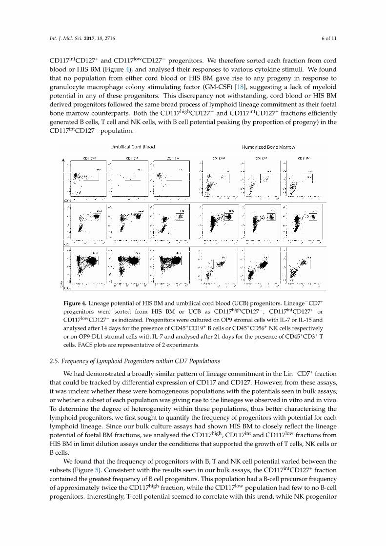
CD117intCD127+ and CD117lowCD127− progenitors. We therefore sorted each fraction from cordblood or HIS BM (Figure 4), and analysed their responses to various cytokine stimuli. We foundthat no population from either cord blood or HIS BM gave rise to any progeny in response togranulocyte macrophage colony stimulating factor (GM-CSF) [18], suggesting a lack of myeloidpotential in any of these progenitors. This discrepancy not withstanding, cord blood or HIS BMderived progenitors followed the same broad process of lymphoid lineage commitment as their foetalbone marrow counterparts. Both the CD117highCD127− and CD117intCD127+ fractions efficientlygenerated B cells, T cell and NK cells, with B cell potential peaking (by proportion of progeny) in theCD117intCD127− population.
Int. J. Mol. Sci. 2017, 18, 2716 6 of 10
macrophage colony stimulating factor (GM-CSF) [18], suggesting a lack of myeloid potential in any of these progenitors. This discrepancy not withstanding, cord blood or HIS BM derived progenitors followed the same broad process of lymphoid lineage commitment as their foetal bone marrow counterparts. Both the CD117highCD127− and CD117intCD127+ fractions efficiently generated B cells, T cell and NK cells, with B cell potential peaking (by proportion of progeny) in the CD117intCD127− population.
Figure 4. Lineage potential of HIS BM and umbilical cord blood (UCB) progenitors. Lineage−CD7+ progenitors were sorted from HIS BM or UCB as CD117highCD127−, CD117intCD127+ or CD117lowCD127− as indicated. Progenitors were cultured on OP9 stromal cells with IL-7 or IL-15 and analysed after 14 days for the presence of CD45+CD19+ B cells or CD45+CD56+ NK cells respectively or on OP9-DL1 stromal cells with IL-7 and analysed after 21 days for the presence of CD45+CD3+ T cells. FACS plots are representative of 2 experiments.
2.5. Frequency of Lymphoid Progenitors within CD7 Populations
We had demonstrated a broadly similar pattern of lineage commitment in the Lin−CD7+ fraction that could be tracked by differential expression of CD117 and CD127. However, from these assays, it was unclear whether these were homogeneous populations with the potentials seen in bulk assays, or whether a subset of each population was giving rise to the lineages we observed in vitro and in vivo. To determine the degree of heterogeneity within these populations, thus better characterising the lymphoid progenitors, we first sought to quantify the frequency of progenitors with potential for each lymphoid lineage. Since our bulk culture assays had shown HIS BM to closely reflect the lineage potential of foetal BM fractions, we analysed the CD117high, CD117int and CD117low fractions from HIS BM in limit dilution assays under the conditions that supported the growth of T cells, NK cells or B cells.
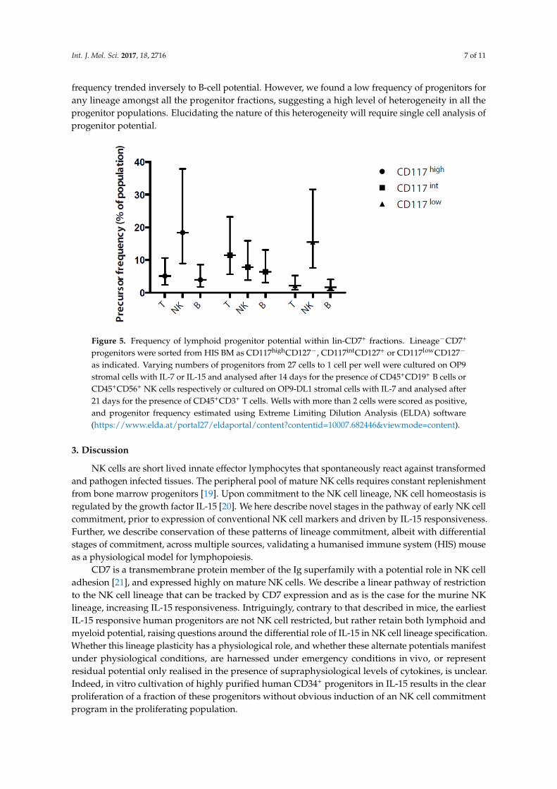
We found that the frequency of progenitors with B, T and NK cell potential varied between the subsets (Figure 5). Consistent with the results seen in our bulk assays, the CD117intCD127+ fraction contained the greatest frequency of B cell progenitors. This population had a B-cell precursor frequency of approximately twice the CD117high fraction, while the CD117low population had few to no B-cell progenitors. Interestingly, T-cell potential seemed to correlate with this trend, while NK progenitor frequency trended inversely to B-cell potential. However, we found a low frequency of progenitors for any lineage amongst all the progenitor fractions, suggesting a high level of heterogeneity in all the progenitor populations. Elucidating the nature of this heterogeneity will require single cell analysis of progenitor potential.
Figure 4. Lineage potential of HIS BM and umbilical cord blood (UCB) progenitors. Lineage−CD7+
progenitors were sorted from HIS BM or UCB as CD117highCD127−, CD117intCD127+ orCD117lowCD127− as indicated. Progenitors were cultured on OP9 stromal cells with IL-7 or IL-15 andanalysed after 14 days for the presence of CD45+CD19+ B cells or CD45+CD56+ NK cells respectivelyor on OP9-DL1 stromal cells with IL-7 and analysed after 21 days for the presence of CD45+CD3+ Tcells. FACS plots are representative of 2 experiments.
2.5. Frequency of Lymphoid Progenitors within CD7 Populations
We had demonstrated a broadly similar pattern of lineage commitment in the Lin−CD7+ fractionthat could be tracked by differential expression of CD117 and CD127. However, from these assays,it was unclear whether these were homogeneous populations with the potentials seen in bulk assays,or whether a subset of each population was giving rise to the lineages we observed in vitro and in vivo.To determine the degree of heterogeneity within these populations, thus better characterising thelymphoid progenitors, we first sought to quantify the frequency of progenitors with potential for eachlymphoid lineage. Since our bulk culture assays had shown HIS BM to closely reflect the lineagepotential of foetal BM fractions, we analysed the CD117high, CD117int and CD117low fractions fromHIS BM in limit dilution assays under the conditions that supported the growth of T cells, NK cells orB cells.
We found that the frequency of progenitors with B, T and NK cell potential varied between thesubsets (Figure 5). Consistent with the results seen in our bulk assays, the CD117intCD127+ fractioncontained the greatest frequency of B cell progenitors. This population had a B-cell precursor frequencyof approximately twice the CD117high fraction, while the CD117low population had few to no B-cellprogenitors. Interestingly, T-cell potential seemed to correlate with this trend, while NK progenitor
Int. J. Mol. Sci. 2017, 18, 2716 7 of 11
frequency trended inversely to B-cell potential. However, we found a low frequency of progenitors forany lineage amongst all the progenitor fractions, suggesting a high level of heterogeneity in all theprogenitor populations. Elucidating the nature of this heterogeneity will require single cell analysis ofprogenitor potential.Int. J. Mol. Sci. 2017, 18, 2716 7 of 10
Figure 5. Frequency of lymphoid progenitor potential within lin-CD7+ fractions. Lineage−CD7+ progenitors were sorted from HIS BM as CD117highCD127−, CD117intCD127+ or CD117lowCD127− as indicated. Varying numbers of progenitors from 27 cells to 1 cell per well were cultured on OP9 stromal cells with IL-7 or IL-15 and analysed after 14 days for the presence of CD45+CD19+ B cells or CD45+CD56+ NK cells respectively or cultured on OP9-DL1 stromal cells with IL-7 and analysed after 21 days for the presence of CD45+CD3+ T cells. Wells with more than 2 cells were scored as positive, and progenitor frequency estimated using Extreme Limiting Dilution Analysis (ELDA) software (https://www.elda.at/portal27/eldaportal/content?contentid=10007.682446&viewmode=content).
3. Discussion
NK cells are short lived innate effector lymphocytes that spontaneously react against transformed and pathogen infected tissues. The peripheral pool of mature NK cells requires constant replenishment from bone marrow progenitors [19]. Upon commitment to the NK cell lineage, NK cell homeostasis is regulated by the growth factor IL-15 [20]. We here describe novel stages in the pathway of early NK cell commitment, prior to expression of conventional NK cell markers and driven by IL-15 responsiveness. Further, we describe conservation of these patterns of lineage commitment, albeit with differential stages of commitment, across multiple sources, validating a humanised immune system (HIS) mouse as a physiological model for lymphopoiesis.
CD7 is a transmembrane protein member of the Ig superfamily with a potential role in NK cell adhesion [21], and expressed highly on mature NK cells. We describe a linear pathway of restriction to the NK cell lineage that can be tracked by CD7 expression and as is the case for the murine NK lineage, increasing IL-15 responsiveness. Intriguingly, contrary to that described in mice, the earliest IL-15 responsive human progenitors are not NK cell restricted, but rather retain both lymphoid and myeloid potential, raising questions around the differential role of IL-15 in NK cell lineage specification. Whether this lineage plasticity has a physiological role, and whether these alternate potentials manifest under physiological conditions, are harnessed under emergency conditions in vivo, or represent residual potential only realised in the presence of supraphysiological levels of cytokines, is unclear. Indeed, in vitro cultivation of highly purified human CD34+ progenitors in IL-15 results in the clear proliferation of a fraction of these progenitors without obvious induction of an NK cell commitment program in the proliferating population.
A common source of variability in studies of human haematopoiesis is that range of progenitor sources used, as access to human tissue varies widely. Further, there remains a need for a model that resembles the physiological pathway of human haematopoietic cell generation as in vivo manipulation of the immune system, such as for cancer immunotherapy, becomes increasingly common. We here demonstrate an identical pattern of lineage restriction in HIS BM as that seen in foetal BM, the primary site of human lymphopoiesis, suggesting the haematopoietic lineages that
Figure 5. Frequency of lymphoid progenitor potential within lin-CD7+ fractions. Lineage−CD7+
progenitors were sorted from HIS BM as CD117highCD127−, CD117intCD127+ or CD117lowCD127−
as indicated. Varying numbers of progenitors from 27 cells to 1 cell per well were cultured on OP9stromal cells with IL-7 or IL-15 and analysed after 14 days for the presence of CD45+CD19+ B cells orCD45+CD56+ NK cells respectively or cultured on OP9-DL1 stromal cells with IL-7 and analysed after21 days for the presence of CD45+CD3+ T cells. Wells with more than 2 cells were scored as positive,and progenitor frequency estimated using Extreme Limiting Dilution Analysis (ELDA) software(https://www.elda.at/portal27/eldaportal/content?contentid=10007.682446&viewmode=content).
3. Discussion
NK cells are short lived innate effector lymphocytes that spontaneously react against transformedand pathogen infected tissues. The peripheral pool of mature NK cells requires constant replenishmentfrom bone marrow progenitors [19]. Upon commitment to the NK cell lineage, NK cell homeostasis isregulated by the growth factor IL-15 [20]. We here describe novel stages in the pathway of early NK cellcommitment, prior to expression of conventional NK cell markers and driven by IL-15 responsiveness.Further, we describe conservation of these patterns of lineage commitment, albeit with differentialstages of commitment, across multiple sources, validating a humanised immune system (HIS) mouseas a physiological model for lymphopoiesis.
CD7 is a transmembrane protein member of the Ig superfamily with a potential role in NK celladhesion [21], and expressed highly on mature NK cells. We describe a linear pathway of restrictionto the NK cell lineage that can be tracked by CD7 expression and as is the case for the murine NKlineage, increasing IL-15 responsiveness. Intriguingly, contrary to that described in mice, the earliestIL-15 responsive human progenitors are not NK cell restricted, but rather retain both lymphoid andmyeloid potential, raising questions around the differential role of IL-15 in NK cell lineage specification.Whether this lineage plasticity has a physiological role, and whether these alternate potentials manifestunder physiological conditions, are harnessed under emergency conditions in vivo, or representresidual potential only realised in the presence of supraphysiological levels of cytokines, is unclear.Indeed, in vitro cultivation of highly purified human CD34+ progenitors in IL-15 results in the clearproliferation of a fraction of these progenitors without obvious induction of an NK cell commitmentprogram in the proliferating population.
Int. J. Mol. Sci. 2017, 18, 2716 8 of 11
A common source of variability in studies of human haematopoiesis is that range of progenitorsources used, as access to human tissue varies widely. Further, there remains a need for a model thatresembles the physiological pathway of human haematopoietic cell generation as in vivo manipulationof the immune system, such as for cancer immunotherapy, becomes increasingly common. We heredemonstrate an identical pattern of lineage restriction in HIS BM as that seen in foetal BM, the primarysite of human lymphopoiesis, suggesting the haematopoietic lineages that develop in these mice do soalong pathways that mirror their physiological counterparts in humans. Thus, we suggest the HISmouse model as an ideal tool to study the stages of human haematopoiesis.
In limit dilution assays, we found surprisingly low precursor potential amongst all precursorfractions. While this may be in part due to the limitations of the assay in efficiently supportingthe development of lymphoid cells from all potential progenitors, it also suggested the progenitorfractions are highly heterogeneous. Traditionally, such heterogeneity has been delineated by thefurther subsetting of the precursor fractions by surface markers. However, the relative lack of availablereagents and known surface markers in humans may preclude such fine subsetting. Given recentadvances in genomic technology, we suggest the progenitors we have described here form the basisfor isolation of progenitors for single cell RNA sequencing, which would provide further insights intoprogenitor heterogeneity and regulation. Although such work is beyond the scope of this study, ourdescription of the frequency of progenitors with B, NK and T cell lineage in each fraction and ourdescription of the consistency of progenitor potential across different sources of human haematopoietictissue lays an exciting foundation for correlating progenitor potential with the gene expression profileof cells within these populations, thus identifying regulators of this process.
4. Materials and Methods
4.1. Mice
Balb/C immunodeficient mice have been previously described [11,22]. Experimental mice werespecific-pathogen-free (SPF) and all animal work was conducted with standard operating proceduresapproved by The Walter and Eliza Hall Institute of Medical Research animal ethics committeesAEC2015.017 (1/3/2015). Mice with human immune systems were generated by irradiating newborn(3–5 day-old) mice with 3 Gy total body irradiation from a γ source, and were injected intra-hepatically(i.h.) with 5 × 10⁵–3× 10⁶ CD34+ human cord blood cells obtained from the Bone Marrow DonorInstitute/Cord Blood Research Unit (Murdoch Children’s Research Institute, Parkville, VIC, Australia)following approval from The Walter and Eliza Hall Institute of Medical Research Human ResearchEthics Committee.
4.2. In Vitro Assays of Progenitor Potential
Progenitors were isolated as indicated and were cultured on either MS-5 or OP9 stromal cells withIL-7 (50 ng/mL) to determine B cell potential; on MS-5 stromal cells with granulocyte macrophagecolony stimulating factor (GM-CSF (25 ng/mL) to determine myeloid potential; on either MS-5 or OP9stromal cells with IL-15 (50 ng/mL) to determine NK cell potential; or on OP9-DLI stromal cells withIL-7 (50 ng/mL) to determine T cell potential. Cells were split 1:2 onto fresh stromal cells and freshcytokine media if required. Myeloid assays were analysed after 7 days for the presence of CD33+CD14+
monocytes and CD33+CD16+ granulocytes; B cell and NK cell assays were analysed after 14 daysfor the presence of CD45+CD19+ B cells or CD45+CD56+ or NKp46+ NK cells respectively; and T cellassays analysed at 21 days for the presence of CD45+CD3+ T cells. All cultures were analysed byflow cytometry as described below. Foetal bone marrow cultures were performed in 48 well plates inDMEM/10% AB serum + IL-15 (50 ng/mL) or on OP9-DL1 with IL-7 (50 ng/mL) and IL-15 (50 ng/mL).Cells were purified as indicated and labelled with 5 µM carboxyfluorescein succinimidyl ester (CFSE)(Molecular Probes, Eugene, OR, USA).

Int. J. Mol. Sci. 2017, 18, 2716 9 of 11
4.3. Flow Cytometry Analysis for Cell-Surface and Intracellular Markers
The following anti-human mAb were used to stain cell suspension for FACS analysis: CD3 (SK7),CD4 (SK3), CD34 (581), CD8 (SK1), CD19 (HIB19), CD10 (HI10a), CD38 (HB7), NKp46 (9E2), CD16(3G8), CD161 (DX12), CD56 (B159), HLA-DR (L243), CD244.2 (2B4), HLA-A/B/C (G46-2.6), CD117(YB5.B8), CD14 (M5E2), CD122 (Mik-3), TCR-β (T10B9.1A-31), CD127 (hIL-7R-M21), CD11c (B-ly6),CD7 (M-T701), CD45 (2D1) from (BD Bioscience, CA, USA). All washings and reagent dilutions weredone with phosphate buffered saline (PBS) containing 2% foetal calf serum (FCS). All acquisitionswere performed using FACSVerse cytometers and cell sorting was perform using FACS ARIA, allmachines were interfaced to the FACS-Diva software (BD Bioscience). Data analysis was performedusing FlowJo software from TreeStar Inc. (Ashland, OR, USA).
4.4. Cell Preparation
Umbilical cord blood was obtained from the Bone Marrow Donor Institute Cord Blood Bank(Murdoch Children’s Research Institute, Parkville, VIC, Australia). Experiments were approved by TheWalter & Eliza Hall Institute of Medical Research Human Research Ethics Committee (HREC12/08).Magnetic enrichment of CD34+ cells (>98% pure) was performed by using the CD34 Progenitor CellIsolation Kit (Miltenyi Biotech, Auburn, CA, USA), after preparation of single-cell suspension andisolation of mononuclear cells by density gradient centrifugation over Ficoll-Hypaque (NycomedPharma, Roskilde, Denmark). Cell suspensions were prepared in PBS medium with 2% foetalcalf serum. Single cell suspensions of murine organs were prepared as previously described [23].Human foetal bone marrow was a kind gift from Professor Hergen Spits (AMC, Amsterdam-Zuidoost,The Netherlands) and obtained from elective abortions approved by the Medical and Ethical Committeeat AMC-UvA. Human NK cells were purified from donor blood buffy coat prepared by density gradientcentrifugation over Ficoll-Hypaque (Nycomed Pharma, Zürich, Switzerland) using anti-CD56 magneticbeads (Miltenyi Biotech, Bergisch Gladbach, Germany). Purified cells (CD34+) were loaded with 5 µMCFSE (Invitrogen, Carlsbad, CA, USA) or 5 µM cell trace violet (CTV) and cultured at 1 × 10⁵ cells/mLin StemSpan (STEMCELLS Technologies, Vancouver, Canada) with 10% human serum and either50 ng/mL recombinant human IL-15 (rhIL-15; Peprotech, NJ, USA) or human thrombopoietin (TPO),fms-like tyrosine kinase 3 ligand (FLT3L) and stem cell factor (SCF) (100 ng/mL).
4.5. Statistical Analysis
Statistical analyses were performed using GraphPad Prism (GraphPad Software, San Diego, CA,USA). All data were subjected to two-tailed unpaired Student t test analysis. Limit dilution assayswere analysed using extreme limit dilution assay (ELDA) software (Hu and Smyth, 2009).
Acknowledgments: We acknowledge the Bloemenhove Clinic (Heemstede, The Netherlands) for providing foetaltissues and Hergen Spits (AMC, Amsterdam-Zuidoost, The Netherlands) for the kind gift of foetal bone marrowand reagents. This work is supported project grants from the National Health and Medical Research Council(NHMRC) of Australia (1049407, 1066770, 1057852, 1027472 to N.D.H), an NHMRC CDF2 fellowship (NDH) aswell as an NHMRC Independent Research Institute Infrastructure Support scheme grant and a Victorian StateGovernment Operational Infrastructure Scheme grant. NDH is a recipient of research grants from the followingorganisations: Ian Potter Foundation (Australia), Harry J Lloyd Charitable Trust (USA), Melanoma ResearchAlliance (USA) and Cancer Research Institute (USA).
Author Contributions: Priyanka Sathe and Nicholas D. Huntington designed and performed experiments andwrote the paper; Rebecca Delconte and Swee Heng Milon Pang performed experiments and Ngaire Elwoodsupplied reagents.
Conflicts of Interest: The authors declare no conflict of interest.
Int. J. Mol. Sci. 2017, 18, 2716 10 of 11
References
1. Mori, Y.; Iwasaki, H.; Kohno, K.; Yoshimoto, G.; Kikushige, Y.; Okeda, A.; Miyamoto, T. Identification of thehuman eosinophil lineage-committed progenitor: Revision of phenotypic definition of the human commonmyeloid progenitor. J. Exp. Med. 2009, 206, 183–193. [CrossRef] [PubMed]
2. Manz, M.G.; Miyamoto, T.; Akashi, K.; Weissman, I.L. Prospective isolation of human clonogenic commonmyeloid progenitors. Proc. Natl. Acad. Sci. USA 2002, 99, 11872–11877. [CrossRef] [PubMed]
3. Galy, A.; Travis, M.; Cen, D.; Chen, B. Human T, B, natural killer, and dendritic cells arise from a commonbone marrow progenitor cell subset. Immunity 1995, 3, 459–473. [CrossRef]
4. Galy, A.H.; Cen, D.; Travis, M.; Chen, S.; Chen, B.P. Delineation of T-progenitor cell activity within the CD34+compartment of adult bone marrow. Blood 1995, 85, 2770–2778. [PubMed]
5. Hao, Q.L.; Zhu, J.; Price, M.A.; Payne, K.J.; Barsky, L.W.; Crooks, G.M. Identification of a novel, humanmultilymphoid progenitor in cord blood. Blood 2001, 97, 3683–3690. [CrossRef] [PubMed]
6. Ishikawa, F.; Niiro, H.; Iino, T.; Yoshida, S.; Saito, N.; Onohara, S.; Harada, M. The developmental programof human dendritic cells is operated independently of conventional myeloid and lymphoid pathways. Blood2007, 110, 3591–3660. [CrossRef] [PubMed]
7. Sanchez, M.J.; Muench, M.O.; Roncarolo, M.G.; Lanier, L.L.; Phillips, J.H. Identification of a commonT/natural killer cell progenitor in human fetal thymus. J. Exp. Med. 1994, 180, 569–576. [CrossRef] [PubMed]
8. Renoux, V.M.; Zriwil, A.; Peitzsch, C.; Michaëlsson, J.; Friberg, D.; Soneji, S.; Sitnicka, E. Identification ofa Human Natural Killer Cell Lineage-Restricted Progenitor in Fetal and Adult Tissues. Immunity 2015,43, 394–407. [CrossRef] [PubMed]
9. Kovanen, P.E.; Leonard, W.J. Cytokines and immunodeficiency diseases: Critical roles of thegamma(c)-dependent cytokines interleukins 2, 4, 7, 9, 15, and 21, and their signaling pathways. Immunol. Rev.2004, 202, 67–83. [CrossRef] [PubMed]
10. Huntington, N.D.; Puthalakath, H.; Gunn, P.; Naik, E.; Michalak, E.M.; Smyth, M.J.; Motoyama, N. Interleukin15-mediated survival of natural killer cells is determined by interactions among Bim, Noxa and Mcl-1.Nat. Immunol. 2007, 8, 856–863. [CrossRef] [PubMed]
11. Huntington, N.D.; Legrand, N.; Alves, N.L.; Jaron, B.; Weijer, K.; Plet, A.; Di Santo, J.P. IL-15trans-presentation promotes human NK cell development and differentiation in vivo. J. Exp. Med. 2009,206, 25–34. [CrossRef] [PubMed]
12. Mrozek, E.; Anderson, P.; Caligiuri, M.A. Role of interleukin-15 in the development of human CD56+ naturalkiller cells from CD34+ hematopoietic progenitor cells. Blood 1996, 87, 2632–2640. [PubMed]
13. Carotta, S.; Pang, S.H.; Nutt, S.L.; Belz, G.T. Identification of the earliest NK-cell precursor in the mouse BM.Blood 2011, 117, 5449–5452. [CrossRef] [PubMed]
14. Delconte, R.B.; Shi, W.; Sathe, P.; Ushiki, T.; Seillet, C.; Minnich, M.; Busslinger, M. The Helix-Loop-HelixProtein ID2 Governs NK Cell Fate by Tuning Their Sensitivity to Interleukin-15. Immunity 2016, 44, 103–115.[CrossRef] [PubMed]
15. Rosmaraki, E.E.; Douagi, I.; Roth, C.; Colucci, F.; Cumano, A.; Di Santo, J.P. Identification of committed NKcell progenitors in adult murine bone marrow. Eur. J. Immunol. 2001, 31, 1900–1909. [CrossRef]
16. Carayol, G.; Robin, C.; Bourhis, J.H.; Bennaceur-Griscelli, A.; Chouaib, S.; Coulombel, L.; Caignard, A.NK cells differentiated from bone marrow, cord blood and peripheral blood stem cells exhibit similarphenotype and functions. Eur. J. Immunol. 1998, 28, 1991–2002. [CrossRef]
17. Kyoizumi, S.; Kubo, Y.; Kajimura, J.; Yoshida, K.; Hayashi, T.; Nakachi, K.; Kusunoki, Y. Fate DecisionBetween Group 3 Innate Lymphoid and Conventional NK Cell Lineages by Notch Signaling in HumanCirculating Hematopoietic Progenitors. J. Immunol. 2017, 199, 2777–2793. [CrossRef] [PubMed]
18. Sathe, P.; Huntington, N.D.; (The Walter and Eliza Hall Institute of Medical Research, 1G Royal Parade,Parkville, VIC 3052, Australia). Personal communication, 2017.
19. Huntington, N.D.; Vosshenrich, C.A.; Di Santo, J.P. Developmental pathways that generate natural-killer-celldiversity in mice and humans. Nat. Rev. Immunol. 2007, 7, 703–714. [CrossRef] [PubMed]
20. Huntington, N.D. The unconventional expression of IL-15 and its role in NK cell homeostasis.Immunol. Cell Biol. 2014, 92, 210–213. [CrossRef] [PubMed]
21. Rabinowich, H.; Pricop, L.; Herberman, R.B.; Whiteside, T.L. Expression and function of CD7 molecule onhuman natural killer cells. J. Immunol. 1994, 152, 517–526. [PubMed]

Int. J. Mol. Sci. 2017, 18, 2716 11 of 11
22. Huntington, N.D.; Alves, N.L.; Legrand, N.; Lim, A.; Strick-Marchand, H.; Mention, J.J.; Guzman, C. IL-15transpresentation promotes both human T-cell reconstitution and T-cell-dependent antibody responsesin vivo. Proc. Natl. Acad. Sci. USA 2011, 108, 6217–6222. [CrossRef] [PubMed]
23. Huntington, N.D.; Xu, Y.; Nutt, S.L.; Tarlinton, D.M. A requirement for CD45 distinguishes Ly49D-mediatedcytokine and chemokine production from killing in primary natural killer cells. J. Exp. Med. 2005,201, 1421–1433. [CrossRef] [PubMed]
© 2017 by the authors. Licensee MDPI, Basel, Switzerland. This article is an open accessarticle distributed under the terms and conditions of the Creative Commons Attribution(CC BY) license (http://creativecommons.org/licenses/by/4.0/).