Embed Size (px)
Citation preview
SHORT REPORT Open Access
Identification of novel antiviral of fungus-derived brefeldin A against dengue virusesMuhareva Raekiansyah1, Mihoko Mori2,3, Kenichi Nonaka2,3, Masanobu Agoh1, Kazuro Shiomi2,3,Atsuko Matsumoto2,3 and Kouichi Morita1*
Abstract
Microbial natural products possess a wide range of biological and biochemical potential. Among them, fungal secondarymetabolites are one of the most important sources for discovering new drugs or lead compounds. In the present study,we explored substances produced by the strain Penicillium sp. FKI-7127 for its antiviral activity. We identified brefeldin Aas a novel antiviral agent against dengue viruses. The inhibitory effect of brefeldin A was confirmed by virus titer andimmunofluorescence assay. Brefeldin A inhibited dengue viruses regardless of serotypes and other related virusesincluding Zika virus and Japanese encephalitis virus. Time-of-addition study showed that brefeldin A exerts itsantiviral effect at an early stage of the dengue virus (DENV) life cycle. These studies demonstrate that (i) brefeldinA could be used as a lead compound for drug development of anti-DENV and other related viruses and (ii) fungalmetabolites are a potential and valuable source for dengue virus drug discovery.
Keywords: Dengue virus, Antiviral, Brefeldin A, Secondary metabolite, Fungus
IntroductionDengue virus (DENV) is an important mosquito-bornepathogen for causing dengue fever (DF) and denguehemorrhagic fever (DHF). DF is relatively mild, but DHFleads to the life-threatening dengue shock syndrome [1].It is estimated that there are 390 million dengueinfections per year, of which 96 million manifestapparently [2]. At present, no specific antiviral therapyfor treatment of dengue disease is available. Thus, drugdiscovery research for dengue is of importance.Natural products are valuable materials for the
discovery and development of new drugs for treatingmany diseases since they possess a wide range ofstructural and functional diversity [3]. Among variedsources of natural products, secondary metabolitesproduced by fungi have been recognized as an importantsource of lead structures for new drugs [4].Some bioactive compounds that are isolated and charac-
terized from metabolites of soil-borne and endophyticfungi have led to the development of drugs such as anti-cancer drug Taxol which originated from endophytic-
fungal metabolites [5]. Furthermore, a lot of antibacterialsubstances have been demonstrated from extracts andpure substances obtained from culture broth or fungalbiomass [6]. Some studies have identified substances thatinhibit viruses [7, 8]. However, there are only few reportsabout DENV compounds found from fungal metabolitesso far [9].In this study, we examined anti-DENV activity of
secondary metabolites produced by a fungal strain, Peni-cillium sp. FKI-7127. We isolated and identified brefel-din A (BFA) as a novel antiviral agent against DENVs.Inhibition of BFA on Japanese encephalitis virus (JEV)and Zika virus (ZIKV) was also demonstrated.
Materials and methodsCell lines and virusesVero cells were maintained in a minimum essentialmedium supplemented with 10% fetal calf serum. Thecells were grown at 37 °C with 5% CO2. The strains usedfor this study were patient-derived DENV1–4 from thePhilippines, strains 99st, 00st-22A, SLMC50, andSLMC318, respectively; Zika virus strain 976; andJapanese encephalitis virus (JEV) Beijing strain.
* Correspondence: [email protected] of Virology, Institute of Tropical Medicine, Nagasaki University,1-12-4 Sakamoto, Nagasaki 852-8523, JapanFull list of author information is available at the end of the article
Tropical Medicineand Health
© The Author(s). 2017 Open Access This article is distributed under the terms of the Creative Commons Attribution 4.0International License (http://creativecommons.org/licenses/by/4.0/), which permits unrestricted use, distribution, andreproduction in any medium, provided you give appropriate credit to the original author(s) and the source, provide a link tothe Creative Commons license, and indicate if changes were made. The Creative Commons Public Domain Dedication waiver(http://creativecommons.org/publicdomain/zero/1.0/) applies to the data made available in this article, unless otherwise stated.
Raekiansyah et al. Tropical Medicine and Health (2017) 45:32 DOI 10.1186/s41182-017-0072-7
Microorganisms and culture of the fungal strainThe fungal strain FKI-7127 was isolated from a soilaround the root of Angelica keiskei collected in KouzuIsland, Tokyo, Japan. To observe the morphologicalcharacteristics, this strain was incubated on Miura’smedium (LcA). From the results of morphologicalobservation, the producing strain FKI-7127 wasclassified as genus Penicillium. The strain Penicilliumsp. FKI-7127 was maintained on an LcA slant. Aloopful of spores of this strain was inoculated into atest tube, containing 10 ml of a seed medium consist-ing of 2% glucose, 0.5% Polypepton, 0.2% yeast ex-tract, 0.2% KH2PO4, 0.05% MgSO4·7H2O, and 0.1%agar, and incubated for 3 days. One milliliter of theseed culture was inoculated into each of two 500-mlErlenmeyer flasks containing 100 ml of a productionmedium consisting of 3% soluble starch, 1.0% gly-cerol, 2% soybean meal, 0.3% dry yeast, 0.3% KCl,0.2% CaCO3, 0.05% KH2PO4, 0.05% MgSO4·7H2O,and 0.03% quercetin dihydrate, and the productionculture was incubated for 6 days. Fifty percent ofethanol extract of cultured broth were prepared forantiviral test.
Antigen detection ELISATo evaluate the antiviral activity of a sample, Vero cellswere seeded in 96-well plates (1 × 10⁴ cells/well) andinfected with DENV at multiplicity of infection (MOI) of0.5 in the presence of samples/compound or 0.1%dimethyl sulfoxide (DMSO). The cells were incubated for3 days when infected culture fluid (ICF) was harvestedand subjected for antigen detection enzyme-linked im-munosorbent assay (ELISA) to determine dengue virusantigen level as described previously [10]. The result wasexpressed as a percent of inhibition which determined as(OD value of DMSO-treated cells) − (OD value ofcompound-treated cells) × 100% divided by (OD value ofDMSO-treated cells).
Cells viability assayVero cells in 96-well plates were treated with samplesfor 5 days. Cell viability was evaluated by MTT [3-(4,5-dimethylthiazol-2-yl)-2,5-diphenyl tetrazolium bromide]according to manufacturer’s instruction (Promega).
Isolation and identification of brefeldin ACultured broth was extracted with 200 ml of ethyl alcohol(EtOH). After the mycelia were separated by centrifuga-tion, the extract was evaporated to remove EtOH. A partof the aqueous residue was applied to a Seppak plus ODSC18 cartridge and eluted with H2O–CH3CN system to
give five fractions (pass through 100:0, 70:30, 40:60, and0:100 each 3 ml). Brefeldin A was detected in both 40:60fraction and 0:100 fraction by high-performance liquidchromatography (HPLC) analysis, and identification ofbrefeldin A was achieved by high-resolution electrosprayionization mass spectrometry (HR-ESI-MS) and nuclearmagnetic resonance (NMR) measurement.
Immunofluorescence assayVero cells in a 24-well plate were infected withDENV-2 and added with BFA. After 48 h, infectedcells were recovered, washed with PBS, and spottedonto a glass slide. Immunostaining was done asdescribed previously [11].
Time-of-addition studiesTime-of-addition studies were performed in 96-well platecells as follows. (i) Pre-infection assay: Vero cells weretreated with 125 nM BFA or 0.1% DMSO as control for2 h at 37 °C prior to being washed twice with PBS and in-fected with DENV-2 at an MOI of 10. After 1.5 h virus ad-sorption, the cells were washed twice with PBS andincubated in fresh media for 24 h before ICFs were har-vested for virus quantification. (ii) During-infection assay:Vero cells were infected with DENV-2 in the presence ofBFA. After 1.5 h virus adsorption, inoculum was removedand the cells were washed twice. The cells were then incu-bated with fresh media for 24 h before ICFs were har-vested for virus quantification. (iii) After-infection assay:Vero cells were infected with DENV-2. After virus adsorp-tion and washing, BFA or 0.1% DMSO as control wasadded at seven different time points postinfection (0, 2, 4,6, 8, 12, and 18 h). The ICFs were harvested at 24 hpi forvirus quantification.
Virus titration and focus reduction assayVirus titers were determined using Vero cells in 96-wellplates as described previously [11]. In brief, virus stock orICFs were diluted tenfold in the MEM and inoculated tothe cells. After 60 min of virus adsorption, the MEM con-taining 2% FCS and 1.25% methylcellulose was overlaid onthe cells. The cells were then incubated for 2 to 4 days be-fore subjected to focus staining. For focus staining,12D11/7E8 monoclonal antibody and HRP-conjugatedgoat anti-mouse IgG + M were used as a primary and sec-ondary antibody, respectively. The infected cells were vi-sualized with 3,3′-diaminobenzidine, tetrahydrochloride(DAB). For focus reduction assay, Vero cells in 96-wellplates were infected with DENV-1, 2, 3, 4, ZIKV, or JEV atan MOI of 0.5 in the presence of BFA at different concen-trations. After 48 h incubation, ICFs were harvested. A
Raekiansyah et al. Tropical Medicine and Health (2017) 45:32 Page 2 of 7
hundred microliters of diluted ICFs (100× or 1000× dilu-tion) was infected into fresh Vero cells in 96-well platesand incubate for another 2 to 4 days. The cells were thensubjected for focus staining as described above. Experi-ments were performed twice, duplicating each.
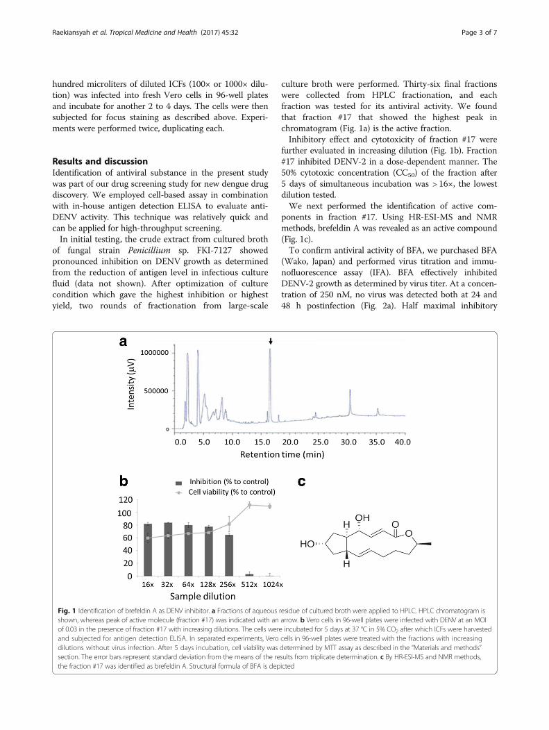
Results and discussionIdentification of antiviral substance in the present studywas part of our drug screening study for new dengue drugdiscovery. We employed cell-based assay in combinationwith in-house antigen detection ELISA to evaluate anti-DENV activity. This technique was relatively quick andcan be applied for high-throughput screening.In initial testing, the crude extract from cultured broth
of fungal strain Penicillium sp. FKI-7127 showedpronounced inhibition on DENV growth as determinedfrom the reduction of antigen level in infectious culturefluid (data not shown). After optimization of culturecondition which gave the highest inhibition or highestyield, two rounds of fractionation from large-scale
culture broth were performed. Thirty-six final fractionswere collected from HPLC fractionation, and eachfraction was tested for its antiviral activity. We foundthat fraction #17 that showed the highest peak inchromatogram (Fig. 1a) is the active fraction.Inhibitory effect and cytotoxicity of fraction #17 were
further evaluated in increasing dilution (Fig. 1b). Fraction#17 inhibited DENV-2 in a dose-dependent manner. The50% cytotoxic concentration (CC50) of the fraction after5 days of simultaneous incubation was > 16×, the lowestdilution tested.We next performed the identification of active com-
ponents in fraction #17. Using HR-ESI-MS and NMRmethods, brefeldin A was revealed as an active compound(Fig. 1c).To confirm antiviral activity of BFA, we purchased BFA
(Wako, Japan) and performed virus titration and immu-nofluorescence assay (IFA). BFA effectively inhibitedDENV-2 growth as determined by virus titer. At a concen-tration of 250 nM, no virus was detected both at 24 and48 h postinfection (Fig. 2a). Half maximal inhibitory
Fig. 1 Identification of brefeldin A as DENV inhibitor. a Fractions of aqueous residue of cultured broth were applied to HPLC. HPLC chromatogram isshown, whereas peak of active molecule (fraction #17) was indicated with an arrow. b Vero cells in 96-well plates were infected with DENV at an MOIof 0.03 in the presence of fraction #17 with increasing dilutions. The cells were incubated for 5 days at 37 °C in 5% CO2 after which ICFs were harvestedand subjected for antigen detection ELISA. In separated experiments, Vero cells in 96-well plates were treated with the fractions with increasingdilutions without virus infection. After 5 days incubation, cell viability was determined by MTT assay as described in the “Materials and methods”section. The error bars represent standard deviation from the means of the results from triplicate determination. c By HR-ESI-MS and NMR methods,the fraction #17 was identified as brefeldin A. Structural formula of BFA is depicted
Raekiansyah et al. Tropical Medicine and Health (2017) 45:32 Page 3 of 7
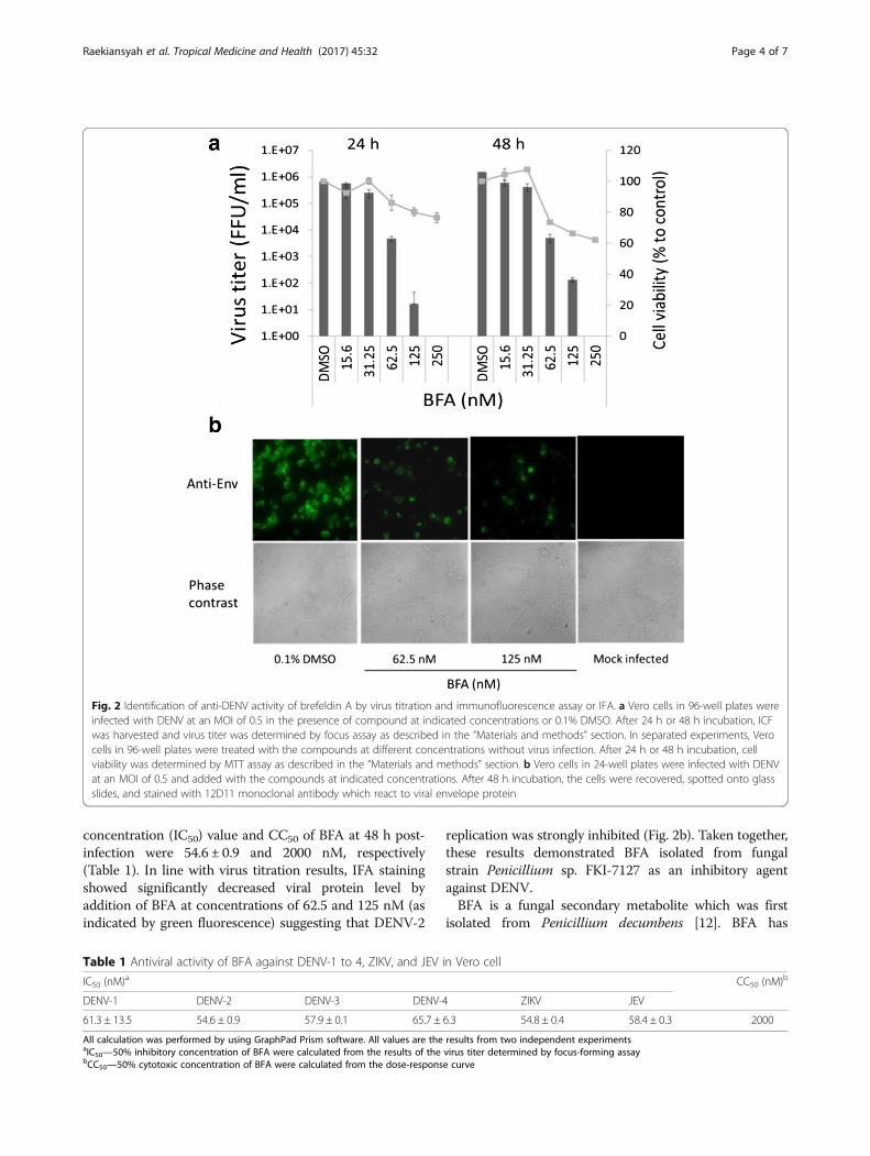
concentration (IC50) value and CC50 of BFA at 48 h post-infection were 54.6 ± 0.9 and 2000 nM, respectively(Table 1). In line with virus titration results, IFA stainingshowed significantly decreased viral protein level byaddition of BFA at concentrations of 62.5 and 125 nM (asindicated by green fluorescence) suggesting that DENV-2
replication was strongly inhibited (Fig. 2b). Taken together,these results demonstrated BFA isolated from fungalstrain Penicillium sp. FKI-7127 as an inhibitory agentagainst DENV.BFA is a fungal secondary metabolite which was first
isolated from Penicillium decumbens [12]. BFA has
Table 1 Antiviral activity of BFA against DENV-1 to 4, ZIKV, and JEV in Vero cell
IC50 (nM)a CC50 (nM)b
DENV-1 DENV-2 DENV-3 DENV-4 ZIKV JEV
61.3 ± 13.5 54.6 ± 0.9 57.9 ± 0.1 65.7 ± 6.3 54.8 ± 0.4 58.4 ± 0.3 2000
All calculation was performed by using GraphPad Prism software. All values are the results from two independent experimentsaIC50—50% inhibitory concentration of BFA were calculated from the results of the virus titer determined by focus-forming assaybCC50—50% cytotoxic concentration of BFA were calculated from the dose-response curve
Fig. 2 Identification of anti-DENV activity of brefeldin A by virus titration and immunofluorescence assay or IFA. a Vero cells in 96-well plates wereinfected with DENV at an MOI of 0.5 in the presence of compound at indicated concentrations or 0.1% DMSO. After 24 h or 48 h incubation, ICFwas harvested and virus titer was determined by focus assay as described in the “Materials and methods” section. In separated experiments, Verocells in 96-well plates were treated with the compounds at different concentrations without virus infection. After 24 h or 48 h incubation, cellviability was determined by MTT assay as described in the “Materials and methods” section. b Vero cells in 24-well plates were infected with DENVat an MOI of 0.5 and added with the compounds at indicated concentrations. After 48 h incubation, the cells were recovered, spotted onto glassslides, and stained with 12D11 monoclonal antibody which react to viral envelope protein
Raekiansyah et al. Tropical Medicine and Health (2017) 45:32 Page 4 of 7

various biological actions including antitumor andantibacterial activities [13]. BFA has also been re-ported to have antiviral activities against some virusesincluding poliovirus [14] and Rotavirus [15]. Recently,Zhou et al. demonstrated the inhibitory effect of BFAon Japanese encephalitis virus (JEV) in BHK-21 cells[16]. To our knowledge, this is the first report todemonstrate antiviral activity of BFA on DENV.Antiviral activity of BFA against all DENV serotypes
as well as two related viruses including ZIKV and JEVwas also analyzed by focus reduction assay. Additionof BFA reduced focus number in a dose-dependentmanner which indicates virus inhibition. BFA inhib-ited not only DENV-2 but also all other serotypes.Furthermore, strong inhibition of ZIKV and JEV byBFA was also demonstrated (Fig. 3). Determined byvirus titer, the EC50 of BFA against DENV-1, 3, and4, ZIKV, and JEV at 48 h postinfection/treatmentwere 61.32 ± 13.5, 57.9 ± 0.1, 65.7 ± 6.3, 54.8 ± 0.4, and58.4 ± 0.3 nM, respectively (Table 1).In order to identify the window in the DENV repli-
cation cycle when BFA exerts its antiviral effect, time-
of-addition studies were performed in different treat-ments (Fig. 4a). During a single flavivirus life cycle,viral proteins are translated from genomic RNA inthe first 1–5 h postinfection (hpi) followed by viralRNA synthesis which occurs after 5 hpi and progenyvirus assembly and release after 12 hpi [17]. Asshown in Fig. 4b, BFA does not interfere in theDENV entry process in the host cells. A significantreduction of the DENV titer was observed when BFAwas added at the 0-hpi up to 4-hpi time points. After4 hpi, inhibitory effect of BFA was gradually reduced.Addition of BFA at 18 hpi resulted in complete lossof inhibition of DENV replication. These results sug-gested that BFA inhibits DENV at an early phase inthe viral replication cycle that occurs after viral entry.The time-of-addition study results are in line with
an already known mode of action of BFA. BFA hasbeen known to inhibit protein transport from theendoplasmic reticulum (ER) to the Golgi apparatusindirectly by interfering with the function of the Golgiapparatus [18]. BFA disturbs maturation and egress ofherpes simplex virus particles during infection [19].BFA has also been reported to interfere processingand secretion of the envelope of glycoproteins ofHIV-1 in T-lymphoblast cells leading to inhibition ofviral particle formation [20]. In case of poliovirus,BFA inhibits viral RNA synthesis by preventing theformation of secretory vesicles [21]. We speculatethat, like other enveloped viruses, BFA inhibits thematuration of the DENV, ZIKV, and JEV by directlyblocking the trafficking of glycoprotein from the ERto Golgi apparatus leading to the prevention offormation and release of the viruses from infectedcells. In addition in our study, no inhibitory effect ofBFA on DENV was shown in C6/36 cells (data notshown) indicating that BFA could not block intracel-lular protein transport in mosquito cell line.Despite the fact that BFA possesses antiviral activity as
demonstrated in this study and other previous studies, itstoxicity would become a crucial issue in order to developit as antiviral agents. Toxicity of BFA indeed is not unex-pected because it targets Golgi apparatus which eventuallycauses cell death. However, BFA could be served as a leadcompound. In the future, it can be structurally and pheno-typically optimized by reducing its toxicity. Furthermore,its antiviral activity can also be improved through deriva-tive analyses or another approaches.Aside from its antiviral property, mechanism of
disruption of the proper vesicular transport between ERand Golgi by BFA which is a critical step for the viralreplication and release could provide a new tool tocharacterize the lifecycle of the virus. It is also possibleto help researchers develop novel inhibitors of DENVand other viruses.
Fig. 3 Inhibitory effect of BFA on all DENV serotypes, ZIKV, and JEV.Vero cells in 96-well plates were infected with DENV-1, 2, 3, and 4,ZIKV, or JEV in the presence of BFA at indicated concentration or0.1% DMSO. Forty-eight hours after infection/treatment, ICFs wereharvested and then infected into fresh cells for focus reduction assayas described in the “Materials and methods” section. The imageshown is a representative result from two experiments
Raekiansyah et al. Tropical Medicine and Health (2017) 45:32 Page 5 of 7

ConclusionIn conclusion, here we isolated, identified, and character-ized an anti-DENV agent of fungus-derived BFA which ispotentially used as a lead compound for drug develop-ment of anti-DENV and other related viruses. Fungal sec-ondary metabolites are a potential and valuable source indrug screening for the development of antiviral agents.
AbbreviationsBFA: Brefeldin A; CC50: The 50% cytotoxic concentration; DENV: Dengue virus;DF: Dengue fever; DHF: Dengue hemorrhagic fever; DMSO: Dimethylsulfoxide; ELISA: Enzyme-linked immunosorbent assay; HPLC:High-performance liquid chromatography; IC50: Half maximal inhibitory
concentration; IFA: Immunofluorescence assay; JEV: Japanese encephalitisvirus; ZIKV: Zika virus
AcknowledgementsNot applicable.
FundingThis research was supported by the Japan Initiative for Global Research Networkon Infectious Diseases (J-GRID) of Japan Agency for Medical Research andDevelopment (AMED).
Availability of data and materialsNot applicable.
Fig. 4 Time-of-addition studies. a Schematic illustration of time-of-addition studies for treatment with BFA. Vero cells in 96-well plates weretreated with 125 nM BFA at different time points before, during, and after DENV infection (MOI of 5). For after-infection assay, BFA was added atseven different time points after virus exposure. b After 24 h postinfection, released virus titer was determined by focus assay as described in the“Materials and methods” section. Error bars represent standard errors of two independent experiments
Raekiansyah et al. Tropical Medicine and Health (2017) 45:32 Page 6 of 7
Authors’ contributionsKM and MR conceived and designed the study. MR, MM, KN, MA, and AMperformed the experiments. MR, MM, KS, AM, and KM analyzed the data. MR,MM, KN, KS, and KM wrote the paper. All authors read and approved thefinal manuscript.
Ethics approval and consent to participateNot applicable.
Consent for publicationNot applicable.
Competing interestsThe authors declare that they have no competing interests.
Publisher’s NoteSpringer Nature remains neutral with regard to jurisdictional claims inpublished maps and institutional affiliations.
Author details1Department of Virology, Institute of Tropical Medicine, Nagasaki University,1-12-4 Sakamoto, Nagasaki 852-8523, Japan. 2Kitasato Institute for LifeSciences, Kitasato University, 5-9-1 Shirokane, Minato-ku, Tokyo 108-8641,Japan. 3Graduate School of Infection Control Sciences, Kitasato University,5-9-1 Shirokane, Minato-ku, Tokyo 108-8641, Japan.
Received: 18 August 2017 Accepted: 9 October 2017
References1. Simmons CP, Farrar JJ, Nguyen VV, Wills B. Dengue. N Engl J Med.
2012;366:1423–32.2. Bhatt S, Gething PW, Brady OJ, Messina JP, Farlow AW, Moyes CL, et al. The
global distribution and burden of dengue. Nature. 2013;496:504–7.3. Cragg GM, Newman DJ. Natural products: a continuing source of novel
drug leads. Biochim Biophys Acta. 2013;1830:3670–95.4. Demain AL, Martens E. Production of valuable compounds by molds and
yeasts. J Antibiot. 2017;70:347–60.5. Strobel G, Yang X, Sears J, Kramer R, Sidhu RS, Hess WM. Taxol from Pestalotiopsis
microspora, an endophytic fungus of Taxus wallachiana. Microbiology.1996;142:435–40.
6. Radić N, Strukelj B. Endophytic fungi: the treasure chest of antibacterialsubstances. Phytomedicine. 2012;19:1270–84.
7. Bunyapaiboonsri T, Yoiprommarat S, Srikitikulchai P, Srichomthong K, LumyongS. Oblongolides from the endophytic fungus Phomopsis sp. BCC 9789. J NatProd. 2010;73:55–9.
8. Roy BG. Potential of small-molecule fungal metabolites in antiviral chemotherapy.Antiviral Chem Chemother. 2017;25:20–52.
9. Estoppey D, Lee CM, Janoschke M, Lee BH, Wan KF, Dong H, et al. Thenatural product cavinafungin selectively interferes with Zika and denguevirus replication by inhibition of the host signal peptidase. Cell Rep.2017;19:451–60.
10. Ngwe Tun MM, Kyaw AK, Makki N, Muthugala R, Nabeshima T, Inoue S, etal. Characterization of the 2013 dengue epidemic in Myanmar with denguevirus 1 as the dominant serotype. Infect Genet Evol. 2016;43:31–7.
11. Raekiansyah M, Espada-Murao LA, Okamoto K, Kubo T, Morita K.Dengue virus neither directly mediates hyperpermeability nor enhancestumor necrosis factor-α-induced permeability in vitro. Jpn J Infect Dis.2014;67:86–94.
12. Singleton VL, Bohonos N, Ullstrup AJ. Decumbin, a new compound from aspecies of PenicilIium. Nature. 1958;181:1072–3.
13. Betina V. Biological effects of the antibiotic brefeldin A (decumbin,cyanein, ascotoxin, synergisidin): a retrospective. Folia Microbiol (Praha).1992;37:3–11.
14. Cuconati A, Molla A, Wimmer E, Brefeldin A. Inhibits cell-free, de novosynthesis of poliovirus. J Virol. 1998;72:6456–64.
15. Mirazimi A, von Bonsdorff CH, Svensson L. Effect of brefeldin A on rotavirusassembly and oligosaccharide processing. Virology. 1996;217:554–63.
16. Zhou J, Wang SQ, Wei JC, Zhang XM, Gao ZC, Liu K, et al. Mx is notresponsible for the antiviral activity of interferon-α against Japaneseencephalitis virus. Viruses. 2017;9:5.
17. Chambers TJ, Hahn CS, Galler R, Rice CM. Flavivirus genome organization,expression, and replication. Annu Rev Microbiol. 1990;44:649–88.
18. Lippincott-Schwartz J, Yuan LC, Bonifacino JS, Klausner RD. Rapid redistributionof Golgi proteins into the ER in cells treated with brefeldin A: evidence formembrane cycling from Golgi to ER. Cell. 1989;56:801–13.
19. Cheung P, Banfield BW, Tufaro F, Brefeldin A. Arrests the maturation andegress of herpes simplex virus particles during infection. J Virol.1991;65:1893–904.
20. Pal R, Mumbauer S, Hoke GM, Takahashi A, Sarngadharan MG, Brefeldin A.Inhibits the processing and secretion of envelope glycoproteins of humanimmunodeficiency virus type 1. AIDS Res Hum Retrovir. 1991;7:707–12.
21. Maynell LA, Kirkegaard K, Klymkowsky MW. Inhibition of poliovirus RNAsynthesis by brefeldin A. J Virol. 1992;66:1985–94.
• We accept pre-submission inquiries
• Our selector tool helps you to find the most relevant journal
• We provide round the clock customer support
• Convenient online submission
• Thorough peer review
• Inclusion in PubMed and all major indexing services
• Maximum visibility for your research
Submit your manuscript atwww.biomedcentral.com/submit
Submit your next manuscript to BioMed Central and we will help you at every step:
Raekiansyah et al. Tropical Medicine and Health (2017) 45:32 Page 7 of 7