Embed Size (px)
Citation preview

TECHNICAL ADVANCE Open Access
Identification of new biomarkers for AcuteRespiratory Distress Syndrome byexpression-based genome-wide associationstudyDmitry N. Grigoryev1*, Dilyara I. Cheranova2, Suman Chaudhary2, Daniel P. Heruth2, Li Qin Zhang2 and Shui Q. Ye2,3
Abstract
Background: Accumulated to-date gene microarray data on Acute Respiratory Distress Syndrome (ARDS) in theGene Expression Omnibus (GEO) represent a rich source for identifying new unsuspected targets and mechanismsof ARDS. The recently developed expression-based genome-wide association study (eGWAS) for analysis of GEOdata was successfully used for analysis of gene expression of comparatively noncomplex adipose tissue, 75 % ofwhich is represented by adipocytes. Although lung tissue is more heterogenic and does not possess a prevalentcell type for driving gene expression patterns, we hypothesized that eGWAS of ARDS samples will generatebiologically meaningful results.
Methods: The eGWAS was conducted according to (Proc Natl Acad Sci U S A 109:7049-7054, 2012) and genes wereranked according to p values of chi-square test.
Results: The search of GEO retrieved 487 ARDS related entries. These entries were filtered for multiple qualitativeand quantitative conditions and 219 samples were selected: mouse nsham/ARDS = 67/92, rat n = 13/13, human cellsn = 11/11, canine n = 6/6 with the following ARDS model distributions: mechanical ventilation (MV)/cyclic stretchn = 11; endotoxin (LPS) treatment n = 8; MV + LPS n = 3; distant organ injury induced ARDS n = 3; chemicallyinduced ARDS n = 2; Staphylococcus aureus induced ARDS n = 2; and one experiment each for radiation and shockinduced ARDS. The eGWAS of this dataset identified 42 significant (Bonferroni threshold P < 1.55 × 10⁻⁶) genes.66.6 % of these genes, were associated previously with lung injury and include the well known ARDS genes such asIL1R2 (P = 4.42 × 10⁻¹⁹), IL1β (P = 3.38 × 10⁻¹⁷), PAI1 (P = 9.59 × 10⁻¹⁴), IL6 (P = 3.57 × 10⁻¹²), SOCS3 (P = 1.05 × 10⁻¹⁰),and THBS1 (P = 2.01 × 10⁻⁹). The remaining genes were new ARDS candidates. Expression of the most prominentlyupregulated genes, CLEC4E (P = 4.46 × 10⁻¹⁴) and CD300LF (P = 2.31 × 10⁻¹⁶), was confirmed by real time PCR. Theformer was also validated by in silico pathway analysis and the latter by Western blot analysis.
Conclusions: Our first in the field application of eGWAS in ARDS and utilization of more than 120 publicly availablemicroarray samples of ARDS not only justified applicability of eGWAS to complex lung tissue, but also discovered 14new candidate genes which associated with ARDS. Detailed studies of these new candidates might lead toidentification of unsuspected evolutionarily conserved mechanisms triggered by ARDS.
Keywords: Expression-based genome-wide association studies, Acute respiratory distress syndrome, Geneexpression, Biomarkers, Microarray, Genomics
* Correspondence: [email protected] of Translational Studies and Personalized Medicine, MoscowInstitute of Physics and Technology, Dolgoprudny, Moscow Region, RussianFederationFull list of author information is available at the end of the article
© 2015 Grigoryev et al. Open Access This article is distributed under the terms of the Creative Commons Attribution 4.0International License (http://creativecommons.org/licenses/by/4.0/), which permits unrestricted use, distribution, andreproduction in any medium, provided you give appropriate credit to the original author(s) and the source, provide a link tothe Creative Commons license, and indicate if changes were made. The Creative Commons Public Domain Dedication waiver(http://creativecommons.org/publicdomain/zero/1.0/) applies to the data made available in this article, unless otherwise stated.
Grigoryev et al. BMC Pulmonary Medicine (2015) 15:95 DOI 10.1186/s12890-015-0088-x

BackgroundAcute Respiratory Distress Syndrome (ARDS) is a devas-tating illness associated with systemic inflammatory re-sponse to infection or severe injury [1]. Sepsis, trauma,shock, surgery and other causes of systemic inflamma-tion can lead to ARDS. The clinical presentation ofARDS is characterized by profuse pulmonary edema,acute lung inflammation with recruitment of neutrophilsand disruption of the alveolar-capillary barrier. Severalstudies by our group replicated these characteristics ofARDS in both cell culture (cyclic stretch) and rodentmodels (mechanical ventilation, endotoxin (LPS) treat-ment, and ARDS induced by kidney ischemia reperfu-sion injury) [2, 3]. The role of inflammation in ARDShas typically been studied in relation to either infectionand sepsis or trauma and visceral organ injury. Althoughprogress has been made in the understanding of ARDSetiologies, there is pausity of studies that focus on com-mon responses of the lung to both septic and asepticchallenges.While in the last decade, a large amount of data from
different models of ARDS was collected and becameavailable to the scientific community, the systematic ana-lysis of this remarkable data substrate was not activelyconducted. Therefore, we intended to analyze this bigdata using newly developed expression-based genome-wide association studies (eGWAS), which was testedsuccessfully on adipose tissue [4]. However, the majorityof cells in adipose tissue is represented by adipocytes
(75 %), with the other 25 % shared between vasculatureand connective tissue [5]. The lung alveolar tissue onthe other hand does not have a prevalent cell type,which might drive the transcriptional response to an in-jury, and is comprised of type I epithelial cells (8 %), al-veolar macrophages (9 %), type II epithelial cells (16 %),capillary endothelial cells (30 %), and cells of the intersti-tial space (37 %) [6].Therefore, we hypothesized that despite variable con-
tribution to gene expression by different alveolar celltypes, eGWAS will be efficient for the analysis of thetranscriptional response by lung tissue to an injury. Totest this hypothesis we collected and simultaneously ana-lyzed more than 120 publicly available microarray sam-ples of ARDS.
MethodsDataThe ARDS datasets were collected from Gene ExpressionOmnibus (GEO, NCBI, (http://www.ncbi.nlm.nih.gov/geo)public functional genomics data repository. The GEOdatabase was searched for “acute lung injury,” whichreturned 60 hits, and “lung injury,” which returned 487hits (as of September 2014). Most of these entries were in-dividual samples that duplicated samples, which werealready combined into existing data sets. The eliminationof these duplicates reduced the obtained data to 48 sets(Additional file 1: Table S1). These sets were further fil-tered down to 31 data sets using the following criteria:
Fig. 1 eGWAS of combined 8 ARDS models. The plot depicts chi-square P values (−log10) on the y axis arranged by chromosomal location (x axis).P values for each gene were calculated for 97 sham-operated or vehicle exposed control microarray samples and 122 ARDS microarray samplesas the likelihood of finding repeated differential expression compared with expected using χ2 analysis. Out of 32160 tested gene entries, 42 genesdemonstrated a significant differential expression. The dashed line indicates the Bonferroni threshold (P = 1.55 × 10⁻⁶). The gene symbols indicateknown ARDS genes that are most significantly associated with ARDS throughout all tested models, where underscored symbols indicate newcandidate genes
Grigoryev et al. BMC Pulmonary Medicine (2015) 15:95 Page 2 of 7

1) the array samples must represent genome wide studiesby the established microarray platforms with the numberof interrogated sequences >5000, thus excluding customplatforms designed for a specific organ or pathway, whichintroduces tissue or biological process bias; 2) the experi-mental settings must contain untreated (either placebo orsham) and unmodified (wild type) subjects; 3) biologicalreplicate n ≥ 3 per each sham or experimental group; 4)proper signal distribution as identified by the significanceof microarray (SAM) plot.Authors believe that all data uploaded from GEO in-
volving human subjects have been obtained with the ap-proval of appropriate ethics committees by Silva et al.and Eltzschig et al. (Additional file 1: Table S1).
Expression-based genome-wide association study (eGWAS)The eGWAS was conducted as described previously [4].Briefly, to estimate differences between groups of samplesfrom ARDS subjects and sham controls, raw postquantita-tion microarray data were reanalyzed by SAM 2.0 soft-ware. Then the P values from the number of positive/negative experiments for each gene and sum of the num-ber of positive/negative experiments for all other geneswere calculated using a 2 × 2 chi-square or a Fisher’s exacttest. The probe IDs across different microarray platformsfor mouse, rat, and human were linked using the array in-formation library universal navigator (AILUN) tool (http://ailun.stanford.edu). The probes that remained unmatchedby the AILUN tool were linked to the AILUN createdmouse-rat-human dataset via their gene symbol entries.Given that canine and human samples were represented
by only 1 and 2 experiments, respectively the species-related weighting algorithm [7] was not applicable.
Pathway analysisThe pathway analysis, which identifies the most relevantbiological processes to a specific list of candidate genes,was conducted using the Ingenuity Pathways KnowledgeBase tool (IPA, Ingenuity Systems, Inc., Redwood City,CA.) as described previously [3].
Automated literature searchPubMatrix (multiplex literature mining tool) analysis wasconducted as described previously [7]. We restricted oursearch to human symbols approved by HUGO Gene No-menclature Committee (HGNC), which were enriched byall aliases and former (discontinued) symbols for selectedcandidate genes (http://www.genenames.org).
Cecal ligation and puncture modelAll mouse experiments were approved by the University ofMissouri Kansas City Institutional Animal Care and UserCommittee. To confirm the unversal nature of the responseto the ARDS stimuli by our candidates, we employed the
Table 1 PubMatrix analysis
Symbol Lung Lung injury Acute lung injury
IL1R2 11 3 1
IL1B 59 11 4
CLEC4D 1 1 0
CD300LF 0 0 0
CLEC4E 1 0 0
PAI1 548 159 91
LILRB4 4 0 0
S100A9 88 8 2
CCR1 84 10 6
CXCL2 600 250 134
IL6 332 55 24
PLAUR 146 16 6
CCRN4L 0 0 0
CH25H 3 0 0
NFIL3 2 1 1
S100A8 96 12 6
MMP8 20 5 4
SOCS3 82 15 8
BCL3 2 1 1
MAFF 3 0 0
THBS1 140 12 1
RHOU 3 0 0
CXCL3 13 3 2
GADD45G 10 0 0
SPHK1 32 6 0
SAMSN1 3 0 0
ZFP36 11 0 0
CXCL1 458 163 91
CDKN1A 714 35 5
ARG2 23 1 0
CCL3 305 54 26
JUNB 55 3 0
PLA2G7 2 0 0
ARID5A 0 0 0
SLPI 184 28 10
APOLD1 1 0 0
CSF2RB 18 1 0
FPR2 42 6 4
ADM 440 21 8
OSMR 7 0 0
TREM1 39 21 3
TNFAIP3 16 5 3
Grigoryev et al. BMC Pulmonary Medicine (2015) 15:95 Page 3 of 7
mouse septic model - cecal ligation and puncture (CLP),which was not present in the GEO data collection(Additional file 1: Table S1). We selected the most com-monly used time point (24 h after injury) for the CLP septicmodel [8]. Animals (n= 6) were anesthetized and laparot-omy performed. The cecum was exposed and ligated. Inthree mice, the cecum was punctured twice using 16Gneedle and then squeezed to extrude contents in a 2 mm offecal amount. Mice (n = 3) without the puncture were usedas controls (shams). Wounds were closed. After 24 h micewere sacrificed and the lungs were collected for histological,real-time RT-PCR, and Western blot studies. All mouseexperiments were approved by the University of MissouriKansas City Institutional Animal Care and User Committee.
Histological studies and Real-time RT-PCRFor histological studies, lungs were perfused first withPBS followed by 4 % paraformaldehyde, and then em-bedded in paraffin. 5 μm thick slices were prepared andstained with H&E according to standard protocol.The real time PCR of mRNAs extracted from lung tissue
was conducted as previously described [7]. Slight modifi-cations were made according to the manufacturer’s newprotocol. Briefly, the 384-well microtiter plate setting of aViiA™ 7 Real-Time PCR System (Applied Biosystems) wasemployed. TaqMan® Predeveloped Assay Reagent mouseβ-actin (REF 435234E, probe dye VIC-MGB) was used asan internal control for normalization. TaqMan® Gene Ex-pression assay for mouse Clec4e and Cd300lf were pur-chased from Applied Biosystems Inc. (Mm 01183703_m1and Mm 00467508_m1, respectively). All experimentalprotocols were based on manufactures’ recommendationusing the TaqMan® Universal Master Mix II (P/N4440039). A relative quantitative method was used to cal-culate corresponding transcript levels relative to actin
expression. The significance of the obtained differ-ence in signal between sham and CLP mice wasassessed using t-tests where p < 0.05 was consideredsignificant.
Western blottingImmediately after euthanizing the mice, the lungs wereperfused with PBS prior to tissue proteins extraction withlysis buffer. Protein concentration was determined byBCA assay (Thermo Scientific, prod # 23227). Equal totalproteins (20 μg) for each lung cell lysate sample were ana-lyzed by SDS-PAGE followed by Western blotting onPVDF membrane. Anti-CD300LF (Proteintech Group,Inc, cat# 13334-1-AP) antibody was used. Monoclonalanti-actin antibody (Santa Cruz, cat. # SC-47778) wasused as a loading control. Protein signal was revealed byFluorChem M chemiluminescence (Proteinsimple™).
Results and discussionThe search of the GEO repository returned submissionsrelated to lung injury from the past 8 years with a totalof 487 samples of in vivo and in vitro models of ARDS,which covered the most common biological experimen-tal models: rodent, canine, and cultures of human cells.The filtering for inclusion criteria outlined in Materialsand Methods retained a total of 97 control and 122ARDS samples: mouse nsham/ARDS = 67/92, rat n = 13/13,human cells n = 11/11, canine n = 6/6. All these sampleswere derived from 31 in vivo and in vitro experimentalsettings (ES) that were grouped into 8 commonmodels: 1) mechanical ventilation (MV)/cyclic stretchES = 11; 2) LPS treatment ES = 8; 3) MV + LPS ES = 3;4) distant organ injury induced ARDS ES = 3; 5)
A CB
Sham CLP Cd300lf Clec4e
Fol
d ch
ange
CLP
vs
sham
(lo
g 10)
1
10
100
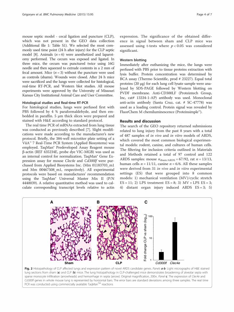
Fig. 2 Histopathology of CLP affected lungs and expression pattern of novel ARDS candidate genes. Panels a-b: Light micrographs of H&E stainedlung sections from sham (a) and CLP (b) mice. The lung histopathology in CLP-challenged mice demonstrates broadening of alveolar septa withsparse monocyte infiltration (arrowheads) and hemorrhage in septa (arrow). Original magnification, 200×. Panel c: The expression of Clec4e andCd300lf genes in whole mouse lung is represented by horizontal bars. The error bars are standard deviations among three samples. The real timePCR was conducted using commercially available TaqManTM reactions
Grigoryev et al. BMC Pulmonary Medicine (2015) 15:95 Page 4 of 7

chemically induced ARDS ES = 2; 6) Staphylococcusaureus induced ARDS ES = 2; and one experiment foreach 7) radiation and 8) shock induced ARDS (Additionalfile 1: Table S1).Cross-referencing of these microarrays resulted in a
dataset of 32160 gene entries. eGWAS analysis rankedthese genes first by the likelihood that repeated differen-tial expression for a given gene was due to chance, thencontrolled for multiple-hypothesis testing using Bonfer-roni threshold (P < 1.55 × 10⁻⁶), which identified 42 can-didate genes (Fig. 1, Additional file 2: Table S2).To identify relevance of our candidates to ARDS, we
linked 42 genes to 3 terms: “lung”, “lung injury”, and
“acute lung injury” using the PubMatrix tool. This ap-proach identified 23 candidates (54.7 %) as previouslylinked to ARDS genes (at least one citation with theterm “acute lung injury”), which justifies the suitabilityof eGWAS for the analysis of lung injury (Table 1). Theactual Score(d) and fold changes for these genes in eachmodel can be found in Additional file 3: Table S3.This approach also identified 5 novel genes (at least
one citation with the term “lung injury”) and 14 newgenes (0 links to “lung injury”). Surprisingly, among thetop five genes identified in our study to be associatedwith ARDS, only IL1R2 and IL1B were known ARDSgenes [3], the other three genes were a novel C-type
- cytokine
- unknown
- phosphatase
- trans-membranereceptor
A
B
- CD300LF
Sham CLP CLP
- Actin
- CD300LF
Sham CLP CLP
- Actin
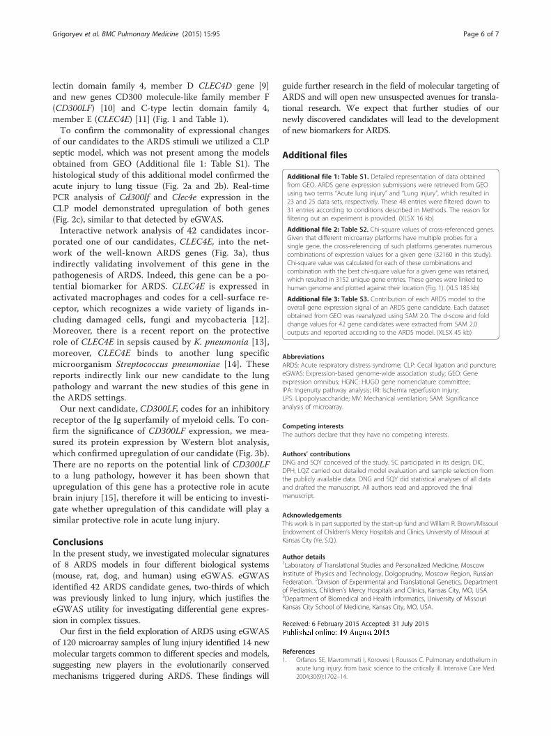
Fig. 3 In silico and in vitro validation of Clec4e and Cd300lf gene candidates. Panel a: The top gene network generated by IPA for 42 ARDS genecandidates. This network was reduced to genes that posses at least three established connections to members of the network. Relationshipsbetween genes are represented by solid lines (binding), solid arrows (direct activation), or broken arrows (indirect activation). Arrows point to theelement on which an action is performed. Black molecules represent new candidate genes. Grey molecules represent significant ARDS genes.Panel b: The Western blot analysis of Cd300lf protein expression. Each lane contains 20 μg of lung tissue lysate. The top bands represent signalsof Cd300lf protein in the sham and two CLP challenged mice. The bottom bands represent signals of actin, which was used as a loading control
Grigoryev et al. BMC Pulmonary Medicine (2015) 15:95 Page 5 of 7
lectin domain family 4, member D CLEC4D gene [9]and new genes CD300 molecule-like family member F(CD300LF) [10] and C-type lectin domain family 4,member E (CLEC4E) [11] (Fig. 1 and Table 1).To confirm the commonality of expressional changes
of our candidates to the ARDS stimuli we utilized a CLPseptic model, which was not present among the modelsobtained from GEO (Additional file 1: Table S1). Thehistological study of this additional model confirmed theacute injury to lung tissue (Fig. 2a and 2b). Real-timePCR analysis of Cd300lf and Clec4e expression in theCLP model demonstrated upregulation of both genes(Fig. 2c), similar to that detected by eGWAS.Interactive network analysis of 42 candidates incor-
porated one of our candidates, CLEC4E, into the net-work of the well-known ARDS genes (Fig. 3a), thusindirectly validating involvement of this gene in thepathogenesis of ARDS. Indeed, this gene can be a po-tential biomarker for ARDS. CLEC4E is expressed inactivated macrophages and codes for a cell-surface re-ceptor, which recognizes a wide variety of ligands in-cluding damaged cells, fungi and mycobacteria [12].Moreover, there is a recent report on the protectiverole of CLEC4E in sepsis caused by K. pneumonia [13],moreover, CLEC4E binds to another lung specificmicroorganism Streptococcus pneumoniae [14]. Thesereports indirectly link our new candidate to the lungpathology and warrant the new studies of this gene inthe ARDS settings.Our next candidate, CD300LF, codes for an inhibitory
receptor of the Ig superfamily of myeloid cells. To con-firm the significance of CD300LF expression, we mea-sured its protein expression by Western blot analysis,which confirmed upregulation of our candidate (Fig. 3b).There are no reports on the potential link of CD300LFto a lung pathology, however it has been shown thatupregulation of this gene has a protective role in acutebrain injury [15], therefore it will be enticing to investi-gate whether upregulation of this candidate will play asimilar protective role in acute lung injury.
ConclusionsIn the present study, we investigated molecular signaturesof 8 ARDS models in four different biological systems(mouse, rat, dog, and human) using eGWAS. eGWASidentified 42 ARDS candidate genes, two-thirds of whichwas previously linked to lung injury, which justifies theeGWAS utility for investigating differential gene expres-sion in complex tissues.Our first in the field exploration of ARDS using eGWAS
of 120 microarray samples of lung injury identified 14 newmolecular targets common to different species and models,suggesting new players in the evolutionarily conservedmechanisms triggered during ARDS. These findings will
guide further research in the field of molecular targeting ofARDS and will open new unsuspected avenues for transla-tional research. We expect that further studies of ournewly discovered candidates will lead to the developmentof new biomarkers for ARDS.
Additional files
Additional file 1: Table S1. Detailed representation of data obtainedfrom GEO. ARDS gene expression submissions were retrieved from GEOusing two terms “Acute lung injury” and “Lung injury”, which resulted in23 and 25 data sets, respectively. These 48 entries were filtered down to31 entries according to conditions described in Methods. The reason forfiltering out an experiment is provided. (XLSX 16 kb)
Additional file 2: Table S2. Chi-square values of cross-referenced genes.Given that different microarray platforms have multiple probes for asingle gene, the cross-referencing of such platforms generates numerouscombinations of expression values for a given gene (32160 in this study).Chi-square value was calculated for each of these combinations andcombination with the best chi-square value for a given gene was retained,which resulted in 3152 unique gene entries. These genes were linked tohuman genome and plotted against their location (Fig. 1). (XLS 185 kb)
Additional file 3: Table S3. Contribution of each ARDS model to theoverall gene expression signal of an ARDS gene candidate. Each datasetobtained from GEO was reanalyzed using SAM 2.0. The d-score and foldchange values for 42 gene candidates were extracted from SAM 2.0outputs and reported according to the ARDS model. (XLSX 45 kb)
AbbreviationsARDS: Acute respiratory distress syndrome; CLP: Cecal ligation and puncture;eGWAS: Expression-based genome-wide association study; GEO: Geneexpression omnibus; HGNC: HUGO gene nomenclature committee;IPA: Ingenuity pathway analysis; IRI: Ischemia reperfusion injury;LPS: Lipopolysaccharide; MV: Mechanical ventilation; SAM: Significanceanalysis of microarray.
Competing interestsThe authors declare that they have no competing interests.
Authors’ contributionsDNG and SQY conceived of the study. SC participated in its design, DIC,DPH, LQZ carried out detailed model evaluation and sample selection fromthe publicly available data. DNG and SQY did statistical analyses of all dataand drafted the manuscript. All authors read and approved the finalmanuscript.
AcknowledgementsThis work is in part supported by the start-up fund and William R. Brown/MissouriEndowment of Children’s Mercy Hospitals and Clinics, University of Missouri atKansas City (Ye, S.Q.).
Author details1Laboratory of Translational Studies and Personalized Medicine, MoscowInstitute of Physics and Technology, Dolgoprudny, Moscow Region, RussianFederation. 2Division of Experimental and Translational Genetics, Departmentof Pediatrics, Children’s Mercy Hospitals and Clinics, Kansas City, MO, USA.3Department of Biomedical and Health Informatics, University of MissouriKansas City School of Medicine, Kansas City, MO, USA.
Received: 6 February 2015 Accepted: 31 July 2015
References1. Orfanos SE, Mavrommati I, Korovesi I, Roussos C. Pulmonary endothelium in
acute lung injury: from basic science to the critically ill. Intensive Care Med.2004;30(9):1702–14.
Grigoryev et al. BMC Pulmonary Medicine (2015) 15:95 Page 6 of 7

2. Grigoryev DN, Finigan JH, Hassoun P, Garcia JG. Science review: searchingfor gene candidates in acute lung injury. Crit Care. 2004;8(6):440–7.
3. Grigoryev DN, Liu M, Hassoun HT, Cheadle C, Barnes KC, Rabb H. The localand systemic inflammatory transcriptome after acute kidney injury.J Am Soc Nephrol. 2008;19(3):547–58.
4. Kodama K, Horikoshi M, Toda K, Yamada S, Hara K, Irie J, et al. Expression-based genome-wide association study links the receptor CD44 in adiposetissue with type 2 diabetes. Proc Natl Acad Sci U S A. 2012;109(18):7049–54.
5. Cinti S. Transdifferentiation properties of adipocytes in the adipose organ.Am J Physiol Endocrinol Metab. 2009;297(5):E977–86.
6. Crapo JD, Barry BE, Gehr P, Bachofen M, Weibel ER. Cell number and cellcharacteristics of the normal human lung. Am Rev Respir Dis.1982;126(2):332–7.
7. Grigoryev DN, Cheranova DI, Heruth DP, Huang P, Zhang LQ, Rabb H, et al.Meta-analysis of molecular response of kidney to ischemia reperfusioninjury for the identification of new candidate genes. BMC Nephrol.2013;14:231.
8. Toscano MG, Ganea D, Gamero AM. Cecal ligation puncture procedure.J Vis Exp. 2011;51
9. Steichen AL, Binstock BJ, Mishra BB, Sharma J. C-type lectin receptor Clec4dplays a protective role in resolution of Gram-negative pneumonia. J LeukocBiol. 2013;94:393.
10. Can I, Tahara-Hanaoka S, Shibuya A. Expression of a splicing isoform ofMAIR-V (CD300LF), an inhibitory immunoglobulin-like receptor on myeloidcells. Hybridoma (Larchmt). 2008;27(1):59–61.
11. Yamasaki S, Matsumoto M, Takeuchi O, Matsuzawa T, Ishikawa E, Sakuma M,et al. C-type lectin Mincle is an activating receptor for pathogenic fungus,Malassezia. Proc Natl Acad Sci U S A. 2009;106(6):1897–902.
12. Miyake Y, Ishikawa E, Ishikawa T, Yamasaki S. Self and nonself recognitionthrough C-type lectin receptor, Mincle. Self/nonself. 2010;1(4):310–3.
13. Sharma A, Steichen AL, Jondle CN, Mishra BB, Sharma J. Protective Role ofMincle in Bacterial Pneumonia by Regulation of Neutrophil MediatedPhagocytosis and Extracellular Trap Formation. J Infect Dis. 2014;209:1837.
14. Rabes A, Zimmermann S, Reppe K, Lang R, Seeberger PH, Suttorp N, et al.The C-type lectin receptor Mincle binds to Streptococcus pneumoniae butplays a limited role in the anti-pneumococcal innate immune response.PLoS One. 2015;10(2):e0117022.
15. Peluffo H, Ali-Ruiz D, Ejarque-Ortiz A, Heras-Alvarez V, Comas-Casellas E,Martinez-Barriocanal A, et al. Overexpression of the immunoreceptor CD300fhas a neuroprotective role in a model of acute brain injury. Brain Pathol.2012;22(3):318–28.
Submit your next manuscript to BioMed Centraland take full advantage of:
• Convenient online submission
• Thorough peer review
• No space constraints or color figure charges
• Immediate publication on acceptance
• Inclusion in PubMed, CAS, Scopus and Google Scholar
• Research which is freely available for redistribution
Submit your manuscript at www.biomedcentral.com/submit
Grigoryev et al. BMC Pulmonary Medicine (2015) 15:95 Page 7 of 7