Embed Size (px)
Citation preview
Identification of Methylated Proteins in the Yeast Small RibosomalSubunit: A Role for SPOUT Methyltransferases in Protein ArginineMethylationBrian D. Young,§ David I. Weiss,§ Cecilia I. Zurita-Lopez,§ Kristofor J. Webb,§ Steven G. Clarke,*,§
and Anne E. McBride*,§,‡
§Department of Chemistry and Biochemistry and the Molecular Biology Institute, UCLA, Los Angeles, California 90095, UnitedStates‡Department of Biology, Bowdoin College, Brunswick, Maine 04011, United States
*S Supporting Information
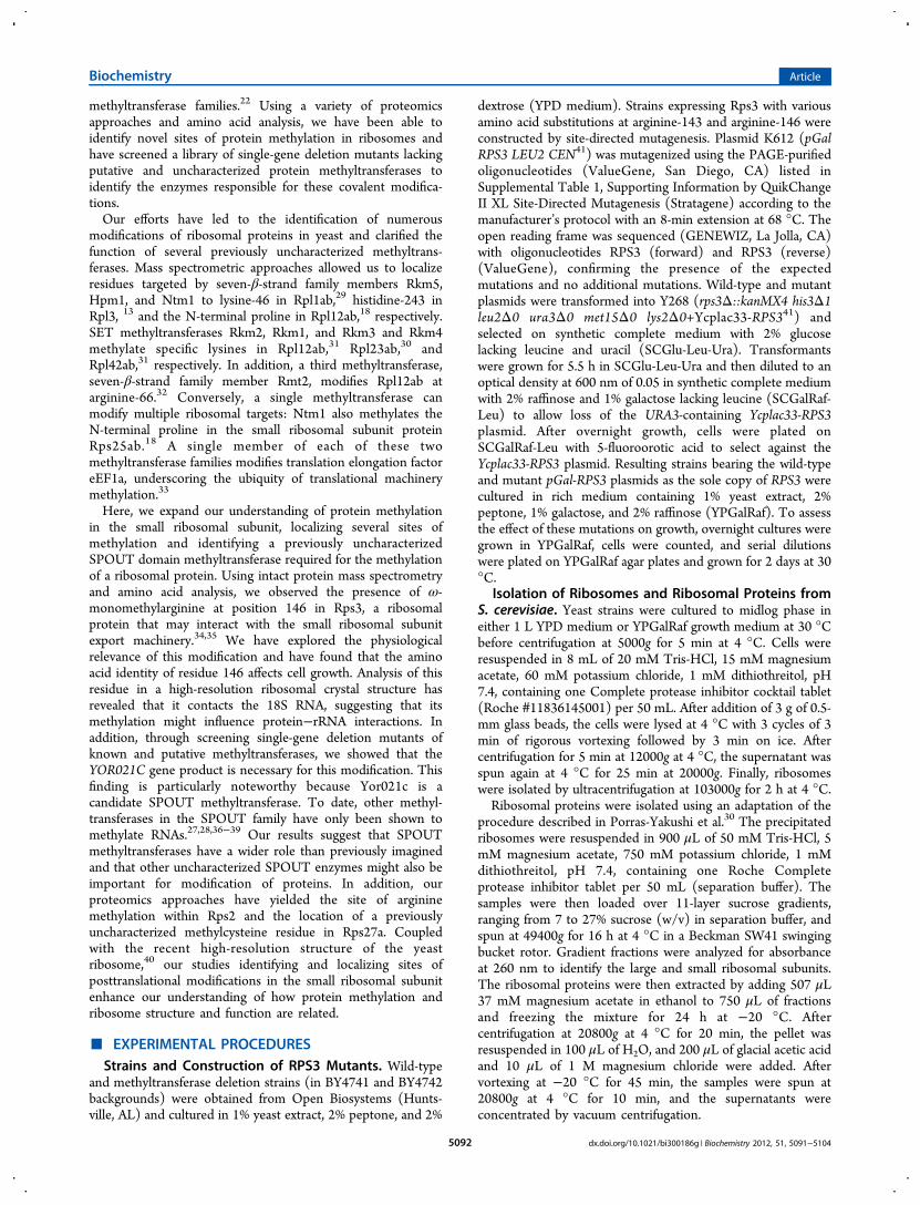
ABSTRACT: We have characterized the posttranslationalmethylation of Rps2, Rps3, and Rps27a, three small ribosomalsubunit proteins in the yeast Saccharomyces cerevisiae, usingmass spectrometry and amino acid analysis. We found thatRps2 is substoichiometrically modified at arginine-10 by theRmt1 methyltransferase. We demonstrated that Rps3 isstoichiometrically modified by ω-monomethylation at argi-nine-146 by mass spectrometric and site-directed mutagenicanalyses. Substitution of alanine for arginine at position 146 isassociated with slow cell growth, suggesting that the aminoacid identity at this site may influence ribosomal function and/or biogenesis. Analysis of the three-dimensional structure of Rps3in S. cerevisiae shows that arginine-146 makes contacts with the small subunit rRNA. Screening of deletion mutants encodingpotential yeast methyltransferases revealed that the loss of the YOR021C gene results in the absence of methylation of Rps3. Wedemonstrated that recombinant Yor021c catalyzes ω-monomethylarginine formation when incubated with S-adenosylmethionineand hypomethylated ribosomes prepared from a YOR021C deletion strain. Interestingly, Yor021c belongs to the family ofSPOUT methyltransferases that, to date, have only been shown to modify RNA substrates. Our findings suggest a wider role forSPOUT methyltransferases in nature. Finally, we have demonstrated the presence of a stoichiometrically methylated cysteineresidue at position 39 of Rps27a in a zinc-cysteine cluster. The discovery of these three novel sites of protein modification withinthe small ribosomal subunit will now allow for an analysis of their functional roles in translation and possibly other cellularprocesses.
In nature, the library of stereochemical possibilities formacromolecules is vastly expanded by covalent modifica-
tions. Modifications of nucleic acids facilitate changes in geneexpression and guide rRNA processing.1,2 Protein function isalso modulated by the posttranslational covalent modificationsof amino acid residues.3 These stereochemical changes can alterphysical interactions within the protein and with other proteins,affecting signaling,4,5 enzyme activity,6 turnover,7 and local-ization.8 Families of enzymes have evolved to catalyze theformation and removal of these modifications, allowing precisecontrol of a wide array of biochemical pathways. For proteins,methylation reactions, along with phosphorylation andacetylation reactions, represent major modification pathways.9
Many amino acid side chains are known to be methylated ineukaryotes, including those of lysine,10 arginine,11,12 histidine,13
glutamic acid,14 and glutamine15 residues. In addition, there canbe methylation of the N- and C-termini of proteins.16−18
Methylation of proteins is integral for epigenetics,19 cellularsignaling,4,20 and other processes, including translation.21
We have been interested in exploring the methylation ofproteins involved in translation and identifying novel enzymescatalyzing these modifications. In Saccharomyces cerevisiae,nearly 90 methyltransferases have been identified andpredicted.22,23 Nearly half of them modify components of thetranslational apparatus rRNA, tRNAs, ribosomal proteins,and other proteins involved in translation. The majority ofthese enzymes belong to the seven β-strand methyltransferasefamily characterized by a series of four well-conserved motifs.24
Different members of this family catalyze methylation of a widerange of substrates, including nucleic acids, proteins, smallmolecules, and lipids.22,23 The other major subgroups ofmethyltransferases are the SET25,26 and SPOUT27,28 families,which, to date, have been shown to methylate proteins at lysineresidues and RNAs, respectively. Our group has previously usedbioinformatics to identify new putative members of these
Received: February 9, 2012Revised: May 23, 2012Published: May 31, 2012
Article
pubs.acs.org/biochemistry
© 2012 American Chemical Society 5091 dx.doi.org/10.1021/bi300186g | Biochemistry 2012, 51, 5091−5104
methyltransferase families.22 Using a variety of proteomicsapproaches and amino acid analysis, we have been able toidentify novel sites of protein methylation in ribosomes andhave screened a library of single-gene deletion mutants lackingputative and uncharacterized protein methyltransferases toidentify the enzymes responsible for these covalent modifica-tions.Our efforts have led to the identification of numerous
modifications of ribosomal proteins in yeast and clarified thefunction of several previously uncharacterized methyltrans-ferases. Mass spectrometric approaches allowed us to localizeresidues targeted by seven-β-strand family members Rkm5,Hpm1, and Ntm1 to lysine-46 in Rpl1ab,29 histidine-243 inRpl3, 13 and the N-terminal proline in Rpl12ab,18 respectively.SET methyltransferases Rkm2, Rkm1, and Rkm3 and Rkm4methylate specific lysines in Rpl12ab,31 Rpl23ab,30 andRpl42ab,31 respectively. In addition, a third methyltransferase,seven-β-strand family member Rmt2, modifies Rpl12ab atarginine-66.32 Conversely, a single methyltransferase canmodify multiple ribosomal targets: Ntm1 also methylates theN-terminal proline in the small ribosomal subunit proteinRps25ab.18 A single member of each of these twomethyltransferase families modifies translation elongation factoreEF1a, underscoring the ubiquity of translational machinerymethylation.33
Here, we expand our understanding of protein methylationin the small ribosomal subunit, localizing several sites ofmethylation and identifying a previously uncharacterizedSPOUT domain methyltransferase required for the methylationof a ribosomal protein. Using intact protein mass spectrometryand amino acid analysis, we observed the presence of ω-monomethylarginine at position 146 in Rps3, a ribosomalprotein that may interact with the small ribosomal subunitexport machinery.34,35 We have explored the physiologicalrelevance of this modification and have found that the aminoacid identity of residue 146 affects cell growth. Analysis of thisresidue in a high-resolution ribosomal crystal structure hasrevealed that it contacts the 18S RNA, suggesting that itsmethylation might influence protein−rRNA interactions. Inaddition, through screening single-gene deletion mutants ofknown and putative methyltransferases, we showed that theYOR021C gene product is necessary for this modification. Thisfinding is particularly noteworthy because Yor021c is acandidate SPOUT methyltransferase. To date, other methyl-transferases in the SPOUT family have only been shown tomethylate RNAs.27,28,36−39 Our results suggest that SPOUTmethyltransferases have a wider role than previously imaginedand that other uncharacterized SPOUT enzymes might also beimportant for modification of proteins. In addition, ourproteomics approaches have yielded the site of argininemethylation within Rps2 and the location of a previouslyuncharacterized methylcysteine residue in Rps27a. Coupledwith the recent high-resolution structure of the yeastribosome,40 our studies identifying and localizing sites ofposttranslational modifications in the small ribosomal subunitenhance our understanding of how protein methylation andribosome structure and function are related.
■ EXPERIMENTAL PROCEDURESStrains and Construction of RPS3 Mutants. Wild-type
and methyltransferase deletion strains (in BY4741 and BY4742backgrounds) were obtained from Open Biosystems (Hunts-ville, AL) and cultured in 1% yeast extract, 2% peptone, and 2%
dextrose (YPD medium). Strains expressing Rps3 with variousamino acid substitutions at arginine-143 and arginine-146 wereconstructed by site-directed mutagenesis. Plasmid K612 (pGalRPS3 LEU2 CEN41) was mutagenized using the PAGE-purifiedoligonucleotides (ValueGene, San Diego, CA) listed inSupplemental Table 1, Supporting Information by QuikChangeII XL Site-Directed Mutagenesis (Stratagene) according to themanufacturer’s protocol with an 8-min extension at 68 °C. Theopen reading frame was sequenced (GENEWIZ, La Jolla, CA)with oligonucleotides RPS3 (forward) and RPS3 (reverse)(ValueGene), confirming the presence of the expectedmutations and no additional mutations. Wild-type and mutantplasmids were transformed into Y268 (rps3Δ::kanMX4 his3Δ1leu2Δ0 ura3Δ0 met15Δ0 lys2Δ0+Ycplac33-RPS341) andselected on synthetic complete medium with 2% glucoselacking leucine and uracil (SCGlu-Leu-Ura). Transformantswere grown for 5.5 h in SCGlu-Leu-Ura and then diluted to anoptical density at 600 nm of 0.05 in synthetic complete mediumwith 2% raffinose and 1% galactose lacking leucine (SCGalRaf-Leu) to allow loss of the URA3-containing Ycplac33-RPS3plasmid. After overnight growth, cells were plated onSCGalRaf-Leu with 5-fluoroorotic acid to select against theYcplac33-RPS3 plasmid. Resulting strains bearing the wild-typeand mutant pGal-RPS3 plasmids as the sole copy of RPS3 werecultured in rich medium containing 1% yeast extract, 2%peptone, 1% galactose, and 2% raffinose (YPGalRaf). To assessthe effect of these mutations on growth, overnight cultures weregrown in YPGalRaf, cells were counted, and serial dilutionswere plated on YPGalRaf agar plates and grown for 2 days at 30°C.
Isolation of Ribosomes and Ribosomal Proteins fromS. cerevisiae. Yeast strains were cultured to midlog phase ineither 1 L YPD medium or YPGalRaf growth medium at 30 °Cbefore centrifugation at 5000g for 5 min at 4 °C. Cells wereresuspended in 8 mL of 20 mM Tris-HCl, 15 mM magnesiumacetate, 60 mM potassium chloride, 1 mM dithiothreitol, pH7.4, containing one Complete protease inhibitor cocktail tablet(Roche #11836145001) per 50 mL. After addition of 3 g of 0.5-mm glass beads, the cells were lysed at 4 °C with 3 cycles of 3min of rigorous vortexing followed by 3 min on ice. Aftercentrifugation for 5 min at 12000g at 4 °C, the supernatant wasspun again at 4 °C for 25 min at 20000g. Finally, ribosomeswere isolated by ultracentrifugation at 103000g for 2 h at 4 °C.Ribosomal proteins were isolated using an adaptation of the
procedure described in Porras-Yakushi et al.30 The precipitatedribosomes were resuspended in 900 μL of 50 mM Tris-HCl, 5mM magnesium acetate, 750 mM potassium chloride, 1 mMdithiothreitol, pH 7.4, containing one Roche Completeprotease inhibitor tablet per 50 mL (separation buffer). Thesamples were then loaded over 11-layer sucrose gradients,ranging from 7 to 27% sucrose (w/v) in separation buffer, andspun at 49400g for 16 h at 4 °C in a Beckman SW41 swingingbucket rotor. Gradient fractions were analyzed for absorbanceat 260 nm to identify the large and small ribosomal subunits.The ribosomal proteins were then extracted by adding 507 μL37 mM magnesium acetate in ethanol to 750 μL of fractionsand freezing the mixture for 24 h at −20 °C. Aftercentrifugation at 20800g at 4 °C for 20 min, the pellet wasresuspended in 100 μL of H2O, and 200 μL of glacial acetic acidand 10 μL of 1 M magnesium chloride were added. Aftervortexing at −20 °C for 45 min, the samples were spun at20800g at 4 °C for 10 min, and the supernatants wereconcentrated by vacuum centrifugation.
Biochemistry Article
dx.doi.org/10.1021/bi300186g | Biochemistry 2012, 51, 5091−51045092
Expression and Purification of Recombinant YOR021CGene Product. Genomic DNA from S. cerevisiae was extractedfrom a 10-mL culture grown to saturation. The YOR021C openreading frame was amplified by PCR with a forward primer 5′-CACCATGAAGTACATTATTGAGCATATGG and a reverseprimer 5′-CTACATCAACAGATCGTCCAAAC. The productswere fractionated on a 1.1% agarose gel at 120 V for 60 min.The band containing the YOR021C open reading frame was cutfrom the gel and the DNA was purified using a QIAquick GelExtraction Kit (QIAGEN, catalog no. 28704). Using theChampion pET100 Directional TOPO Expression Kit(Invitrogen), the DNA was inserted into a pET100/D-TOPOvector encoding a His-tagged N-terminal linker sequenceMRG SHHHHHHGMA SMTGGQQMGRD L YD -DDDKDHPFT that is followed by the complete sequence ofthe YOR021C open reading frame, including the initiatormethionine residue, that is regulated by the T7 promoter.TOP10 chemically competent Escherichia coli cells were thentransformed with the plasmid and plated onto LB platescontaining 100 μg/mL ampicillin to select for coloniescontaining the plasmid. Plasmid DNA was sequenced onboth strands to ensure proper insertion of the YOR021C genewithout mutation (GENEWIZ, La Jolla, CA). Per the protocolof the Champion pET100 Directional TOPO Expression kit,the plasmid was used to transform competent BL21 Star (DE3)E. coli cells. These cells were then grown at 37 °C to an opticaldensity of 0.5 at 600 nm in 1 L of LB containing 100 μg/mLampicillin. At this time, isopropyl-ß-D-thiogalactopyranosidewas added to give a concentration of 0.4 mM and theincubation continued for 18 h at 18 °C. Cells were pelleted for5 min at 5000g at 4 °C, washed in 10 mL of water, and thenresuspended in 30 mL of 50 mM sodium phosphate, pH 8.0,500 mM sodium chloride, and 5% glycerol along with 1Complete protease inhibitor tablet (Roche Diagnostics) and 11μL of β-mercaptoethanol. Using an Emulsiflex C-3 emulsifier(Avestin), the cells were lysed and the supernatant wasrecovered after centrifugation at 20000g for 15 min at 4 °C.The supernatant was applied onto a 5-mL HisTrap HP nickelaffinity column (GE Healthcare, catalog no. 17-5248-01) andthe recombinant Yor021c protein was recovered using agradient of 5−500 mM imidazole. The final protein preparation(2.6 mg/mL), in a buffer of 50 mM sodium phosphate, 260mM imidazole chloride, 300 mM sodium chloride, 5% glycerol,pH 8.0, was stored at −20 °C.In Vivo Labeling of Ribosomal Proteins from S.
cerevisiae and Analysis by High-Resolution Cation-Exchange Chromatography. BY4742 wild-type ribosomeswere labeled in vivo by incubating intact cells with S-adenosyl-L-[methyl-3H]-L-methionine. Cells were grown in 500 mL ofYPD medium to an optical density at 600 nm of 0.6, pelleted at5000g for 5 min, and washed twice in water. Cells were thenresuspended in 304 μL of S-adenosyl-L-[methyl-3H]-L-methio-nine (PerkinElmer, 75−85 Ci/mmol, 0.55 mCi/mL in 10 mMH2SO4/ethanol (9:1, v/v)) and incubated in 40 mL of YPDmedium in a rotary shaker for 30 min at 30 °C. Labeled cellswere pelleted, washed, and stored at −80 °C until lysis andisolation of the protein fraction of the ribosomal small subunitas described above. Rps3 was purified by HPLC and acidhydrolyzed as described previously.13 Purified Rps3 was placedin a 6 × 50-mm glass vial and dried by vacuum centrifugation.The protein was acid hydrolyzed by addition of 50 μL of 6 MHCl to the vial and 200 μL of 6 M HCl into the reactionchamber (Eldex Laboratories, catalog no. 1163). The vial was
heated for 20 h in vacuo at 110 °C using a Pico-Tag Vapor-Phase apparatus (Waters). After 20 h, residual HCl wasremoved by vacuum centrifugation. The hydrolyzed aminoacids were resuspended in 50 μL of H2O and 500 μL of 0.2 Msodium citrate buffer, pH 2.2. This material was loaded onto acation-exchange chromatography column (0.9-cm inner diam-eter × 10-cm column length; PA-35 sulfonated polystyrenebeads; 6−12 μm, Benson Co.) along with 1.0 μmol of thefollowing standards: N-ε-monomethyllysine hydrochloride(MMK, Bachem, #E-2155); 1-methyl-L-histidine (1-Me-His,Sigma, #M-9005); asymmetric NG,NG-dimethylarginine hydro-chloride (ADMA, Sigma, #D4268); symmetric NG, NG′-dimethyl-L-arginine di(p-hydroxyazobenzene-p′-sulfonate(SDMA, Sigma, #D0390) and NG-methyl-L-arginine acetatesalt (MMA, Sigma, #M7033). The amino acids were eluted at55 °C with 0.35 M sodium citrate buffer, pH 5.27 at 1 mL/minand 1-min fractions were collected. The column wasregenerated with 0.2 N NaOH for 25 min and equilibratedwith 0.35 M sodium citrate buffer, pH 5.27 for 25 min prior toeach run. The eluted standards were identified via a ninhydrinassay. Briefly, 30 μL of each column fraction was mixed with200 μL of water and 100 μL of a solution of 20 mg/mLninhydrin and 3 mg/mL hydrindantin in a solvent of 75% (v/v)dimethyl sulfoxide and 25% (v/v) 4 M lithium acetate, pH 4.2in a 96-well clear flat bottom plate. The plates were then heatedin a 100 °C oven for 10 min, and the absorbance at 570 nm wasread in a Molecular Devices SpectraMax M5 plate reader (0.9-cm path length). Radioactivity was measured by mixing 970 μLof sample with 400 μL of H2O and 5 mL of fluor (Safety Solve,Research Products International, catalog no. 111177). Sampleswere counted on a Beckman LS6500 instrument three times for5 min.
Localization of the Rps3 Methylation Site by MassSpectrometry Analysis of Cyanogen Bromide CleavageProducts. Purified ribosomal proteins of the small subunitwere isolated from wild-type BY4742, as detailed above, andRps3 was purified by HPLC and digested with cyanogenbromide as described previously.13 Briefly, 20 mg of cyanogenbromide was added to purified protein in 70 μL of H2O/acetonitrile/trifluoroacetic acid (1:1:0.05) such that the molarratio of cyanogen bromide to methionine residues wasapproximately 1000:1. After an overnight incubation in theabsence of light, the digest was infused into a hybrid linear iontrap/FTICR mass spectrometer (LTQ FT Ultra, ThermoScientific, San Jose, CA) operating in MS-only mode. TheXtract algorithm in the Xcalibur software suite (ThermoScientific) was used to deconvolute the resulting spectra andcalculate the uncharged monoisotopic masses.
Intact Protein Mass Spectrometry Analysis of IsolatedRibosomal Proteins. The extracted ribosomal proteins fromthe small ribosomal subunit were analyzed by liquidchromatography−mass spectrometry using an electrosprayQSTAR Elite time-of-flight mass spectrometer (AppliedBiosystems) calibrated with peptide standards. For the wild-type and mutant BY4742 strains, the chromatography and massspectrometry method used has been described previously.13 Forthe other strains, a slightly modified chromatography methodwas used. Briefly, 10 μL of the samples was mixed with 30 μL ofH2O and injected onto a PLRP-S column (150 mm × 1 mm, 5-μm particle size, 300-Å pore size) (Polymer Laboratories,Amherst, MA). Mobile phase A contained 2% acetonitrile and0.1% formic acid while mobile phase B contained 98%acetonitrile and 0.1% formic acid. The following gradient was
Biochemistry Article
dx.doi.org/10.1021/bi300186g | Biochemistry 2012, 51, 5091−51045093
used with a flow rate of 50 μL/min: t = 0−3 min, 5% B; t = 13min, 25% B; t = 38 min, 62.5% B; t = 38.1−49 min, 95% B; t =49.1−60 min; 5% B. Spectra were reconstructed usingMagTran 1.03.42
Sample Preparation of In Vitro Methylation Reactionsfor Cation-Exchange Chromatography. Reaction mixtures(300 μL) were transferred to 6 × 50-mm glass vials andproteins were precipitated by incubation with an equal volumeof 25% (wt/vol) trichloroacetic acid for 1 h at roomtemperature. The protein pellet from centrifugation at 4000gfor 1 h at room temperature was washed with 200 μL ofacetone at 0 °C. Acid hydrolysis of the pellet was performed asdescribed above.Localization of the Rps27a Methylation Site by Top-
Down Mass Spectrometry using Collisionally ActivatedDissociation and Electron-Capture Dissociation. Rps27aprotein isolated and purified by HPLC from wild-type BY4742was infused into the hybrid linear ion trap/FTICR massspectrometer and fragmented using collisionally activateddissociation and electron-capture dissociation as describedpreviously.13 The spectra were deconvoluted, as describedabove, with the Xtract algorithm. The resulting unchargedmonoisotopic masses were analyzed with the ProSight PTMonline tool using the Rps27a and Rps27b protein sequencesand a mass error range of 5.5 ppm.Localization of the Rps2 Methylation Site by Bottom-
Up Mass Spectrometry. Rps2 protein isolated and purifiedby HPLC from wild-type strain BY4742 was digested withchymotrypsin (Roche) and analyzed by liquid chromatography-tandem mass spectrometry as described previously.13 Briefly,the protein was loaded onto a Biobasic C18 column (3.5 cm ×100 μm, 5-μm particle size, 300-Å pore size) (MicrotechScientific, Fontana, CA). The peptides were loaded at 5 μL/min and were eluted at 300 nL/min with the followinggradient: t = 0, 2% B; t = 40 min, 60% B; t = 50 min, 80% B.Mobile phase A contained 1% acetonitrile and 0.1% formic acidwhile mobile phase B contained 100% acetonitrile and 0.1%formic acid. The eluted peptides were directed into the hybridlinear ion trap/FTICR mass spectrometer, which was operatedin data-dependent mode with a full high-resolution scan (100 000 resolution at m/z= 400) followed by six MS/MSexperiments using the low-resolution linear trap and collision-ally activated dissociation. For the MS/MS experiments, theintensity threshold was 5000 and the m/z range was 300−2000.The data were processed using Mascot (Matrix Science, UK).
■ RESULTSRps3 Is Monomethylated at Arginine-146. Rps3 is an
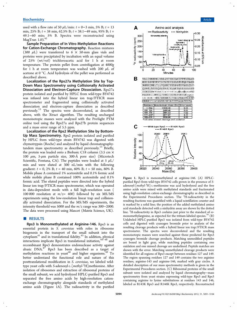
essential protein in S. cerevisiae with roles in ribosomebiogenesis in the transport of the small subunit into thecytoplasm41 and in translational fidelity.43 In addition, physicalinteractions implicate Rps3 in translational initiation,44−46 andrecombinant Rps3 demonstrates endonuclease activity againstabasic DNA.47 Rps3 has been described as a target ofmethylation reactions in yeast48 and higher organisms.49 Tobetter understand the functional role and nature of thisposttranslational modification in S. cerevisiae, we labeled wild-type yeast cells with S-adenosyl-L-[methyl-3H]methionine. Afterisolation of ribosomes and extraction of ribosomal proteins ofthe small subunit, we acid hydrolyzed HPLC-purified Rps3 andseparated the free amino acids by high-resolution cation-exchange chromatography alongside standards of methylatedamino acids (Figure 1A). The radioactivity in the purified
Figure 1. Rps3 is monomethylated at arginine-146. (A) HPLC-purified Rps3 from wild-type BY4742 cells grown in the presence of S-adenosyl-[methyl-3H]-L-methionine was acid hydrolyzed and the freeamino acids were mixed with methylated standards and fractionatedusing high-resolution cation-exchange chromatography as described inthe Experimental Procedures section. The 3H-radioactivity in theresulting fractions was quantified with a liquid scintillation counter andis marked by a solid line; the position of the added methylated aminoacid standards detected with a ninhydrin assay are shown by the dottedline. 3H-radioactivity in Rps3 coelutes just prior to the standard of ω-monomethylarginine, as expected for the tritium-labeled species.79 (B)Unlabeled HPLC-purified Rps3 was isolated from wild-type BY4742cells and digested with cyanogen bromide prior to analysis of theresulting cleavage products with a hybrid linear ion trap/FTICR massspectrometer. The spectra were deconvoluted and the resultingmonoisotopic masses were searched against those predicted for Rps3cyanogen bromide cleavage products. Matching unmodified peptidesare boxed in light gray, while matching peptides containing oneoxidation and one missed cleavage are underlined. Peptide matches areshown with the error. Matching unmethylated cleavage products wereidentified for all regions of Rps3 except between residues 127 and 149.The region spanning residues 127 and 149 contains the two arginineresidues, arginine-143 and arginine-146, marked with gray circles. Adetailed description of our mass spectrometry methods is given in theExperimental Procedures section. (C) Ribosomal proteins of the smallsubunit were isolated and analyzed by liquid chromatography−massspectrometry from yeast strains expressing wild-type Rps3 and Rps3containing arginine to lysine substitutions at residues 143 and 146,labeled as R143K Rps3 and R146K Rps3, respectively. Reconstructed
Biochemistry Article
dx.doi.org/10.1021/bi300186g | Biochemistry 2012, 51, 5091−51045094
protein eluted in the expected position of ω-monomethylargi-nine and had a retention profile distinct from 1-methylhistidine, monomethyllysine, asymmetric dimethylarginine,and symmetric dimethylarginine. These results revealed thatRps3 in S. cerevisiae contains an ω-monomethylated arginineresidue.To identify the site of arginine methylation within the
protein, we analyzed cyanogen bromide cleavage products ofHPLC-purified Rps3 by high-resolution mass spectrometry. Wewere able to detect all of the unmodified cyanogen bromidecleavage products of Rps3 with the exception of the peptidesspanning residues 127−149 and 177−188 (Figure 1B). Whenwe expanded our search to include peptides containing missedcleavages and oxidations, we found an unmethylated singlyoxidized peptide containing residues 177−239. Taken together,these results suggested that the site of arginine methylation isbetween positions 127 and 149.To further localize the site of monomethylation in Rps3, we
used site-directed mutagenesis, constructing strains expressingwild-type and mutant Rps3 with arginine to lysine amino acidsubstitutions at arginine-143 and arginine-146, the two arginineresidues present in the polypeptide between residues 127 and149. We isolated ribosomes from these strains, purified proteinsof the small subunit by sucrose density centrifugation, andanalyzed the extracted proteins by liquid chromatography−intact protein mass spectrometry (Figure 1C). In the strainexpressing wild-type Rps3 under a GAL promoter, we identifieda 26384.3-Da species, consistent with the mass of mono-methylated Rps3 after the previously described loss of theinitiator methionine residue (error = 45 ppm).50,51 Signifi-cantly, the unmethylated protein is not observed; we detectedno ions corresponding to a mass of 26370 Da in twoindependent experiments. In the strain expressing Rps3 withan arginine to lysine substitution at position 143, we identified a26355.8-Da species, which is consistent with the mass expectedfor monomethylated Rps3 with an arginine to lysine amino acidsubstitution (error = 64 ppm), indicating that arginine-143 isnot the site of methylation. For a similar mutant strain with thearginine to lysine substitution at position 146, however, a26342.0-Da species was observed. This mass is consistent withthat of unmethylated Rps3 with the arginine to lysinesubstitution (error = 56 ppm), revealing that arginine-146 isthe site of monomethylation in S. cerevisiae. In two replicateintact mass determinations, we did not detect ions correspond-ing to a methylated species of 26356 Da. Collectively, ouramino acid analysis of Rps3, mass spectrometry analyses ofcyanogen bromide digests, and mass spectrometry of intactwild-type and mutant Rps3 indicate that Rps3 is stoichiometri-cally ω-monomethylated at arginine-146, although minoramounts of unmethylated Rps3 species may have escapeddetection.Arginine-146 in Rps3 Interacts with the 18S rRNA in
Crystal Structures of the Small Ribosomal Subunit.
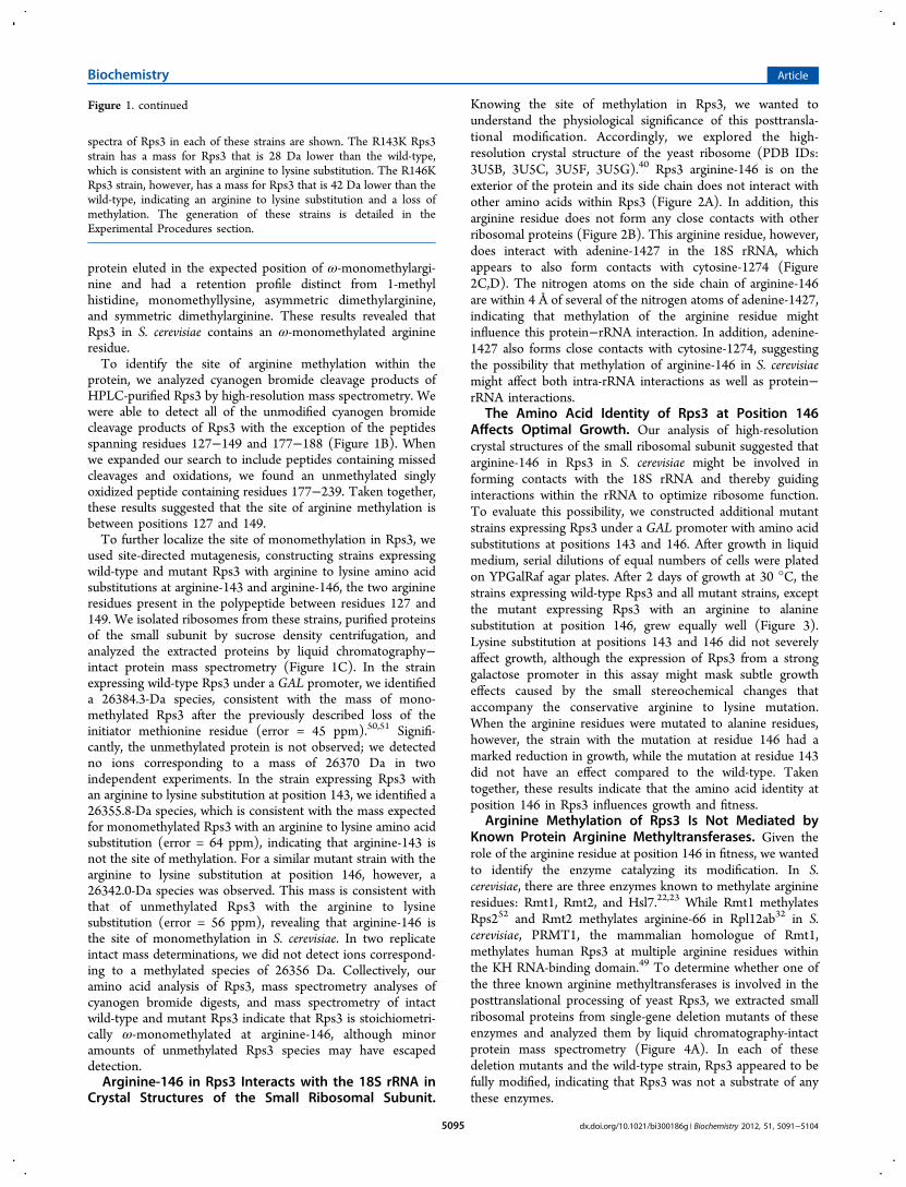
Knowing the site of methylation in Rps3, we wanted tounderstand the physiological significance of this posttransla-tional modification. Accordingly, we explored the high-resolution crystal structure of the yeast ribosome (PDB IDs:3U5B, 3U5C, 3U5F, 3U5G).40 Rps3 arginine-146 is on theexterior of the protein and its side chain does not interact withother amino acids within Rps3 (Figure 2A). In addition, thisarginine residue does not form any close contacts with otherribosomal proteins (Figure 2B). This arginine residue, however,does interact with adenine-1427 in the 18S rRNA, whichappears to also form contacts with cytosine-1274 (Figure2C,D). The nitrogen atoms on the side chain of arginine-146are within 4 Å of several of the nitrogen atoms of adenine-1427,indicating that methylation of the arginine residue mightinfluence this protein−rRNA interaction. In addition, adenine-1427 also forms close contacts with cytosine-1274, suggestingthe possibility that methylation of arginine-146 in S. cerevisiaemight affect both intra-rRNA interactions as well as protein−rRNA interactions.
The Amino Acid Identity of Rps3 at Position 146Affects Optimal Growth. Our analysis of high-resolutioncrystal structures of the small ribosomal subunit suggested thatarginine-146 in Rps3 in S. cerevisiae might be involved informing contacts with the 18S rRNA and thereby guidinginteractions within the rRNA to optimize ribosome function.To evaluate this possibility, we constructed additional mutantstrains expressing Rps3 under a GAL promoter with amino acidsubstitutions at positions 143 and 146. After growth in liquidmedium, serial dilutions of equal numbers of cells were platedon YPGalRaf agar plates. After 2 days of growth at 30 °C, thestrains expressing wild-type Rps3 and all mutant strains, exceptthe mutant expressing Rps3 with an arginine to alaninesubstitution at position 146, grew equally well (Figure 3).Lysine substitution at positions 143 and 146 did not severelyaffect growth, although the expression of Rps3 from a stronggalactose promoter in this assay might mask subtle growtheffects caused by the small stereochemical changes thataccompany the conservative arginine to lysine mutation.When the arginine residues were mutated to alanine residues,however, the strain with the mutation at residue 146 had amarked reduction in growth, while the mutation at residue 143did not have an effect compared to the wild-type. Takentogether, these results indicate that the amino acid identity atposition 146 in Rps3 influences growth and fitness.
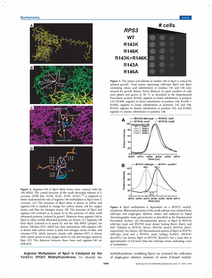
Arginine Methylation of Rps3 Is Not Mediated byKnown Protein Arginine Methyltransferases. Given therole of the arginine residue at position 146 in fitness, we wantedto identify the enzyme catalyzing its modification. In S.cerevisiae, there are three enzymes known to methylate arginineresidues: Rmt1, Rmt2, and Hsl7.22,23 While Rmt1 methylatesRps252 and Rmt2 methylates arginine-66 in Rpl12ab32 in S.cerevisiae, PRMT1, the mammalian homologue of Rmt1,methylates human Rps3 at multiple arginine residues withinthe KH RNA-binding domain.49 To determine whether one ofthe three known arginine methyltransferases is involved in theposttranslational processing of yeast Rps3, we extracted smallribosomal proteins from single-gene deletion mutants of theseenzymes and analyzed them by liquid chromatography-intactprotein mass spectrometry (Figure 4A). In each of thesedeletion mutants and the wild-type strain, Rps3 appeared to befully modified, indicating that Rps3 was not a substrate of anythese enzymes.
Figure 1. continued
spectra of Rps3 in each of these strains are shown. The R143K Rps3strain has a mass for Rps3 that is 28 Da lower than the wild-type,which is consistent with an arginine to lysine substitution. The R146KRps3 strain, however, has a mass for Rps3 that is 42 Da lower than thewild-type, indicating an arginine to lysine substitution and a loss ofmethylation. The generation of these strains is detailed in theExperimental Procedures section.
Biochemistry Article
dx.doi.org/10.1021/bi300186g | Biochemistry 2012, 51, 5091−51045095
Arginine Methylation of Rps3 Is Catalyzed by theYor021c SPOUT Methyltransferase. To identify the
methyltransferase modifying Rps3, we screened the collectionof single-gene deletion mutants of seven β-strand methyl-
Figure 2. Arginine-146 in Rps3 likely forms close contacts with the18S rRNA. The crystal structure of the small ribosomal subunit of S.cerevisiae (PDB IDs: 3U5B, 3U5C, 3U5F, 3U5G) 40 is explored tobetter understand the role of arginine-146 methylation in Rps3 from S.cerevisiae. (A) The structure of Rps3 alone is shown in yellow andarginine-146 is marked in orange for carbon atoms, red for oxygenatoms, and blue for nitrogen atoms. (B) The structure of Rps3 andarginine-146 (colored as in panel A) in the presence of other smallribosomal proteins (colored in green). Distances from arginine-146 inRps3 to other nearby ribosomal proteins are shown. (C) Arginine-146from Rps3 (colored as in panel A) and the 18S rRNA (purple) areshown. Adenine-1427, which has close interactions with arginine-146,is shown with carbon atoms in pink and nitrogen atoms in blue, andcytosine-1274, which interacts closely with adenine-1427, is shownwith carbon atoms in teal, oxygen atoms in red, and nitrogen atoms inblue. (D) The distances between these bases and arginine-146 areindicated.
Figure 3. The amino acid identity at residue 146 in Rps3 is critical foroptimal growth. Yeast strains expressing wild-type Rps3 and Rps3containing amino acid substitutions at residues 143 and 146 wereassayed for growth fitness. Serial dilutions of equal numbers of cellswere plated and grown at 30 °C as described in the ExperimentalProcedures section. R143K, arginine to lysine substitution at position143; R146K, arginine to lysine substitution at position 146; R143K +R146K, arginine to lysine substitutions at positions 143 and 146;R143A, arginine to alanine substitution at position 143; and R146A,arginine to alanine substitution at position 146.
Figure 4. Rps3 methylation is dependent on a SPOUT methyl-transferase. Ribosomal proteins of the small subunit were isolated fromwild-type and single-gene deletion strains and analyzed by liquidchromatography−mass spectrometry as described in the ExperimentalProcedures section. (A) Reconstructed spectra of Rps3 in BY4742wild-type yeast and BY4742 yeast strains lacking Rmt1, Rmt2, andHsl7 (labeled as BY4742 Δrmt1, BY4742 Δrmt2, BY4742 Δhsl7,respectively) are shown. (B) Reconstructed spectra of Rps3 in BY4741wild-type yeast and a BY4741 strain lacking Yor021c (BY4741Δyor021c) are shown. Rps3 in BY4741 Δyor021c has a mass that isapproximately 14 Da lower than the wild-type strain, indicating a lossof methylation.
Biochemistry Article
dx.doi.org/10.1021/bi300186g | Biochemistry 2012, 51, 5091−51045096
transferases described previously13 for loss of Rps3 methylation.None of the deletions tested appeared to affect the modificationstate of Rps3 (data not shown). A previous study showed thatYar1, an ankyrin-repeat protein, interacts physically with Rps3and Yor021c through a two-hybrid screen.35 Yor021c has beenidentified as a putative SPOUT family methyltransferasethrough bioinformatics analysis.22,28 We isolated ribosomalproteins of the small ribosomal subunit from a deletion strain
lacking this enzyme and wild-type strains (both strains in theBY4741 and BY4742 backgrounds, although the latter is notshown) (Figure 4B). Significantly, we detected no Rps3methylation in the absence of Yor021c. We also tested deletionstrains of two other uncharacterized putative SPOUTmethyltransferases, YGR283C and YMR310C. Neither ofthem, however, was required for Rps3 modification (data not
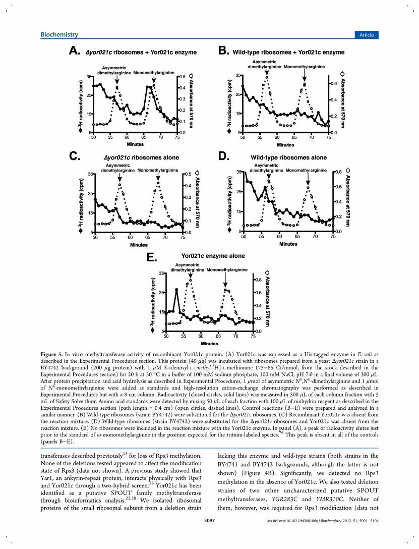
Figure 5. In vitro methyltransferase activity of recombinant Yor021c protein. (A) Yor021c was expressed as a His-tagged enzyme in E. coli asdescribed in the Experimental Procedures section. This protein (40 μg) was incubated with ribosomes prepared from a yeast Δyor021c strain in aBY4742 background (200 μg protein) with 1 μM S-adenosyl-L-[methyl-3H]-L-methionine (75−85 Ci/mmol, from the stock described in theExperimental Procedures section) for 20 h at 30 °C in a buffer of 100 mM sodium phosphate, 100 mM NaCl, pH 7.0 in a final volume of 300 μL.After protein precipitation and acid hydrolysis as described in Experimental Procedures, 1 μmol of asymmetric NG,NG-dimethylarginine and 1 μmolof NG-monomethylarginine were added as standards and high-resolution cation-exchange chromatography was performed as described inExperimental Procedures but with a 8-cm column. Radioactivity (closed circles, solid lines) was measured in 500 μL of each column fraction with 5mL of Safety Solve fluor. Amino acid standards were detected by mixing 50 μL of each fraction with 100 μL of ninhydrin reagent as described in theExperimental Procedures section (path length = 0.4 cm) (open circles, dashed lines). Control reactions (B−E) were prepared and analyzed in asimilar manner. (B) Wild-type ribosomes (strain BY4742) were substituted for the Δyor021c ribosomes. (C) Recombinant Yor021c was absent fromthe reaction mixture. (D) Wild-type ribosomes (strain BY4742) were substituted for the Δyor021c ribosomes and Yor021c was absent from thereaction mixture. (E) No ribosomes were included in the reaction mixture with the Yor021c enzyme. In panel (A), a peak of radioactivity elutes justprior to the standard of ω-monomethylarginine in the position expected for the tritium-labeled species.79 This peak is absent in all of the controls(panels B−E).
Biochemistry Article
dx.doi.org/10.1021/bi300186g | Biochemistry 2012, 51, 5091−51045097
shown). These results indicate that Yor021c likely catalyzes themonomethylation of arginine-146 in Rps3.The catalytic activity of recombinant Yor021c purified from
E. coli was tested with ribosomes isolated from the YOR021Cdeletion strain that lack methylation of Rps3. As shown inFigure 5A, incubation of the recombinant protein with suchhypomethylated ribosomes and S-adenosyl-[methyl-3H]-L-me-thionine results in the formation of 3H-ω-monomethylarginine.Radiolabeled ω-monomethylarginine was not detected inexperiments using wild-type ribosomes, where the Rps3 proteinwas already methylated, as a methyl-accepting substrate (Figure5B). We also did not detect the formation of 3H-ω-monomethylarginine in control experiments lacking theYor021c enzyme or ribosomes (Figure 5C−E). These experi-ments provide evidence that Rps3 is a substrate for the SPOUTmethyltransferase Yor021c.The methylation of Rps3 by Yor021c suggests that SPOUT
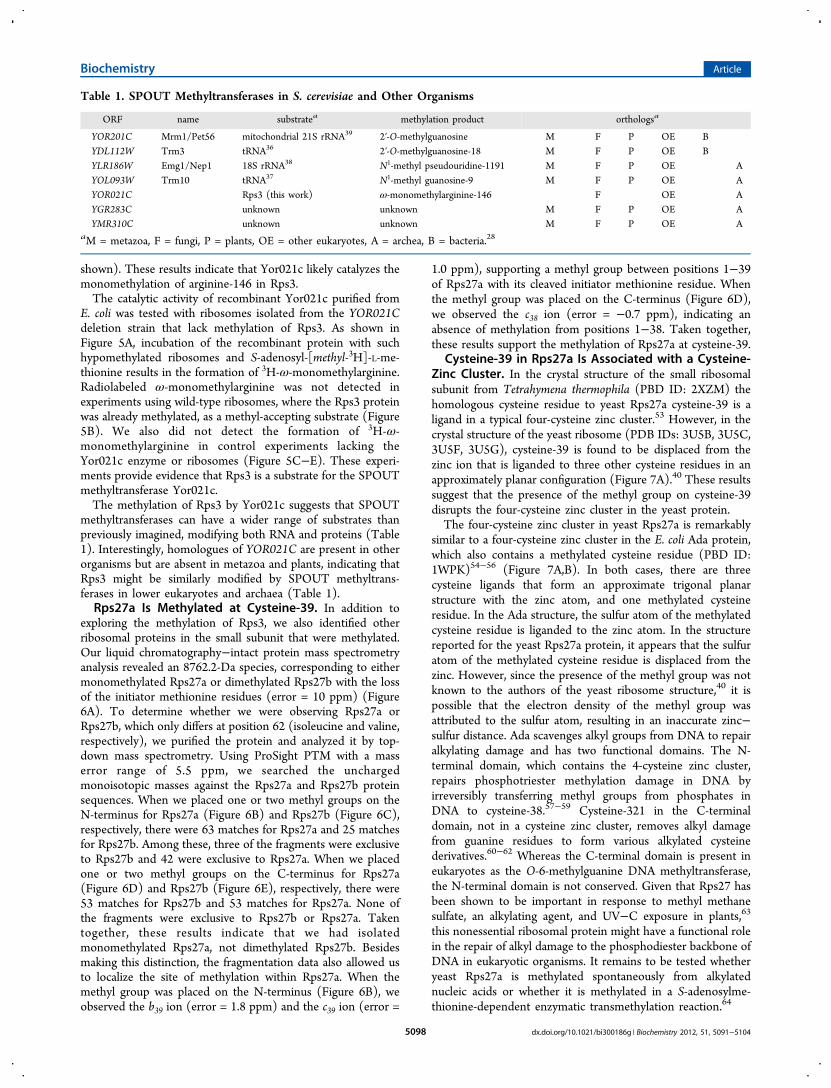
methyltransferases can have a wider range of substrates thanpreviously imagined, modifying both RNA and proteins (Table1). Interestingly, homologues of YOR021C are present in otherorganisms but are absent in metazoa and plants, indicating thatRps3 might be similarly modified by SPOUT methyltrans-ferases in lower eukaryotes and archaea (Table 1).Rps27a Is Methylated at Cysteine-39. In addition to
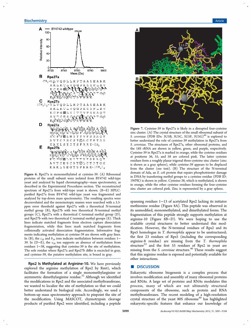
exploring the methylation of Rps3, we also identified otherribosomal proteins in the small subunit that were methylated.Our liquid chromatography−intact protein mass spectrometryanalysis revealed an 8762.2-Da species, corresponding to eithermonomethylated Rps27a or dimethylated Rps27b with the lossof the initiator methionine residues (error = 10 ppm) (Figure6A). To determine whether we were observing Rps27a orRps27b, which only differs at position 62 (isoleucine and valine,respectively), we purified the protein and analyzed it by top-down mass spectrometry. Using ProSight PTM with a masserror range of 5.5 ppm, we searched the unchargedmonoisotopic masses against the Rps27a and Rps27b proteinsequences. When we placed one or two methyl groups on theN-terminus for Rps27a (Figure 6B) and Rps27b (Figure 6C),respectively, there were 63 matches for Rps27a and 25 matchesfor Rps27b. Among these, three of the fragments were exclusiveto Rps27b and 42 were exclusive to Rps27a. When we placedone or two methyl groups on the C-terminus for Rps27a(Figure 6D) and Rps27b (Figure 6E), respectively, there were53 matches for Rps27b and 53 matches for Rps27a. None ofthe fragments were exclusive to Rps27b or Rps27a. Takentogether, these results indicate that we had isolatedmonomethylated Rps27a, not dimethylated Rps27b. Besidesmaking this distinction, the fragmentation data also allowed usto localize the site of methylation within Rps27a. When themethyl group was placed on the N-terminus (Figure 6B), weobserved the b39 ion (error = 1.8 ppm) and the c39 ion (error =
1.0 ppm), supporting a methyl group between positions 1−39of Rps27a with its cleaved initiator methionine residue. Whenthe methyl group was placed on the C-terminus (Figure 6D),we observed the c38 ion (error = −0.7 ppm), indicating anabsence of methylation from positions 1−38. Taken together,these results support the methylation of Rps27a at cysteine-39.
Cysteine-39 in Rps27a Is Associated with a Cysteine-Zinc Cluster. In the crystal structure of the small ribosomalsubunit from Tetrahymena thermophila (PBD ID: 2XZM) thehomologous cysteine residue to yeast Rps27a cysteine-39 is aligand in a typical four-cysteine zinc cluster.53 However, in thecrystal structure of the yeast ribosome (PDB IDs: 3U5B, 3U5C,3U5F, 3U5G), cysteine-39 is found to be displaced from thezinc ion that is liganded to three other cysteine residues in anapproximately planar configuration (Figure 7A).40 These resultssuggest that the presence of the methyl group on cysteine-39disrupts the four-cysteine zinc cluster in the yeast protein.The four-cysteine zinc cluster in yeast Rps27a is remarkably
similar to a four-cysteine zinc cluster in the E. coli Ada protein,which also contains a methylated cysteine residue (PBD ID:1WPK)54−56 (Figure 7A,B). In both cases, there are threecysteine ligands that form an approximate trigonal planarstructure with the zinc atom, and one methylated cysteineresidue. In the Ada structure, the sulfur atom of the methylatedcysteine residue is liganded to the zinc atom. In the structurereported for the yeast Rps27a protein, it appears that the sulfuratom of the methylated cysteine residue is displaced from thezinc. However, since the presence of the methyl group was notknown to the authors of the yeast ribosome structure,40 it ispossible that the electron density of the methyl group wasattributed to the sulfur atom, resulting in an inaccurate zinc−sulfur distance. Ada scavenges alkyl groups from DNA to repairalkylating damage and has two functional domains. The N-terminal domain, which contains the 4-cysteine zinc cluster,repairs phosphotriester methylation damage in DNA byirreversibly transferring methyl groups from phosphates inDNA to cysteine-38.57−59 Cysteine-321 in the C-terminaldomain, not in a cysteine zinc cluster, removes alkyl damagefrom guanine residues to form various alkylated cysteinederivatives.60−62 Whereas the C-terminal domain is present ineukaryotes as the O-6-methylguanine DNA methyltransferase,the N-terminal domain is not conserved. Given that Rps27 hasbeen shown to be important in response to methyl methanesulfate, an alkylating agent, and UV−C exposure in plants,63
this nonessential ribosomal protein might have a functional rolein the repair of alkyl damage to the phosphodiester backbone ofDNA in eukaryotic organisms. It remains to be tested whetheryeast Rps27a is methylated spontaneously from alkylatednucleic acids or whether it is methylated in a S-adenosylme-thionine-dependent enzymatic transmethylation reaction.64
Table 1. SPOUT Methyltransferases in S. cerevisiae and Other Organisms
ORF name substratea methylation product orthologsa
YOR201C Mrm1/Pet56 mitochondrial 21S rRNA39 2′-O-methylguanosine M F P OE BYDL112W Trm3 tRNA36 2′-O-methylguanosine-18 M F P OE BYLR186W Emg1/Nep1 18S rRNA38 N1-methyl pseudouridine-1191 M F P OE AYOL093W Trm10 tRNA37 N1-methyl guanosine-9 M F P OE AYOR021C Rps3 (this work) ω-monomethylarginine-146 F OE AYGR283C unknown unknown M F P OE AYMR310C unknown unknown M F P OE A
aM = metazoa, F = fungi, P = plants, OE = other eukaryotes, A = archea, B = bacteria.28
Biochemistry Article
dx.doi.org/10.1021/bi300186g | Biochemistry 2012, 51, 5091−51045098
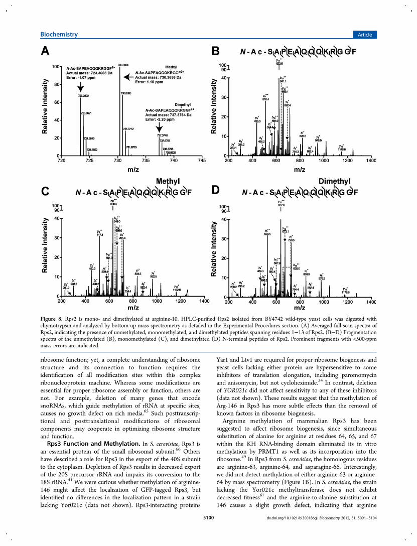
Rps2 Is Methylated at Arginine-10. We have previouslyexplored the arginine methylation of Rps2 by Rmt1, whichfacilitates the formation of a single monomethylarginine orasymmetric dimethylarginine residue.52 Although we identifiedthe modifications in Rps2 and the associated methyltransferase,we wanted to localize the site of methylation so that we couldbetter understand its biological role. Accordingly, we used abottom-up mass spectrometry approach to pinpoint the site ofthe modification. Using MASCOT, chymotrypsin cleavageproducts of purified Rps2 were identified, including a peptide
spanning residues 1−13 of acetylated Rps2 lacking its initiatormethionine residue (Figure 8A). This peptide was observed inits unmodified, monomethylated, and dimethylated forms. Thefragmentation of this peptide strongly supports methylation atarginine-10 (Figure 8B−D). We were hoping to use theavailable crystal structures to better understand this mod-ification. However, the N-terminal residues of Rps2 and itsRps5 homologue in T. thermophila appear to be unstructured;the first 23 residues of Rps5 (including the correspondingarginine-8 residue) are missing from the T. thermophilastructure53 and the first 33 residues of Rps2 in yeast aremissing from the S. cerevisiae structure.40 These results suggestthat this arginine residue is exposed and potentially available forother interactions.
■ DISCUSSIONEukaryotic ribosome biogenesis is a complex process thatinvolves modification and assembly of many ribosomal proteinsand RNAs. A large set of proteins and RNAs modulates thisprocess, many of which are not ultimately structuralcomponents of the ribosome, such as protein and RNAmethyltransferases. The recent modeling of a high-resolutioncrystal structure of the yeast 80S ribosome40 has highlightedeukaryotic-specific features that enhance our knowledge of
Figure 6. Rps27a is monomethylated at cysteine-39. (A) Ribosomalproteins of the small subunit were isolated from BY4742 wild-typeyeast and analyzed by liquid chromatography−mass spectrometry, asdescribed in the Experimental Procedures section. The reconstructedspectrum of Rps27a from wild-type yeast is shown. (B−E) HPLC-purified Rps27a from BY4742 wild-type yeast was fragmented andanalyzed by top-down mass spectrometry. The resulting spectra weredeconvoluted and the monoisotopic masses were searched with a 5.5-ppm error threshold against Rps27a with a theoretical N-terminalmethyl group (B), Rps27b with two theoretical N-terminal methylgroups (C), Rps27a with a theoretical C-terminal methyl group (D),and Rps27b with two theoretical C-terminal methyl groups (E). Thicklines indicate matched fragments from electron capture dissociationfragmentation, while thin lines mark matched fragments fromcollisionally activated dissociation fragmentation. Informative frag-ments indicating methylation at cysteine-39 are shown with gray lines.In (B), the c39 and b39 ions indicate methylation between residues 1−39. In (D−E), the c38 ion supports an absence of methylation fromresidues 1−38, suggesting that cysteine-39 is the site of methylation.The sole residue where Rps27a and Rps27b differ is encircled in gray,and cysteine-39, the putative methylation site, is boxed in gray.
Figure 7. Cysteine-39 in Rps27a is likely in a disrupted four-cysteinezinc cluster. (A) The crystal structure of the small ribosomal subunit ofS. cerevisiae (PDB IDs: 3U5B, 3U5C, 3U5F, 3U5G)40 is explored tobetter understand the role of cysteine-39 methylation in Rps27a fromS. cerevisiae. The structures of Rps27a, other ribosomal proteins, andthe 18S rRNA are shown in yellow, green, and purple, respectively.Cysteine-39 in Rps27a is marked in orange, while the cysteine residuesat positions 36, 55, and 58 are colored pink. The latter cysteineresidues form a roughly planar trigonal three-cysteine zinc cluster (zincis shown as a gray sphere), while cysteine-39 appears to be displacedfrom the cluster (see text). (B) The structure of the N-terminaldomain of Ada, an E. coli protein that repairs phosphotriester damagein DNA by transferring methyl groups to a cysteine residue (PDB ID:1WPK) is shown in yellow. Cysteine-38, which is methylated, is shownin orange, while the other cysteine residues forming the four-cysteinezinc cluster are colored pink. Zinc is represented by a gray sphere.
Biochemistry Article
dx.doi.org/10.1021/bi300186g | Biochemistry 2012, 51, 5091−51045099
ribosome function; yet, a complete understanding of ribosomestructure and its connection to function requires theidentification of all modification sites within this complexribonucleoprotein machine. Whereas some modifications areessential for proper ribosome assembly or function, others arenot. For example, deletion of many genes that encodesnoRNAs, which guide methylation of rRNA at specific sites,causes no growth defect on rich media.65 Such posttranscrip-tional and posttranslational modifications of ribosomalcomponents may cooperate in optimizing ribosome structureand function.Rps3 Function and Methylation. In S. cerevisiae, Rps3 is
an essential protein of the small ribosomal subunit.66 Othershave described a role for Rps3 in the export of the 40S subunitto the cytoplasm. Depletion of Rps3 results in decreased exportof the 20S precursor rRNA and impairs its conversion to the18S rRNA.41 We were curious whether methylation of arginine-146 might affect the localization of GFP-tagged Rps3, butidentified no differences in the localization pattern in a strainlacking Yor021c (data not shown). Rps3-interacting proteins
Yar1 and Ltv1 are required for proper ribosome biogenesis andyeast cells lacking either protein are hypersensitive to someinhibitors of translation elongation, including paromomycinand anisomycin, but not cycloheximide.34 In contrast, deletionof YOR021c did not affect sensitivity to any of these inhibitors(data not shown). These results suggest that the methylation ofArg-146 in Rps3 has more subtle effects than the removal ofknown factors in ribosome biogenesis.Arginine methylation of mammalian Rps3 has been
suggested to affect ribosome biogenesis, since simultaneoussubstitution of alanine for arginine at residues 64, 65, and 67within the KH RNA-binding domain eliminated its in vitromethylation by PRMT1 as well as its incorporation into theribosome.49 In Rps3 from S. cerevisiae, the homologous residuesare arginine-63, arginine-64, and asparagine-66. Interestingly,we did not detect methylation of either arginine-63 or arginine-64 by mass spectrometry (Figure 1B). In S. cerevisiae, the strainlacking the Yor021c methyltransferase does not exhibitdecreased fitness67 and the arginine-to-alanine substitution at146 causes a slight growth defect, indicating that arginine
Figure 8. Rps2 is mono- and dimethylated at arginine-10. HPLC-purified Rps2 isolated from BY4742 wild-type yeast cells was digested withchymotrypsin and analyzed by bottom-up mass spectrometry as detailed in the Experimental Procedures section. (A) Averaged full-scan spectra ofRps2, indicating the presence of unmethylated, monomethylated, and dimethylated peptides spanning residues 1−13 of Rps2. (B−D) Fragmentationspectra of the unmethylated (B), monomethylated (C), and dimethylated (D) N-terminal peptides of Rps2. Prominent fragments with <500-ppmmass errors are indicated.
Biochemistry Article
dx.doi.org/10.1021/bi300186g | Biochemistry 2012, 51, 5091−51045100
methylation of Rps3 might have a distinct function from itsmammalian homologue. In mammals, Rps3 has a wide range ofextraribosomal roles,57 and it is possible that some of thesemight be modulated by arginine methylation in S. cerevisiae. Forexample, Rps3 has been described to have an endonucleaseactivity for abasic DNA in both S. cerevisiae and mammals,47,68
raising the possibility that methylation of arginine-146 in Rps3might regulate this repair process. In addition, the ability ofRps3 to autoregulate the stability of its mRNA43 suggests thatmethylation of arginine-146 might influence its interaction withRPS3 mRNA and mRNA turnover.The wild-type growth of Δyor021c cells and of strains that
expressed Rps3 with an arginine to lysine substitution at residue146 under standard growth conditions, suggests that, ratherthan being crucial for basic ribosome function, methylation maycooperate with other modifications or factors to optimize theinteractions between arginine-146 and the 18S rRNA. Ouranalysis of the high-resolution structure of the yeast ribosome40
indicates that that arginine-146 closely interacts with adenine-1427, which is within 4 Å of cytosine-1274. Interestingly, one ofthe nonessential snoRNAs, snR56, directs 2′-O-methylation atguanosine-1425, two nucleotides 5′ to adenine-1427,65 and theessential 40S ribosomal assembly factor Enp1 binds twonucleotides 3′ to adenine-1427, suggesting the significance ofmolecular interactions in this region. Strains expressing Rps3with an arginine to alanine amino acid substitution at residue146 exhibited a growth defect, indicating the importance of theamino acid identity at this position for optimal cellular growth.These interactions may become important for enhancingribosome function or assembly under certain environmentalconditions or in the absence of other factors or modifications.SPOUT Methyltransferases and Translational Machi-
nery. A striking observation in this work was the dependenceof Rps3 methylation on the Yor021c SPOUT candidatemethyltransferase. The many enzymes that facilitate ribosomebiogenesis include ribonucleases that cleave precursor rRNAs,kinases that modulate ribosome function, and methyltrans-ferases that modify rRNA and ribosomal proteins. Analyses oftranscriptional profiles across multiple growth conditionsdemonstrated that many of these genes are coregulated witheach other and with rRNA and ribosomal protein genes,forming an rRNA and ribosome biosynthesis (RRB) regulon.69
On the basis of multiple genome- and proteome-wide data sets,a protein-function prediction algorithm predicted GO termsrelated to ribosome biogenesis or function for 16 of thepreviously uncharacterized RRB regulon genes.70 Interestingly,five of these 16 genes are predicted to encode methyltrans-ferases: YBR271W, YNL022C, YGR283C, YMR310C, andYOR021C.22 Ybr271w and Ynl022c belong to the large familyof seven β-strand methyltransferase genes, which includes all ofthe identified arginine methyltransferases in the eukaryoticdomain.22,23 The shared seven β-strand family structure allowsmodification of a variety of substrates; enzymes in this familyalso methylate proteins on lysine, histidine, and carboxylgroups, as well as RNA, DNA, and small molecules.22,23
Ygr283c, Ymr310c, and Yor021c, however, are members of theSPOUT methyltransferase family.22,28 Although there is lessprimary sequence similarity among the SPOUT familymembers than within the seven β-strand family, conservedstructural features have allowed identification of SPOUTmethyltransferases in all three domains of life.28
All SPOUT family members analyzed previously methylateRNAs (Table 1). The largest SPOUT cluster of orthologous
groups (COG) includes 2′-O-ribose methyltransferases, repre-sented by Mrm1 and Trm3 in yeast.28 Ygr283c and Ymr310cbelong to a distinct COG, which has no members of knownfunction. The proteins within the COG containing Yor021calso have no known function, although this COG is closelyrelated to the COG containing yeast Trm10, a guanine tRNAmethyltransferase that modifies the N1 site. The association ofYor021c with monomethylation of arginine-146 of Rps3indicates that SPOUT methyltransferases may have a broaderrange of substrates than previously imagined. Indeed, thenitrogen atom at position 1 in the guanine base that ismethylated by Trm10 is structurally similar to the terminalnitrogen atom of the guanidino group of an arginine residue,suggesting the SPOUT enzymes might methylate both guanineand arginine. Such substrate variability is also seen amongmembers of the HemK family of seven-beta strand methyl-transferases, which include enzymes that methylate thechemically similar nitrogen atoms of the 6-amino group ofadenine bases and the side chain amide of glutamine residues.71
Proteins in the Yor021c COG have a C-terminal extensionbeyond the SPOUT domain, in contrast to an N-terminalextension in the Trm10 COG.28 Crystal structures of SPOUTmethyltransferases have revealed the presence of flexible linkersbetween the SPOUT and terminal domains, as well as the rolesof these domains in substrate binding.72,73 The unique C-terminal domain of these Yor021c-related proteins maytherefore guide methylation of the non-RNA substrate Rps3.In light of our direct demonstration of the methyltransferaseactivity of Yor021c on Rps3, we suggest the gene designationSFM1 (SPOUT family methyltransferase 1) for the S. cerevisiaeYOR021C open reading frame.
Rps27a and Rps2 Methylation. Our studies also led tothe identification of a methylcysteine residue at position 39 inRps27a. Rps27a is a nonessential ribosomal protein and hasbeen suggested to be important for the synthesis of the 18SrRNA.74 Our analysis of the crystal structure of Rps27a in theyeast small ribosomal subunit suggests that a four cysteine zinccluster is present that may be similar to the one found in the N-terminal domain of the E. coli Ada protein that allows the repairof DNA phosphotriester damage by accepting methyl groups.54
Eukaryotes lack the N-terminal region of Ada, and we wonderwhether Rps27a might function in repairing these deleteriousDNA lesions57 in eukaryotes because its absence causesincreased sensitivity to UV light and alkylating agents in A.thaliana.63 Such a role, however, would require a population ofunmethylated Rps27a. We have not observed such a species inribosomes, although it might be present in the nucleus orcytosol.In addition, we have identified the site of arginine
methylation in Rps2 by bottom-up mass spectrometry. Theshort arginine-glycine-rich region of S. cerevisiae Rps2 is variablymono- or asymmetrically dimethylated by Rmt1 on arginine-10within an RGGF sequence, a common substrate recognitionmotif for Rmt1-catalyzed methylation in S. cerevisiae.75 Incontrast, S. pombe Rps2 is asymmetrically dimethylated in vivoon eight arginines in its RG-rich N-terminus by PRMT3.76 Invitro methylation of mammalian Rps2 by PRMT3 also occursprimarily on a stretch of eight RG dipeptides downstream oftwo RGGF peptides.77 Similar to our results with S. cerevisiaeRps3, S. pombe Rps2 with arginine-to-lysine substitutions canreplace wild-type Rps2 whereas alanine substitutions decreaseRps2 function,76 indicating the greater importance of aminoacid identity rather than methylation. Our previous mass
Biochemistry Article
dx.doi.org/10.1021/bi300186g | Biochemistry 2012, 51, 5091−51045101
spectrometry analysis of intact S. cerevisiae Rps2 revealed thepresence of only one or two methyl groups per molecule,suggesting that the two downstream RG peptides are notmethylated in this organism that lacks a PRMT3 homologue.78
The physiological role of arginine-10 and the functionalsignificance of RG region length in Rps2 remain to bedetermined; to date, the N-terminus of Rps2 has not beenresolved in eukaryotic ribosome structures, suggesting itsflexibility and the potential for interactions with other factorsthat might influence ribosome function.In this paper, we enhance our understanding of methylation
of proteins in the small ribosomal subunit in S. cerevisiae. Weidentified the sites of methylation modifications in Rps3,Rps27a, and Rps2 by a variety of analytical methods. Ouranalysis of available crystal structures has enabled us to suggestfunctional roles for these modifications, including methylatedarginine-146 in Rps3 as a mediator of 18S rRNA interactionsand cysteine-39 in Rps27a as a potential acceptor for alkylatingdamage of DNA. Finally, we have demonstrated that theYor021c SPOUT methyltransferase is a protein argininemethyltransferase that methylates Rps3. To date, SPOUTmethyltransferases have been shown to only modify RNA andour observations indicate that these enzymes are also capable ofmethylating ribosomal proteins. Our findings, which comple-ment other efforts to identify sites of ribosomal proteinmodification and the enzymes that catalyze these modifications,are crucial for interpreting new high-resolution ribosome crystalstructures and will help build our understanding of ribosomalstructure and function.
■ ASSOCIATED CONTENT
*S Supporting InformationA list of oligonucleotides used to generate and verify Rps3mutants is provided as Supplemental Table S1. This material isavailable free of charge via the Internet at http://pubs.acs.org.
■ AUTHOR INFORMATION
Corresponding Author*(S.G.C.) Address: Department of Chemistry and Biochemis-try and the Molecular Biology Institute, UCLA, 607 Charles E.Young Drive East, Los Angeles, California 90095, UnitedStates. Tel: 310 825-8754. Fax: 310 825-1968. E-mail: [email protected]. (A.E.M.) Address: Department of Biology,Bowdoin College, 6500 College Station, Brunswick, Maine04011, United States. Tel: 207 798-7109. Fax: 207 725-3405. E-mail: [email protected].
FundingThis work was supported by National Institutes of HealthGrant GM026020 to S.G.C. and by a Senior Scholar in AgingAward from the Ellison Medical Foundation to S.G.C.
NotesThe authors declare no competing financial interest.
■ ACKNOWLEDGMENTS
We thank our colleagues in the Molecular InstrumentationCenter and Pasarow Mass Spectrometry Laboratory at UCLA,especially Julian Whitelegge, Kym Faull, Pete Souda, and GreggCzerwieniec, for their expertise and guidance with our massspectrometry studies. We also thank Philipp Milkereit for RPS3strains and plasmids.
■ REFERENCES(1) Li, E., Beard, C., and Jaenisch, R. (1993) Role for DNAmethylation in genomic imprinting. Nature 366, 362−365.(2) Bonnerot, C., Pintard, L., and Lutfalla, G. (2003) Functionalredundancy of Spb1p and a snR52-dependent mechanism for the 2′-O-ribose methylation of a conserved rRNA position in yeast.Mol. Cell 12,1309−1315.(3) Walsh, C. T. (2006) Posttranslational Modification of Proteins:Expanding Nature’s Inventory, Roberts and Company Publishers.(4) Ortega-Gutierrez, S., Leung, D., Ficarro, S., Peters, E. C., andCravatt, B. F. (2008) Targeted disruption of the PME-1 gene causesloss of demethylated PP2A and perinatal lethality in mice. PLoS One 3,e2486.(5) Olsen, J. V., Blagoev, B., Gnad, F., Macek, B., Kumar, C.,Mortensen, P., and Mann, M. (2006) Global, in vivo, and site-specificphosphorylation dynamics in signaling networks. Cell 127, 635−648.(6) Thorsness, P. E., and Koshland, D. E., Jr. (1987) Inactivation ofisocitrate dehydrogenase by phosphorylation is mediated by thenegative charge of the phosphate. J. Biol. Chem. 262, 10422−10425.(7) Breitschopf, K., Haendeler, J., Malchow, P., Zeiher, A. M., andDimmeler, S. (2000) Posttranslational modification of Bcl-2 facilitatesits proteasome-dependent degradation: molecular characterization ofthe involved signaling pathway. Mol. Cell. Biol. 20, 1886−1896.(8) Bergo, M. O., Leung, G. K., Ambroziak, P., Otto, J. C., Casey, P.J., and Young, S. G. (2000) Targeted inactivation of theisoprenylcysteine carboxyl methyltransferase gene causes mislocaliza-tion of K-Ras in mammalian cells. J. Biol. Chem. 275, 17605−17610.(9) Clarke, S. G., Tamanoi, F. (2006) In The Enzymes. Proteinmethyltransferases, Academic Press, Amsterdam, Vol 24.(10) Del Rizzo, P. A., and Trievel, R. C. (2011) Substrate andproduct specificities of SET domain methyltransferases. Epigenetics 6,1059−1067.(11) Bedford, M. T., and Clarke, S. G. (2009) Protein argininemethylation in mammals: who, what, and why. Mol. Cell 33, 1−13.(12) Bachand, F. (2007) Protein arginine methyltransferases: fromunicellular eukaryotes to humans. Eukaryot. Cell 6, 889−898.(13) Webb, K. J., Zurita-Lopez, C. I., Al-Hadid, Q., Laganowsky, A.,Young, B. D., Lipson, R. S., Souda, P., Faull, K. F., Whitelegge, J. P.,and Clarke, S. G. (2010) A novel 3-methylhistidine modification ofyeast ribosomal protein Rpl3 is dependent upon the YIL110Wmethyltransferase. J. Biol. Chem. 285, 37598−37606.(14) Muppirala, U. K., Desensi, S., Lybrand, T. P., Hazelbauer, G. L.,and Li, Z. (2009) Molecular modeling of flexible arm-mediatedinteractions between bacterial chemoreceptors and their modificationenzyme. Protein Sci. 18, 1702−1714.(15) Heurgue-Hamard, V., Champ, S., Mora, L., Merkulova-Rainon,T., Kisselev, L. L., and Buckingham, R. H. (2005) The glutamineresidue of the conserved GGQ motif in Saccharomyces cerevisiae releasefactor eRF1 is methylated by the product of the YDR140w gene. J.Biol. Chem. 280, 2439−2445.(16) Anderson, J. L., Henriksen, B. S., Gibbs, R. A., and Hrycyna, C.A. (2005) The isoprenoid substrate specificity of isoprenylcysteinecarboxylmethyltransferase: development of novel inhibitors. J. Biol.Chem. 280, 29454−29461.(17) Stanevich, V., Jiang, L., Satyshur, K. A., Li, Y., Jeffrey, P. D., Li,Z., Menden, P., Semmelhack, M. F., and Xing, Y. (2011) Thestructural basis for tight control of PP2A methylation and function byLCMT-1. Mol. Cell 41, 331−342.(18) Webb, K. J., Lipson, R. S., Al-Hadid, Q., Whitelegge, J. P., andClarke, S. G. (2010) Identification of protein N-terminal methyl-transferases in yeast and humans. Biochemistry 49, 5225−5235.(19) Wang, H., Huang, Z. Q., Xia, L., Feng, Q., Erdjument-Bromage,H., Strahl, B. D., Briggs, S. D., Allis, C. D., Wong, J., Tempst, P., andZhang, Y. (2001) Methylation of histone H4 at arginine 3 facilitatingtranscriptional activation by nuclear hormone receptor. Science 293,853−857.(20) Hrycyna, C. A., Sapperstein, S. K., Clarke, S., and Michaelis, S.(1991) The Saccharomyces cerevisiae STE14 gene encodes a
Biochemistry Article
dx.doi.org/10.1021/bi300186g | Biochemistry 2012, 51, 5091−51045102
methyltransferase that mediates C-terminal methylation of a-factor andRAS proteins. EMBO J. 10, 1699−1709.(21) Polevoda, B., and Sherman, F. (2007) Methylation of proteinsinvolved in translation. Mol. Microbiol. 65, 590−606.(22) Petrossian, T., and Clarke, S. (2009) Bioinformatic identificationof novel methyltransferases. Epigenomics 1, 163−175.(23) Wlodarski, T., Kutner, J., Towpik, J., Knizewski, L., Rychlewski,L., Kudlicki, A., Rowicka, M., Dziembowski, A., and Ginalski, K.(2011) Comprehensive structural and substrate specificity classifica-tion of the Saccharomyces cerevisiae methyltransferome. PLoS One 6,e23168.(24) Petrossian, T. C., and Clarke, S. G. (2009) Multiple motifscanning to identify methyltransferases from the yeast proteome. Mol.Cell. Proteomics 8, 1516−1526.(25) Trievel, R. C., Beach, B. M., Dirk, L. M., Houtz, R. L., andHurley, J. H. (2002) Structure and catalytic mechanism of a SETdomain protein methyltransferase. Cell 111, 91−103.(26) Rea, S., Eisenhaber, F., O’Carroll, D., Strahl, B. D., Sun, Z. W.,Schmid, M., Opravil, S., Mechtler, K., Ponting, C. P., Allis, C. D., andJenuwein, T. (2000) Regulation of chromatin structure by site-specifichistone H3 methyltransferases. Nature 406, 593−599.(27) Anantharaman, V., Koonin, E. V., and Aravind, L. (2002)SPOUT: a class of methyltransferases that includes spoU and trmDRNA methylase superfamilies, and novel superfamilies of predictedprokaryotic RNA methylases. J. Mol. Microbiol. Biotechnol. 4, 71−75.(28) Tkaczuk, K. L., Dunin-Horkawicz, S., Purta, E., and Bujnicki, J.M. (2007) Structural and evolutionary bioinformatics of the SPOUTsuperfamily of methyltransferases. BMC Bioinformatics 8, 73.(29) Webb, K. J., Al-Hadid, Q., Zurita-Lopez, C. I., Young, B. D.,Lipson, R. S., and Clarke, S. G. (2011) The ribosomal L1 protuberancein yeast is methylated on a lysine residue catalyzed by a seven-beta-strand methyltransferase. J. Biol. Chem. 286, 18405−18413.(30) Porras-Yakushi, T. R., Whitelegge, J. P., Miranda, T. B., andClarke, S. (2005) A novel SET domain methyltransferase modifiesribosomal protein Rpl23ab in yeast. J. Biol. Chem. 280, 34590−34598.(31) Webb, K. J., Laganowsky, A., Whitelegge, J. P., and Clarke, S. G.(2008) Identification of two SET domain proteins required formethylation of lysine residues in yeast ribosomal protein Rpl42ab. J.Biol. Chem. 283, 35561−35568.(32) Chern, M. K., Chang, K. N., Liu, L. F., Tam, T. C., Liu, Y. C.,Liang, Y. L., and Tam, M. F. (2002) Yeast ribosomal protein L12 is asubstrate of protein-arginine methyltransferase 2. J. Biol. Chem. 277,15345−15353.(33) Lipson, R. S., Webb, K. J., and Clarke, S. G. (2010) Two novelmethyltransferases acting upon eukaryotic elongation factor 1A inSaccharomyces cerevisiae. Arch. Biochem. Biophys. 500, 137−143.(34) Seiser, R. M., Sundberg, A. E., Wollam, B. J., Zobel-Thropp, P.,Baldwin, K., Spector, M. D., and Lycan, D. E. (2006) Ltv1 is requiredfor efficient nuclear export of the ribosomal small subunit inSaccharomyces cerevisiae. Genetics 174, 679−691.(35) Loar, J. W., Seiser, R. M., Sundberg, A. E., Sagerson, H. J., Ilias,N., Zobel-Thropp, P., Craig, E. A., and Lycan, D. E. (2004) Geneticand biochemical interactions among Yar1, Ltv1 and Rps3 define novellinks between environmental stress and ribosome biogenesis inSaccharomyces cerevisiae. Genetics 168, 1877−1889.(36) Cavaille, J., Chetouani, F., and Bachellerie, J. P. (1999) Theyeast Saccharomyces cerevisiae YDL112w ORF encodes the putative 2′-O-ribose methyltransferase catalyzing the formation of Gm18 intRNAs. RNA 5, 66−81.(37) Jackman, J. E., Montange, R. K., Malik, H. S., and Phizicky, E. M.(2003) Identification of the yeast gene encoding the tRNA m1Gmethyltransferase responsible for modification at position 9. RNA 9,574−585.(38) Meyer, B., Wurm, J. P., Kotter, P., Leisegang, M. S., Schilling, V.,Buchhaupt, M., Held, M., Bahr, U., Karas, M., Heckel, A., Bohnsack,M. T., Wohnert, J., and Entian, K. D. (2011) The Bowen-Conradisyndrome protein Nep1 (Emg1) has a dual role in eukaryoticribosome biogenesis, as an essential assembly factor and in the
methylation of Psi1191 in yeast 18S rRNA. Nucleic Acids Res. 39,1526−1537.(39) Sirum-Connolly, K., and Mason, T. L. (1993) Functionalrequirement of a site-specific ribose methylation in ribosomal RNA.Science 262, 1886−1889.(40) Ben-Shem, A., Garreau de Loubresse, N., Melnikov, S., Jenner,L., Yusupova, G., and Yusupov, M. (2011) The structure of theeukaryotic ribosome at 3.0 Å resolution. Science 334, 1524−1529.(41) Ferreira-Cerca, S., Poll, G., Gleizes, P. E., Tschochner, H., andMilkereit, P. (2005) Roles of eukaryotic ribosomal proteins inmaturation and transport of pre-18S rRNA and ribosome function.Mol. Cell 20, 263−275.(42) Zhang, Z., and Marshall, A. G. (1998) A universal algorithm forfast and automated charge state deconvolution of electrospray mass-to-charge ratio spectra. J. Am. Soc. Mass Spectrom. 9, 225−233.(43) Hendrick, J. L., Wilson, P. G., Edelman, II, Sandbaken, M. G.,Ursic, D., and Culbertson, M. R. (2001) Yeast frameshift suppressormutations in the genes coding for transcription factor Mbf1p andribosomal protein S3: evidence for autoregulation of S3 synthesis.Genetics 157, 1141−1158.(44) Chiu, W. L., Wagner, S., Herrmannova, A., Burela, L., Zhang, F.,Saini, A. K., Valasek, L., and Hinnebusch, A. G. (2010) The C-terminalregion of eukaryotic translation initiation factor 3a (eIF3a) promotesmRNA recruitment, scanning, and, together with eIF3j and the eIF3bRNA recognition motif, selection of AUG start codons. Mol. Cell. Biol.30, 4415−4434.(45) Cuchalova, L., Kouba, T., Herrmannova, A., Danyi, I., Chiu, W.L., and Valasek, L. (2010) The RNA recognition motif of eukaryotictranslation initiation factor 3g (eIF3g) is required for resumption ofscanning of posttermination ribosomes for reinitiation on GCN4 andtogether with eIF3i stimulates linear scanning. Mol. Cell. Biol. 30,4671−4686.(46) Passmore, L. A., Schmeing, T. M., Maag, D., Applefield, D. J.,Acker, M. G., Algire, M. A., Lorsch, J. R., and Ramakrishnan, V. (2007)The eukaryotic translation initiation factors eIF1 and eIF1A induce anopen conformation of the 40S ribosome. Mol. Cell 26, 41−50.(47) Jung, S. O., Lee, J. Y., and Kim, J. (2001) Yeast ribosomalprotein S3 has an endonuclease activity on AP DNA. Mol. Cells 12,84−90.(48) Lhoest, J., Lobet, Y., Costers, E., and Colson, C. (1984)Methylated proteins and amino acids in the ribosomes ofSaccharomyces cerevisiae. Eur. J. Biochem. 141, 585−590.(49) Shin, H. S., Jang, C. Y., Kim, H. D., Kim, T. S., Kim, S., and Kim,J. (2009) Arginine methylation of ribosomal protein S3 affectsribosome assembly. Biochem. Biophys. Res. Commun. 385, 273−278.(50) Takakura, H., Tsunasawa, S., Miyagi, M., and Warner, J. R.(1992) NH2-terminal acetylation of ribosomal proteins of Saccha-romyces cerevisiae. J. Biol. Chem. 267, 5442−5445.(51) Arnold, R. J., Polevoda, B., Reilly, J. P., and Sherman, F. (1999)The action of N-terminal acetyltransferases on yeast ribosomalproteins. J. Biol. Chem. 274, 37035−37040.(52) Lipson, R. S., Webb, K. J., and Clarke, S. G. (2010) Rmt1catalyzes zinc-finger independent arginine methylation of ribosomalprotein Rps2 in Saccharomyces cerevisiae. Biochem. Biophys. Res.Commun. 391, 1658−1662.(53) Rabl, J., Leibundgut, M., Ataide, S. F., Haag, A., and Ban, N.(2011) Crystal structure of the eukaryotic 40S ribosomal subunit incomplex with initiation factor 1. Science 331, 730−736.(54) Takinowaki, H., Matsuda, Y., Yoshida, T., Kobayashi, Y., andOhkubo, T. (2006) The solution structure of the methylated form ofthe N-terminal 16-kDa domain of Escherichia coli Ada protein. ProteinSci. 15, 487−497.(55) He, C., Hus, J. C., Sun, L. J., Zhou, P., Norman, D. P., Dotsch,V., Wei, H., Gross, J. D., Lane, W. S., Wagner, G., and Verdine, G. L.(2005) A methylation-dependent electrostatic switch controls DNArepair and transcriptional activation by E. coli Ada. Mol. Cell 20, 117−129.(56) Myers, L. C., Terranova, M. P., Nash, H. M., Markus, M. A., andVerdine, G. L. (1992) Zinc binding by the methylation signaling
Biochemistry Article
dx.doi.org/10.1021/bi300186g | Biochemistry 2012, 51, 5091−51045103
domain of the Escherichia coli Ada protein. Biochemistry 31, 4541−4547.(57) Jones, G. D., Le Pla, R. C., and Farmer, P. B. (2010)Phosphotriester adducts (PTEs): DNA’s overlooked lesion. Muta-genesis 25, 3−16.(58) McCarthy, T. V., and Lindahl, T. (1985) Methyl phospho-triesters in alkylated DNA are repaired by the Ada regulatory proteinof E. coli. Nucleic Acids Res. 13, 2683−2698.(59) Teo, I., Sedgwick, B., Demple, B., Li, B., and Lindahl, T. (1984)Induction of resistance to alkylating agents in E. coli: the Ada+ geneproduct serves both as a regulatory protein and as an enzyme for repairof mutagenic damage. EMBO J. 3, 2151−2157.(60) Nakabeppu, Y., Kondo, H., Kawabata, S., Iwanaga, S., andSekiguchi, M. (1985) Purification and structure of the intact Adaregulatory protein of Escherichia coli K12, O6-methylguanine-DNAmethyltransferase. J. Biol. Chem. 260, 7281−7288.(61) Graves, R. J., Li, B. F., and Swann, P. F. (1989) Repair of O6-methylguanine, O6-ethylguanine, O6-isopropylguanine and O4-methylthymine in synthetic oligodeoxynucleotides by Escherichia coliAda gene O6-alkylguanine-DNA-alkyltransferase. Carcinogenesis 10,661−666.(62) Moore, M. H., Gulbis, J. M., Dodson, E. J., Demple, B., andMoody, P. C. (1994) Crystal structure of a suicidal DNA repairprotein: the Ada O6-methylguanine-DNA methyltransferase from E.coli. EMBO J. 13, 1495−1501.(63) Revenkova, E., Masson, J., Koncz, C., Afsar, K., Jakovleva, L.,and Paszkowski, J. (1999) Involvement of Arabidopsis thalianaribosomal protein S27 in mRNA degradation triggered by genotoxicstress. EMBO J. 18, 490−499.(64) Selmer, T., Kahnt, J., Goubeaud, M., Shima, S., Grabarse, W.,Ermler, U., and Thauer, R. K. (2000) The biosynthesis of methylatedamino acids in the active site region of methyl-coenzyme M reductase.J. Biol. Chem. 275, 3755−3760.(65) Lowe, T. M., and Eddy, S. R. (1999) A computational screen formethylation guide snoRNAs in yeast. Science 283, 1168−1171.(66) Fingen-Eigen, M., Domdey, H., and Kohrer, K. (1996) Theribosomal protein gene RPS3 is an essential single copy gene of theyeast Saccharomyces cerevisiae. Biochem. Biophys. Res. Commun. 223,397−403.(67) Deutschbauer, A. M., Jaramillo, D. F., Proctor, M., Kumm, J.,Hillenmeyer, M. E., Davis, R. W., Nislow, C., and Giaever, G. (2005)Mechanisms of haploinsufficiency revealed by genome-wide profilingin yeast. Genetics 169, 1915−1925.(68) Kim, J., Chubatsu, L. S., Admon, A., Stahl, J., Fellous, R., andLinn, S. (1995) Implication of mammalian ribosomal protein S3 in theprocessing of DNA damage. J. Biol. Chem. 270, 13620−13629.(69) Wade, C. H., Umbarger, M. A., and McAlear, M. A. (2006) Thebudding yeast rRNA and ribosome biosynthesis (RRB) reguloncontains over 200 genes. Yeast 23, 293−306.(70) Nariai, N., Kolaczyk, E. D., and Kasif, S. (2007) Probabilisticprotein function prediction from heterogeneous genome-wide data.PLoS One 2, e337.(71) Clarke, S. (2002) The methylator meets the terminator. Proc.Natl. Acad. Sci. U. S. A. 99, 1104−1106.(72) Elkins, P. A., Watts, J. M., Zalacain, M., van Thiel, A., Vitazka, P.R., Redlak, M., Andraos-Selim, C., Rastinejad, F., and Holmes, W. M.(2003) Insights into catalysis by a knotted TrmD tRNA methyl-transferase. J. Mol. Biol. 333, 931−949.(73) Pleshe, E., Truesdell, J., and Batey, R. T. (2005) Structure of aclass II TrmH tRNA-modifying enzyme from Aquifex aeolicus. ActaCrystallogr. Sect. F: Struct. Biol. Cryst. Commun. 61, 722−728.(74) Baudin-Baillieu, A., Tollervey, D., Cullin, C., and Lacroute, F.(1997) Functional analysis of Rrp7p, an essential yeast proteininvolved in pre-rRNA processing and ribosome assembly. Mol. Cell.Biol. 17, 5023−5032.(75) McBride, A. E., Cook, J. T., Stemmler, E. A., Rutledge, K. L.,McGrath, K. A., and Rubens, J. A. (2005) Arginine methylation ofyeast mRNA-binding protein Npl3 directly affects its function, nuclear
export, and intranuclear protein interactions. J. Biol. Chem. 280,30888−30898.(76) Perreault, A., Gascon, S., D’Amours, A., Aletta, J. M., andBachand, F. (2009) A methyltransferase-independent function forRmt3 in ribosomal subunit homeostasis. J. Biol. Chem. 284, 15026−15037.(77) Swiercz, R., Person, M. D., and Bedford, M. T. (2005)Ribosomal protein S2 is a substrate for mammalian PRMT3 (proteinarginine methyltransferase 3). Biochem. J. 386, 85−91.(78) Lipson, R. S., Webb, K. J., and Clarke, S. G. (2010) Rmt1catalyzes zinc-finger independent arginine methylation of ribosomalprotein Rps2 in Saccharomyces cerevisiae. Biochem. Biophys. Res.Commun. 391, 1658−1662.(79) Fisk, J. C., Zurita-Lopez, C., Sayegh, J., Tomasello, D. L., Clarke,S. G., and Read, L. K. (2010) TbPRMT6 is a type I protein argininemethyltransferase that contributes to cytokinesis in Trypanosomabrucei. Eukaryot. Cell 9, 866−877.
Biochemistry Article
dx.doi.org/10.1021/bi300186g | Biochemistry 2012, 51, 5091−51045104