Embed Size (px)
Citation preview
Identification of Metabolic Routes and Catabolic Enzymes Involved in Phytoremediation of the Nitro-
Substituted Explosives TNT, RDX, and HMX Final Technical Report
SERDP Project Number CU1317
Prof. Jerald L. Schnoor
Benoit Van Aken Laura B. Brentner
Sachiyo Tanaka Brittany Flokstra Jong Moon Yoon
Civil and Environmental Engineering
The University of Iowa Iowa City, IA
July 31, 2006
Approved for public release; distribution is unlimited

This report was prepared under contract to the Department of Defense Strategic Environmental Research and Development Program (SERDP). The publication of this report does not indicate endorsement by the Department of Defense, nor should the contents be construed as reflecting the official policy or position of the Department of Defense. Reference herein to any specific commercial product, process, or service by trade name, trademark, manufacturer, or otherwise, does not necessarily constitute or imply its endorsement, recommendation, or favoring by the Department of Defense.
SERDP Final Technical Report 02 CU13-17
ii
Table of Contents
List of Figures and Tables.................................................................................................. iv Acknowlegements............................................................................................................. vii I. Executive Summary......................................................................................................... 1 II.Objectives........................................................................................................................ 4 III. Background ................................................................................................................... 4 IV. Materials and Methods ................................................................................................. 7
Chemicals.................................................................................................................... 7 Task 1. Degradation experimenents using cuttings, cell/tissue cultures........................ 7
Hybrid Poplar Trees.................................................................................................... 7 Plant Tissues Cultures................................................................................................. 7 Degradation of RDX by Tissue Cultures .................................................................... 8 Degradation of 14CH2Oand 14CH3OH by Tissue Cultures...................................... 9 Leaching and Extraction ............................................................................................. 9
Task 2. Metabolite Identification.................................................................................... 9 Analyses...................................................................................................................... 9 RDX Metabolite Analysis with LC/MS.................................................................... 10
Task 3. Degradation Experiments using Crude Extracts .............................................. 11 Plant Leaf Crude Extracts ......................................................................................... 11 Plant Root crude extracts .......................................................................................... 11 Degradation of RDX by Crude Extracts ................................................................... 11
Task 4. Enzyme Activities and Purification ................................................................ 12 DNA and RNA extractions ....................................................................................... 12 Reverse-transcriptase (RT) real-time PCR ............................................................... 12 Primer design and sequence analysis........................................................................ 13 Data analysis ............................................................................................................. 14 PCR and sequencing ................................................................................................. 14 Primer design and sequence analysis of GSTs in Poplar Plantlets ........................... 15 Plant crude extract GST-enzymatic activity assay.................................................... 15
Task 5. Toxicity Tests................................................................................................... 16 Exposure of poplar plantlets to TNT and RDX ........................................................ 16 Microtox set-up......................................................................................................... 16 Microtox analysis...................................................................................................... 17
Task 6. Degradation Experiments using Purified Enzymes.......................................... 17 In vitro conjugation of TNT using purified GST...................................................... 17
V. Results and Accomplishments ..................................................................................... 18 Degradation of RDX by Poplar Tissue Cultures (Task 1) ............................................ 18 Metabolism of Formaldehyde and Methanol by TissueCultures (Task 1) ................... 19 Synthesis of 14C-U-ring labeled HMX (Task 2) ........................................................... 19 Degradation of RDX by Poplar Crude Extracts (Task 3) ............................................. 20 Identification of Polar Metabolites in RDX Transformation (Task 2) ......................... 20 Metabolism of Formaldehyde and Methanol by Tissue Cultures (Task 1, 2) .............. 20 TNT Metabolites in poplar plantlet hydroponic solution (Task 1, 2) ........................... 27 Exposure of poplar plantlets to TNT (Task 5) .............................................................. 28 Plant exposure to RDX (Task 5)................................................................................... 28
SERDP Final Technical Report 02 CU13-17
iii
Microtox and Plant Tissue Culture (in vitro) Results for TNT (Task 5) ...................... 29 Microtox and Plant Tissue Culture (in vitro) Results for RDX (Task 5)...................... 33 Microtox and Plant Tissue Culture (in vitro) Results for HMX (Task 5)..................... 35 GST sequences from P. trichocarpa for Gene Expression studies (Task 4) ................ 35 Analyses of GST expression in Poplar Plantlets Exposed to TNT (Task 4) ................ 37 Enzymatic Assay of GST Activity in Poplar Plantlets Exposed to TNT (Task 4) ....... 39 In vitro conjugation of TNT by purified GST .............................................................. 40 Gene Expression Analysis in RDX-Exposed Poplar Plantlets (Task 4) ....................... 41
VI. Conclusions................................................................................................................. 50 References......................................................................................................................... 54 Appendices........................................................................................................................ 59
A. List of Explosive Contaminated Military Sites whose Climate is Supportive of the Growth of Poplars ......................................................................................................... 59 B. List of Technical Publications.................................................................................. 59
Journal Articles ......................................................................................................... 59 Conferences/Symposiums/Abstracts......................................................................... 60
Acronyms: CYP (cytochrome P450 monooxygenases); DNX (hexahydro-1,3-dinitroso-5-nitro-1,3,5- triazine); GST (glutathione-S-transferase); HMX (octahydro-1,3,5,7-tetranitro-1,3,5,7-tetrazocine); MNX (hexahydro-1-nitroso-3,5-dinitro-1,3,5- triazine); PTC (plant tissue culture); RDX (1,3,5-trinitro-1,3,5-triazine); TNT (2,4,6-trinitrotoluene)
SERDP Final Technical Report 02 CU13-17
iv
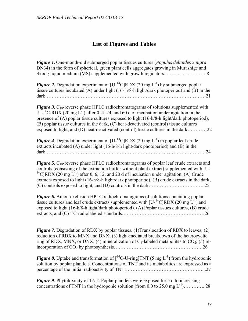
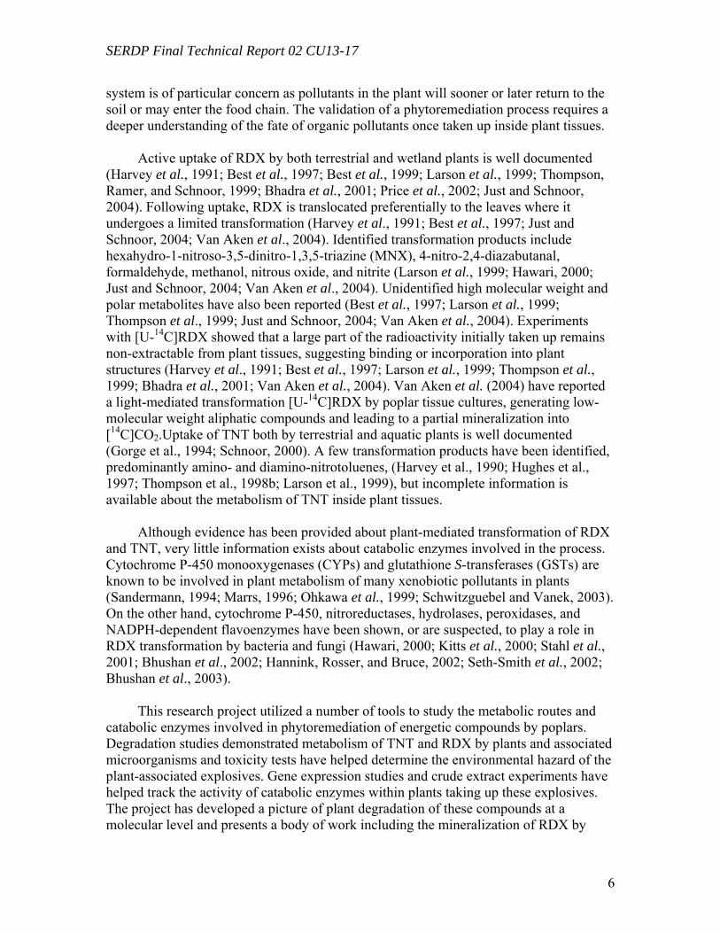
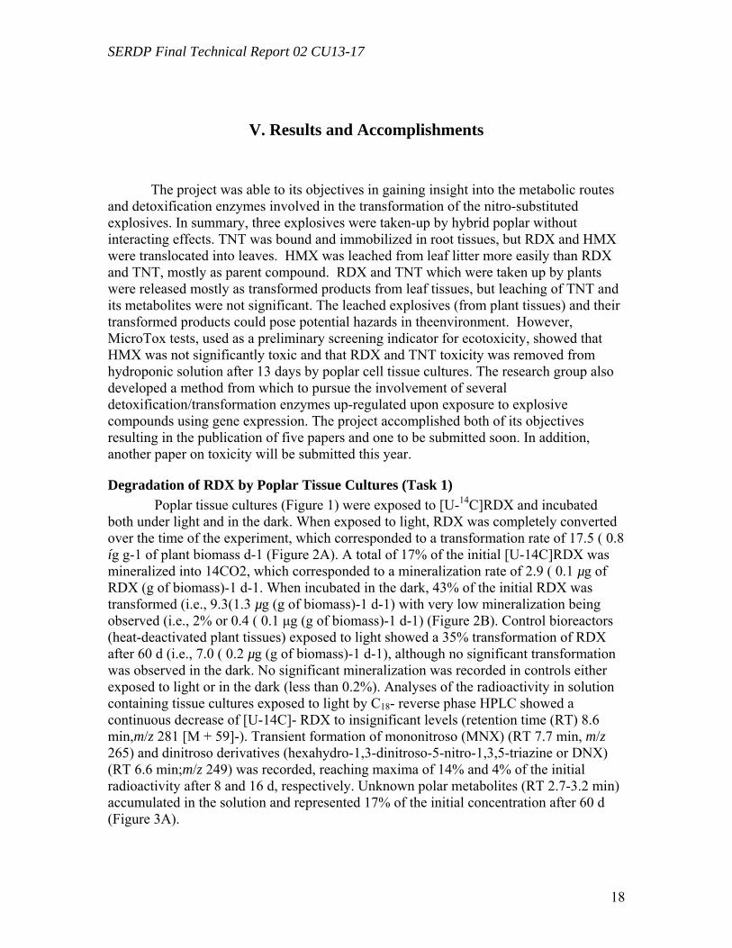
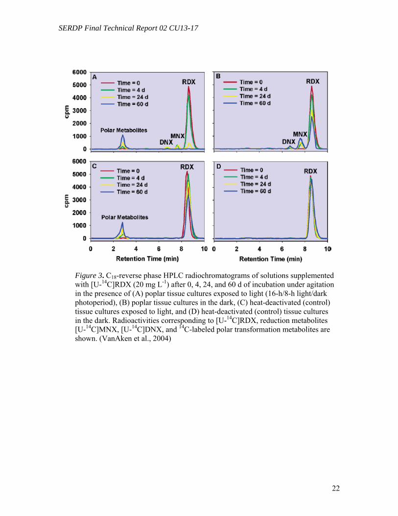
List of Figures and Tables Figure 1. One-month-old submerged poplar tissues cultures (Populus deltoides x nigra DN34) in the form of spherical, green plant cells aggregates growing in Murashige and Skoog liquid medium (MS) supplemented with growth regulators. …………………….8 Figure 2. Degradation experiment of [U-14C]RDX (20 mg L-1) by submerged poplar tissue cultures incubated (A) under light (16- h/8-h light/dark photoperiod) and (B) in the dark………………………………………………………………………………………21 Figure 3. C18-reverse phase HPLC radiochromatograms of solutions supplemented with [U-14C]RDX (20 mg L-1) after 0, 4, 24, and 60 d of incubation under agitation in the presence of (A) poplar tissue cultures exposed to light (16-h/8-h light/dark photoperiod), (B) poplar tissue cultures in the dark, (C) heat-deactivated (control) tissue cultures exposed to light, and (D) heat-deactivated (control) tissue cultures in the dark…………22 Figure 4. Degradation experiment of [U-14C]RDX (20 mg L-1) in poplar leaf crude extracts incubated (A) under light (16-h/8-h light/dark photoperiod) and (B) in the dark………………………………………………………………………………………24 Figure 5. C18-reverse phase HPLC radiochromatograms of poplar leaf crude extracts and controls (consisting of the extraction buffer without plant extract) supplemented with [U-14C]RDX (20 mg L-1) after 0, 6, 12, and 20 d of incubation under agitation. (A) Crude extracts exposed to light (16-h/8-h light/dark photoperiod), (B) crude extracts in the dark, (C) controls exposed to light, and (D) controls in the dark……………………………..25 Figure 6. Anion-exclusion HPLC radiochromatograms of solutions containing poplar tissue cultures and leaf crude extracts supplemented with [U-14C]RDX (20 mg L-1) and exposed to light (16-h/8-h light/dark photoperiod). (A) Poplar tissues cultures, (B) crude extracts, and (C) 14C-radiolabeled standards……………………………………………26 Figure 7. Degradation of RDX by poplar tissues. (1)Translocation of RDX to leaves; (2) reduction of RDX to MNX and DNX; (3) light-mediated breakdown of the heterocyclic ring of RDX, MNX, or DNX; (4) mineralization of C1-labeled metabolites to CO2; (5) re-incorporation of CO2 by photosynthesis……………………………………………….26 Figure 8. Uptake and transformation of [14C-U-ring]TNT (5 mg L-1) from the hydroponic solution by poplar plantlets. Concentrations of TNT and its metabolites are expressed as a percentage of the initial radioactivity of TNT…………………………………………...27 Figure 9. Phytotoxicity of TNT. Poplar plantlets were exposed for 5 d to increasing concentrations of TNT in the hydroponic solution (from 0.0 to 25.0 mg L-1)…………..28
SERDP Final Technical Report 02 CU13-17
v
Figure 10. Distribution of the radioactivity in poplar plantlets exposed to 50 mg L-1 of [U-14C]RDX in the hydroponic solution……………………………………………...….29 Figure 11. Microtox® 5-minute test EC50 percent concentrations over the duration of the experiment beginning with 20 mg/L TNT in solution with poplar tree plant tissue cultures in sterile, 250 mL flasks………………………………………………………...31 Figure 12. LSC results from experimental and positive control samples for the duration of the experiment………………………………………………………...………………32 Figure 13. HPLC results from experimental flask samples for the duration of the experiment……………………………………………………………………………….32 Figure 14. Microtox® 5-minute test EC50 percent concentrations over the duration of the experiment beginning with 20 mg/L RDX in solution with poplar tree plant tissue cultures in sterile, 250 mL flasks………………………………………………………...33 Figure 15. LSC results from experimental and positive control samples for the duration of the experiment………………………………………………………………………..34 Figure 16. HPLC results from experimental flask samples for the duration of the experiment………………………………………………………………………………34 Figure 17. Phylogenetic tree showing the relationship of poplar (Populus trichocarpa) glutathione S-transferase (GST) putative genes listed in the JGI Poplar Genome database (translated sequences). …………………………………….………………………….....36 Figure 18. Expression of glutathione S-transferase genes GST173 (A) and GST180 (B) in roots of Populus trichocarpa hydroponic plantlets exposed to TNT (5.0 mg L-1). ….38 Figure 19. Expression of the glutathione S-transferase gene GST180 in roots of Populus trichocarpa hydroponic plantlets exposed to TNT (5.0 mg L-1) using two different primer pairs designed to amplify genomic and cDNA (gst180) and exclusively cDNA (gst180x)…………………………………………………………………………………39 Figure 20. Enzymatic activity of glutathione S-transferase (GST) in roots of Populus trichocarpa hydroponic plantlets exposed to TNT (5.0 mg L-1)………………….……..40 Figure 21. In vitro conjugation of TNT and CDNB with GSH catalyzed by purified glutathione S-transferase from equine liver (Sigma); LC-MS-MS analysis……….…….41 Figure 22. Levels of genes expression in leaves (A) and roots (B) of Populus deltoides × nigra, DN34 plantlets exposed to RDX (50 mg L-1) for 24 h……………………………44
SERDP Final Technical Report 02 CU13-17
vi
Figure 23. Contribution of co-extracted genomic DNA (gDNA) to the recorded amplification of cDNA in leaves (A) and roots (B) by RT real-time PCR………………45 Figure 24. Comparison of gene expression levels recorded using CYP180 and NTR2, and CYP180X and NTR2X primers in four root samples. “X” primers, CYP180X and NTR2X, were designed to amplify selectively cDNA, “regular” primers, CYP180 and NTR2, gave amplification products from both cDNA and gDNA……………………...46 Figure 25. The ratio of the radioactivity leached from leaves (A) and roots (B) to the radioactivity taken up by whole plants………………………………………………….49 Figure 26. Schematic diagrams of fate of (a) TNT; (b) RDX; (c) HMX in plants following uptake…………………………………………………………………………50 Table 1. Mass balances for radioactivity from initial [U-14C]RDX (20mg L-1) after 60d of treatment with tissue cultures exposed to both light (16h/8h light/dark) and dark………22 Table 2. Mass balances for radioactivity from 14CH2O and 14CH3OH after 16d of incubation with tissue cultures exposed to both light (16h/8h light/dark) and dark……..22 Table 3. Microtox® effective concentration levels of toxicity………………………29 Table 4. Selected genes of Populus deltoides × nigra, DN34 identified from corresponding known sequences of Arabidopsis thaliana……………………………43 Table 5. Mass balance recovery of 14C radiolabel from whole plants exposed to TNT, RDX, and HMX by combusting analysis……………………………………………...47 Table 6. Mass balance of 14C from biooxidation and LSC analysis after 5 days of extraction using different solvents……………………………………………………..48
SERDP Final Technical Report 02 CU13-17
vii
Acknowlegements
Lead Performer Dr. Jerald L. Schnoor The University of Iowa Civil and Environmental Engineering 4112 Seamans Center Iowa City, IA 52242 E-Mail: [email protected] Phone: 319- 335-5649 Fax: 319- 335-5660 Co-PI Dr. Benoit Van Aken University of West Virginia Civil and Environmental Engineering PO Box 6103 Morgantown, WV 26506-6103 Phone: 304-293-3031 Fax: 304-293-7109 Email: [email protected] COR Dr. Charles M. Reynolds U.S. Army ERDC/CRREL 72 Lyme Road Hanover, NH 03755-1290 USA E-Mail: [email protected] Phone: 603-646-4394 Fax: 603-646-4785 Student Research Assistants Laura B. Brentner [email protected] Brittany Flockstra [email protected] Sachiyo Tanaka [email protected] The University of Iowa Civil and Environmental Engineering 4105 Seamans Center Iowa City, IA 52242
Dr. Jong Moon Yoon [email protected] Current address: Center for Sustainable Environmental Technologies 281 Metals Development Iowa State University Ames, IA 50010
SERDP Final Technical Report 02 CU13-17
1
I. Executive Summary
The manufacturing and testing of energetic compounds TNT, RDX and HMX for military purposes has led to widespread contamination of soils and groundwater in the United States and across Europe. The compounds have been shown to be toxic and are considered pollutants. Phytoremediation has been shown to provide a cost-effective alternative to classical technologies for cleaning up nitro-substituted explosive-contaminated sites, which generally requires excavation followed by incineration or landfilling. This research project investigated potential detoxification pathways of these compounds once they are taken up by poplar trees. Poplar trees are a model plant for phytoremediation. They have been well studied in phytoremediation research and make good candidates because of their high transpiration rate, their ability to tap into ground water, their fast growth rates, and the information available as a result of the completion of the genome sequence for Populus trichocarpa. Based on the high level of contamination of testing and training ranges, due to periodic inputs of energetic materials, the only reliable in-situ biological treatment method is phytoremediation. This project provides a better understanding of the metabolic pathways and catabolic enzymes underlying phytotransformation of nitro-substituted explosives. The project evaluated effects of the toxicity of TNT, RDX and HMX in relation to poplar trees and poplar plant tissues as well as analyzing gene expression and transformation products from poplar degradation of these explosive compounds. Once taken up inside plants, toxic pollutants may follow several routes, including translocation to other parts of the plant, enzymatic transformations, storage into organelles, conjugation, and binding to plant macromolecules. Therefore, the environment relevance of a phytoremediation system is of particular concern as pollutants in the plant will sooner or later return to the soil or may enter the food chain. The validation of a phytoremediation process requires a deeper understanding of the fate of organic pollutants once taken up inside plant tissues. Based on the observation that microbes, i.e. bacteria and fungi, possess catabolic enzymes able to transform nitroaromatic and heterocyclic explosives into non or less toxic products, we assume that higher plant tissues, known to house similar enzymes, are also able to achieve efficient detoxification of nitro-substituted pollutants, either by chemical transformation to less harmful metabolites or by binding to plant materials.
Plant enzymes likely involved in phytotransformation and/or binding of nitro-substituted pollutants include: -Nitroreductases, which catalyze the initial reduction of nitro groups into nitroso, hydroxylamino, or amino groups, converting HMX, RDX, and TNT into more easily degradable and less toxic metabolites. -Cytochrome P450 mono-oxygenases and peroxidases, which catalyze the catabolic oxidation of HMX, RDX, and TNT, but more likely of their reduction derivatives. -Glutathione S-transferases, which catalyze the conjugation of activated (i.e. reduced) derivatives from HMX, RDX, and TNT, yielding less biologically harmful adducts.
SERDP Final Technical Report 02 CU13-17
2
Degradation experiments were performed by incubating TNT, RDX, and HMX in the presence of whole plants (in vivo experiments) and in the presence of cell cultures, tissues cultures or enzyme crude extracts (in vitro experiments). Identification and quantification of nitro-substituted pollutants and related metabolites in the different plant fractions will allow us to clarify the metabolic pathways and provide clues for the enzymes potentially involved in the process. Toxicity assessments on in vivo and in vitro treatments of nitro-substituted pollutants by plant materials were performed in order to evaluate the ecological relevance of the phytoremediation process. Gene expression studies were used to gain insight into the involvement of detoxification enzymes which may play a role in the transformation of explosives inside poplar plant tissues. Poplar tissue cultures and leaf crude extracts (Populus deltoides x nigra DN-34) were exposed to uniformly ring-labeled [U-14C]RDX and incubated under light and in the dark. Poplar tissue cultures were able to partially reduce RDX to hexahydro-1-nitroso-3,5-dinitro-1,3,5- triazine (MNX) and hexahydro-1,3-dinitroso-5-nitro-1,3,5- triazine (DNX), regardless of the presence or absence of light. However, further transformation of RDX, MNX, and DNX required exposure to light and resulted in the formation of formaldehyde (CH2O), methanol (CH3OH), and carbon dioxide (CO2). Similarly, transformation of RDX by poplar leaf crude extracts required exposure to light. Neither reduction of RDX to MNX and DNX nor mineralization into CO2 were recorded in crude extracts, even when exposed to light, suggesting that both processes were light-independent and required intact plant cells. Control experiments without plant material showed that RDX was partially transformed abiotically, by the sole action of light, but to a lesser extent than in the presence of plant crude extracts, suggesting the intervention of plant subcellular structures through a light-mediated mechanism. Poplar tissue cultures were also shown to mineralize 14CH2O and 14CH3-OH, regardless of the presence or absence of light. These results suggest that transformation of [U-14C]RDX by plant tissue cultures may occur through a three-step process, involving (i) a light-independent reduction of RDX to MNX and DNX by intact plant cells; (ii) a plant/light mediated breakdown of the heterocyclic ring of RDX, MNX, or DNX into C1-labeled metabolites (CH2O and CH3OH); and (iii) a further light-independent mineralization of C1- labeled metabolites by intact plant cells. This is the first time that a significant mineralization of RDX into CO2 by light exposed plant tissue cultures has been reported.
Following uptake and degradation by poplar plants, leaf litter dropped by deciduous plants presents a potential source of explosives exposure through leaching. HMX was shown to leach from leaves in an earlier study (Yoon et al., 2002), but the fate of RDX and TNT following uptake and leaching from leaf and root tissues had not been previously investigated. The uptake and fate of TNT, RDX, and HMX by hybrid poplars in hydroponic systems were compared, and exposed leaves were leached with water to simulate potential exposure pathways from groundwater in the field. TNT was removed faster from solution than nitramine explosives. Most of radioactivity remained in root tissues for 14C-TNT, but in leaves for 14C-RDX and 14C-HMX. Radiolabel recovery for TNT and HMX was over 94 %, but that of RDX decreased over time, suggesting a loss of volatile products. A considerable fraction (45.5 %) of radioactivity taken up by whole plants exposed to 14C-HMX was released into deionized water mostly as parent
SERDP Final Technical Report 02 CU13-17
3
compound after 5 days of leaching. About a quarter (24.0 %) and 1.2 % were leached for RDX and TNT, respectively, mostly as transformed products. Leached radioactivity from roots was insignificant in all cases (< 2%). This is the first report that small amounts of transformation products of RDX leach from dried leaves following uptake by poplars, and such behavior for HMX was reported earlier and is reconfirmed here. All three compounds differ substantially in their fate and transport during the leaching process. The expression of genes potentially involved in the metabolism of toxic explosives was analyzed by reverse-transcriptase (RT) real-time PCR. Poplar plants (Populus deltoides × nigra, DN34) growing under hydroponic conditions exposed to 50 mg L-1 of RDX for 24 hours were compared to non-exposed controls. Genes under study were selected by reference to corresponding genes previously shown to be up-regulated in the model plant Arabidopsis thaliana by exposure TNT (Ekman et al., 2003). Target genes investigated include several genes encoding for enzymes known to be involved in the detoxification of xenobiotic pollutants, such as glutathione S-transferases (GSTs), cytochrome P-450s (CYPs), NADPH-dependent reductases, and peroxidases. Starting from A. thaliana TNT-inducible genes, corresponding Populus sequences were retrieved from the JGI Poplar Genome Project database and they were used to design gene-specific primers. 18S ribosomal DNA (rDNA) was used as an internal standard and recorded gene expression levels were normalized by reference to non-exposed plants. In three separate experiments, 5 genes were found to be significantly amplified in leaf tissues by exposure to RDX, including GST (9.7 fold), CYP (1.6 fold), reductases (1.6 to 1.7 fold), and peroxidase (1.7 fold). In root tissues, only a single GST gene was found to be significantly amplified by exposure to RDX (2.0-fold). These results show for the first time that exposure of poplar plants to RDX results in the induction of several genes potentially involved in explosive detoxification. Hydroponic poplar plants (Populus trichocarpa) were exposed to the toxic explosive TNT and RNA extracted from root tissues was used to quantify the expression of two GST genes by reverse-transcriptase real-time PCR. Populus GST genes were identified from Arabidopsis thaliana sequences previously shown to be induced by exposure to TNT. Using the resources of the JGI Poplar Genome Project and NCBI databases, Populus GST conserved domains were identified and used to design gene specific primers. Cyclophilin and 18S ribosomal DNA genes were used as internal standards. The expression levels of GSTs in root tissues were quantified after 12 h, 24 h, and 48 h of exposure to 5 mg L-1 of TNT and compared to non-exposed plants. In three separate experiments, exposure to TNT resulted in a significant increase of GST expression, reaching average levels of about 25 and 10-fold for each of the GST genes, respectively. This is the first time that GST genes in poplar trees were shown to be induced by exposure to the toxic explosive TNT.
In summary, three explosives were taken-up by hybrid poplar without interacting
effects. TNT was bound and immobilized in root tissues, but RDX and HMX were translocated into leaves. HMX was leached from leaf litter more easily than RDX and TNT, mostly as parent compound. RDX and TNT which were taken up by plants were released mostly as transformed products from leaf tissues, but leaching of TNT and its
SERDP Final Technical Report 02 CU13-17
4
metabolites were not significant. The leached explosives (from plant tissues) and their transformed products could pose potential hazards in theenvironment. However, MicroTox tests, used as a preliminary screening indicator for ecotoxicity, showed that HMX was not significantly toxic and that RDX and TNT toxicity was removed from hydroponic solution after 13 days by poplar cell tissue cultures.
The research group also developed a method from which to pursue the involvement
of several detoxification/transformation enzymes up-regulated upon exposure to explosive compounds using gene expression. Besides fundamental insights about the metabolism of RDX and TNT by plants, the identification of genes potentially involved in their degradation could have several practical applications, such as to assess the toxicity associated with residual explosive compounds and metabolites inside plant tissues; to monitor soil, groundwater, or plant contamination by energetic compounds; or to develop transgenic plants with enhanced phytoremediation capabilities. These results will continue to support and potentially benefit the use of phytoremediation for the treatment of groundwater contaminated with nitro-substituted explosives. The project accomplished both of its main objectives, gaining insight in the metabolic routes and catabolic enzymes involved in detoxification/transformation of explosive compounds as well as determining phytotoxicity and ecotoxicity of the compounds and their transformed products. Research has resulted in the publication of five papers and one to be submitted soon. Another paper on toxicity will be submitted later this year.
II.Objectives
The main objective of the research project was to determine the metabolic routes and the catabolic enzymes involved in the transformation/detoxification of nitrosubstituted explosives TNT, RDX, and HMX by poplar trees, a model plant for phytoremediation studies. Particular attention was given to biocatalysts known or suspected in the degradation of nitro-substituted explosives by microbes and having corresponding equivalents in plants.
A secondary objective was to investigate the toxicity and the environmental hazard associated with nitro-substituted explosives and their phyto-transformation products, once taken up into poplar tree tissues. Both phytotoxicity and ecotoxicity were considered.
III. Background
Hexahydro-1,3,5-trinitro-1,3,5-triazine (RDX) is today one of the most powerful and widespread conventional explosives. Besides its explosive properties, RDX is known
SERDP Final Technical Report 02 CU13-17
5
to be toxic for all living organisms, including bacteria (Gong et al., 2001), algae (Sunahara et al., 1998), plants (Burken, Shanks, and Thompson, 2000), fishes (Burton, Turley, and Peters, 1994), earthworms (Robidoux et al., 2000), and mammals (Hawari, 2000). Formerly used as a rat poison, RDX is listed by the U.S. Environmental Protection Agency (EPA) as a Class C possible carcinogen (Lachance et al., 1999; Seth-Smith et al., 2002). 2,4,6-trinitrotoluene (TNT) is known to be toxic for all living organisms, including bacteria (Won et al., 1976), algae (Smock et al., 1976), plants (Thomson et al., 1998a), and humans (McNally, 1944). Evidence of mutagenic effects has also been reported (Lachance et al., 1999). TNT is listed as a "priority pollutant" by the U.S. Environmental Protection Agency (EPA) (Keith and Telliard, 1979).
Military activities worldwide have resulted in extensive contamination of soils and groundwater by toxic explosives at firing and training ranges (Hawari, 2000). In addition, the synthesis of explosives at ammunition plants and the destruction of obsolete explosive stocks constitute supplemental sources of pollution. Explosive contaminations of military sites are generally widespread, diffused, and heterogeneous. As a consequence, traditional physiochemical treatments of explosive-contaminated soil and water (open burning/open detonation (OB/OD), adsorption onto activated carbon or resin, advanced photooxidation (UV/O3)) are costly, damaging for the environment, and in most cases unfeasible (Hawari, 2000). Therefore, there is an urgent need for the development of cost-efficient and environmentally friendly alternatives, such as bioremediation.
Phytoremediation, or the use of higher plants for amelioration of contaminated soils and groundwater, is a promising and cost-effective technology for cleaning up moderate to low levels of hydrophilic contaminants over extensive areas (McCutcheon and Schnoor, 2003). Phytoremediation encompasses several processes, including uptake by the roots (or so-called rhizofiltration), enzymatic reaction (phytotransformation), immobilization (phytostabilization), accumulation (phytoextraction), and degradation by symbiotic bacteria (rhizodegradation) (McCutcheon and Schnoor, 2003). According to the "Green Liver" model, xenobiotic pollutants taken up by plants undergo a three-phase transformation process, including the initial "activation" of parent molecules by oxidation, reduction, or hydrolysis reactions; the conjugation of activated compounds with a plant molecule; and the sequestration of resulting conjugates within cellular compartments (Sandermann, 1994; Coleman, Blake-Kalff, and Davies, 1997).
Due to their widespread distribution and valuable physiological characteristics, such as fast growing, high transpiration rate, and ease of clonal propagation, poplar trees emerge as a model plant for phytoremediation studies and applications. With the completion of sequencing the poplar genome (Populus trichocarpa), poplar trees constitute, in addition, a genetic model of forest trees (JGI Poplar Genome Project, 2004). The availability of information on the poplar tree, as well as other well-studied model plants such as Arabidopsis thaliana, provides a good basis from which to study molecular responses in plants exposed to explosive contaminants. Once taken up inside plants, toxic pollutants may follow several routes, including translocation to other parts of the plant, enzymatic transformations, storage into organelles, conjugation, and binding to plant macromolecules. Therefore, the environment relevance of a phytoremediation
SERDP Final Technical Report 02 CU13-17
6
system is of particular concern as pollutants in the plant will sooner or later return to the soil or may enter the food chain. The validation of a phytoremediation process requires a deeper understanding of the fate of organic pollutants once taken up inside plant tissues.
Active uptake of RDX by both terrestrial and wetland plants is well documented
(Harvey et al., 1991; Best et al., 1997; Best et al., 1999; Larson et al., 1999; Thompson, Ramer, and Schnoor, 1999; Bhadra et al., 2001; Price et al., 2002; Just and Schnoor, 2004). Following uptake, RDX is translocated preferentially to the leaves where it undergoes a limited transformation (Harvey et al., 1991; Best et al., 1997; Just and Schnoor, 2004; Van Aken et al., 2004). Identified transformation products include hexahydro-1-nitroso-3,5-dinitro-1,3,5-triazine (MNX), 4-nitro-2,4-diazabutanal, formaldehyde, methanol, nitrous oxide, and nitrite (Larson et al., 1999; Hawari, 2000; Just and Schnoor, 2004; Van Aken et al., 2004). Unidentified high molecular weight and polar metabolites have also been reported (Best et al., 1997; Larson et al., 1999; Thompson et al., 1999; Just and Schnoor, 2004; Van Aken et al., 2004). Experiments with [U-14C]RDX showed that a large part of the radioactivity initially taken up remains non-extractable from plant tissues, suggesting binding or incorporation into plant structures (Harvey et al., 1991; Best et al., 1997; Larson et al., 1999; Thompson et al., 1999; Bhadra et al., 2001; Van Aken et al., 2004). Van Aken et al. (2004) have reported a light-mediated transformation [U-14C]RDX by poplar tissue cultures, generating low-molecular weight aliphatic compounds and leading to a partial mineralization into [14C]CO2.Uptake of TNT both by terrestrial and aquatic plants is well documented (Gorge et al., 1994; Schnoor, 2000). A few transformation products have been identified, predominantly amino- and diamino-nitrotoluenes, (Harvey et al., 1990; Hughes et al., 1997; Thompson et al., 1998b; Larson et al., 1999), but incomplete information is available about the metabolism of TNT inside plant tissues.
Although evidence has been provided about plant-mediated transformation of RDX
and TNT, very little information exists about catabolic enzymes involved in the process. Cytochrome P-450 monooxygenases (CYPs) and glutathione S-transferases (GSTs) are known to be involved in plant metabolism of many xenobiotic pollutants in plants (Sandermann, 1994; Marrs, 1996; Ohkawa et al., 1999; Schwitzguebel and Vanek, 2003). On the other hand, cytochrome P-450, nitroreductases, hydrolases, peroxidases, and NADPH-dependent flavoenzymes have been shown, or are suspected, to play a role in RDX transformation by bacteria and fungi (Hawari, 2000; Kitts et al., 2000; Stahl et al., 2001; Bhushan et al., 2002; Hannink, Rosser, and Bruce, 2002; Seth-Smith et al., 2002; Bhushan et al., 2003).
This research project utilized a number of tools to study the metabolic routes and
catabolic enzymes involved in phytoremediation of energetic compounds by poplars. Degradation studies demonstrated metabolism of TNT and RDX by plants and associated microorganisms and toxicity tests have helped determine the environmental hazard of the plant-associated explosives. Gene expression studies and crude extract experiments have helped track the activity of catabolic enzymes within plants taking up these explosives. The project has developed a picture of plant degradation of these compounds at a molecular level and presents a body of work including the mineralization of RDX by
SERDP Final Technical Report 02 CU13-17
7
poplar plant tissue cultures; degradation of RDX by a phytosymbiont of the poplar; toxicity of TNT, RDX, and HMX in poplar tissue culture and poplar plantlet hydroponic solutions; the regulation of detoxification genes involved in response to RDX in poplar plants; and gene expression of GSTs in poplar plantlets exposed to TNT in hydroponic solution.
IV. Materials and Methods
Chemicals All chemicals were of analytical grade and purchased from Fluka (Ronkonkoma, NY) or Sigma (St Louis,MO). Plant growth regulators, antibiotics, and benomyl were from Sigma. [U-14C]RDX was purchased from DuPont NEN (Boston, MA) and exhibited a specific radioactivity of 8.3 mCi mmol-1. For the degradation experiments [U-14C]RDX was mixed with nonlabeled RDX. The final specific activity was about 1.2 íCi mg-1. 14C-radiolabeled NaH14CO3, H14COOH, 14CH2O, and 14CH3OH were from Sigma and exhibited a specific radioactivity ranging from 5.0 to 20.0 mCi mmol-1. For the degradation experiments, 14CH2O and14CH3OH were mixed with nonlabeled CH2O and CH3OH.Final specific activities were about 50 and 30 íCi mg-1,respectively. TNT 99% purity was from Chem Service (West Chester, PA). Purified GST (equine liver, EC 2.5.1.18, 55 units mg-1 protein was from Sigma. Radioactive [U-ring-14C]TNT was 40 mCi mmol-1 and from PerkinElmer (Boston, MA).
Task 1. Degradation experimenents using cuttings, cell/tissue cultures
Hybrid Poplar Trees Poplar tree (genus Populus, family Salicaceae) is one of the most studied woody
plants due to its economical importance and, recently, its particular interest for phytoremediation applications (e.g., fast growth, large transpiration flux, and regrowth from cut stems). Imperial Carolina hybrid poplar cuttings (Populus deltoides x nigra DN-34) were obtained from Hramoor Nursery (Manistee, MI). Poplar cuttings for TNT experiments (P. trichocarpa, Bear Creek Valley, SW Oregon Drainage) were obtained from Segal Ranch Hybrid Poplars nursery (Grand View, WA). Hydroponic poplar plantlets were produced by growing 8-inch dormant cuttings for two weeks in half-strength modified Hoagland solution under a 16-h/8-h (light/dark) photoperiod (150 µmol s-1 m-2).
Plant Tissues Cultures Plant tissues cultures were initiated and maintained as previously described (Van
Aken and Schnoor, 2002). Briefly, sterilized explants (i.e., pieces of young leaves and stems) from hybrid poplar trees were grown on Murashige and Skoog solid culture medium (MS) (Murashige and Skoog,) supplemented with 2,4-dichlorophenoxyacetic acid (2,4-D) and 6-furfurylaminopurine (kinetin) as growth regulators. After about one
SERDP Final Technical Report 02 CU13-17
8
month of growth under a 16-h/8-h light/dark photoperiod, actively developing calli were introduced into MS liquid medium supplemented with the same combination of growth regulators, and incubated with agitation at 125 rpm and under a 16-h/8-h photoperiod. After 1 month, initial callus material developed in the form of spherical, green plant cell aggregates, 10-20 mm in diameter (Figure 1) (32). While requiring an additional carbon source (i.e., sucrose) to support growth, plant tissue cultures were shown to be photosynthetic (i.e., presence of chloroplasts and production of oxygen when exposed to light). Manipulations were performed under sterile conditions. Before degradation experiments, plant tissue cultures were treated for one week with a combination of antibiotics (50 ígmL-1 gentamycin, 50 ígmL-1 kanamycin, and 100 íg mL-1 streptomycin), fungicide (2.5 íg mL-1 benomyl), and plant cell culture preservative (0.05% v/v PPM; Plant Cell Technology, Washington, DC) to prevent microbial contamination. To ensure the absence of microbial contamination during the degradation experiments, aliquots of the liquid medium and of plant culture extracts were plated both on nutrient agar 2.3% (Difco, Sparks,MD)and on yeastextract agar 2.0% (Difco). Inoculated Petri dishes were incubated for 2 weeks at 37 °C prior to observation. In addition, samples were subject to direct microscopic observations and to specific 16S rDNA quantitative analyses (using real time PCR) (McCown, 1986).
Degradation of RDX by Tissue Cultures About 6-8 cm3 of poplar tissue (3-4 pieces) were introduced in 250-mL conical
flasks tightly sealed with a rubber plug and containing 150 mL of MS liquid medium supplemented with 20 mg L-1 [U-14C]RDX, representing a total radioactivity of about 4.0 íCi per bioreactor. Bioreactors contained a CO2 trap consisting of an open 5-mL glass vial filled with 1mL of 0.5NNaOH and suspended from the rubber stopper. Flasks were incubated with agitation at 60 rpm and exposed to light (16-h/8-h photoperiod) or in the dark. Light sources consisted of arrays of four fluorescent lamps simulating natural sunlight; spectral distribution was 200-700 nm with a maximum of intensity at 350-550
Figure 1. One-month-old submerged poplar tissues cultures (Populus deltoides x nigra DN34) in the form of spherical, green plant cells aggregates growing in Murashige and Skoog liquid medium (MS) supplemented with growth regulators. Cultures were incubated with agitation at 125 rpm and under a 16-h/8-h photoperiod ,10-20 mm in diameter. (VanAken et al., 2004)
SERDP Final Technical Report 02 CU13-17
9
(Instant Sun, Full Spectrum, 40 W; Verilux, Stamford, CT). Light intensity as measured at the top of the liquid medium containing plant tissue cultures was 150(10 µmol s-1m-2 (light meter Quantum/Photometer LI-189; LI-COR Biosciences, Lincoln, NE). Aliquots of liquid medium and the CO2 trap were collected periodically for analysis. Control experiments were performed under the same conditions but contained heat-deactivated (autoclaved) plant tissues. Experiments were conducted in triplicate. Manipulations were performed under sterile conditions.
Degradation of 14CH2Oand 14CH3OH by Tissue Cultures Degradation experiments of 14CH2O and 14CH3OH by poplar tissue cultures were
performed as described above. The liquid medium containing tissue cultures was supplemented separately with 5.0 mg L-1 of 14CH2O and 14CH3OH, representing a total radioactivity of about 2.5 and 1.5 µCi per bioreactor, respectively.
Leaching and Extraction Dry leaves and roots were chopped finely with scissors, and then the radioactivity per
gram was determined by biooxidation. About 0.1 to 0.2 g of the leaf and root tissues were wrapped with 5.5 cm or 7 cm diameter No 1 Whatman filter papers. Ten or 15 mL of deionized water or different organic solvents (methanol, acetone, and acetonitrile) were added to 20 mL vials containing dried plant tissues and filter papers. The vials were shaken on an Innova 2100 digital platform shaker of New Brunswick (Edison, NJ) at 160 RPM for 5 days. Liquid phases were drained and the volume measured. The residues were dried in an incubation chamber at 30 oC for 3 days and air dried for additional 2 days at room temperature. For a mass balance, an aliquot (100 µL) of liquid phases was analyzed via LSC. After drying, residues with filter papers were combusted as described above.
Task 2. Metabolite Identification
Analyses Analysis of RDX and its metabolites was performed by reverse phase HPLC (HP
series 1100; Hewlett-Packard, Palo Alto, CA) on a C18 Supelcosil LC-18 column (4.6 x 250 mm, 5 ím; Supelco, Bellefonte, PA). The mobile phase consisted of acetonitrile (AcCN):0.1% w/v ammonium acetate (NH4CH3COO)30:70 v/v or 50:50 v/v at a flow rate of 1.0 mL min-1. RDX and its metabolites were detected by UV absorbance at 230 nm using a UVvisible photodiode array detector (HP series 1100). Radiolabeled compounds were detected using a Radiomatic Flo-One® radiochromatograph (Packard Bioscience, Meriden, CA) operating with a flow consisting of scintillation cocktail (Ultima-Flo, Packard Bioscience):mobile phase 75:25 v/v at 4mLmin-1. For mass analyses, a Zorbax 80 Å Extended-C18 column (2.1 x 100 mm, 3.5 µm; Agilent, Palo Alto, CA) at a flow rate of 0.25 mL min-1 was used. The mass spectrometer (Agilent 1100 series LC/MSD) was equipped with an electrospray ionization source (ESI) used in negative mode. Operating parameters were as follows: capillary voltage, 3.0 kV; drying gas flow, 12.0 L min-1; nebulizer pressure, 35 psig; drying gas temperature, 350 °C. RDX and its reduction metabolites, MNX and DNX, were detected by their [M + 59]- (acetate
SERDP Final Technical Report 02 CU13-17
10
conjugate) ion masses. Total radioactivity in solution, in extracts, and in CO2 traps was analyzed with a Beckman liquid scintillation counter (LSC) LS 6000IC (BeckmanCoulter, Fullerton, CA) using Ultima GoldXRscintillation cocktail (Packard Bioscience).
Radioactivity associated with exposed tissue cultures was analyzed by combustion
using a biological oxidizer (Harvey OX600; R. J. Harvey Instrument, Hillsdale, NJ) with resulting 14CO2 trapped into 10 mL of 14C scintillation cocktail (R. J. Harvey Instrument). Radiolabeled polar metabolites and 14CO2 resulting from [U-14C]RDX degradation were analyzed byHPLC(HP series 1100; Hewlett- Packard) on an anion exclusion column (OA-1000 Organic Acid, 6.5_300mm;Alltech, Deerfield, IL). The mobile phase was 0.1% acetic acid (CH3COOH) flowing at 0.7 mL min-1. Analytes were monitored by the UV absorbance at 210 nm and the 14C radioactivity using a radiochromatograph as previously described. Identification was done by comparison of retention times with radiolabeled standards. Protein content was analyzed using bicinchoninic acid (BCC) Protein Assay Kit (Sigma) according to the manufacturer protocol. A total of 20 µL of crude extract was added to 1.0 mL of BCC working reagent and incubated 2 h at room temperature. Absorbance at 562 nm was recorded. Standard curve was drawn using bovine serum albumin (BSA) standards (Sigma).
TNT and its metabolites were analyzed by reverse phase high pressure liquid chromatography (HPLC) (HP Series 1100; Hewlett-Packard, Palo Alto, CA) using a C18 Supelcosil® LC-18 column (250 mm × 4.6 mm, 5 µm; Supelco, Bellefonte, PA). The system was equipped with a UV-visible photodiode array detector (HP Series 1100), a mass spectrometer (MS) (Agilent 1100 Series LC/MSD; Agilent, Palo Alto, CA), and a Radiomatic Flo-One® radio-chromatograph (Packard Bioscience, Meriden, CA) for the detection of 14C-radioactive compounds. The mobile phase consisted of acetonitrile:2 mM ammonium acetate (NH4CH3COO) running at a flow rate of 1.0 mL min-1. For mass analyses, a Zorbax 80 Å Extended-C18
® column (100 × 2.1 mm, 3.5 µm; Agilent) was used at flow rate of 0.2 mL min-1. The mass spectrometer was equipped with an electrospray ionization (ESI) source used in negative mode. TNT and its metabolites were detected by their [M-H]- ion masses. LC-MS-MS analyses were performed using the same LC conditions on a Thermo Finnigan LCQ DecaXPPlus® mass spectrometer (Thermo Electron, Waltham, MS) equipped with an ESI source used in negative mode. For MS-MS analyses, the collision energy was 30 %.
For HPLC analyses, aliquots of the Hoagland nutrient solution were mixed with one volume of acetonitrile, vortexed, and filtered on 22 µm prior to injection. For plant root tissues, 100 to 250 mg of fresh samples were ground under liquid nitrogen, mixed with 1 volume of glass beads and 1 volume of acetonitrile (v/w), and homogenized in a bead beater as previously described. Extracts were then sonicated overnight under refrigeration, centrifuged (13,000 rpm), and filtered (22 µm) prior to injection.
RDX Metabolite Analysis with LC/MS For identification of RDX metabolites, 12C, 13C, and 15N-ring labeled RDX solutions
in acetone were spiked into hydroponic solutions to make the initial concentration of
SERDP Final Technical Report 02 CU13-17
11
RDX was 60 mg/L. After 7 days of exposure, about 1.5 g of the dried leaves of poplars was extracted with 10 mL deionized water. After filtration of the extracts with glass fiber filters, they were centrifuged at 12,000 rpm using a Marathon 21K/BR centrifuge of Fisher Scientific (Pittsburgh, PA) at 10 oC. Supernatants were ultrafiltered with 4.45 cm-diameter Amicon YC05 ultrafiltration discs and a stirred Amicon ultrafiltration cell (Millipore, Bedford, MA).
RDX and its metabolites were analyzed by an Agilent mass selective detector with an
Agilent 1100 HPLC (Palo Alto, CA). Negative electrospray (ES) ionization mode was used to produce the deprotonated mass ions, [M-H]-, and acetate adducts. A Zorbax 80A Extend-C18 column (Agilent, 2.1 x 150mm, 5µm) was used to separate compounds with acetonitrile/1 % ammonium acetate (30/70). The flow rate was 250 µL/min and 20 µL was injected for LC/MS analysis.
Task 3. Degradation Experiments using Crude Extracts
Plant Leaf Crude Extracts Plant crude extracts were prepared from young leaves of Populus deltoides x nigra DN-34. Leaves were washed with tap water, surface-sterilized with 70% ethanol, and flash-frozen in liquid nitrogen. Frozen material was ground to a fine powder with a mortar and pestle under liquid nitrogen without being allowed to thaw. Frozen powder was carefully mixed with one volume of prechilled extraction buffer consisting of 15.6 g L-1 Tris-HCl (100 mM), 100 g L-1 glycerol (10% w/v), 370mgL-1 Na-EDTA (1.0 mM), 770 mg L-1 dithiothreitol (5.0 mM), 1.0 g L-1 BSA (0.1% w/v), 10 g L-1 PVP, 1.0% FAD (5.0 íM), and 500 mg L-1 cysteine (0.05% w/v). The mixture was homogenized on ice for 2 min, filtered on cheesecloth, and centrifuged at 13000 rpm for 20 min under refrigeration. Crude extracts were filter sterilized on 22 ím filter units (Millex-GS, Millipore, Bedford, MA) and stored at -80 °C.
Plant Root crude extracts In order to measure GST-enzymatic activities, crude extracts were prepared from the same TNT-exposed root samples. One hundred to 250 mg of frozen ground tissue were mixed thoroughly with 1 volume of glass beads and 4 volumes (v/w) of extraction buffer: Tris(hydroxymethyl)aminomethane (Tris) 15.6 g L-1 (100 mM), glycerol 100 g L-1 (10 % w/v), ethylenediaminetetraacetic acid (EDTA) 370 mg L-1 (1.0 mM), dithiothreitol (DTT) 770 mg L-1 (5.0 mM), bovine serum albumin (BSA) 1.0 g L-1 (0.1 % w/v), polyvinyl pyrrolidone (PVP) 10.0 g L-1 (1.0 % w/v), flavin adenine dinucleotide (FAD) (5.0 µM), and cysteine 500 mg L-1 (0.05 % w/v). The mixture was homogenized in a bead beater as described previously. Extracts were then centrifuged at 13,000 rpm for 5 min, filtered on 22 µm, and kept on ice until enzymatic analysis.
Degradation of RDX by Crude Extracts Experiments were carried out in 30-mL serum bottles (Weathon, Millville, NJ)
containing 10 mL of crude extract supplemented with 0.1 mM NADH and 20 mg L-1 [U-14C]RDX, representing a total radioactivity of about 4.0 µCi per bottle. Bioreactors
SERDP Final Technical Report 02 CU13-17
12
contained aCO2 trap consisting of an open 2.5-mL glass tube filled with 0.5 mL of 0.5 N NaOH. Sampling and incubation were performed as described above. Control experiments were performed under the same conditions but using the extraction buffer without plant material. Experiments were conducted in triplicate. Manipulations were performed under sterile conditions.
Task 4. Enzyme Activities and Purification
DNA and RNA extractions General molecular biology techniques were carried out according to standard protocols (Ausubel et al., 1999; Sambrook and Russel, 2001) or according to the manufacturer's protocol when using commercial kits. Vessels and solutions used for RNA manipulation were treated with diethylpyrocarbonate (DEPC) (Sambrook and Russel, 2001). DNA extraction from poplar roots and leaves was performed using DNeasy® Plant Mini Kit (Qiagen, Valencia, CA).
Total RNA extraction was performed using RNeasy® Plant Mini Kit (Qiagen). RNA extracts were treated with RNAse-free DNase® (Qiagen). The quality of the extracted RNA was controlled by 3.0 %-formaldehyde denaturating agarose gel electrophoresis (AGE) and spectrophotometric analysis (Sambrook and Russel, 2001).
Reverse-transcriptase (RT) real-time PCR Two to 3.0 µg of RNA (as measured by A260) was reverse-transcribed into
complementary DNA (cDNA) using SuperScriptTM Reverse Transcriptase II (Invitrogen, Carlsbad, CA) and 9-mer random primers (Integrated DNA Technologies, Coralville, IA). Controls were run under the same conditions but without primers, without RNA template, or without reverse-transcriptase. Reaction mixtures were treated with 0.3 volume of 0.1 N sodium hydroxide (NaOH) at 65 ºC for RNA digestion (Sambrook and Russel, 2001), neutralized with an equivalent volume of 0.1 N hydrochloric acid (HCl), and purified using QiaQuick® PCR Purification Kit (Qiagen).
RT efficiency was determined by incorporation of deoxy[1',2',5'-3H]cytidine triphosphate ([3H]dCTP, 57 mCi mmol-1; Amersham Biosciences, Piscataway, NJ), added in the RT mixture to a final radioactivity of 100 �Ci mL-1. Ten-�L aliquots of RT mixture were applied on glass fiber filters and air-dried. Filters were washed three times in ice-cold 5.0 % trichloroacetic acid (TCA) containing 20 mM of sodium pyrophosphate (Na4P2O7) (Sambrook et al., 2001). Filters were introduced in LSC vials containing 10 mL of Ultima Gold® scintillation cocktail (Perkin-Elmer, Boston, MA) and the radioactivity was determined by LSC (see above). RT efficiency was determined by the radioactive cDNA precipitated on TCA-washed filters, as compared with filters without TCA treatment.
Real-time PCR analyses were performed on an ABI Prism® 7000 Sequence
Detection System (Applied Biosystems, Foster City, CA) using SYBR® Green PCR Master Mix (Applied Biosystems) and gene-specific primers (see below). DNA
SERDP Final Technical Report 02 CU13-17
13
amplification was detected by fluorescence using SYBR Green PCR Master Mix (Applied Biosystems). Reaction mixture (25 µL) consisted of 12.5 µL of 2X PCR mix, 1.25 µL of each 10-µM primer, and 10 µL of purified cDNA from RT reactions. Cycling conditions were: Initial warming at 50 °C for 2 min; polymerase activation/initial denaturation at 95 °C for 10 min; and 40 cycles consisting of denaturation at 95 °C for 15 sec and annealing/extension at 60 °C for 1 min. After the cycling sequence, a dissociation protocol was run from 60 to 95 °C. The passive reference was ROXTM. Control reactions contained either sterile PCR-water, or control cDNA obtained without reverse-transcriptase. The expression of target genes was determined using the Comparative CT (cycle threshold) method (or ∆∆CT method). The level of gene expression was normalized by the expression of 18S ribosomal DNA (rDNA) used as an internal standard (QuantumRNATM 1718 Universal 18S Internal Standards; Ambion, Austin, TX). Analyses were performed in triplicate and the results were calculated according to the manufacturer's recommendations (Applied Biosystems, 2004). Briefly, a single threshold value for all genes was chosen in the exponential phase of the amplification. ∆CT was calculated by subtracting the CT of the internal standard from the CT of the target gene: ∆CT = CT(target) - CT(internal standard). (The CT value is the number of cycles at which the fluorescent signal reaches the threshold value.) Results were presented in average on triplicate CT values with standard deviations. ∆∆CT was calculated as followed: ∆∆CT = ∆CT(RDX-exposed sample) - ∆CT(non-exposed control). The normalized expression level of target genes was determined by 2exp(-∆∆CT). "Validation" experiments (see below) were performed with different ratios 18S rRNA primer:CompetimerTM (1:0, 1:2, and 1:4) to test the amplification efficiency of the real-time PCR reaction. Optimal results were obtained with 18s rDNA primer without CompetimerTM (i.e., primer:CompetimerTM = 1:0), which was used for subsequent gene expression analyses.
Primer design and sequence analysis Starting from target genes of A. thaliana (Table 1), corresponding protein sequences
were retrieved from the Center for Biotechnology Information (NCBI) database (NCBI Genebank, 2005). Protein sequences were used to find similar DNA sequences in the Poplar Genome Project database using JGI Populus BLAST (tblastn) (JGI Poplar Genome Project, 2005). Gene-specific conserved domains were detected using the ORF Finder and Conserved Domain Database (CDD) on NCBI (Altschul et al., 1997; Marchler-Bauer et al., 2003; NCBI Genebank, 2005). Conserved sequences were used to design gene-specific primers (Primer Express 2.0.0; Applied Biosystems).
For peroxidase genes, Populus sp. protein sequences were found from the NCBI database (NCBI Genebank, 2005) and used to find similar DNA sequences in the Poplar Genome Project database as described above (JGI Poplar Genome Project, 2005). In order to ensure the identity of the retrieved sequences, putative P. trichocarpa genes were compared to similar sequences in NCBI database using BLAST (blastn) (NCBI Genebank, 2005). In order to ensure that designed primers would not amplify potential contaminant DNA (for instance bacterial DNA from plant-associated bacteria), the specificity of the primers was checked against NCBI database (blastn) (NCBI Genebank,
SERDP Final Technical Report 02 CU13-17
14
2005): No bacterial sequence producing significant alignment (e-value less than 1.0) was found.
Data analysis Validation experiments for RT real-time PCR (Comparative CT method). In order to test the amplification efficiency of real-time PCR, serial dilutions of cDNA templates were prepared consisting in 6 concentrations over a 3-magnitude order range: 0.5, 0.2, 0.1, 0.05, 0.02, and 0.01 (Applied Biosystems, 2004). In the absence of bias in efficiencies, the difference between the expression levels of target and internal standard genes, as expressed by the threshold cycle, ∆CT = CT(target) – CT(internal standard), must be constant over the dilution range. In addition, the slope of the trend line ∆CT = a + b log(input RNA) is recommended to be < 0.1 (Applied Biosystems, 2004). Statistical analyses. In order to take into account plant-to-plant variation, gene expression analyses were conducted using cDNA from different exposed and non-exposed plant samples (experimental replicates). In addition, real-time PCR was performed using triplicates of each cDNA samples (analytical replicates) in order to take into account analytical variation.
A standard deviation less than 0.3 is recommended for analytical replicates (n = 3) (Applied Biosystems, 2004). Therefore, standard deviations for exposed and non-exposed plants were tested for normality and the hypothesis of standard deviation < 0.3 was tested at 95 %-confidence using a t-test for normal distributions and the Wilcoxon test for non-normal distributions.
For experimental replicates, a set of RDX-exposed (n = 7) and non-exposed plants
(n = 6) were used in three separate experiments to account for plant-to-plant variation in gene expression response. A cDNA template was generated from leaf and root extracts and RT real-time PCR was performed as described earlier. Gene expression results expressed by 2exp(-∆∆CT) were tested for normality and the hypothesis was tested that gene expression in exposed and non-exposed plants was equal. Hypothesis tests were conducted using both the independent-sample t-test and the non-parametric alternative Mann-Whitney test at 95%-confidence level. Statistical analyses were performed using MINITAB 14 (Minitab, State College, PA).
PCR and sequencing PCR amplification of poplar genomic DNA (gDNA) and complementary DNA (cDNA) was performed using HotStarTaq® Master Mix Kit (Qiagen) on a Mastercycler Gradient® thermocycler (Eppendorf, Hamburg, Germany). Cycling conditions were: Initial activation/denaturation at 95 ºC for 15 min; 30 cycles: Denaturation at 95 ºC for 1 min, elongation at 60 ºC for 1 min, and extension at 72 ºC for 1.5 min; and final extension at 72 ºC for 10 min. PCR products were analyzed by AGE. For sequencing, PCR products were purified using QiaQuick® PCR Purification Kit (Qiagen) and submitted at the University of Iowa DNA Core Facility (Iowa City, IA). Sequencing was carried out by Sanger-based fluorescent identification of bases on an ABI Prism® 3700 electrophoresis detector (Applied Biosystems, Foster City, CA).
SERDP Final Technical Report 02 CU13-17
15
Primer design and sequence analysis of GSTs in Poplar Plantlets Starting from two glutathione S-transferase (GST) genes from A. thaliana
(At1g17170 and At2g29490) and a peptidyl-prolyl cis-trans isomerase (CYC) gene from P. tremuloides (AA063777), corresponding protein sequences (NP173160, NP180510, and AAO63777) were retrieved from the NCBI database. Protein sequences were used to find similar DNA sequences in the JGI Poplar Genome Project database using JGI Populus BLAST (tblastn): GST173, GST180, and CYC063 genes. Conserved domains specific to GST and CYC were detected using ORF (Open Reading Frame) Finder and Conserved Domain Database (CDD) on NCBI (Altschul et al., 1997; Marchler-Bauer et al., 2003). Conserved sequences were used to design real-time gene-specific primers (Primer Express 2.0.0; Applied Biosystems): gst173, gst180, and cyc063. Additional primers for GST180 were designed in coding sequences spanning an intron (i.e., annealing to cDNA and not to gDNA): gst180x. Primers specific to P. trichocarpa 18S rDNA were designed using a similar strategy starting from a sequence of P. tremuloides (AF206999). The corresponding P. trichocarpa sequence was found using the JGI Populus BLAST (blastn): 18srdna.
The four identified putative P. trichocarpa genes were blasted for comparison to similar sequences using NCBI BLAST (blastn).
Primers for real-time PCR were gst173-f180, TGGGAAGCCCATTTGTGAGT; gst173-r290, AATCTGGCTTGGGATCTTTGG; gst180-f157, GTCCCTGTCCTCCTCCACAA; gst180-r260, GGATCTTCAGGCAAGATGGG; gst180x-f274 ATGGCTCGATTCTGGGCTAA, gst180x-r409, GGCAACAATATCTACCAGCCGT cyc063-f234, TGGAACCGGAGGAGAATCAA; cyc063-r350, GACCCATTAGTGCCAGGCC; 18srdna-1598f, CGTCCCTGCCCTTTGTACAC; 18srdna-1659r, ACACTTCACCGGACCATTCAA.
Sequences of 18S rDNA, CYC063, GST173, and GST180 were deposited on NCBI database with the accession numbers AY652861, AY652862, AY652863, and AY652864.
The sequence similarity between the two original, TNT-inducible Arabidopsis GST
genes (Ekman et al., 2003) and the corresponding poplar genes under study was analyzed by comparison to other poplar GST genes. The GST protein sequences retrieved from the JGI Poplar Genome Project (P. trichocarpa GSTs) and NCBI databases (A. thaliana GSTs) were aligned and the tree topology was inferred by the "neighbor-joining" method using ClustalX 1.83 (Thompson et al., 1997).
Plant crude extract GST-enzymatic activity assay In order to measure GST-enzymatic activities, crude extracts were prepared from the
same TNT-exposed root samples. One hundred to 250 mg of frozen ground tissue were mixed thoroughly with 1 volume of glass beads and 4 volumes (v/w) of extraction buffer: Tris(hydroxymethyl)aminomethane (Tris) 15.6 g L-1 (100 mM), glycerol 100 g L-1 (10 % w/v), ethylenediaminetetraacetic acid (EDTA) 370 mg L-1 (1.0 mM), dithiothreitol (DTT)
SERDP Final Technical Report 02 CU13-17
16
770 mg L-1 (5.0 mM), bovine serum albumin (BSA) 1.0 g L-1 (0.1 % w/v), polyvinyl pyrrolidone (PVP) 10.0 g L-1 (1.0 % w/v), flavin adenine dinucleotide (FAD) (5.0 uM), and cysteine 500 mg L-1 (0.05 % w/v). The mixture was homogenized in a bead beater as described previously. Extracts were then centrifuged at 13,000 rpm for 5 min, filtered on 22µm, and kept on ice until enzymatic analysis.
GST activity assay was based on the conjugation reaction of 1-chloro-2,4-dinitrobenzene (CDNB) with GSH (Habig and Jakoby, 1981). The reaction mixture consisted of Tris buffer 6.1 g L-1 (50 mM), pH 6.5; CDNB 203 mg L-1 (1.0 mM); GSH 307 mg L-1 (1.0 mM); and 50 µL GST sample in 1.0 mL. The rate of 1-S-glutathionyl-2,4-dinitrobenzene (GS-DNB) production was monitored at 340 nm. Specific enzymatic activity was expressed in nkat (10-9 mol sec-1) per mg of protein using a molar absorption coefficient λ340 = 9.6 mM-1 cm-1. The protein content was analyzed using the bicinchoninic acid (BCC) Protein Assay Kit (Sigma): Five to 10.0 µL of crude extract were added to 1.0 mL of BCC working reagent and incubated for 2 hours at room temperature. The absorbance at 562 nm was recorded. A standard curve was drawn using BSA standards (Sigma).
Task 5. Toxicity Tests
Exposure of poplar plantlets to TNT and RDX Phytotoxicity experiments were conducted under similar conditions by exposing
pre-grown plantlets to increasing concentrations of TNT (from 0.0 to 25.0 mg L-1) for 120 h. Transpiration volumes were determined daily by gravimetric measurements of the flasks, prior to water the plants. Biomass growth rates were determined by gravimetric measurements of fresh plants at the beginning of the experiment and after 120 h of exposure. Eight plants were used per TNT concentration. A similar research design was used to determine phytotoxicity of RDX.
Microtox set-up For both TNT and RDX explosive experiments, poplar cuttings were placed in
sterile flasks containing the MS solution and 20 mg/L 14C TNT or 20 mg/L 14C RDX. Positive controls contained both the MS solution and the explosive. Negative controls contained only the MS solution and the poplar cuttings.
Explosives, positive and negative controls were all done in triplicate for both
experiments. All sampling, addition of explosives and addition of PTCs were done in a sterile laminar hood environment, with additional flame sterilization. Initial samples were taken immediately after the explosives were added to solution, but before the PTCs were added. The second sample was taken 24 hours after PTCs were added. Additional samples were taken every two days.
SERDP Final Technical Report 02 CU13-17
17
Microtox analysis Samples were placed at 4°C and analyzed within three days of sampling. Analysis included Microtox®, High Performance Liquid Chromatography (HPLC) with Radiochromatography (RC) and Liquid Scintillation Counter (LSC). The Microtox® Basic Test Protocol was performed on a Microtox® Model 500 Analyzer using reagents and bacteria from Azur Environmental. Instrument readings were entered into the MicrotoxOmni® data reduction software for analysis. Further statistical analyses, including sample replicate standard deviation and linear regression analysis were done in Excel and SigmaPlot. Results are given in EC50 concentrations, the effective concentration where 50% of the originally emitted light was reduced. EC50 concentrations in Microtox® are similar to LD50 results reported in toxicity literature.
Samples were analyzed on a Hewlett-Packard Series 1100 high-performance liquid chromatography with variable wavelength detector (HPLC/UV) and a Supelcosil LC-18 column. Samples were filtered through 0.2-µm microfilters and diluted 1:1 with acetonitrile before analysis. Mobile phase through the HPLC column was 50:50 deionized water and acetonitrile with 1% NH4… and a flow rate of 1 mL per minute. Injection volume was 100 µL
LSC tests were performed on a Beckman LS 6000IC scintillation counter
(Fullerton, CA). Samples were placed in a glass vial containing Optima Gold solution.
Task 6. Degradation Experiments using Purified Enzymes
In vitro conjugation of TNT using purified GST TNT was incubated for 120 h in the presence of purified GST from equine liver
(55 units mg-1 protein, Sigma) and reduced glutathione (GSH). The reaction mixture consisted of Tris buffer 3.1 g L-1 (20 mM), pH 6.5; GSH 134 mg L-1 (0.5 µM); GST 20.0 mg L-1; and radio-labeled [U-ring-14C-U]TNT 5.0 mg L-1 (22.0 µM, 540 nCi mL-1) or non labeled TNT 5.0 mg L-1 (22.0 µM) in a total volume of 4.0 mL. As measured by the rate of conjugation of 1-chloro-2,6-dinitrobenzene (CDNB; see below), the reaction mixture exhibited an initial GST activity of 14.0 ± 0.4 nkat mL-1 (1 nkat = 10-9 mol sec-1), which decreased to 12.3 ± 2.0 nkat mL-1 after 120 h. Reaction vials were incubated in the dark and under agitation at 200 rpm. Controls were conducted under the same conditions without enzyme. Similar experiments were performed using a typical substrate for GST: CDNB, and with TNT reduction metabolites: 2-Hydroxyloamino-4,6-dinitrotoluene (2HADNT), 4-hydroxyloamino-2,6-dinitrotoluene (4HADNT), 2-amino-4,6-dinitrotoluene (2ADNT), 4-amino-2,6-dinitrotoluene (4ADNT), 2,4-diamino-6-nitrotoluene (24DANT), and 2,6-diamino-4-nitrotoluene (26DANT)
SERDP Final Technical Report 02 CU13-17
18
V. Results and Accomplishments
The project was able to its objectives in gaining insight into the metabolic routes and detoxification enzymes involved in the transformation of the nitro-substituted explosives. In summary, three explosives were taken-up by hybrid poplar without interacting effects. TNT was bound and immobilized in root tissues, but RDX and HMX were translocated into leaves. HMX was leached from leaf litter more easily than RDX and TNT, mostly as parent compound. RDX and TNT which were taken up by plants were released mostly as transformed products from leaf tissues, but leaching of TNT and its metabolites were not significant. The leached explosives (from plant tissues) and their transformed products could pose potential hazards in theenvironment. However, MicroTox tests, used as a preliminary screening indicator for ecotoxicity, showed that HMX was not significantly toxic and that RDX and TNT toxicity was removed from hydroponic solution after 13 days by poplar cell tissue cultures. The research group also developed a method from which to pursue the involvement of several detoxification/transformation enzymes up-regulated upon exposure to explosive compounds using gene expression. The project accomplished both of its objectives resulting in the publication of five papers and one to be submitted soon. In addition, another paper on toxicity will be submitted this year.
Degradation of RDX by Poplar Tissue Cultures (Task 1) Poplar tissue cultures (Figure 1) were exposed to [U-14C]RDX and incubated
both under light and in the dark. When exposed to light, RDX was completely converted over the time of the experiment, which corresponded to a transformation rate of 17.5 ( 0.8 íg g-1 of plant biomass d-1 (Figure 2A). A total of 17% of the initial [U-14C]RDX was mineralized into 14CO2, which corresponded to a mineralization rate of 2.9 ( 0.1 µg of RDX (g of biomass)-1 d-1. When incubated in the dark, 43% of the initial RDX was transformed (i.e., 9.3(1.3 µg (g of biomass)-1 d-1) with very low mineralization being observed (i.e., 2% or 0.4 ( 0.1 µg (g of biomass)-1 d-1) (Figure 2B). Control bioreactors (heat-deactivated plant tissues) exposed to light showed a 35% transformation of RDX after 60 d (i.e., 7.0 ( 0.2 µg (g of biomass)-1 d-1), although no significant transformation was observed in the dark. No significant mineralization was recorded in controls either exposed to light or in the dark (less than 0.2%). Analyses of the radioactivity in solution containing tissue cultures exposed to light by C18- reverse phase HPLC showed a continuous decrease of [U-14C]- RDX to insignificant levels (retention time (RT) 8.6 min,m/z 281 [M + 59]-). Transient formation of mononitroso (MNX) (RT 7.7 min, m/z 265) and dinitroso derivatives (hexahydro-1,3-dinitroso-5-nitro-1,3,5-triazine or DNX) (RT 6.6 min;m/z 249) was recorded, reaching maxima of 14% and 4% of the initial radioactivity after 8 and 16 d, respectively. Unknown polar metabolites (RT 2.7-3.2 min) accumulated in the solution and represented 17% of the initial concentration after 60 d (Figure 3A).
SERDP Final Technical Report 02 CU13-17
19
Tissue cultures in the dark showed a slower decrease of [U-14C]RDX reaching 51% of the initial concentration at the end of the experiment. Transformation of initial RDX was accompanied with the formation of MNX and DNX, which accumulated in the medium to levels of 15% and 4% of the initial radioactivity, respectively (Figure 3B). In control bioreactors exposed to light, the RDX concentration decreased to 65% of its initial value, and it was accompanied by the formation of unknown polar metabolites (RT 2.7-3.2 min), representing 24% of the initial radioactivity at the end of the experiment (Figure 3C). No significant transformation was observed in controls incubated in the dark (Figure 3D). Mass balances after 60 d of incubation showed that the radioactivity (corresponding to initial [U-14C]-RDX) was distributed into four fractions: (i) liquid medium, 15 ( 2% under light and 73 ( 2% in the dark; (ii) extractable from plant tissues, 13 ( 6% under light and 11 ( 4% in the dark; (iii) plant tissues (non-extractable), 40 ( 5% under light and 4 ( 1% in the dark; and (iv) mineralized into 14CO2, 17 (2% under light and 2 ( 1% in the dark (Table 1).
After 60 d of incubation, mass balances of radioactivity in controls both exposed
to light and in the dark were mainly represented by the radioactivity in solution, 89 ( 2% and 93 ( 5%, respectively; only low levels of radioactivity accounted for the other fractions (less than 2%). (DNPH) and the formation of a CH2O-DNPH adduct (m/z 209 [M – H]-). In addition to formaldehyde and methanol, a third unidentified metabolite (RT 3.6 min) was isolated from tissue culture controls (i.e., without living plant cells) and from crude extracts exposed to light. These results confirm that plant-mediated degradation of RDX occurs and identifies major metabolic products. This meets the first objective of this research project for RDX only.
Metabolism of Formaldehyde and Methanol by TissueCultures (Task 1) Plant tissue cultures were incubated separately in the presence of 14CH2O and 14CH3OH, both under light and in the dark. At the end of the experiment, formaldehyde was mineralized into 14CO2 to an extent of 18 (3% under light and 13 ( 1% in the dark, which corresponded to a mineralization rate of 3.2 ( 0.6 and 2.2 ( 0.5 µg g-1 of plant biomass d-1 respectively. Methanol was mineralized to an extent of 13 (1% under light and 12 (0% in the dark, which corresponded to a mineralization rate of 2.0 (0.0 and 2.1 (0.3 µg (g of biomass)-1 d-1, respectively (Table 2). These results continue to explore metabolic routes involved in the transformation of RDX within plant tissue.
Synthesis of 14C-U-ring labeled HMX (Task 2) Because uptake of HMX by plant is very limited and no metabolites from HMX have been identified so far in plant tissues, and because no toxic effect has been observed at the solubility limit, we did not need to synthesize ring-labeled HMX. We focused mainly
SERDP Final Technical Report 02 CU13-17
20
on TNT and RDX, which are toxic and transformed by poplar plants, and have been shown to induce enzyme expression.
Degradation of RDX by Poplar Crude Extracts (Task 3) Degradation experiments of [U-14C]RDX were carried out with leaf crude extracts both exposed to light and in the dark. Total protein content in crude extracts was 5.2 ( 0.2 mg mL-1. Figure 4A shows that RDX incubated under light was partially transformed both in crude extract mixtures and in controls (consisting of the extraction buffer) to levels of 49 (3%) and 18 (2%) of the initial radioactivity in 18 d of incubation. No significant mineralization of [U-14C]RDX into 14CO2 was recorded. On the other hand, only limited transformation of [U-14C]RDX occurred in crude extracts incubated in the dark (8 (7%)), and no significant transformation was observed in dark controls (2 (1%)) (Figure 4B). Analyses of the radioactivity in crude extracts and in controls exposed to light by C18-reverse phase HPLC showed a quantitative conversion of [U-14C]RDX to polar metabolites (Figure 5A,C), while no significant transformations were detected in the dark (Figure 5B,D). These experiments explored the metabolic routes of transformation in a system closer to that of a whole plant. They demonstrated differences in the transformation process in a fully differentiated plant cell system compared to the cell tissue culture.
Identification of Polar Metabolites in RDX Transformation (Task 2) Polar unidentified 14C-labeled metabolites detected in solutions exposed to light
(i.e., tissue cultures, crude extracts, and controls) were not well-resolved using a reverse phase C18-column (RT 2.7-3.2 min). Alternatively, polar metabolites could be better resolved using an anion-exchange column for organic acids separation. Radiochromatograms of tissue cultures exposed to light showed the presence of two metabolites, identified as formaldehyde (CH2O) (RT 8.3 min) and methanol (CH3OH) (RT 11.0 min) (Figure 6A) by comparison to 14C-radioactive standards (Figure 6C). Formaldehyde generation was confirmed by derivatization with 2,4-dinitrophenylhydrazine (DNPH) and the formation of a CH2O-DNPH adduct (m/z 209 [M – H]-). In addition to formaldehyde and methanol, a third unidentified metabolite (RT 3.6 min) was isolated from tissue culture controls (i.e., without living plant cells) and from crude extracts exposed to light (Figure 6B).
Metabolism of Formaldehyde and Methanol by Tissue Cultures (Task 1, 2) Plant tissue cultures were incubated separately in the presence of 14CH2O and 14CH3OH, both under light and in the dark. At the end of the experiment, formaldehyde was mineralized into 14CO2 to an extent of 18 (3% under light and 13 (1% in the dark, which corresponded to a mineralization rate of 3.2 ( 0.6 and 2.2 ( 0.5 µg g-1 of plant biomass d-1 respectively. Methanol was mineralized to an extent of 13 (1% under light
SERDP Final Technical Report 02 CU13-17
21
and 12( 0% in the dark, which corresponded to a mineralization rate of 2.0 ( 0.0 and 2.1 (0.3 µg (g of biomass)-1 d-1, respectively (Table 2).
Figure 2. Degradation experiment of [U-14C]RDX (20 mg L-1) by submerged poplar tissue cultures incubated (A) under light (16- h/8-h light/dark photoperiod) and (B) in the dark. [U-14C]RDX concentration in solutions containing poplar tissue cultures (closed circles), [U-14C]RDX concentration in control solutions (open circles), radioactivity in solutions containing poplar tissue cultures (closed squares), radioactivity in control solutions (open squares), 14CO2 released from poplar tissue cultures (closed diamonds), and 14CO2 released from control cultures (open diamonds) are shown. Control tissue cultures contained heat-deactivated plant material. [U-14C]-RDX concentrations, radioactivity, and 14CO2
SERDP Final Technical Report 02 CU13-17
22
Figure 3. C18-reverse phase HPLC radiochromatograms of solutions supplemented with [U-14C]RDX (20 mg L-1) after 0, 4, 24, and 60 d of incubation under agitation in the presence of (A) poplar tissue cultures exposed to light (16-h/8-h light/dark photoperiod), (B) poplar tissue cultures in the dark, (C) heat-deactivated (control) tissue cultures exposed to light, and (D) heat-deactivated (control) tissue cultures in the dark. Radioactivities corresponding to [U-14C]RDX, reduction metabolites [U-14C]MNX, [U-14C]DNX, and 14C-labeled polar transformation metabolites are shown. (VanAken et al., 2004)
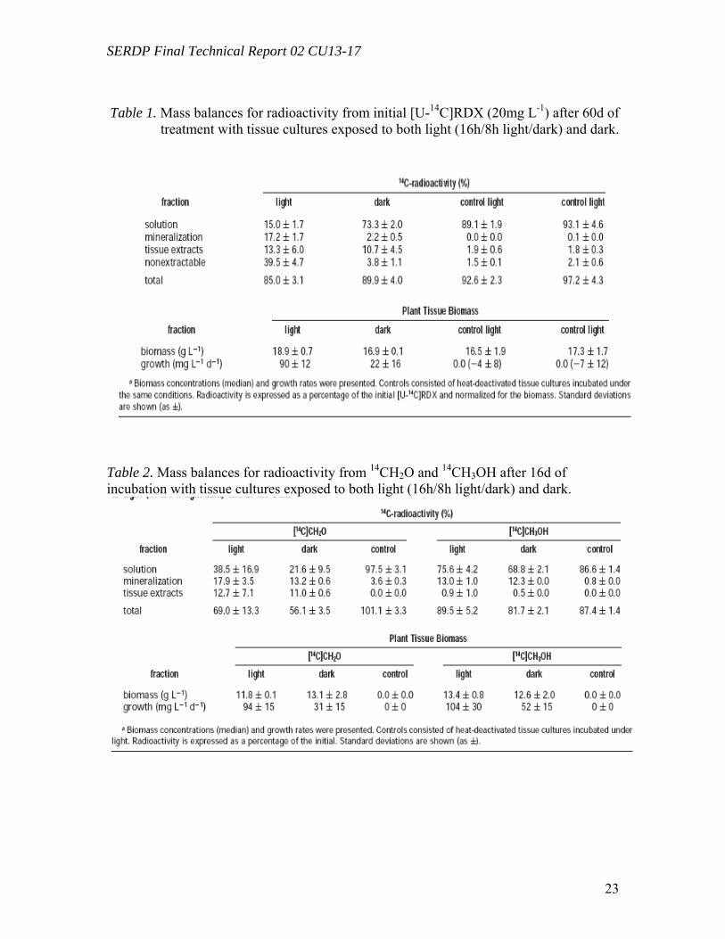
SERDP Final Technical Report 02 CU13-17
23
Table 1. Mass balances for radioactivity from initial [U-14C]RDX (20mg L-1) after 60d of
treatment with tissue cultures exposed to both light (16h/8h light/dark) and dark.
Table 2. Mass balances for radioactivity from 14CH2O and 14CH3OH after 16d of incubation with tissue cultures exposed to both light (16h/8h light/dark) and dark.
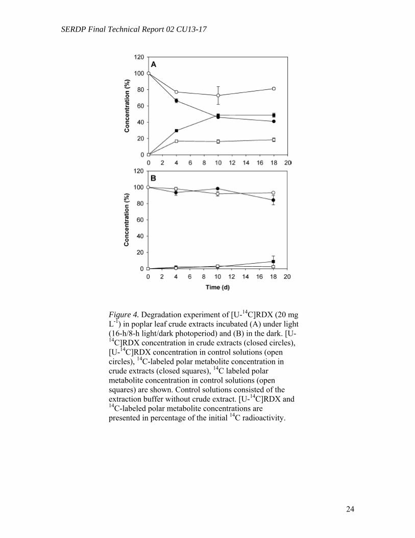
SERDP Final Technical Report 02 CU13-17
24
Figure 4. Degradation experiment of [U-14C]RDX (20 mg L-1) in poplar leaf crude extracts incubated (A) under light (16-h/8-h light/dark photoperiod) and (B) in the dark. [U-14C]RDX concentration in crude extracts (closed circles), [U-14C]RDX concentration in control solutions (open circles), 14C-labeled polar metabolite concentration in crude extracts (closed squares), 14C labeled polar metabolite concentration in control solutions (open squares) are shown. Control solutions consisted of the extraction buffer without crude extract. [U-14C]RDX and 14C-labeled polar metabolite concentrations are presented in percentage of the initial 14C radioactivity.
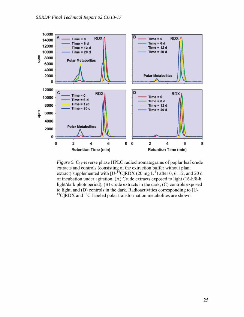
SERDP Final Technical Report 02 CU13-17
25
Figure 5. C18-reverse phase HPLC radiochromatograms of poplar leaf crude extracts and controls (consisting of the extraction buffer without plant extract) supplemented with [U-14C]RDX (20 mg L-1) after 0, 6, 12, and 20 d of incubation under agitation. (A) Crude extracts exposed to light (16-h/8-h light/dark photoperiod), (B) crude extracts in the dark, (C) controls exposed to light, and (D) controls in the dark. Radioactivities corresponding to [U-14C]RDX and 14C-labeled polar transformation metabolites are shown.
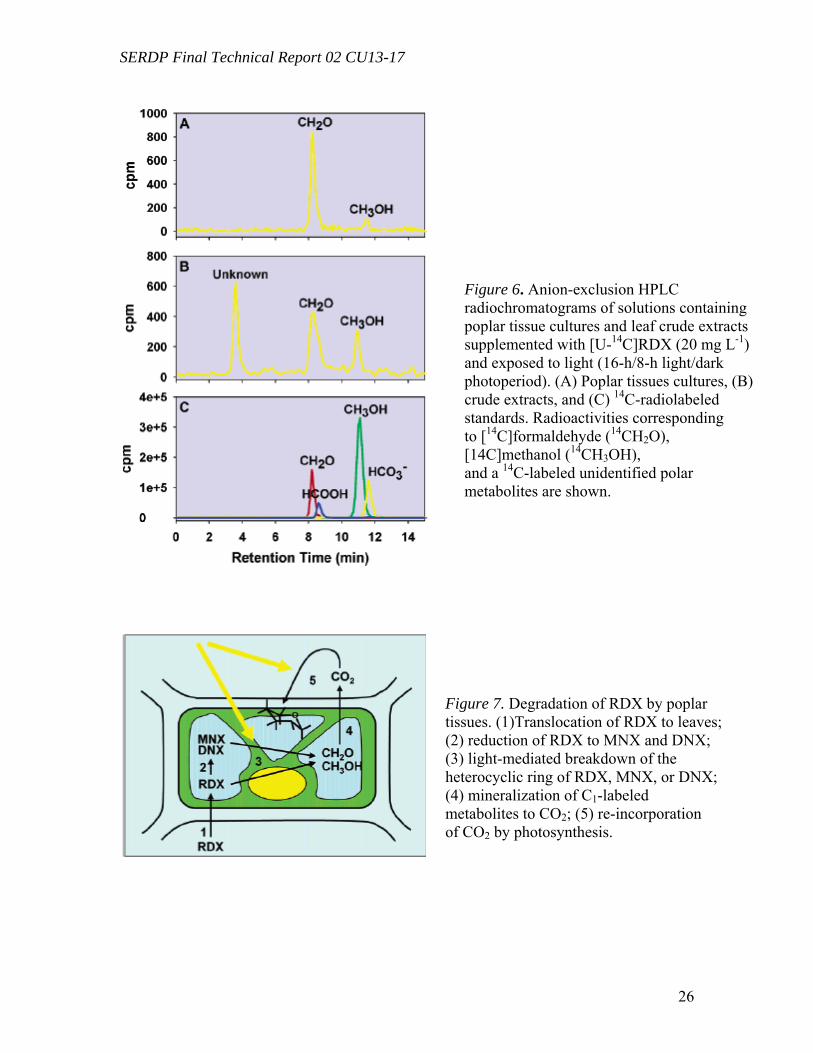
SERDP Final Technical Report 02 CU13-17
26
Figure 6. Anion-exclusion HPLC radiochromatograms of solutions containing poplar tissue cultures and leaf crude extracts supplemented with [U-14C]RDX (20 mg L-1) and exposed to light (16-h/8-h light/dark photoperiod). (A) Poplar tissues cultures, (B) crude extracts, and (C) 14C-radiolabeled standards. Radioactivities corresponding to [14C]formaldehyde (14CH2O), [14C]methanol (14CH3OH), and a 14C-labeled unidentified polar metabolites are shown.
Figure 7. Degradation of RDX by poplar tissues. (1)Translocation of RDX to leaves; (2) reduction of RDX to MNX and DNX; (3) light-mediated breakdown of the heterocyclic ring of RDX, MNX, or DNX; (4) mineralization of C1-labeled metabolites to CO2; (5) re-incorporation of CO2 by photosynthesis.
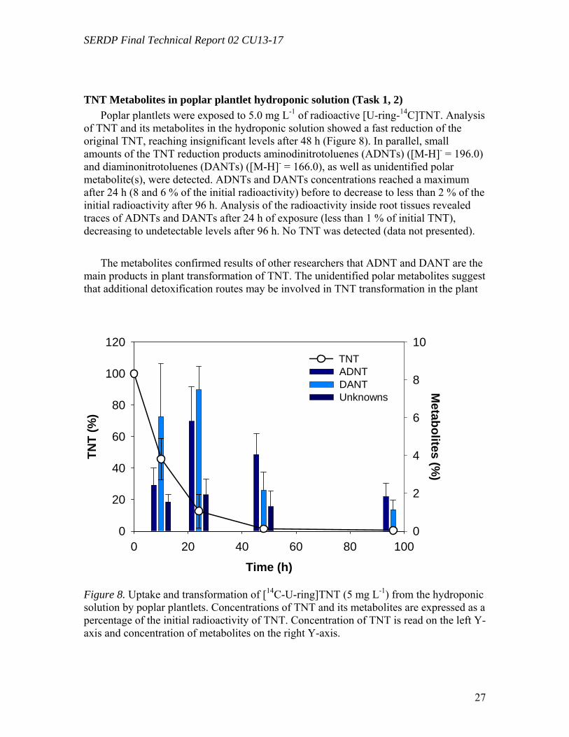
SERDP Final Technical Report 02 CU13-17
27
TNT Metabolites in poplar plantlet hydroponic solution (Task 1, 2) Poplar plantlets were exposed to 5.0 mg L-1 of radioactive [U-ring-14C]TNT. Analysis
of TNT and its metabolites in the hydroponic solution showed a fast reduction of the original TNT, reaching insignificant levels after 48 h (Figure 8). In parallel, small amounts of the TNT reduction products aminodinitrotoluenes (ADNTs) ([M-H]- = 196.0) and diaminonitrotoluenes (DANTs) ([M-H]- = 166.0), as well as unidentified polar metabolite(s), were detected. ADNTs and DANTs concentrations reached a maximum after 24 h (8 and 6 % of the initial radioactivity) before to decrease to less than 2 % of the initial radioactivity after 96 h. Analysis of the radioactivity inside root tissues revealed traces of ADNTs and DANTs after 24 h of exposure (less than 1 % of initial TNT), decreasing to undetectable levels after 96 h. No TNT was detected (data not presented).
The metabolites confirmed results of other researchers that ADNT and DANT are the main products in plant transformation of TNT. The unidentified polar metabolites suggest that additional detoxification routes may be involved in TNT transformation in the plant
Time (h)0 20 40 60 80 100
Metabolites (%
)
0
2
4
6
8
10
TNT
(%)
0
20
40
60
80
100
120
ADNTDANTUnknowns
TNT
Figure 8. Uptake and transformation of [14C-U-ring]TNT (5 mg L-1) from the hydroponic solution by poplar plantlets. Concentrations of TNT and its metabolites are expressed as a percentage of the initial radioactivity of TNT. Concentration of TNT is read on the left Y-axis and concentration of metabolites on the right Y-axis.
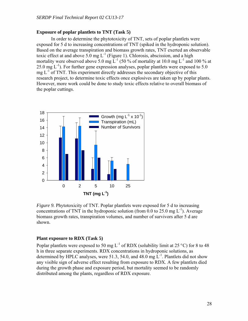
SERDP Final Technical Report 02 CU13-17
28
Exposure of poplar plantlets to TNT (Task 5) In order to determine the phytotoxicity of TNT, sets of poplar plantlets were
exposed for 5 d to increasing concentrations of TNT (spiked in the hydroponic solution). Based on the average transpiration and biomass growth rates, TNT exerted an observable toxic effect at and above 5.0 mg L-1 (Figure 1). Chlorosis, abscission, and a high mortality were observed above 5.0 mg L-1 (50 % of mortality at 10.0 mg L-1 and 100 % at 25.0 mg L-1). For further gene expression analyses, poplar plantlets were exposed to 5.0 mg L-1 of TNT. This experiment directly addresses the secondary objective of this research project, to determine toxic effects once explosives are taken up by poplar plants. However, more work could be done to study toxic effects relative to overall biomass of the poplar cuttings.
TNT (mg L-1)
0 2 5 10 250
2
4
6
8
10
12
14
16
18Growth (mg L-1 x 10⁻¹)Transpiration (mL)Number of Survivors
Figure 9. Phytotoxicity of TNT. Poplar plantlets were exposed for 5 d to increasing concentrations of TNT in the hydroponic solution (from 0.0 to 25.0 mg L-1). Average biomass growth rates, transpiration volumes, and number of survivors after 5 d are shown.
Plant exposure to RDX (Task 5) Poplar plantlets were exposed to 50 mg L-1 of RDX (solubility limit at 25 °C) for 8 to 48 h in three separate experiments. RDX concentrations in hydroponic solutions, as determined by HPLC analyses, were 51.3, 54.0, and 48.0 mg L-1. Plantlets did not show any visible sign of adverse effect resulting from exposure to RDX. A few plantlets died during the growth phase and exposure period, but mortality seemed to be randomly distributed among the plants, regardless of RDX exposure.
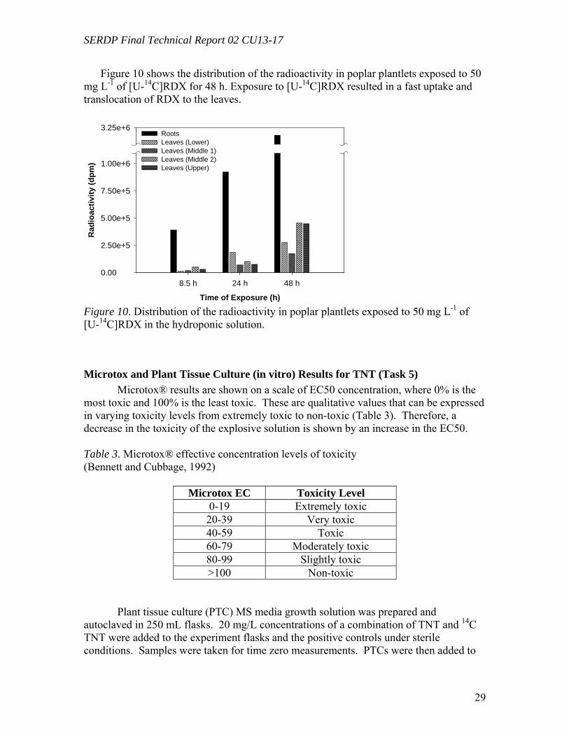
SERDP Final Technical Report 02 CU13-17
29
Figure 10 shows the distribution of the radioactivity in poplar plantlets exposed to 50 mg L-1 of [U-14C]RDX for 48 h. Exposure to [U-14C]RDX resulted in a fast uptake and translocation of RDX to the leaves.
Time of Exposure (h)
8.5 h 24 h 48 h
Rad
ioac
tivity
(dpm
)
0.00
2.50e+5
5.00e+5
7.50e+5
1.00e+6
3.25e+6RootsLeaves (Lower)Leaves (Middle 1)Leaves (Middle 2)Leaves (Upper)
Figure 10. Distribution of the radioactivity in poplar plantlets exposed to 50 mg L-1 of [U-14C]RDX in the hydroponic solution.
Microtox and Plant Tissue Culture (in vitro) Results for TNT (Task 5) Microtox® results are shown on a scale of EC50 concentration, where 0% is the most toxic and 100% is the least toxic. These are qualitative values that can be expressed in varying toxicity levels from extremely toxic to non-toxic (Table 3). Therefore, a decrease in the toxicity of the explosive solution is shown by an increase in the EC50. Table 3. Microtox® effective concentration levels of toxicity (Bennett and Cubbage, 1992)
Microtox EC Toxicity Level 0-19 Extremely toxic 20-39 Very toxic 40-59 Toxic 60-79 Moderately toxic 80-99 Slightly toxic >100 Non-toxic
Plant tissue culture (PTC) MS media growth solution was prepared and autoclaved in 250 mL flasks. 20 mg/L concentrations of a combination of TNT and 14C TNT were added to the experiment flasks and the positive controls under sterile conditions. Samples were taken for time zero measurements. PTCs were then added to
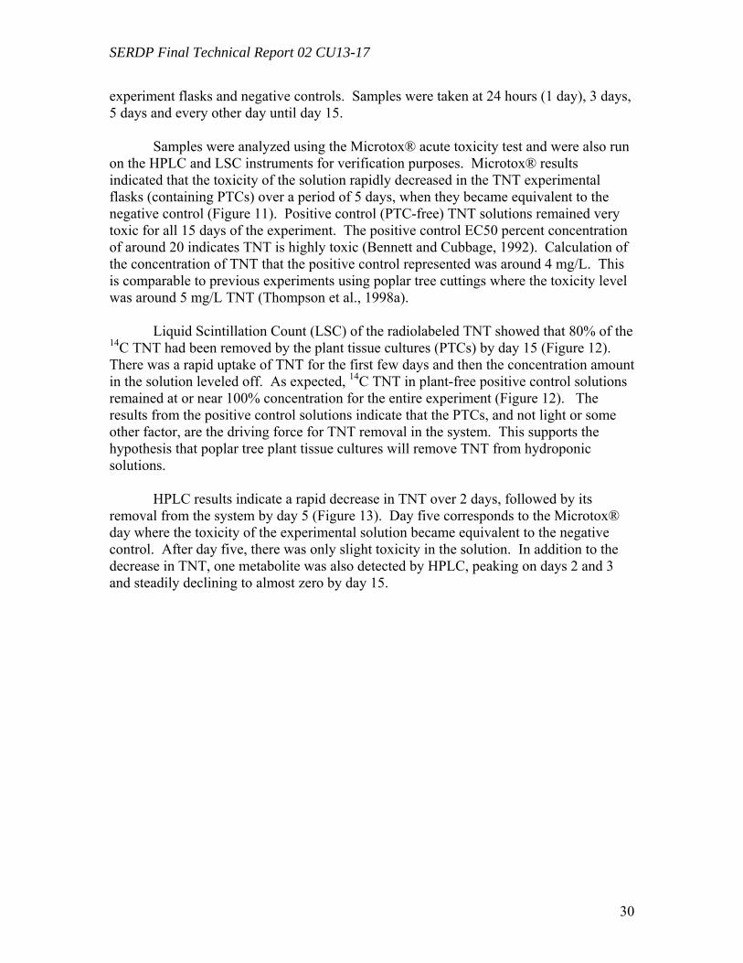
SERDP Final Technical Report 02 CU13-17
30
experiment flasks and negative controls. Samples were taken at 24 hours (1 day), 3 days, 5 days and every other day until day 15. Samples were analyzed using the Microtox® acute toxicity test and were also run on the HPLC and LSC instruments for verification purposes. Microtox® results indicated that the toxicity of the solution rapidly decreased in the TNT experimental flasks (containing PTCs) over a period of 5 days, when they became equivalent to the negative control (Figure 11). Positive control (PTC-free) TNT solutions remained very toxic for all 15 days of the experiment. The positive control EC50 percent concentration of around 20 indicates TNT is highly toxic (Bennett and Cubbage, 1992). Calculation of the concentration of TNT that the positive control represented was around 4 mg/L. This is comparable to previous experiments using poplar tree cuttings where the toxicity level was around 5 mg/L TNT (Thompson et al., 1998a).
Liquid Scintillation Count (LSC) of the radiolabeled TNT showed that 80% of the 14C TNT had been removed by the plant tissue cultures (PTCs) by day 15 (Figure 12). There was a rapid uptake of TNT for the first few days and then the concentration amount in the solution leveled off. As expected, 14C TNT in plant-free positive control solutions remained at or near 100% concentration for the entire experiment (Figure 12). The results from the positive control solutions indicate that the PTCs, and not light or some other factor, are the driving force for TNT removal in the system. This supports the hypothesis that poplar tree plant tissue cultures will remove TNT from hydroponic solutions.
HPLC results indicate a rapid decrease in TNT over 2 days, followed by its removal from the system by day 5 (Figure 13). Day five corresponds to the Microtox® day where the toxicity of the experimental solution became equivalent to the negative control. After day five, there was only slight toxicity in the solution. In addition to the decrease in TNT, one metabolite was also detected by HPLC, peaking on days 2 and 3 and steadily declining to almost zero by day 15.
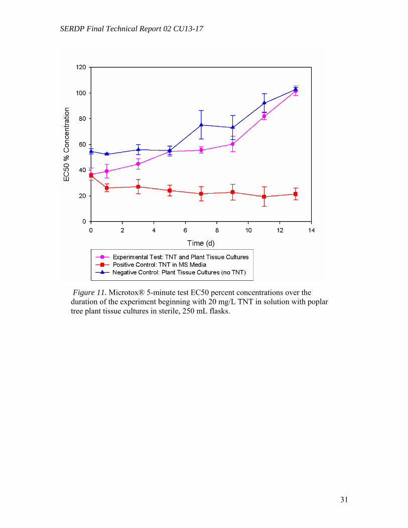
SERDP Final Technical Report 02 CU13-17
31
Figure 11. Microtox® 5-minute test EC50 percent concentrations over the duration of the experiment beginning with 20 mg/L TNT in solution with poplar tree plant tissue cultures in sterile, 250 mL flasks.
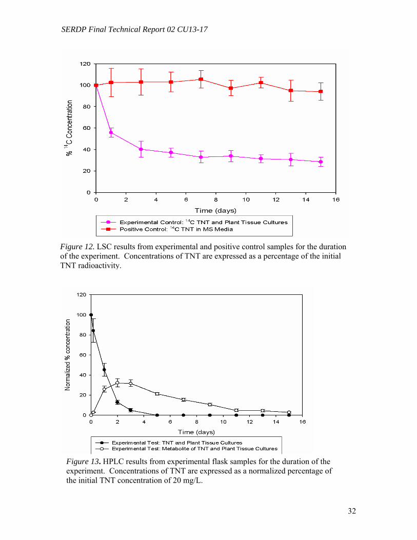
SERDP Final Technical Report 02 CU13-17
32
Figure 13. HPLC results from experimental flask samples for the duration of the experiment. Concentrations of TNT are expressed as a normalized percentage of the initial TNT concentration of 20 mg/L.
Figure 12. LSC results from experimental and positive control samples for the duration of the experiment. Concentrations of TNT are expressed as a percentage of the initial TNT radioactivity.
SERDP Final Technical Report 02 CU13-17
33
Microtox and Plant Tissue Culture (in vitro) Results for RDX (Task 5) Microtox® results showed that toxicity of the RDX solution decreased by about 15% over the entire experiment, but it never reached the non-toxic negative control (Figure 14). Positive control RDX solutions remained toxic, but not as high as TNT, which confirms previous findings of relative toxicity TNT > RDX > HMX. There is a large decrease in the toxicity of the negative control solution after day 15. Liquid Scintillation Count of the radiolabeled RDX showed that 20% of the 14C RDX had been removed by the PTCs by day 21 at a much slower rate than that of the 14C TNT (Figure 15). Positive control RDX solutions remained constant at or near 100% concentration for the entire experiment, which suggests that the plant tissue cultures are the most likely reason the RDX is removed.
The HPLC results are a little more difficult to interpret. The experimental flasks have only a slightly lower concentration of RDX than the positive RDX controls (Figure 16). No metabolites were detected. There is a significant amount of RDX removal (~52%), but this removal is true for both the experimental and positive control flasks. Though the LSC results do not support this trend, photolysis of RDX is suspected in this degradation process.
Figure 14. Microtox® 5-minute test EC50 percent concentrations over the duration of
the experiment beginning with 20 mg/L RDX in solution with poplar tree plant tissue cultures in sterile, 250 mL flasks.
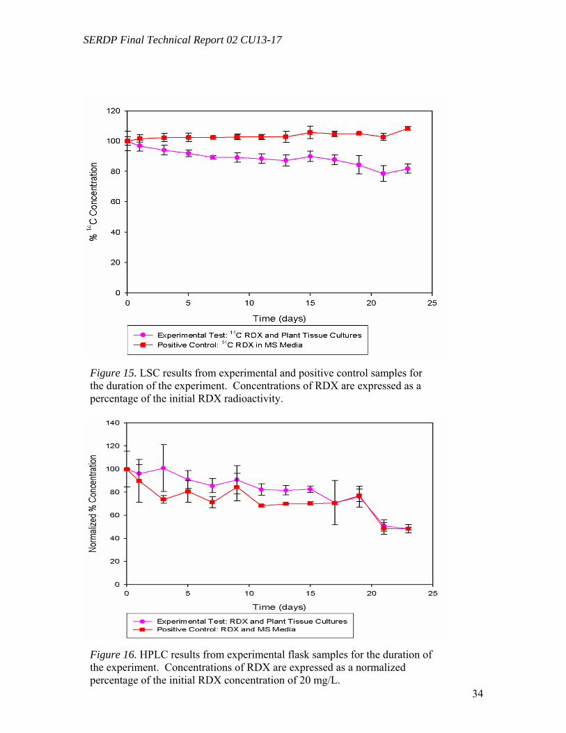
SERDP Final Technical Report 02 CU13-17
34
Figure 15. LSC results from experimental and positive control samples for the duration of the experiment. Concentrations of RDX are expressed as a percentage of the initial RDX radioactivity.
Figure 16. HPLC results from experimental flask samples for the duration of the experiment. Concentrations of RDX are expressed as a normalized percentage of the initial RDX concentration of 20 mg/L.
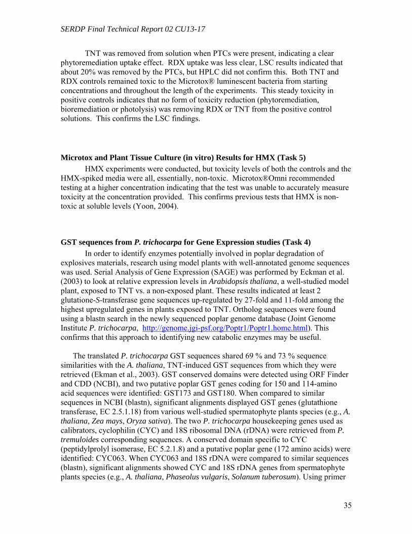
SERDP Final Technical Report 02 CU13-17
35
TNT was removed from solution when PTCs were present, indicating a clear phytoremediation uptake effect. RDX uptake was less clear, LSC results indicated that about 20% was removed by the PTCs, but HPLC did not confirm this. Both TNT and RDX controls remained toxic to the Microtox® luminescent bacteria from starting concentrations and throughout the length of the experiments. This steady toxicity in positive controls indicates that no form of toxicity reduction (phytoremediation, bioremediation or photolysis) was removing RDX or TNT from the positive control solutions. This confirms the LSC findings.
Microtox and Plant Tissue Culture (in vitro) Results for HMX (Task 5) HMX experiments were conducted, but toxicity levels of both the controls and the
HMX-spiked media were all, essentially, non-toxic. Microtox®Omni recommended testing at a higher concentration indicating that the test was unable to accurately measure toxicity at the concentration provided. This confirms previous tests that HMX is non-toxic at soluble levels (Yoon, 2004).
GST sequences from P. trichocarpa for Gene Expression studies (Task 4) In order to identify enzymes potentially involved in poplar degradation of explosives materials, research using model plants with well-annotated genome sequences was used. Serial Analysis of Gene Expression (SAGE) was performed by Eckman et al. (2003) to look at relative expression levels in Arabidopsis thaliana, a well-studied model plant, exposed to TNT vs. a non-exposed plant. These results indicated at least 2 glutatione-S-transferase gene sequences up-regulated by 27-fold and 11-fold among the highest upregulated genes in plants exposed to TNT. Ortholog sequences were found using a blastn search in the newly sequenced poplar genome database (Joint Genome Institute P. trichocarpa, http://genome.jgi-psf.org/Poptr1/Poptr1.home.html). This confirms that this approach to identifying new catabolic enzymes may be useful.
The translated P. trichocarpa GST sequences shared 69 % and 73 % sequence similarities with the A. thaliana, TNT-induced GST sequences from which they were retrieved (Ekman et al., 2003). GST conserved domains were detected using ORF Finder and CDD (NCBI), and two putative poplar GST genes coding for 150 and 114-amino acid sequences were identified: GST173 and GST180. When compared to similar sequences in NCBI (blastn), significant alignments displayed GST genes (glutathione transferase, EC 2.5.1.18) from various well-studied spermatophyte plants species (e.g., A. thaliana, Zea mays, Oryza sativa). The two P. trichocarpa housekeeping genes used as calibrators, cyclophilin (CYC) and 18S ribosomal DNA (rDNA) were retrieved from P. tremuloides corresponding sequences. A conserved domain specific to CYC (peptidylprolyl isomerase, EC 5.2.1.8) and a putative poplar gene (172 amino acids) were identified: CYC063. When CYC063 and 18S rDNA were compared to similar sequences (blastn), significant alignments showed CYC and 18S rDNA genes from spermatophyte plants species (e.g., A. thaliana, Phaseolus vulgaris, Solanum tuberosum). Using primer
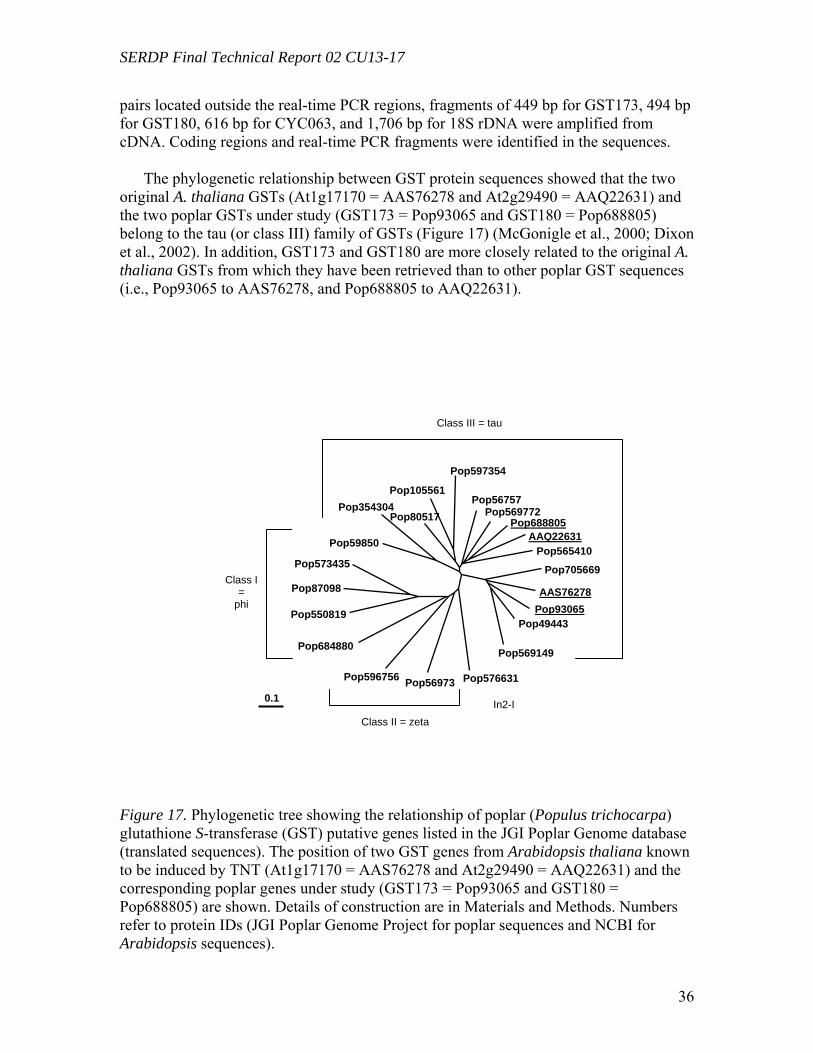
SERDP Final Technical Report 02 CU13-17
36
pairs located outside the real-time PCR regions, fragments of 449 bp for GST173, 494 bp for GST180, 616 bp for CYC063, and 1,706 bp for 18S rDNA were amplified from cDNA. Coding regions and real-time PCR fragments were identified in the sequences.
The phylogenetic relationship between GST protein sequences showed that the two
original A. thaliana GSTs (At1g17170 = AAS76278 and At2g29490 = AAQ22631) and the two poplar GSTs under study (GST173 = Pop93065 and GST180 = Pop688805) belong to the tau (or class III) family of GSTs (Figure 17) (McGonigle et al., 2000; Dixon et al., 2002). In addition, GST173 and GST180 are more closely related to the original A. thaliana GSTs from which they have been retrieved than to other poplar GST sequences (i.e., Pop93065 to AAS76278, and Pop688805 to AAQ22631).
Figure 17. Phylogenetic tree showing the relationship of poplar (Populus trichocarpa) glutathione S-transferase (GST) putative genes listed in the JGI Poplar Genome database (translated sequences). The position of two GST genes from Arabidopsis thaliana known to be induced by TNT (At1g17170 = AAS76278 and At2g29490 = AAQ22631) and the corresponding poplar genes under study (GST173 = Pop93065 and GST180 = Pop688805) are shown. Details of construction are in Materials and Methods. Numbers refer to protein IDs (JGI Poplar Genome Project for poplar sequences and NCBI for Arabidopsis sequences).
0.1
Pop705669
AAS76278 Pop93065
Pop49443
Pop569149
Pop576631Pop56973Pop596756
Pop684880
Pop550819
Pop87098
Pop573435
Pop59850
Pop354304Pop80517
Pop105561
Pop597354
Pop56757Pop569772
Pop688805AAQ22631
Pop565410
Class III = tau
Class II = zeta
Class I =
phi
In2-I
SERDP Final Technical Report 02 CU13-17
37
Analyses of GST expression in Poplar Plantlets Exposed to TNT (Task 4) Gene expression patterns are used here to track enzymatic patterns at the transcription
level. Before an enzyme is expressed, it must be transcribed and therefore quantitative gene expression experiments using reverse-transcriptase real-time PCR can be used to look at the potential for enzyme activity by identifying relative quantities of transcribed genes in exposed vs. non-exposed poplar plants.
In three separate experiments, sets of poplar plantlets were exposed to 5.0 mg L-1 of
TNT (non-exposed plants were used as controls). The expression of GST173 and GST180 was analyzed in root tissues after 12 h, 24h, and 48 h, and compared to non-exposed plants (Figure 18). Both GST genes were expressed at a significantly higher level in exposed tissues: About 18 – 21-fold for GST173 after 12 h (depending on the calibrator used), increasing slightly to 23 – 25-fold after 48 h; for GST180, an induction of about 5 – 6-fold was observed after 4 h, reaching a peak of 9 – 10-fold after 24 h, before to decrease to 4 – 5-fold after 48 h. The expression levels of GST173 and GST180 recorded in control (non-exposed) plants did not change significantly over the course of the experiment, 1.0 – 1.5-fold, with one exception at 2.3-fold (GST180 after 24 h, calibrator CYC063). While statistically significant, a considerable variation of the induction level of GST173 was observed between the three experiments (analytical replicates), as reflected by the high standard deviation: From 4 to 41-fold after 12 h, from 10 to 24-fold after 24 h, and from 7 to 36-fold after 48 h (calibrator 18S rDNA). On the other hand, GST180 showed lower induction levels, but with more consistency between experiments: Three to 8-fold after 12 h, 5 to 13-fold after 24 h, and 4 to 5-fold after 48 h (calibrator 18S rDNA). On the other hand, similar expression levels were recorded using the two calibrators, CYC063 or 18S rDNA, which suggests that their expression was not affected by TNT and that they constituted efficient internal standards (Figure 18). Tentative analysis of GST expression in leaf tissues from the same TNT-exposed plantlets gave less reliable and/or less reproducible results than those recorded in root tissues (data not presented).
In order to ensure that co-extracted genomic DNA (gDNA) did not contribute
significantly to the amplification of cDNA, additional primers (for GST180) were designed, amplifying exclusively cDNA. Figure 19 showed the amplification levels of GST180 using both the “regular” primers, susceptible to amplify both gDNA and cDNA (designed inside a single exon): gst180; and the primers designed to amplify only cDNA (one of the primer sequence being located spanning an intron): gst180x. Expression results using in parallel gst180 and gst180x did not show significant differences, which indicates the absence of contaminating gDNA at a level affecting quantitative gene expression. AGE analyses confirmed that gst180x primers gave PCR products from cDNA, but failed to amplify fragments from gDNA, while “regular” gst180 primers gave products from both cDNA and gDNA.
When used as templates in PCR reactions, cDNA controls (non-transcribed, prepared
without reverse-transcriptase) did not produce recordable PCR amplification, which further confirms the absence of contamination by genomic DNA.The dissociation curves of the real-time PCR products for the four genes under analysis (GST173, GST180,
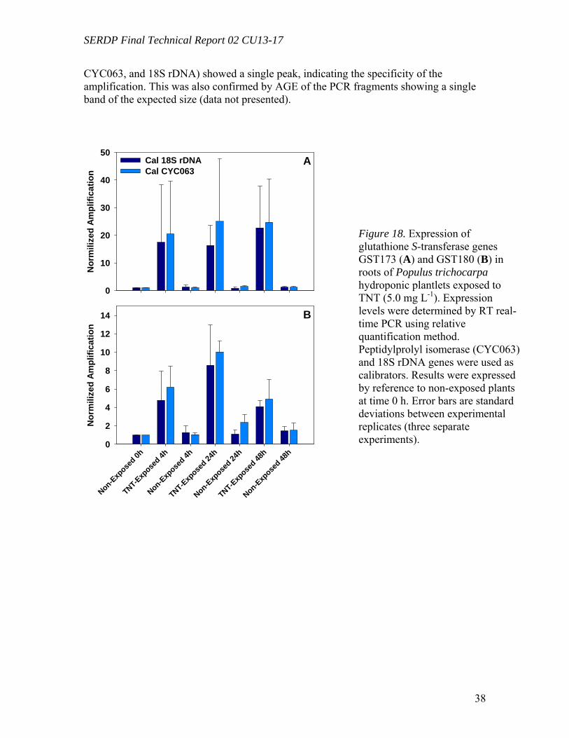
SERDP Final Technical Report 02 CU13-17
38
CYC063, and 18S rDNA) showed a single peak, indicating the specificity of the amplification. This was also confirmed by AGE of the PCR fragments showing a single band of the expected size (data not presented).
Blank Mark
Nor
mili
zed
Ampl
ifica
tion
0
10
20
30
40
50Cal 18S rDNACal CYC063
Non-Exp
osed 0h
TNT-Exp
osed 4h
Non-Exp
osed 4h
TNT-Exp
osed 24
h
Non-Exp
osed 24
h
TNT-Exp
osed 48
h
Non-Exp
osed 48
h
Nor
mili
zed
Ampl
ifica
tion
0
2
4
6
8
10
12
14
A
B
Figure 18. Expression of glutathione S-transferase genes GST173 (A) and GST180 (B) in roots of Populus trichocarpa hydroponic plantlets exposed to TNT (5.0 mg L-1). Expression levels were determined by RT real-time PCR using relative quantification method. Peptidylprolyl isomerase (CYC063) and 18S rDNA genes were used as calibrators. Results were expressed by reference to non-exposed plants at time 0 h. Error bars are standard deviations between experimental replicates (three separate experiments).
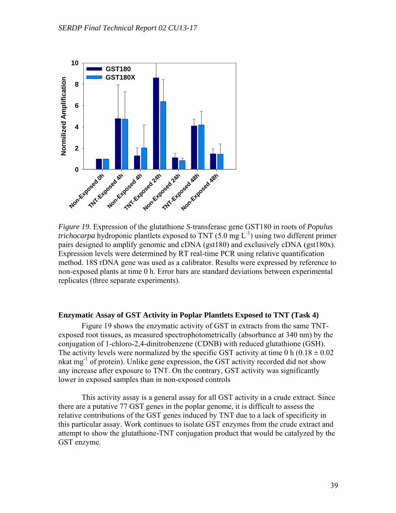
SERDP Final Technical Report 02 CU13-17
39
Blank MarkNon-Exp
osed 0h
TNT-Exp
osed 4h
Non-Exp
osed 4h
TNT-Exp
osed 24
h
Non-Exp
osed 24
h
TNT-Exp
osed 48
h
Non-Exp
osed 48
h
Nor
mili
zed
Ampl
ifica
tion
0
2
4
6
8
10GST180GST180X
Figure 19. Expression of the glutathione S-transferase gene GST180 in roots of Populus trichocarpa hydroponic plantlets exposed to TNT (5.0 mg L-1) using two different primer pairs designed to amplify genomic and cDNA (gst180) and exclusively cDNA (gst180x). Expression levels were determined by RT real-time PCR using relative quantification method. 18S rDNA gene was used as a calibrator. Results were expressed by reference to non-exposed plants at time 0 h. Error bars are standard deviations between experimental replicates (three separate experiments).
Enzymatic Assay of GST Activity in Poplar Plantlets Exposed to TNT (Task 4) Figure 19 shows the enzymatic activity of GST in extracts from the same TNT-
exposed root tissues, as measured spectrophotometrically (absorbance at 340 nm) by the conjugation of 1-chloro-2,4-dinitrobenzene (CDNB) with reduced glutathione (GSH). The activity levels were normalized by the specific GST activity at time 0 h (0.18 ± 0.02 nkat mg-1 of protein). Unlike gene expression, the GST activity recorded did not show any increase after exposure to TNT. On the contrary, GST activity was significantly lower in exposed samples than in non-exposed controls This activity assay is a general assay for all GST activity in a crude extract. Since there are a putative 77 GST genes in the poplar genome, it is difficult to assess the relative contributions of the GST genes induced by TNT due to a lack of specificity in this particular assay. Work continues to isolate GST enzymes from the crude extract and attempt to show the glutathione-TNT conjugation product that would be catalyzed by the GST enzyme.
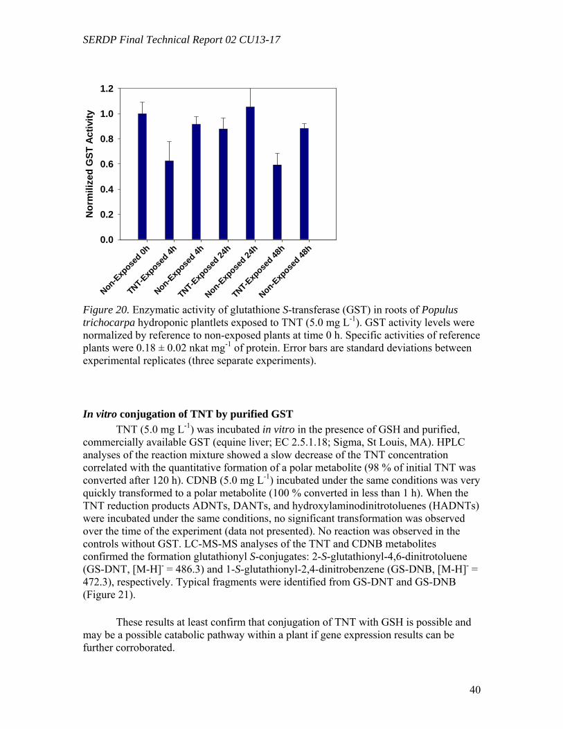
SERDP Final Technical Report 02 CU13-17
40
Blan MarkNon-Exp
osed 0h
TNT-E
xpose
d 4h
Non-Exp
osed 4h
TNT-Exp
osed 24
h
Non-Exp
osed 24
h
TNT-E
xpose
d 48h
Non-Exp
osed 48
h
Nor
mili
zed
GST
Act
ivity
0.0
0.2
0.4
0.6
0.8
1.0
1.2
Figure 20. Enzymatic activity of glutathione S-transferase (GST) in roots of Populus trichocarpa hydroponic plantlets exposed to TNT (5.0 mg L-1). GST activity levels were normalized by reference to non-exposed plants at time 0 h. Specific activities of reference plants were 0.18 ± 0.02 nkat mg-1 of protein. Error bars are standard deviations between experimental replicates (three separate experiments).
In vitro conjugation of TNT by purified GST TNT (5.0 mg L-1) was incubated in vitro in the presence of GSH and purified,
commercially available GST (equine liver; EC 2.5.1.18; Sigma, St Louis, MA). HPLC analyses of the reaction mixture showed a slow decrease of the TNT concentration correlated with the quantitative formation of a polar metabolite (98 % of initial TNT was converted after 120 h). CDNB (5.0 mg L-1) incubated under the same conditions was very quickly transformed to a polar metabolite (100 % converted in less than 1 h). When the TNT reduction products ADNTs, DANTs, and hydroxylaminodinitrotoluenes (HADNTs) were incubated under the same conditions, no significant transformation was observed over the time of the experiment (data not presented). No reaction was observed in the controls without GST. LC-MS-MS analyses of the TNT and CDNB metabolites confirmed the formation glutathionyl S-conjugates: 2-S-glutathionyl-4,6-dinitrotoluene (GS-DNT, [M-H]- = 486.3) and 1-S-glutathionyl-2,4-dinitrobenzene (GS-DNB, [M-H]- = 472.3), respectively. Typical fragments were identified from GS-DNT and GS-DNB (Figure 21). These results at least confirm that conjugation of TNT with GSH is possible and may be a possible catabolic pathway within a plant if gene expression results can be further corroborated.
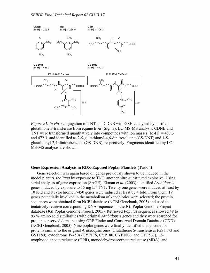
SERDP Final Technical Report 02 CU13-17
41
ClNO2
NO2
HOOC
NH2
NH
OS
NH
O
COOH
HOOC
NH2
NH
OSH
NH
O
COOH
CH3
NO2
NO2
O2N
CH3
NO2O2N
HOOC
NH2
NH
OS
NH
O
COOH
NO2
NO2
CDNB[M-H]- = 201.5
TNT[M-H]- = 226.0
GSH[M-H]- = 306.3
GS-DNT[M-H]- = 486.3
GS-DNB[M-H]- = 472.3
[M-H-213]- = 272.3 [M-H-199]- = 272.3
213 199
Figure 21. In vitro conjugation of TNT and CDNB with GSH catalyzed by purified glutathione S-transferase from equine liver (Sigma); LC-MS-MS analysis. CDNB and TNT were transformed quantitatively into compounds with ion masses [M-H]- = 487.3 and 472.3, and identified as 2-S-glutathionyl-4,6-dinitrotoluene (GS-DNT) and 1-S-glutathionyl-2,4-dinitrobenzene (GS-DNB), respectively. Fragments identified by LC-MS-MS analysis are shown.
Gene Expression Analysis in RDX-Exposed Poplar Plantlets (Task 4) Gene selection was again based on genes previously shown to be induced in the
model plant A. thaliana by exposure to TNT, another nitro-substituted explosive. Using serial analyses of gene expression (SAGE), Ekman et al. (2003) identified Arabidopsis genes induced by exposure to 15 mg L-1 TNT: Twenty one genes were induced at least by 10 fold and 8 cytochrome P-450 genes were induced at least by 4 fold. From them, 19 genes potentially involved in the metabolism of xenobiotics were selected; the protein sequences were obtained form NCBI database (NCBI Genebank, 2005) and used to tentatively retrieve corresponding DNA sequences in the JGI Poplar Genome Project database (JGI Poplar Genome Project, 2005). Retrieved Populus sequences showed 48 to 93 % amino acid similarities with original Arabidopsis genes and they were searched for protein conserved domains using ORF Finder and Conserved Domain Database (CDD) (NCBI Genebank, 2005). Nine poplar genes were finally identified that encode for proteins similar to the original Arabidopsis ones: Glutathione S-transferases (GST173 and GST180), cytochrome P-450s (CYP176, CYP180, CYP1806, and CYP567), 12-oxophytodienoate reductase (OPR), monodehydroascorbate reductase (MDA), and
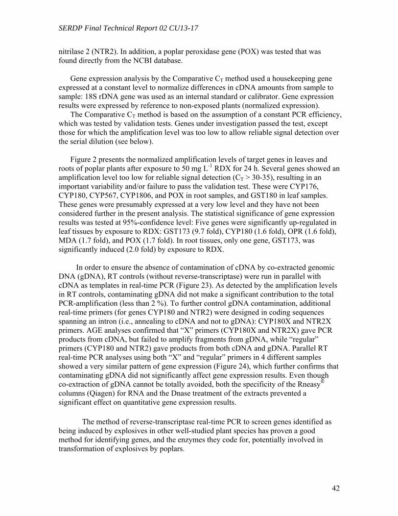
SERDP Final Technical Report 02 CU13-17
42
nitrilase 2 (NTR2). In addition, a poplar peroxidase gene (POX) was tested that was found directly from the NCBI database.
Gene expression analysis by the Comparative CT method used a housekeeping gene expressed at a constant level to normalize differences in cDNA amounts from sample to sample: 18S rDNA gene was used as an internal standard or calibrator. Gene expression results were expressed by reference to non-exposed plants (normalized expression).
The Comparative CT method is based on the assumption of a constant PCR efficiency, which was tested by validation tests. Genes under investigation passed the test, except those for which the amplification level was too low to allow reliable signal detection over the serial dilution (see below).
Figure 2 presents the normalized amplification levels of target genes in leaves and
roots of poplar plants after exposure to 50 mg L-1 RDX for 24 h. Several genes showed an amplification level too low for reliable signal detection (CT > 30-35), resulting in an important variability and/or failure to pass the validation test. These were CYP176, CYP180, CYP567, CYP1806, and POX in root samples, and GST180 in leaf samples. These genes were presumably expressed at a very low level and they have not been considered further in the present analysis. The statistical significance of gene expression results was tested at 95%-confidence level: Five genes were significantly up-regulated in leaf tissues by exposure to RDX: GST173 (9.7 fold), CYP180 (1.6 fold), OPR (1.6 fold), MDA (1.7 fold), and POX (1.7 fold). In root tissues, only one gene, GST173, was significantly induced (2.0 fold) by exposure to RDX.
In order to ensure the absence of contamination of cDNA by co-extracted genomic
DNA (gDNA), RT controls (without reverse-transcriptase) were run in parallel with cDNA as templates in real-time PCR (Figure 23). As detected by the amplification levels in RT controls, contaminating gDNA did not make a significant contribution to the total PCR-amplification (less than 2 %). To further control gDNA contamination, additional real-time primers (for genes CYP180 and NTR2) were designed in coding sequences spanning an intron (i.e., annealing to cDNA and not to gDNA): CYP180X and NTR2X primers. AGE analyses confirmed that “X” primers (CYP180X and NTR2X) gave PCR products from cDNA, but failed to amplify fragments from gDNA, while “regular” primers (CYP180 and NTR2) gave products from both cDNA and gDNA. Parallel RT real-time PCR analyses using both “X” and “regular” primers in 4 different samples showed a very similar pattern of gene expression (Figure 24), which further confirms that contaminating gDNA did not significantly affect gene expression results. Even though co-extraction of gDNA cannot be totally avoided, both the specificity of the Rneasy® columns (Qiagen) for RNA and the Dnase treatment of the extracts prevented a significant effect on quantitative gene expression results. The method of reverse-transcriptase real-time PCR to screen genes identified as being induced by explosives in other well-studied plant species has proven a good method for identifying genes, and the enzymes they code for, potentially involved in transformation of explosives by poplars.
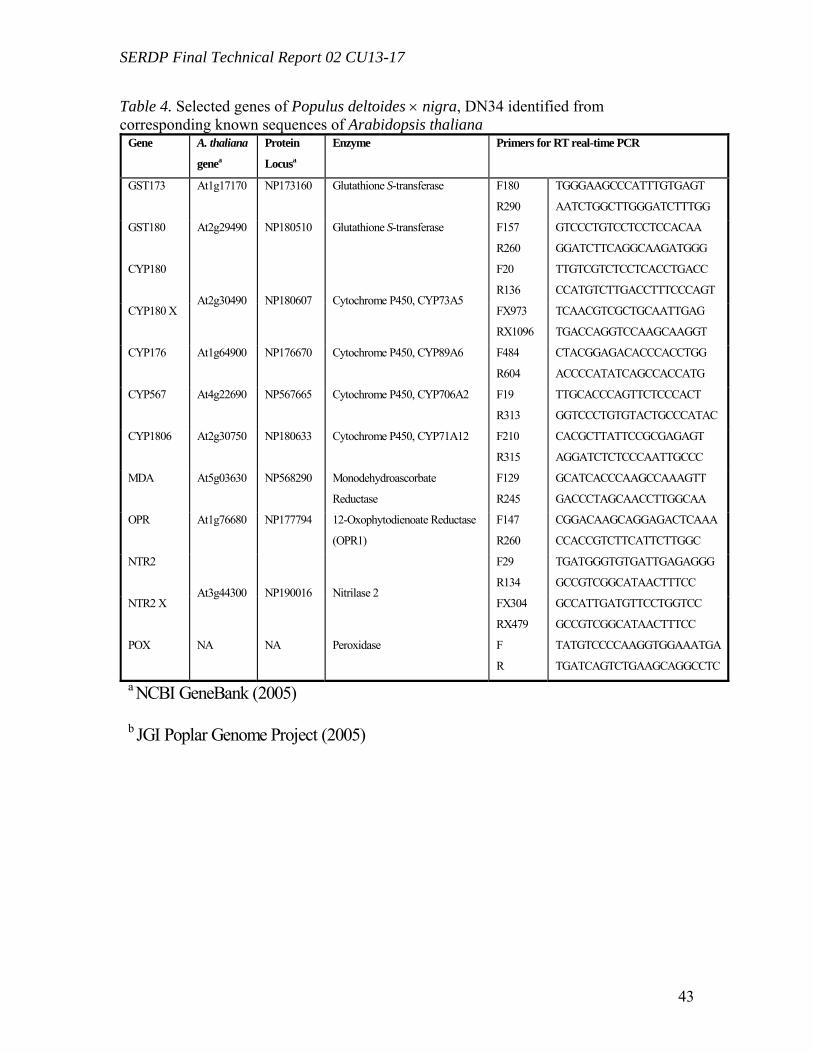
SERDP Final Technical Report 02 CU13-17
43
Table 4. Selected genes of Populus deltoides × nigra, DN34 identified from corresponding known sequences of Arabidopsis thaliana
Gene A. thaliana
genea
Protein
Locusa
Enzyme Primers for RT real-time PCR
GST173 At1g17170 NP173160 Glutathione S-transferase F180
R290
TGGGAAGCCCATTTGTGAGT
AATCTGGCTTGGGATCTTTGG
GST180 At2g29490 NP180510 Glutathione S-transferase F157
R260
GTCCCTGTCCTCCTCCACAA
GGATCTTCAGGCAAGATGGG
CYP180 F20
R136
TTGTCGTCTCCTCACCTGACC
CCATGTCTTGACCTTTCCCAGT
CYP180 X At2g30490 NP180607 Cytochrome P450, CYP73A5
FX973
RX1096
TCAACGTCGCTGCAATTGAG
TGACCAGGTCCAAGCAAGGT
CYP176 At1g64900 NP176670 Cytochrome P450, CYP89A6 F484
R604
CTACGGAGACACCCACCTGG
ACCCCATATCAGCCACCATG
CYP567 At4g22690 NP567665 Cytochrome P450, CYP706A2 F19
R313
TTGCACCCAGTTCTCCCACT
GGTCCCTGTGTACTGCCCATAC
CYP1806 At2g30750 NP180633 Cytochrome P450, CYP71A12 F210
R315
CACGCTTATTCCGCGAGAGT
AGGATCTCTCCCAATTGCCC
MDA At5g03630 NP568290 Monodehydroascorbate
Reductase
F129
R245
GCATCACCCAAGCCAAAGTT
GACCCTAGCAACCTTGGCAA
OPR At1g76680 NP177794 12-Oxophytodienoate Reductase
(OPR1)
F147
R260
CGGACAAGCAGGAGACTCAAA
CCACCGTCTTCATTCTTGGC
NTR2 F29
R134
TGATGGGTGTGATTGAGAGGG
GCCGTCGGCATAACTTTCC
NTR2 X At3g44300 NP190016 Nitrilase 2
FX304
RX479
GCCATTGATGTTCCTGGTCC
GCCGTCGGCATAACTTTCC
POX NA NA Peroxidase F
R
TATGTCCCCAAGGTGGAAATGA
TGATCAGTCTGAAGCAGGCCTC
a NCBI GeneBank (2005)
b JGI Poplar Genome Project (2005)
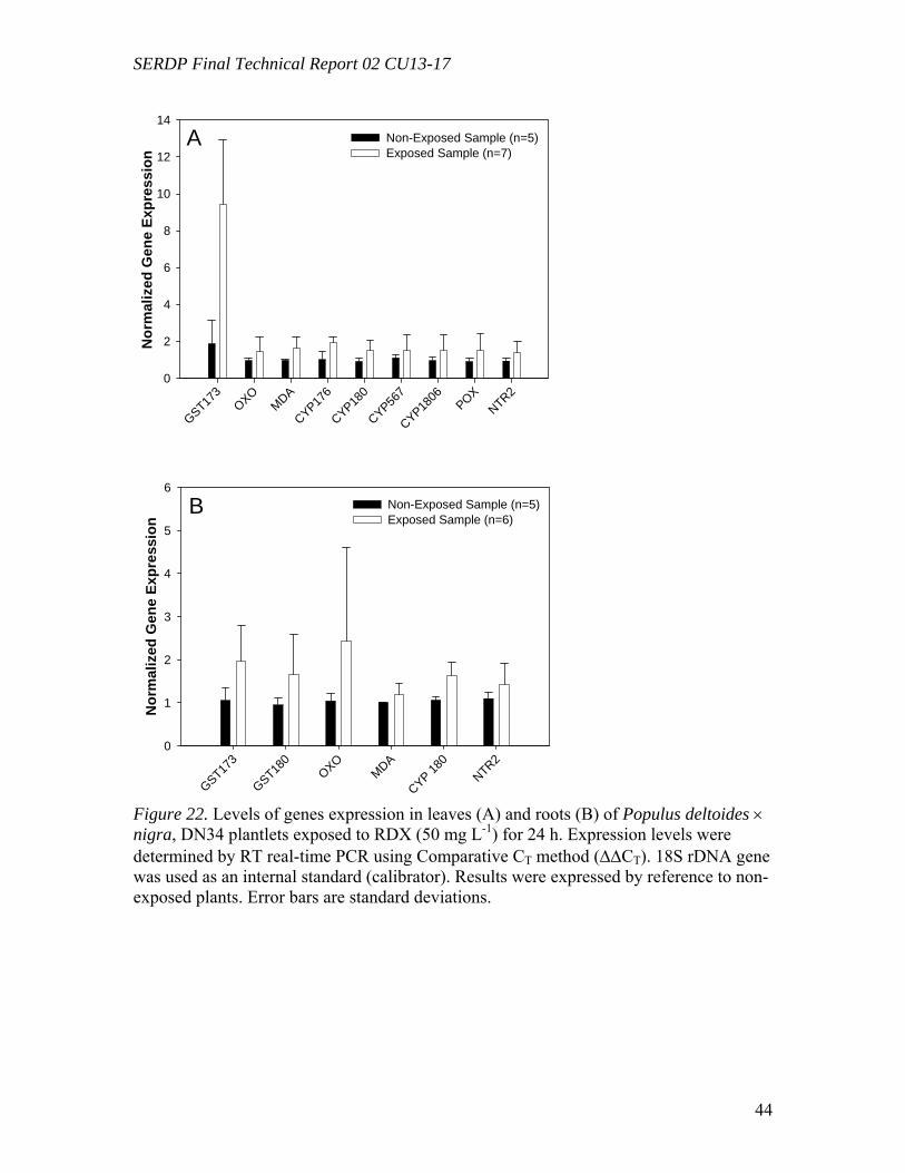
SERDP Final Technical Report 02 CU13-17
44
GST173
OXOMDA
CYP176
CYP180
CYP567
CYP1806
POXNTR2
Nor
mal
ized
Gen
e Ex
pres
sion
0
2
4
6
8
10
12
14Non-Exposed Sample (n=5)Exposed Sample (n=7)
A
GST173
GST180
OXOMDA
CYP 180
NTR2
Nor
mal
ized
Gen
e Ex
pres
sion
0
1
2
3
4
5
6Non-Exposed Sample (n=5)Exposed Sample (n=6)
B
Figure 22. Levels of genes expression in leaves (A) and roots (B) of Populus deltoides × nigra, DN34 plantlets exposed to RDX (50 mg L-1) for 24 h. Expression levels were determined by RT real-time PCR using Comparative CT method (∆∆CT). 18S rDNA gene was used as an internal standard (calibrator). Results were expressed by reference to non-exposed plants. Error bars are standard deviations.
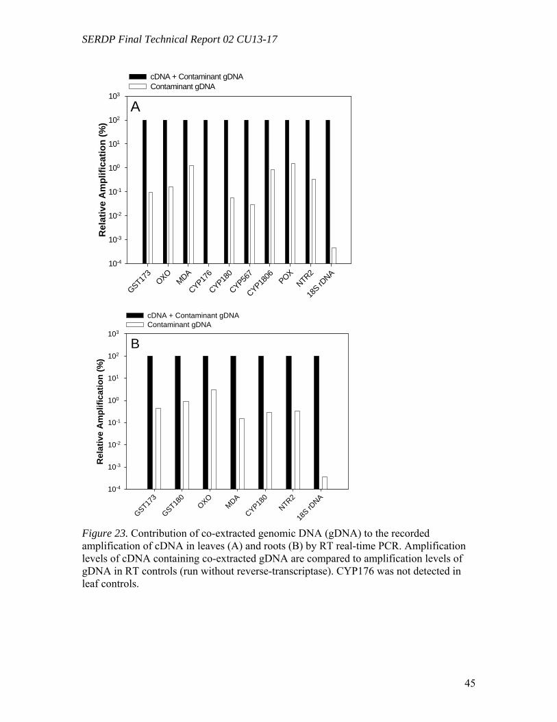
SERDP Final Technical Report 02 CU13-17
45
GST173
OXOMDA
CYP176
CYP180
CYP567
CYP1806 POX
NTR2
18S rD
NA
Rel
ativ
e A
mpl
ifica
tion
(%)
10-4
10-3
10-2
10-1
100
101
102
103
cDNA + Contaminant gDNAContaminant gDNA
A
GST173
GST180
OXOMDA
CYP180
NTR2
18S rD
NA
Rel
ativ
e A
mpl
ifica
tion
(%)
10-4
10-3
10-2
10-1
100
101
102
103
cDNA + Contaminant gDNAContaminant gDNA
B
Figure 23. Contribution of co-extracted genomic DNA (gDNA) to the recorded amplification of cDNA in leaves (A) and roots (B) by RT real-time PCR. Amplification levels of cDNA containing co-extracted gDNA are compared to amplification levels of gDNA in RT controls (run without reverse-transcriptase). CYP176 was not detected in leaf controls.
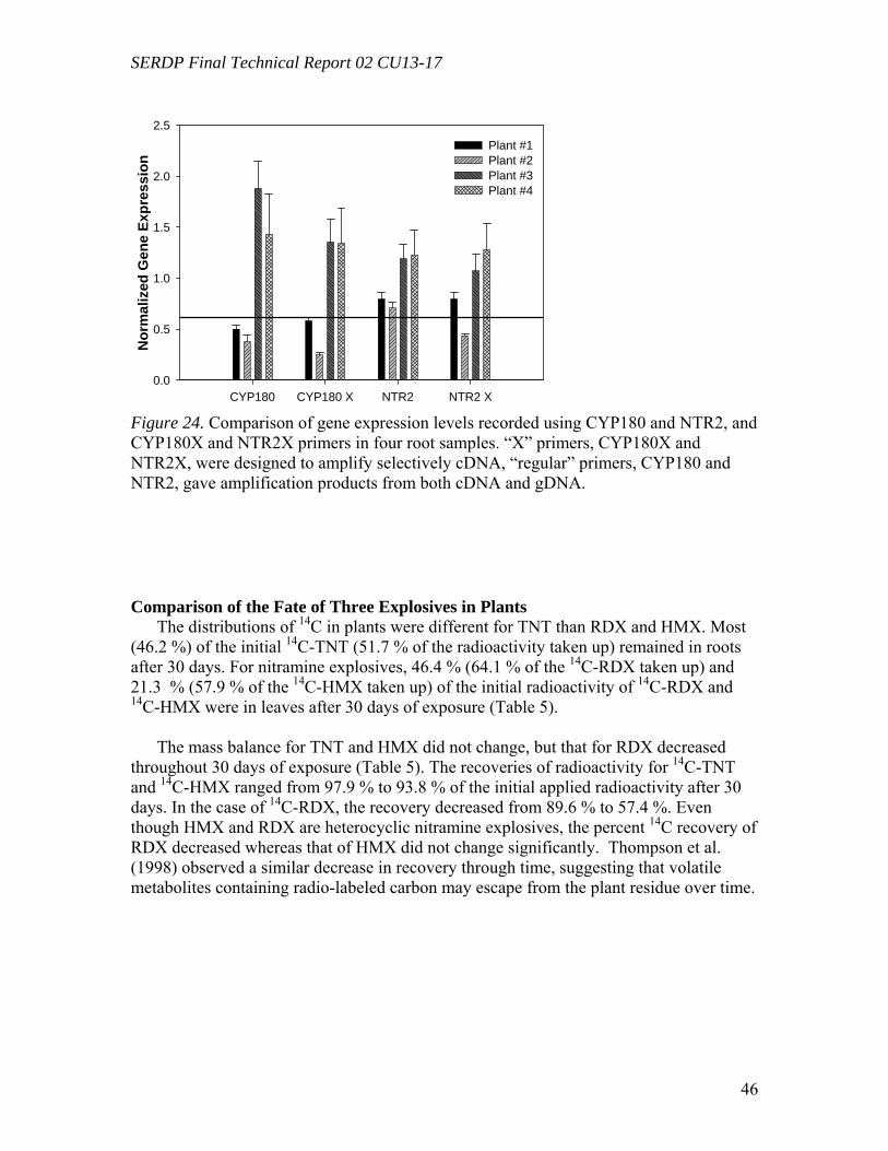
SERDP Final Technical Report 02 CU13-17
46
CYP180 CYP180 X NTR2 NTR2 X
Nor
mal
ized
Gen
e Ex
pres
sion
0.0
0.5
1.0
1.5
2.0
2.5Plant #1Plant #2Plant #3Plant #4
Figure 24. Comparison of gene expression levels recorded using CYP180 and NTR2, and CYP180X and NTR2X primers in four root samples. “X” primers, CYP180X and NTR2X, were designed to amplify selectively cDNA, “regular” primers, CYP180 and NTR2, gave amplification products from both cDNA and gDNA. Comparison of the Fate of Three Explosives in Plants
The distributions of 14C in plants were different for TNT than RDX and HMX. Most (46.2 %) of the initial 14C-TNT (51.7 % of the radioactivity taken up) remained in roots after 30 days. For nitramine explosives, 46.4 % (64.1 % of the 14C-RDX taken up) and 21.3 % (57.9 % of the 14C-HMX taken up) of the initial radioactivity of 14C-RDX and 14C-HMX were in leaves after 30 days of exposure (Table 5).
The mass balance for TNT and HMX did not change, but that for RDX decreased
throughout 30 days of exposure (Table 5). The recoveries of radioactivity for 14C-TNT and 14C-HMX ranged from 97.9 % to 93.8 % of the initial applied radioactivity after 30 days. In the case of 14C-RDX, the recovery decreased from 89.6 % to 57.4 %. Even though HMX and RDX are heterocyclic nitramine explosives, the percent 14C recovery of RDX decreased whereas that of HMX did not change significantly. Thompson et al. (1998) observed a similar decrease in recovery through time, suggesting that volatile metabolites containing radio-labeled carbon may escape from the plant residue over time.
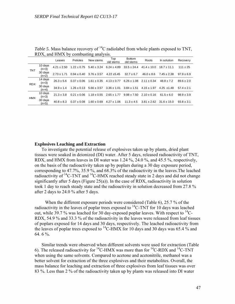
SERDP Final Technical Report 02 CU13-17
47
Table 5. Mass balance recovery of 14C radiolabel from whole plants exposed to TNT, RDX, and HMX by combusting analysis.
Leaves Petioles New stems Top old stems
Bottom old stems Roots In solution Recovery
10 days (n=3) 4.21 ± 2.50 1.22 ± 0.75 5.40 ± 3.24 6.24 ± 4.89 33.5 ± 24.4 41.4 ± 10.0 18.7 ± 11.1 111 ± 25
TNT 30 days
(n=2) 2.73 ± 1.71 0.94 ± 0.40 3.76 ± 3.57 4.22 ±5.45 32.7 ± 6.7 46.0 ± 8.6 7.45 ± 2.38 97.9 ± 6.9
14 days (n=3) 26.3 ± 5.6 0.37 ± 0.06 1.61 ± 0.35 4.13 ± 0.77 6.26 ± 1.08 2.11 ± 0.34 48.8 ± 7.2 89.6 ± 2.0
RDX 30 days
(n=4) 34.9 ± 1.4 1.26 ± 0.13 5.66 ± 3.57 3.36 ± 1.01 3.84 ± 1.51 4.15 ± 1.97 4.25 ±1.48 57.4 ± 2.1
10 days (n=3) 21.3 ± 3.8 0.21 ± 0.06 1.18 ± 0.55 2.65 ± 1.77 9.98 ± 7.50 2.10 ± 0.16 61.5 ± 6.0 98.9 ± 3.9
HMX 30 days
(n=5) 40.8 ± 8.3 0.37 ± 0.08 1.60 ± 0.69 4.27 ± 1.06 11.3 ± 4.5 3.81 ± 2.62 31.6 ± 15.0 93.8 ± 3.1
Explosives Leaching and Extraction To investigate the potential release of explosives taken up by plants, dried plant
tissues were soaked in deionized (DI) water. After 5 days, released radioactivity of TNT, RDX, and HMX from leaves in DI water was 1.24 %, 24.0 %, and 45.5 %, respectively, on the basis of the radioactivity taken up by poplars during a 30 day exposure period, corresponding to 47.7%, 35.9 %, and 68.3% of the radioactivity in the leaves.The leached radioactivity of 14C-TNT and 14C-HMX reached steady state in 2 days and did not change significantly after 5 days (Figure 25(a)). In the case of RDX, radioactivity in solution took 1 day to reach steady state and the radioactivity in solution decreased from 27.8 % after 2 days to 24.0 % after 5 days.
When the different exposure periods were considered (Table 6), 25.7 % of the
radioactivity in the leaves of poplar trees exposed to 14C-TNT for 10 days was leached out, while 39.7 % was leached for 30 day-exposed poplar leaves. With respect to 14C-RDX, 54.9 % and 33.3 % of the radioactivity in the leaves were released from leaf tissues of poplars exposed for 14 days and 30 days, respectively. The leached radioactivity from the leaves of poplar trees exposed to 14C-HMX for 10 days and 30 days was 65.4 % and 64. 6 %.
Similar trends were observed when different solvents were used for extraction (Table
6). The released radioactivity for 14C-HMX was more than for 14C-RDX and 14C-TNT when using the same solvents. Compared to acetone and acetonitrile, methanol was a better solvent for extraction of the three explosives and their metabolites. Overall, the mass balance for leaching and extraction of three explosives from leaf tissues was over 83 %. Less than 2 % of the radioactivity taken up by plants was released into DI water
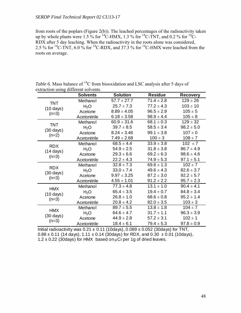
SERDP Final Technical Report 02 CU13-17
48
from roots of the poplars (Figure 2(b)). The leached percentages of the radioactivity taken up by whole plants were 1.5 % for 14C-HMX, 1.3 % for 14C-TNT, and 0.2 % for 14C-RDX after 5 day leaching. When the radioactivity in the roots alone was considered, 2.5 % for 14C-TNT, 6.0 % for 14C-RDX, and 37.3 % for 14C-HMX were leached from the roots on average.
Table 6. Mass balance of 14C from biooxidation and LSC analysis after 5 days of extraction using different solvents.
Solvents Solution Residue Recovery Methanol 57.7 ± 27.7 71.4 ± 2.8 129 ± 26
H2O 25.7 ± 7.3 77.2 ± 4.3 103 ± 10 Acetone 8.89 ± 4.05 96.5 ± 2.9 105 ± 5
TNT (10 days)
(n=3) Acetonitrile 6.18 ± 3.58 98.9 ± 4.4 105 ± 8 Methanol 60.9 ± 31.6 68.1 ± 0.3 129 ± 32
H2O 39.7 ± 8.5 58.5 ± 3.4 98.2 ± 5.0 Acetone 8.24 ± 3.46 99.1 ± 3.8 107 ± 0
TNT (30 days)
(n=2) Acetonitrile 7.49 ± 2.68 100 ± 3 108 ± 7 Methanol 68.5 ± 4.4 33.9 ± 3.8 102 ± 7
H2O 54.9 ± 2.5 31.8 ± 3.8 86.7 ± 4.9 Acetone 29.3 ± 6.6 69.2 ± 6.3 98.6 ± 4.6
RDX (14 days)
(n=3) Acetonitrile 22.2 ± 4.3 74.9 ± 5.3 97.1 ± 5.1 Methanol 32.8 ± 7.3 69.6 ± 1.3 102 ± 7
H2O 33.0 ± 7.4 49.6 ± 4.3 82.6 ± 3.7 Acetone 9.97 ± 3.25 87.2 ± 3.0 92.2 ± 5.7
RDX (30 days)
(n=3) Acetonitrile 4.55 ± 1.01 91.2 ± 2.2 95.7 ± 2.3 Methanol 77.3 ± 4.8 13.1 ± 1.0 90.4 ± 4.1
H2O 65.4 ± 3.5 19.4 ± 0.7 84.8 ± 3.4 Acetone 26.8 ± 1.0 68.6 ± 0.8 95.2 ± 1.4
HMX (10 days)
(n=3) Acetonitrile 20.8 ± 4.2 82.0 ± 3.5 103 ± 3 Methanol 89.7 ± 5.5 13.8 ± 1.8 104 ± 7
H2O 64.6 ± 4.7 31.7 ± 1.1 96.3 ± 3.9 Acetone 44.9 ± 2.8 57.2 ± 3.1 102 ± 1
HMX (30 days)
(n=3) Acetonitrile 18.4 ± 6.1 79.4 ± 5.3 97.8 ± 0.9
Initial radioactivity was 0.21 ± 0.11 (10days), 0.089 ± 0.052 (30days) for TNT, 0.88 ± 0.11 (14 days), 1.11 ± 0.14 (30days) for RDX, and 0.30 ± 0.01 (10days), 1.2 ± 0.22 (30days) for HMX based on µCi per 1g of dried leaves.
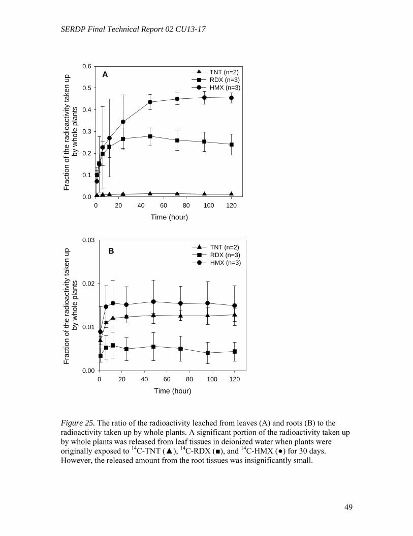
SERDP Final Technical Report 02 CU13-17
49
Time (hour)0 20 40 60 80 100 120
Frac
tion
of th
e ra
dioa
ctiv
ity ta
ken
up
by w
hole
pla
nts
0.0
0.1
0.2
0.3
0.4
0.5
0.6TNT (n=2)RDX (n=3)HMX (n=3)
Time (hour)0 20 40 60 80 100 120
Frac
tion
of th
e ra
dioa
ctiv
ity ta
ken
up
by w
hole
pla
nts
0.00
0.01
0.02
0.03TNT (n=2) RDX (n=3) HMX (n=3)
Figure 25. The ratio of the radioactivity leached from leaves (A) and roots (B) to the radioactivity taken up by whole plants. A significant portion of the radioactivity taken up by whole plants was released from leaf tissues in deionized water when plants were originally exposed to 14C-TNT (▲), 14C-RDX (■), and 14C-HMX (●) for 30 days. However, the released amount from the root tissues was insignificantly small.
A
B
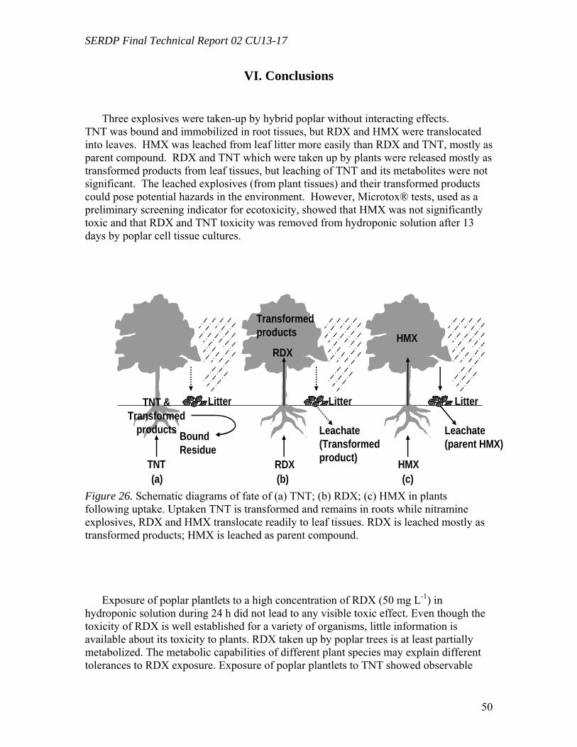
SERDP Final Technical Report 02 CU13-17
50
VI. Conclusions
Three explosives were taken-up by hybrid poplar without interacting effects. TNT was bound and immobilized in root tissues, but RDX and HMX were translocated into leaves. HMX was leached from leaf litter more easily than RDX and TNT, mostly as parent compound. RDX and TNT which were taken up by plants were released mostly as transformed products from leaf tissues, but leaching of TNT and its metabolites were not significant. The leached explosives (from plant tissues) and their transformed products could pose potential hazards in the environment. However, Microtox® tests, used as a preliminary screening indicator for ecotoxicity, showed that HMX was not significantly toxic and that RDX and TNT toxicity was removed from hydroponic solution after 13 days by poplar cell tissue cultures.
TNT RDX
TNT & Transformed
products Bound Residue
RDX
HMX
HMXTransformed products
Litter Litter Litter
Leachate(Transformed product)
Leachate(parent HMX)
(a) (b) (c) Figure 26. Schematic diagrams of fate of (a) TNT; (b) RDX; (c) HMX in plants following uptake. Uptaken TNT is transformed and remains in roots while nitramine explosives, RDX and HMX translocate readily to leaf tissues. RDX is leached mostly as transformed products; HMX is leached as parent compound.
Exposure of poplar plantlets to a high concentration of RDX (50 mg L-1) in
hydroponic solution during 24 h did not lead to any visible toxic effect. Even though the toxicity of RDX is well established for a variety of organisms, little information is available about its toxicity to plants. RDX taken up by poplar trees is at least partially metabolized. The metabolic capabilities of different plant species may explain different tolerances to RDX exposure. Exposure of poplar plantlets to TNT showed observable

SERDP Final Technical Report 02 CU13-17
51
toxic effects after 5 mg/L, with a high rate of mortality after 20 mg/L. This suggests TNT would elicit a stronger phytotoxic effect. This was confirmed by Microtox® studies, which also demonstrated, the bioluminescent bacteria indicated a stronger toxic response to TNT. The results suggest that plants may be able to withstand higher concentrations of RDX, though it may not stimulate as strong of a toxic response (i.e. induction of genes or enzymes) as would be expected by TNT. This is evidenced in gene expression studies discussed below. Information about the phytoxicity and degradation of these compounds in hydroponic solutions allows research to focus in on the potential for detoxification enzymatic reactions to occur.
Degradation experiments with living and heat-deactivated tissue cultures exposed to
light and in the dark indicated that RDX transformation involves three distinct mechanisms: The chemical reduction of RDX, the breakdown of the RDX heterocyclic ring into single carbon metabolites, and the mineralization of C1-labeled metabolites into CO2. The RDX reduction products, MNX and DNX, were only detected in the presence of living tissue cultures (i.e., neither in heat-deactivated controls nor in active crude extracts), both exposed to light and in the dark. This suggests that the initial chemical reduction of RDX was performed by poplar cells through a light-independent mechanism. The absence of reduction metabolites detected in crude extracts both exposed to light and in the dark indicated that the reduction process required intact plant cells. This is the first time that the second-reduction metabolite DNX was detected in plant tissues
While RDX and its reduction metabolites, MNX and DNX, were transformed over the time course of the experiment in plant tissue cultures when exposed to light, they accumulated in the dark, advocating further transformation of remaining RDX (and reduction metabolites) through a light-mediated mechanism. This is supported by the significant transformation of RDX observed in active crude extracts and in controls (i.e., heat-inactivated tissue cultures and extraction buffers) exposed to light and not in the dark. Two polar metabolites from RDX degradation were identified as methanol and formaldehyde, which indicates at least a partial breakdown of the RDX heterocyclic ring into single carbon subunits. Light-mediated transformation of RDX in the presence of plant subcellular structures (i.e., in crude extracts) was significantly enhanced by comparison to pure abiotic photosensitization (i.e., in controls), which indicates that plant subcellular fractions played a role in the process. UV-A and visible light do not affect directly most biomolecules and cellular components. However, light absorbing molecules, termed sensitizers, mediate photodegradation of biomolecules (termed photodynamic degradation to distinguish it from photosensitization) (38-40). Sensitizers absorb a photon, resulting in an energy-rich state (triplet state) and undergo transfer of energy (electron) either directly to a substrate (type I reaction) or to molecular oxygen producing the reactive singlet oxygen 1O2 (type II reaction). Transferring energy, sensitizers return to ground level and are ready to accept another photon in a catalytic fashion. Many plant molecules (which can be part of larger structures,such as proteins and photosystems) are sensitizers: alkaloids (berberine), benzofurans, chlorophylls, flavines, polyhydroquinones, pyridoxal, etc. (Spikes, 1998). Efficient sensitizers are capable to mediate photodynamic degradation of a wide range of biological molecules, including acids, alcohols, carbohydrates, N-heterocycles, phenols, nucleic acids, steroids, proteins, etc. (Spikes,
SERDP Final Technical Report 02 CU13-17
52
1998). While very little information is available about photodynamic degradation of xenobiotics compounds, it is very likely that light-sensitive pollutants, such as RDX, are susceptible to such mechanisms in light exposed environments.
Uptake experiments using radioactive [U-14C]RDX showed rapid uptake and
translocation of RDX or transformed metabolites into leaf tissues. While this has been previously demonstrated over longer periods of time (Thompson et al., 1999), it confirms that a response in leaf tissues may be expected even after only 24 h of exposure. Based on preliminary experiments, the transcriptional response to RDX in poplar plants peaked between 24 and 48 h of exposure. Because the variability between individual plants increased significantly after 24 h of exposure, gene expression was analyzed at this time point. TNT uptake experiments showed a similar rapid initial uptake of TNT, with the occurrence of unidentified polar metabolites occurring after 24 and 48 h of exposure, supporting a similar time frame for gene expression studies.
Gene expression analysis is recognized as a valuable tool to provide insights into
functionality of enzymes. Despite post transcriptional regulations, abundance of proteins correlates generally well to the expression levels of coding genes (Ghaemmaghami et al., 2003). Selection of poplar genes to study TNT and RDX metabolism in poplars was based on TNT-inducible genes previously identified in A. thaliana (Ekman et al., 2003). A central assumption in this approach is that plants respond in a similar manner to the nitro-substituted explosives RDX and TNT. Despite significant differences in their structure and transformation metabolism (Hawari, 2000), both RDX and TNT are toxic, hydrophobic, energetic compounds, which bear 3 reactive nitro groups on a cyclic core. Typical biotransformation of both RDX and TNT begins by the reduction of nitro groups, generating reduction derivatives, which are the most commonly detected metabolites. Another central assumption is that similar genes are at work and induced by RDX in the laboratory model A. thaliana and in Populus sp., which constitutes a model of forest trees and a model for phytoremediation studies. Sterky et al. (2004) showed a considerable similarity between Arabidopsis and Populus genomes, which has already allowed the transfer of functional information from Arabidopsis to Populus (Nanjo et al., 2004). Nearly all gene families found in one species have homologs in the other. Populus sp. has an average genome size of 550 Mbp and is estimated to contain about 58,000 genes, over twice as many as the Arabidopsis genome, which further supports the likelihood to find homologs of Arabidopsis genes in Populus (Nanjo et al., 2004; Wullschlege, Jansson, and Taylor, 2002). Finally, genomic DNA is highly conserved among the Populus genus (Sterky et al., 2004), which allows P. trichocarpa database to be applicable to various Populus species.
Work with TNT focused on expression of GST genes by poplar trees as a response to
exposure to the toxic explosive. GSTs are enzymes known to be involved in the transformation of a large variety of toxic chemicals (Pflugmacher et al., 2000; Schroder et al., 2001; Maars, 1996; Edwards and Dixon, 2004), and they also are suspected to be (at least partially) responsible for the detoxification of TNT (Hughes et al., 1997; Bhadra et al., 1999). Even thought no direct evidence of such a mechanism has been obtained, we showed here that a purified, commercially-available GST was able to catalyze in vitro the

SERDP Final Technical Report 02 CU13-17
53
conjugation of TNT with GSH. The induction levels of the two Populus GST genes recorded in TNT-exposed roots were of the same order of magnitude as those previously reported in TNT-exposed A. thaliana roots (18 – 25-fold for GST173 vs. 27.5-fold for At1g17170 (the corresponding A. thaliana gene), and 4 – 10-fold for GST180 vs. >11.0-fold for At2g29490; Ekman et al., 2003). The decrease of GST180 induction observed after 24 h may reflect a toxic effect; significant deleterious effects, including the reduction of transpiration and growth rates, chlorosis, and abscission, were observed for exposure to 5.0 mg L-1 and higher. Alternatively, this reduction of the expression level may be correlated with the disappearance of TNT from the hydroponic solution observed after 24 h.
GST173 showed a 10-fold induction in RDX-exposed leaf tissues and a 2-fold
induction in root tissues. These results are consistent with Mezzari et al. (2005) that reported a 5-fold induction of GST genes in A. thaliana seedlings exposed to RDX (67 mg L-1). However, competitive confocal imaging of glutathione S-bimane indicated that RDX did not compete strongly for GSH sites in Arabadopsis roots (Mezzari et al., 2005). GSTs catalyze the conjugation of electrophilic substrates with reduced GSH, resulting in more soluble and less toxic conjugates. In addition, GSTs are induced by various stressors, like pathogen infection, wounding, heavy metal, and heat shock (Marrs, 1996; Frova, 2003), where they play a role in scavenging active oxygen species (AOS) (Levine et al., 1994; Tenhaken et al., 1995). Since AOS are also suspected to be produced by nitroso radicals generated during the reduction of nitro-substituted explosives, this may explain why GSTs are induced by RDX. GST173 expression levels in exposed root tissues were significantly lower than those recorded in leaf tissues; even thought root tissues were directly exposed to RDX. This discrepancy may be explained by the observation that RDX was primary translocated to the leaves after being taken up by plants.
A cytochrome P-450 gene (CYP180) and a peroxidase gene (POX) were induced 1.6
fold and 1.7 fold in RDX-exposed leaves, respectively. Cytochrome P-450s and peroxidases have been reported to be involved in the transformation of RDX by bacteria and fungi (Fernando and Aust, 1991; Tekoah and Abeliovich, 1999; Coleman, Spain, and Duxbury, 2002). However, the involvement of these genes in RDX transformation by Populus sp. has not been established. Monodehydroascorbate reductase (MDA) genes are involved in plant responses to oxidative stresses (Ekman et al., 2003). The observed 1.6-fold induction by RDX in leaves can be the result of exposure to nitro-substituted explosives, known to produce an oxidative stress. 12-oxophytodienoate reductase (OPR) is a NADPH-dependent flavoenzyme closely related to nitrate ester reductase, which plays a role in the bacterial reduction of nitro groups of TNT (Williams and Bruce, 2000). A low-level induction of OPRs has also been reported in A. thaliana seedlings exposed to RDX (Mezzari et al., 2005). OPRs are also likely to be involved in TNT reduction by A. thaliana (Ekman et al., 2003).
Using RT real-time PCR, this research demonstrates for the first time that exposure of
Populus plants to the toxic explosive RDX results in the up-regulation of several genes frequently involved in detoxification mechanisms, which suggests their involvement in
SERDP Final Technical Report 02 CU13-17
54
the metabolism of RDX and related explosives. It has also demonstrated the up-regulation by exposure to TNT of two GST genes. However, the actual activity of the translated enzymes in RDX transformation by poplar plants has to be confirmed. Further investigations should try to demonstrate the correlation between gene transcription and protein synthesis or enzymatic activity, as well as the enzymatic transformation of RDX.
Poplar growth is supported by climates across the United States and would be
applicable to nearly any explosive-contaminated military site. A small list of climate-supportive sites is provided in Appendix A, however this list is not all-inclusive. Poplars are model phytoremediation plants because of their climate versatility as well as their rapid transpiration and growth rates. They are deciduous and thrive in temperate climates, but phytoremediation technologies are applicable throughout the U.S. Therefore, the knowledge gained through this project regarding the detoxification of explosive compounds in poplar plantlets taking up these compounds through transpiration streams could potentially serve to benefit a diverse array of contaminated military bases in the U.S. Future work will look to consider the limitations at specific explosives-contaminated military field sites with varying soil properties to determine site-specific limitations in the application of poplar phytoremediaion. This next work will investigate mechanisms of bringing the contaminant to the plant for uptake before the reactions studied in this project take place. The accomplishments of the completed project’s research serve to provide a better understanding of phytoremediation processes leading to the improvement of soils and groundwater in a variety of climates.
References
Altschul SF, Madden TL, Schaffer AA, Zhang JH, Zhang Z, Miller W, Lipman DJ (1997) Gapped blast and psi-blast - A new generation of protein database search programs. Nucl Acids Res 25: 3389-3402 Ambion (2005) Use of Internal and External Standards or Reference RNAs for Accurate Quantitation of RNA Levels. Technical Bulletin #151. Ambion. http://www.ambion.com/techlib/tb/tb_151.html Ampleman, G., Thiboutot, S., Lavigne, J., Marois, A., Hawari, J., Jones, A. M., and Rho, D. 1995. Synthesis of 14C-labelled hexahydro-1,3,5-trinitro-1,3,5-triazine (RDX), 2,4,6-trinitrotoluene (TNT), nitrocellulose (NC), and glycidylazide polymer (GAP) for use in assessing the biodegradation potential of these energetic compounds. J. Labeled. Comp. Rad. 36, 559-577. Applied Biosystems (2002) ABI Prism 7000 Sequence Detection System. User Guide. Applied Biosystems, Foster City Ausubel FM, Brent R, Kingston RE, Moore DD, Seidman JG, Smith JA, Struhl K (1999) Short Protocols in Molecular Biology, 4th Ed. John Wiley, New York Bhadra R, Wayment D, Hughes J, Shank J (1999) Confirmation of conjugation processes during TNT metabolism by axenic plant roots. Environ Sci Technol 33: 446-452
SERDP Final Technical Report 02 CU13-17
55
Bhadra, R., Wayment, D. G., Williams, R. K., Barman, S. N., Stone, M. B., Hughes, J. B., and Shanks, J. V. 2001. Studies on plant-mediated fate of the explosives RDX and HMX. Chemosphere 44, 1259-1264 Bennett, J., and Cubbage, J. (1992) Review and Evaluation of Microtox Test for
Freshwater Sediments. In. Olympia: Washington State Department of Ecology, 1-28. Best, E. P. H., Sprecher, S. L., Larson, S. L., Fredrickson, H. L., and Bader, D. F. 1999. Environmental behavior of explosives in groundwater from the Milan Army Ammunition Plant in aquatic and wetland plant treatments. Uptake and fate of TNT and RDX in plants. Chemosphere 39, 2057-2072. Best, E. P. H., Zappi, M. E., Fredrickson, H. L., Sprecher, S. L., Larson, S. L., and Ochman, M. 1997. Screening of aquatic and wetland plant species for phytoremediation of explosives-contaminated groundwater from the Iowa Army Ammunition Plant. Ann. NY. Acad. Sci. 829, 179-194. Bhushan, B., Halasz, A., Thiboutot, S., Ampleman, G., Spain, J. C., and Hawari, H. 2002. Biotransformation of hexahydro-1,3,5-trinitro-1,3,5-tiazine catalyzed by a NAD(P)H: Nitrate oxidoreductase from Aspergillus niger. Environ. Sci. Technol. 36, 3104-3108. Bhushan, B., Trott, S., Spain, J. C., Halasz, A., Pacquet, L., and Hawari, J. 2003. Biotransformation of hexahydro-1,3,5-trinitro-1,3,5-triazine (RDX) by a rabbit liver cytochrome P-450: Insight into the mechanism of RDX biodegradation by Rhodococcus sp. Strain DN22. Appl. Environ. Microbiol. 69, 1347-1351. Brentner, Laura B.; Tanaka, Sachiyo; Merchie, Kate M.; Schnoor, Jerald L.; and
VanAken, Benoit. Expression of Glutathione Transferases in poplar trees exposed to 2,4,6-trinitrotoluene. (submitted for publication, Environmental Science and Technology)
Burken, J. G., Shanks, J. V., and Thompson, P. L. 2000. Phytoremediation and plant metabolism of explosives and nitroaromatic compounds. In: Biodegradation of Nitroaromatic Compounds and Explosives, pp. 239-276. (Spain, J. C., Hughes, J. B., and Knackmuss, H. J., Eds.). New York, Lewis Publishers. Burton, D. T., Turley, S. D., and Peters, G. T. 1994. The acute and chronic toxicity of hexahydro-1,3,5-trinitro-1,3,5-triazine (RDX) to the fathead minnow (Pimephales promelas). Chemosphere 29, 567-579. Coleman J, Blake-Kalff M, Davies T (1997) Detoxification of xenobiotics by plants: Chemical modification and vacuolar compartimentation. Trends in Plant Science 2: 144-151 Coleman, N. V., Spain, J. C., Duxbury, T. 2002. Evidence that RDX biodegradation by Rhodococcus strain DN22 is plasmid-borne and involves a cytochrome P-450. J. Appl. Microbiol. 93, 463-472. Dixon DP, Lapthorn A, Edwards R (2002) Plant glutathione transferases. Genome Biol 3: 3004.1-3004.10 Edwards R, Dixon DP (2004) Metabolism of natural and xenobiotic substrates by the plant glutathione S-transferase superfamily. In Sandermann H, ed, Molecular Ecotoxicology of Plants. Springer, Berlin, pp 17-50
SERDP Final Technical Report 02 CU13-17
56
Ekman DR, Lorenz WW, Przybyla AE, Wolfe NL, Dean JFD (2003) SAGE analysis of transcriptome responses in Arabidopsis roots exposed to 2,4,6-trinitrotoluene. Plant Physiol 133: 1397-1406 Fernando, T., and Aust, S. D. 1991. Biodegradation of munition waste, TNT (2,4,6-
trinitrotoluene) and RDX (hexahydro-1,3,5-trinitro-1,3,5-triazine) by Phanerochaete chrysosporium. In: Emerging Technologies in Hazardous Waste Management II, pp. 214–232. (Tedder, D. W., and Pohland, F. G., Eds.). Washington, D.C., American Chemical Society.
Frova, C. 2003. The plant glutathione transferase gene family: Genomic structure, functions, expression, and evolution. Physiol. Plant. 119, 469-479.
Ghaemmaghami S, Huh W, Bower K, Howson RW, Belle A, Dephoure N, O'shea EK, Weissman JS (2003) Global analysis of protein expression in yeast. Nature 425: 737-741 Gong, P., Hawari, J., Thiboutot, S., Ampleman, G., and Sunahara, G. I. 2001.
Ecotoxicological effects of hexahydro-1,3,5-trinitro-1,3,5-triazine on soil microbial activities. Environ. Toxicol. Chem. 20, 947-951.
Gorge E, Brandt S, Werner D (1994) Uptake and metabolism of 2,4,6-trinitrotoluene in higher plants. Environ Sci Pollut Res 1: 229-233 Habig W, Jakoby W (1981) Assays for differentiation of glutathione S- transferases. In W Jakoby, ed, Methods in Enzymology, Vol 77. Academic Press, New York, p 398 Hannink, N. K., Rosser, S. J., and Bruce, N. C. 2002. Phytoremediation of explosives.
Crit. Rev. Plant. Sci. 21, 511-538. Harvey S, Fellows R, Cataldo D, Bean R (1990) Analysis of 2,4,6-trinitrotoluene and its
transformation products in soils and plant tissues by high-performance liquid chromatography. J Chromatogr 518: 361-374
Hawari, J. 2000. Biodegradation of RDX and HMX: From basic research to field application. In: Biodegradation of Nitroaromatic Compounds and Explosives, pp. 277-310. (Spain, J. C., and Knackmuss, H. J., Eds.). New York, Lewis Publishers.
Hoagland, D. R., and Arnon, D. I. 1950. The Water-Culture Method for Growing Plants without Soil. California Agriculture Experiment Station, Berkeley.
Hughes JB, Shanks J, Vanderford M, Lauritzen J, Bhadra R (1997) Transformation of TNT by aquatic plants and plant tissue cultures. Environ Sci Technol 31: 266-271 Joint Genome Institute (JGI) Poplar Genome Project (2005) http://genome.jgi- psf.org/Poptr1/Poptr1.home.html Just, C. L., and Schnoor, J. L. 2004. Phytophotolysis of hexahydro-1,3,5-trinitro-1,3,5-
triazine (RDX) in leaves of reed canary grass. Environ. Sci. Technol. 38, 290-295. Keith LH, Telliard, WA (1979) Priority pollutants: A perspective view. Environ Sci Technol 13: 416-423 Kitts, C. L., Green, C. E., Otley, R. A., Alvarez, M. A., and Unkefer, P. J. 2000. Type I
nitroreductases in soil Enterobacteria reduce TNT (2,4,6-trinitrotoluene) and RDX (hexahydro-1,3,5-trinitro-1,3,5-triazine). Can. J. Microbiol. 46, 278-282.
Lachance B, Robidoux PY, Hawari J, Ampleman G, Thiboutot S, Sunahara GI (1999) Cytotoxic and genotoxic effects of energetic compounds on bacterial and mammalian cells in vitro. Mut Res - Genet Toxicol Environ Mut 444: 25-39 Larson S, Jones R, Escalon L, Parker D (1999) Classification of explosives
SERDP Final Technical Report 02 CU13-17
57
transformation products in plant tissues. Environ Tox Chem 18: 1270-1276. Levine, A., Tenhaken, R., Dixon, R., and Lamb, C. 1994. H2O2 from the oxidative burst orchestrates the plant hypersensitive disease resistance response. Cell 79, 583-593. Marchler-Bauer A, Anderson JB, DeWeese-Scott C, Fedorova ND, Geer LY, He SQ, Hurwitz DI, Jackson JD, Jacobs AR, Lanczycki CJ, Liebert CA, Liu CL, Madej T, Marchler GH, Mazumder R, Nikolskaya AN, Panchenko AR, Rao BS, Shoemaker BA, Simonyan V, Song JS, Thiessen PA, Vasudevan S, Wang YL, Yamashita RA, et al. (2003) CDD: A curated Entrez database of conserved domain alignments. Nucl Acids Res 31: 383-387 Marrs KA (1996) The functions and regulation of glutathione S-transferases in plants. Ann Rev Plant Physiol Plant Mol Biol 47: 127-158 McCutcheon, S. C., and Schnoor, J. L. 2003. Overview of phytotransformation and
control of wastes. In: Phytoremediation Transformation and Control of Contaminants, pp. 3-58. (McCutcheon, S. C., and Schnoor, J. L., Eds.). Hoboken, New Jersey, John Wiley.
McGonigle B, Keeler SJ, Lau SMC, Koeppe MK, O'Keefe DP (2000) A genomics approach to the comprehensive analysis of the glutathione S-transferase gene family in soybean and maize. Plant Physiol 124: 1105-1120. McNally W (1944) Three deaths from TNT (trinitrotoluene) poisoning. Indust Med 13: 491-494 Mezzari, M. P., Walters, K., Jelinkova M., Shih M. C., Just C. L., Schnoor, J. L. 2005. Gene expression and microscopic analysis of Arabidopsis exposed to chloroacetanilide herbicides and explosive compounds. A phytoremediation approach. Plant Physiol. 138, 858-869. Nanjo, T., Futamura, N., Nishiguchi, M., Igasaki, T., Shinozaki, K., and Shinohara, K. 2004. Characterization of full-length enriched expressed sequence tags of stress- treated poplar leaves. Plant Cell Physiol. 45, 1738-1748. National Center for Biotechnology Information (NCBI) (2005) http://www.ncbi.nlm.nih.gov Ohakawa, H., Tsujii, H., Shimooji, M., Imajuku, Y., and Imaishi, H. 1999. Cytochrome
P-450 biodiversity and plant protection. J. Pest. Sci. 24, 197-203. Pflugmacher S, Schroder P, Sandermann H (2000) Taxonomic distribution of plant glutathione S-transferases acting on xenobiotics. Phytochemistry 54: 267-273 Price, R. A., Pennington, J. C., Larson, S. L., Neumann, D., and Hayes, C. A. 2002. Uptake of RDX and TNT by agronomic plants. Soil Sed. Contam. 11, 307-326 Robidoux, P. Y., Svendsen, C., Caumartin, J., Hawari, J., Ampleman, G., Thiboutot, S., Weeks, J. M., and Sunahara, G. I. 2000. Chronic toxicity of energetic compounds in soil determined using the earthworm (Eisenia andrei) reproduction test. Environ. Toxicol. Chem. 19, 1764-1773 Sambrook J, Russel DW (2001) Molecular Cloning: A Laboratory Manual, 3rd Ed. Spring Harbor, New York Sandermann HJ (1994) Higher plant metabolism of xenobiotics: The "green liver" concept. Pharmacogenetics 4: 225-241 Schnoor JL (2000) Degradation by plants - Phytoremediation. In Rehm HJ, Reed G, eds, Biotechnology, Vol 11b. Wiley-VCH, Weinheim Schroder P, Scheer C, Belford E (2001) Metabolism of organic xenobiotics in plants:
SERDP Final Technical Report 02 CU13-17
58
Conjugating enzymes and metabolic end points. Minerva Biotechnol 13: 85-91 Schwitzguebel, J. P., and Vanek, T. 2003. Some fundamental advances for xenobiotic
chemicals. In: Phytoremediation: Transformation and Control of Contaminants, pp. 123-157. (McCutcheon, S. C., and Schnoor, J. L., Eds.). Hoboken, New Jersey, John Wiley.
Seth-Smith, H. M. B., Rosser, S. J., Basran, A., Travis, E. R., Dabbs, E. R., Nicklin, S., and Bruce, N. C. 2002. Cloning, sequencing, and characterization of the hexahydro-1,3,5-trinitro-1,3,5-triazine degradation gene cluster from Rhodococcus rhodochrous. Appl. Environ. Microbiol. 68, 4764-4771.
Smock LA, Stoneburner DL, Clark JR (1976) The toxic effects of trinitrotoluene (TNT) and its primary degradation products on two species of algae and the fathead minnow. Water Res 10: 537-543 Spain JC (2000) Biodegradation of nitroaromatic compounds and explosives:
Introduction. In Spain JC, Hughes JB, Knackmuss HJ, eds, Biodegradation of Nitroaromatic Compounds and Explosives. Lewis Publishers, New York
Stahl, J. D., Van Aken, B., Cameron, M. D., and Aust, S. D. 2001. Hexahydro-1,3,5-trinitro-1,3,5-triazine (RDX) biodegradation in liquid and solid-state matrices by Phanerochaete chrysosporium. Biodegradation J. 13-25.
Sterky F, Bhalerao RR, Unneberg P, Segerman B, Nilsson P, Brunner AM, Charbonnel- Campaa L, Lindvall JJ, Tandre K, Strauss SH, Sundberg B, Gustafsson P, Uhlen M, Bhalerao RP, Nilsson O, Sandberg G, Karlsson J, Lundeberg J, Jansson S (2004) A Populus EST resource for plant functional genomics. Proc Natl Acad Sci USA 101: 13951-13956 Sunahara, G. I., Dodard, S., Sarrazin, M., Paquet, L., Ampleman, G., Thiboutot, S.,
Hawari, J., and Renoux, A. Y. 1998. Development of a soil extraction procedure for ecotoxicity characterization of energetic compounds. Ecotox. Environ. Safe. 39, 185-194.
Tanaka, S; Brentner, LB.; Merchie, KM.; Yoon, JM; Schnoor, J L.; and Van Aken, B. 2006. Analysis of gene expression in poplar trees (Populus deltoides × nigra, DN34) exposed to the toxic explosive hexahydro-1,3,5-trinitro-1,3,5-triazine (RDX). International Journal of Phytoremediation. (accepted for publication)
Tenhaken, R., Levine, A., Brisson, L. F., Dixon, R. A., and Lamb, C. 1995. Function of the oxidative burst in hypersensitive disease resistance. Proc. Natl. Acad. Sci. USA. 92, 4158-4163.
Thompson JD, Gibson TJ, Plewniak F, Jeanmougin F, Higgins DG (1997) The ClustalX windows interface: Flexible strategies for multiple sequence alignment aided by quality analysis tools. Nucl Acids Res 25: 4876-4882 Thompson P, Ramer L, Schnoor J (1998b) Uptake and transformation of TNT by hybrid poplar trees. Environ Sci Technol 32: 975-980 Thompson P, Raner L, Guffey A, Schnoor J (1998a) Decreased transpiration in poplar trees exposed to 2,4,6-trinitrotoluene. Environ Tox Chem 17: 902-906 Tuskan J, DiFazio SP, Teichmann T (2002) Poplar genomics is getting popular: The impact of the Poplar Genome Project on tree research. Plant Biol 5: 1-3 Van Aken, B. and Schnoor, J. L. 2002. Evidence of perchlorate (ClO4
-) reduction in plant tissues (poplar tree) using radio-labeled 36ClO4
-. Environ. Sci. Technol. 36, 2789-2788.
SERDP Final Technical Report 02 CU13-17
59
Van Aken, B., Yoon, J. M., Just, C. L., and Schnoor, J. L. 2004. Metabolism and mineralization of hexahydro-1,3,5-trinitro-1,3,5-triazine inside poplar tissues (Populus deltoides × nigra DN34). Environ. Sci. Technol. 38, 4572-4579.
Velculescu V, Zhang L, Volgelstein B, Kinzler K (1995) Serial analysis of gene expression. Science 270: 484-487 Won WD, DiSalvo LH, Ng J (1976) Toxicity and mutagenicity of 2,4,6-trinitrotoluene
and its microbial metabolites. Appl Enviro Microbiol 31: 576-580 Williams, R. E., and Bruce, N. C. 2000. The role of nitrate ester reductase enzymes in the
biodegradation of explosives. In: Biodegradation of Nitroaromatic Compounds and Explosives, pp. 161-184. (Spain, J. C., Hughes, J. B., and Knackmuss, H. J., Eds.). New York, Lewis Publishers.
Wullschleger SD, Jansson S, Taylor G (2002) Genomics and forest biology: Populus emerges as the perennial favorite. Plant Cell 14: 2651-2655 Yoon, J.M. (2004) The fate of explosive chemicals (HMX, TNT, RDX) in plants and
water following uptake by hybrid poplar. PhD Dissertation. In Civil and Environmental Engineering. Iowa City: University of Iowa, p. 138.
Yoon, J.M., Oh, B.T., Just, C.L., and Schnoor, J.L. (2002) Uptake and leaching of octahydro-1,3,5,7-tetranitro-1,3,5,7- tetrazocine by hybrid poplar trees. Environmental Science & Technology 36: 4649-4655
Yoon, JM; Van Aken, B; and Schnoor, JL. 2006. Leaching of contaminated leaves following uptake and phytoremediation of RDX, HMX and TNT by Poplar. International J. of Phytoremediation 8(1):81-94
Appendices
A. List of Explosive Contaminated Military Sites whose Climate is Supportive of the Growth of Poplars Camp Edwards, Otis Air National Guard Base, MA Camp LeJeune Marine Corps Base, NC Eglin Air Force Base, FL Iowa Army Ammunition Plant, Middleton, IA Joliet Army Ammunition Plant, Joliet, IL
B. List of Technical Publications
Journal Articles Brentner, LB.; Tanaka, S; Merchie, KM.; Schnoor, JL.; and VanAken, B. Expression of Glutathione Transferases in poplar trees exposed to 2,4,6-trinitrotoluene. (submitted for publication, Environmental Science and Technology) Just, C. L., and Schnoor, J. L. 2004. Phytophotolysis of hexahydro-1,3,5-trinitro-1,3,5-triazine (RDX) in leaves of reed canary grass. Environ. Sci. Technol. 38, 290-295.
SERDP Final Technical Report 02 CU13-17
60
Mezzari, M. P., Walters, K., Jelinkova M., Shih M. C., Just C. L., Schnoor, J. L. 2005. Gene expression and microscopic analysis of Arabidopsis exposed to chloroacetanilide herbicides and explosive compounds. A phytoremediation approach. Plant Physiol. 138, 858-869. Van Aken, B., Yoon, J. M., Just, C. L., and Schnoor, J. L. 2004. Metabolism and mineralization of hexahydro-1,3,5-trinitro-1,3,5-triazine inside poplar tissues (Populus deltoides × nigra DN34). Environ. Sci. Technol. 38, 4572-4579. Tanaka, Sachiyo; Brentner, Laura B.; Merchie, Kate M.; Yoon, Joon Yoon; Schnoor, Jerald L.; and Van Aken, Benoit. 2006. Analysis of gene expression in poplar trees (Populus deltoides × nigra, DN34) exposed to the toxic explosive hexahydro-1,3,5-trinitro-1,3,5-triazine (RDX). International Journal of Phytoremediation. (accepted for publication) Yoon, JM; Van Aken, B; and Schnoor, JL. 2006. Leaching of contaminated leaves following uptake and phytoremediation of RDX, HMX and TNT by Poplar. International J. of Phytoremediation 8(1):81-94
Conferences/Symposiums/Abstracts Tanaka, S*.; Merchie, K.; Shihm M-C; Van Aken, B; Schnoor, J.L. Use of microarray analysis in Arabidopsis leaves exposed to RDX as a screening technique for determination of RDX upregulated genes in poplar (short lecture). International Conference on Environmental Biotechnology, Leipzig, Germany. July 9-14, 2006. Brentner, LB*; Van Aken, B.; Merchie, K; and Schnoor, JL. Investigating molecular responses in poplar phytoremediation of TNT: glutathione transferases (short lecture) International Conference on Environmental Biotechnology, Leipzig, Germany. July 9-14, 2006 Brentner, LB*; Van Aken, B.; Merchie, K; and Schnoor, JL. Investigating molecular responses in poplar phytoremediation of TNT: glutathione transferases. (poster) Research Open House, The College of Engineering, The University of Iowa. April 20-21, 2006 Award: Best Poster, Civil and Environmental Engineering Schnoor, JL* (keynote lecture) 231st American Chemical Society Meeting and Exposition, Atlanta, GA. March 26-30, 2006 Schnoor, JL*. Phytotechnologies: Theory and Practice. (keynote address) SERDP/ESTCP Partners in Environmental Technology Technical Symposium and Workshop, Washington, DC. November 29-December 1, 2005
SERDP Final Technical Report 02 CU13-17
61
Tanaka, S*; Brentner, LB; Van Aken, B; and Schnoor, JL. Gene expression and metabolism of hexahydro-1,3,5-trinitro-1,3,5-triazine (RDX) in poplar (Populus deltoides × nigra DN34) (poster) SERDP/ESTCP Partners in Environmental Technology Technical Symposium and Workshop, Washington, DC. November 29-December 1, 2005 Brentner, LB*; Van Aken, B; and Schnoor, JL. Glutathione-S-transferases involved in degradation of 2,4,6-trinitrotoluene in poplar trees. (poster) 14th Biocatalysis and Bioprocessing Conference, The University of Iowa, Iowa City, IA. October 24-26, 2005 Flokstra, B; Van Aken, B; and Schnoor, JL. Poplar Tree Plant Tissue Cultures as a Model System for Determining Toxicity in the Phytoremediation of TNT and RDX. (poster) 14th Biocatalysis and Bioprocessing Conference, The University of Iowa, Iowa City, IA. October 24-26, 2005 Schnoor, JL*; Van Aken, B; Yoon, JM; Tanaka, ST; Brentner, LB; and Flokstra, B. Phytoremediation: From the Molecular to the Field Scale. (keynote lecture) 14th Biocatalysis and Bioprocessing Conference, The University of Iowa, Iowa City, IA. October 24-26, 2005 Tanaka, S; Van Aken, B; and Schnoor, JL. Analysis of gene expression in poplar trees (Populus deltoides x nigra DN34) exposed to the toxic explosive hexahydro-1,3,5-trinitro-1,3,5-triazine (RDX). (poster) 14th Biocatalysis and Bioprocessing Conference, The University of Iowa, Iowa City, IA. October 24-26, 2005 Brentner, LB*; Van Akan, B; and Schnoor, JL. Expression of glutathione-S-transferases in poplar trees (Populus trichocarpa) exposed to 2,4,6-trinitrotoluene. (poster) Plant Root Genomics Consortium Proteomics Symposium, Danforth Plant Science Center, St Louis, MO. April 21-22, 2005 Schnoor, JL*; Van Aken, B; Yoon, JM; Tanaka, ST; Brentner, LB; and Flokstra, B. Phytoremediation: From the Molecular to the Field Scale (keynote lecture) U.S. EPA 3rd International Phytotechnologies Conference, Atlanta, GA. April 19-22, 2005 Brentner, LB*; Van Akan, B; and Schnoor, JL. Expression of glutathione-S-transferases in poplar trees (Populus trichocarpa) exposed to 2,4,6-trinitrotoluene. (poster) 13th Biocatalysis and Bioprocessing Conference, The University of Iowa, Iowa City, IA. October 25-27, 2004 Tanaka, S*; Van Aken, B; and Schnoor, JL. Involvement of phytohormones in phytoremediation of toxic explosives RDX and TNT. (poster) 13th Biocatalysis and Bioprocessing Conference, The University of Iowa, Iowa City, IA. October 25-27, 2004 Van Aken*, Flokstra, BR; Yoon, J-M; and Schnoor, JL.Implication of an Endosymbiotic Methylobacterium sp. in the Phytoremediation of Toxic Explosives RDX and HMX by Poplar Tree Tissue Cultures (Populus deltoides × nigra) (poster) Partners in
SERDP Final Technical Report 02 CU13-17
62
Environmental Technology Conference (SERDP meeting) Washington, D.C. December, 2003 Flokstra, BR*; Van Aken, B; Yoon, J-M; and Schnoor, JL. The Use of Microtox® Toxicity Test for the Assessment of Phytoremediation of the Toxic Explosive TNT by Plant Tissue Cultures (Populus deltoides x nigra DN34). (poster) U.S. EPA Phytotechnologies Conference in Chicago, IL. March 3-5, 2003